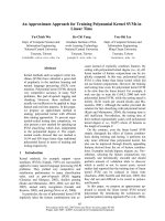
Báo cáo khoa học: Val216 decides the substrate specificity of a-glucosidase in Saccharomyces cerevisiae doc
Bạn đang xem bản rút gọn của tài liệu. Xem và tải ngay bản đầy đủ của tài liệu tại đây (362.23 KB, 7 trang )
Val216 decides the substrate specificity of a-glucosidase
in
Saccharomyces cerevisiae
Keizo Yamamoto
1
, Akifumi Nakayama
2
, Yuka Yamamoto
1,
* and Shiro Tabata
1
1
Department of Chemistry, Nara Medical University, Japan;
2
Nara Prefectural Institute for Hygiene and Environment, Japan
Differences in the s ubstrate s pecificity o f a-glucosidases
should be due to the differences in the su bstrate binding and
the catalytic domains of the enzymes. To elucidate such
differences of enzymes hydrolyzing a-1,4- and a-1,6-glu-
cosidic linkages, two a-glucosidases, maltase and isomaltase,
from Saccharomyces c erevisiae were cloned a nd analyzed.
The cloned yeast isomaltase and maltase consisted of 589
and 584 amino a cid residues, respectively. There w as 72.1%
sequence identity with 165 amino acid a lterations between
the two a- glucosida ses. These two a-glucosidase genes
were subcloned into the pKP1500 expression vector and
expressed in Escherichia coli. The purified a-glucosidases
showed the same substrate specificities as those of their
parent native glucosidases. Chimeric enzymes constructed
from isomaltase by exchanging with maltase fragments were
characterized by their substrate specificities. When the con-
sensus region I I, which is one of the f our regions conserved in
family 13 (a-amylase family), is replaced with the maltase
type, the chimeric enzymes a lter to hydrolyze maltose. T hree
amino a cid r esidues i n consensus region I I w ere d ifferent i n
the t wo a-glucosidases. Thus, we modified Val216, G ly217,
and Ser218 of isom altase to the m altase-type amino acids by
site-directed mutagenesis. The Val216 mutant was altered
to hydrolyze both maltose and isomaltose but neither
the Gly217 nor the Ser218 mutant changed their substrate
specificity, indicating that Val216 is an important residue
discriminating the a-1,4- and 1,6-glucosidic linkages of
substrates.
Keywords: family 13; a-glucosidase; Saccharomyces cere-
visiae; s ite-directed mutagenesis; substrate s pecificity.
Glucosyl hydrolases (EC 3.2.1 ) are key e nzymes of
carbohydrate metabolism that were found in the three
major kingdoms, and are categorized into 57 structural
families [1,2]. Family 13 (a-amylase family) includes
enzymes such as a-amylase, a-glucosidase, pullulanase,
cyclodextrin glucanotransferase, and 1,4-a-
D
-glucan
branching enzyme, specifically acting on a-1,4- and
a-1 ,6-O-glucosidic linkages [ 1]. Many primary structures
of the members of family 13 from various origins are now
available, and have been compared to each other. The
existence of four highly conserved regions (regions I–IV)
and three acidic residues located in the conserved regions as
catalytic residues has been r eported [3–7]. Further more,
computing s econdary structure analysis indicated that
specific structural features of the catalytic (b/a)
8
-barrel
domain exist in these enzymes [8–10].
The relationship of sequence and structure to substrate
specificity i n family 13 enzymes, particularly a-amylase,
cyclomaltodextrinase, and neo pullulanase, has been well
studied [11–13]. Despite the fact that many a-glucosidases
with diverse substrate specificities h ave been purified and
cloned from mammals, plants, and m icroorganisms, i t i s s till
not clear which amino acid residues of a-glucosidase
recognize t he difference between a-1,4- a nd a-1,6-glucosidic
bonds contained in saccharides.
Yeast contains two a-glucosidases, a-1,4-glucosidase
(E.C. 3.2.1.20, maltase) and oligo-1,6-glucosidase
(E.C. 3.2.1.10, isomaltase), which act preferentially on
maltose or isomaltose and methyl a-
D
-glucopyranoside
(a-mg), respectively. The expression of these e nzymes is
controlled by different polymeric genes, MAL or MGL,
separately [14–16]. Maltase (the MAL6 product of
Saccharomyces carlsbergensis) preferentially hydrolyzed
maltose but neither isomaltose nor a-mg, whereas isomaltase
hydrolyzes isomaltose and a-mg but not maltose [17,18].
Thus, we focused on the structure–function relationship of
the two a-glucosidases f rom Saccharomyces as a model in
respect of the difference in t heir substrate s pecificities.
The yeast genome directory which was constructed by
Goffeau et al. revealed t he existence of many homologo us
open reading frames of a-glucosidase [19]. The comp lete
nucleotide sequence of the MAL gene of Sacchar omyces has
been determined [20], w hereas it is not clear w hich open
reading frame corresponds to the MGL gene.
In this study, we cloned the genes encoding isomaltase
and m altase by means of a RT-PCR method and expressed
them in Escherichia coli. Subsequently, f rom a comparison
of the p rimary structures of the two a-glucosidases, chimeric
enzymes were constructed by exchange parts of maltase and
isomaltase genes including any one of the four conserved
Correspondence to K. Yamamoto, Department of Chemistry, Nara
Medical University, Shijo, Kashihara, Nara 634–8521, Japan.
Fax/Tel.: +81 744 29 8810, E-mail:
Abbreviations: a-mg, methyl a-
D
-glucopyranoside; a-pNPG, p-nitro-
phenyl a-
D
-glucopyranoside.
Enzymes: a-1,4-glucosidase (mal tase) (E.C. 3.2.1.20); oligo-
1,6-glucosidase ( isomaltase) (E.C. 3.2.1.10).
*Present address: Department o f General Medicine, Nara Medical
University, J apan.
(Received 2 April 2004, r evised 17 J une 2004, accepted 5 J uly 2004)
Eur. J. Biochem. 271, 3414–3420 (2004) Ó FEBS 2004 doi:10.1111/j.1432-1033.2004.04276.x
regions f or family 13 members. In addition, we constructed
mutants of isomaltase by replacing three amino acid
residues after Asp215 in consensus region II with residues
of the maltase type using site-directed mutagenesis. The
substrate specificities o f all mutant enzymes were examined.
We found that one amino acid residue in consensus region
II decided the substrate specificity of isomaltase.
Materials and methods
Materials
The yeast strains used w ere Saccharomyces cerevisiae
D-346 (ATCC 56960) and 727–14C (ATCC 56959). The
bacterial strains and plasmids used were Escherichia coli
JM109, KP3998 [21], pUC18 and pKP1500 [21].
Hydroxyapatite (Gigapite) was purchased from Seika-
gaku Kogyo and hydroxyapatite (Micro-Prep Ceramic
Hydroxyapatite, type I) was from B io-Rad. Maltose,
isomaltose, a-mg, and p-nitrophenyl a-
D
-glucopyranoside
were from Nakalai T esque, Japan. A site-directed muta-
genesis kit (Quickchange
TM
) was obtained from S tratagene
and the bicinchoninic acid protein assay reagent was from
Pierce Chemicals. La-Taq polymerase was purchased from
Takara Syuzo and restriction endonucleases and T4 DNA
ligase were from Takara S yuzo, or New En gland Biolabs.
Reverse transcriptase was used from the Expand
TM
Reverse Transcriptase kit from Boehringer Mannheim.
The Marathon kit was purchased from Clontech Labor-
atory. Oligonucleotides were synthesized by Takara Syuzo
Custom Service.
Assay method for enzyme activity a-glucosidase activity
was determined by measuring the release of p-nitrophenol
from p-nitrophenyl a-
D
-glucopyranoside ( a-pNPG) accord-
ing t o the method described previously [22]. When maltose,
isomaltose, and methyl a-
D
-glucopyranoside (a-mg) were
used as substrates, the enzyme activity was determined as
the rate of hydrolysis of the substrate by measuring the
release of glucose according to the enzymatic method of
NADP
+
reduction using hexokinase and glucose-6-phos-
phate dehydrogenase [23].
Cloning of the isomaltase gene from
S. cerevisiae
The production of maltase and isomaltase of S. cerevisiae
was induced by adding maltose and a-mg to the culture
medium, respectively [ 22]. In t he case of the cloning of the
isomaltase gene, total RNA w as prepared from S . cerevisiae
D-346 grown o n a medium including 3% (w/v) a-mg by the
method of Chomczynski & S acchi [24]. The mRNA was
purified from the to tal RNA using Oligotex-dT 30 (Super)
(Takara Syuzo) according to the manufacturer’s instruc-
tions. Double-stranded cDNA was constructed from
poly(A) RNA with the oligo-dT primer u sing the Expand
TM
Reverse Transcriptase kit. The N-terminal amino acid
sequence, TISSAHPETEPK, which was determined from
purified yeast isomaltase, matched the ORF YGR287c on
chromosome VII of S. cerev isiae [19]. Therefore, the iso-
maltase gene was amplified from the c DNA library by PCR
using t he N-terminal sequence of ORF YGR287c and oligo-
dT as primers . The 1 .8 kb RT-PCR product was ligated to
plasmid pUC 18 after digestion with SmaI and introduced
into E. coli JM109. The insert was sequenced by the dideoxy
method [25] using t he Dye T erminator C ycle Sequencing F S
Ready Reaction kit (Applied Biosystems). To verify the 5 ¢
end sequence, 5¢ RACE was performed using the Marathon
kit with the AP1 primer and a gene-specific pr imer
(5¢-AGATTGCCTTTCTACAGTCTTCATTC-3¢) accord-
ing to the manufacturer’s p rotocol. The 5¢-RACE product
was sequenced by a direct sequencing method.
Subcloning into the pKP1500 expression vector
Forward and reverse primers were designed based on the
5¢-and3¢-terminal nucleotide sequences of the isomaltase
gene (MGL) for cloning into plasmid pKP1500. The
forward p rimer 5¢-ATGACTATTTCTTCTGCACAT
CCAGAGACAGAAC-3¢ con tains the initiation codon,
while the reverse primer 5¢-CTTTCTGCAGACTCA
TTCGCTGATATATATTC-3¢ linked a PstI restriction site
to the termination codon. PCR was carried out on the
isomaltase gene cloned above. PCR products were digested
with PstI. Simultaneously, pKP1500 was digested with
EcoRI and PstI, and then the EcoRI site was blunted by the
use of a Blunting kit (Takara Syuzo). The vector and
the insert MGL gene were ligated with T4 ligase followed by
transformation into E. coli JM109. The cells were plated on
Luria–Bertani agar supplemented with 40 lgÆmL
)1
5-bromo-4-chloro-3-indolyl-a-
D
-glucopyranoside (Boehrin-
ger Mannheim), 50 lgÆmL
)1
ampicillin, and 1 m
M
isopro-
pyl t hio-b-
D
-galactoside and then one da y later several b lue
colonies appeared. One of the clones expressing isomaltase
was selected. The plasmid containing the isomaltose gene
was designated pYIM.
Cloning of the maltase gene from
S. cerevisiae
For cloning of the maltase gene, S. cerevisiae 727–14C was
grown in medium containing 3% (w/v) maltose. The
mRNA and cDNA were pr epared using the same procedure
as described above.
The gene-specific primers were synthesized based on the
information of Hong & Marmur [20]. The reverse primer was
modified by introducing a HindIII site seven bases down-
stream from the s top codon. PCR with La-Taq polymerase
was carried out on the cDNA prepared from S. cerevisiae
727–14C. The 1 .8 kb PCR fragment w as digest ed wit h
HindIII. Plasmid pKP1500 was digested with EcoRI and
HindIII, and then the Eco RI site was blunted by the use of a
Blunting kit (Takara Syuzo). The fragment was inserted into
the pKP1500 vector and the resulting plasmid was intro-
duced into E. coli KP3998. Several transformants containing
the 1.8 kb insert were selected and sequenced. The plasmid
carrying the maltase gene was designated p YMA.
Expression of recombinant enzymes in
E. coli
and
purification of the enzymes
The E. coli transformant carrying pYIM (or pYMA) was
inoculated into PYG medium [21] supplemented with
50 lgÆmL
)1
of ampicillin and i ncubated at 37 °C. Isopropyl
thio-b-
D
-galactoside (final 1 m
M
) was added when cell
density at A
660
reached 0.5 and the culture was further
incubated for 12 h.
Ó FEBS 2004 Substrate specificity of a-glucosidase (Eur. J. Biochem. 271) 3415
Cells were resuspended in 50 m
M
Tris/HCl buffer
(pH 7 .5) a nd sonicated. The cell-free extract was applied
to a Q AE-Toyopearl column equilibrated with 50 m
M
Tris/
HCl (pH 7.5) and the column was washed with the same
buffer containing 20 m
M
NaCl. The enzyme was eluted with
a linear gradient o f NaCl (20–150 m
M
) in the same buffer.
Active fractions were pooled and applied to a column of
Gigapite equilibrated with 20 m
M
sodium phosphate buffer
(pH 7 .0). The enzyme was eluted with a linear gradient
of sodium phosphate buffer up t o 1 50 m
M
. T he act ive
fractions were collected and dialyzed against 40 m
M
sodium
phosphate buffer ( pH 6.8), t hen subjected to c hromato-
graphy on hydroxyapatite (Micro-Prep Ceramic Hydroxy-
apatite type I). The purified enzyme was eluted at 250 m
M
phosphate buffer (pH 6.8) by a linear gradient of
40–320 m
M
phosphate.
Construction of chimeric enzymes from recombinant
maltase and isomaltase
Chimeric enzymes were constructed by exchanging nucleo-
tide fra gm ents between t he maltase and isomaltase genes at
a single restriction site on the plasmid or by inserting a
fragment which was introduced at a unique restriction site
by PCR.
Chimeric enzymes MAa/IMb and IMa/MAb were con-
structed by exchanging two Mun I/BglII fragments of pYIM
and pYMA which were cleaved at single restriction sites
with both of these restriction enzymes. The chimeric
enzyme, Mun/Bpu was constructed by inserting a fragment,
which was amplified by PCR with the forward p ri-
mer 5¢-AGAAGCCATT
GCTGAGCAATTTTTGTTC-3¢
(underlining i ndicates t he Bpu1102I restriction site) and t he
reverse primer 5¢-AAA
AAGCTTGCACTAATTTTATTT
GAC-3¢ (underliningindicates the HindIII restriction site and
stop codon, respectively) and pYMA as a template, into IMa/
MAb at Bpu1102I/HindIII. Other c himeric enzymes, M un/
Bst, Mun/Pst, and Pst/Bst were constructed by the same
method described for the Mun/Bpu c himera. The chimeric
enzymes are shown in a sche matic diagram in Fig. 2.
Site-directed mutagenesis
Site-directed mutagenesis (Asp215 fi Ala,Val216 fi Th r,
Gly217 fi Ala, and Ser218 fi Gly of isomaltase) was
carried out by the use of the Quick Change
TM
Site-Directed
Mutagenesis k it and DNA from pYIM as a template and
two additional mutagenic oligonucleotide primers for each
amino acid substitution according to t he instruction man-
ual. The sites to which the mutation was introduced were
sequenced to confirm that only the expected mutation had
occurred.
Results and Discussion
Cloning of yeast a-glucosidases
Two a-glucosidase genes, encoding isomaltase and maltase,
were isolated from an S. cerevisiae cDNA library using the
PCR technique. In the case of isomaltase, the N-terminal
amino a cid sequence, TISSAHPETEPK, matched O RF
YGR287c on chromosome VII of S. cerevisiae [19]. More-
over, s ix peptides, i ncluding the N -terminal amino acid
sequence (TISSAHPETEPK, GSAWTFDEK, NGPRI
HEFHQEM, LYTSASR, FRYNLVP, and TLKW
PWEGR) obtained from t he native isomaltase were in
accord with their nucleotide sequences of the ORF. B ased on
this in formation, a 1.8 kb fragment was a mplified from the
cDNA library by PCR using 5¢-sequence o f the ORF and
oligo d T as p rimers, and was inserted into pUC18. Sequen-
cing of the 1 .8 kb insert revealed an open r eading frame o f
1770 bp including a stop codon, TGA. The 5 89 amino acid
protein deduced from the ORF was c omp letely identical to
the amino acid sequence deduced from ORF Y GR287c. The
sequence data for isomaltase is availab le from the DNA Data
Bank of Japan with accession number AB109221.
The entire coding region of the insert was amplified by
PCR and subcloned into the pKP1500 expression vector,
and the resulting plasmid was introduced into E. coli
JM109. Expression of the gene was screened by a plate
assay using 5-bromo-4-chloro-3-indolyl-a-
D
-glucopyrano-
side. Several blue colonies were found to hydrolyze
a-pNPG. The expression of isomaltase in these clones w as
confirmed by their ability to hydrolyze isomaltose and a-mg
but not maltose. The plasmid containing the isomaltase
gene was designated pYIM.
The maltase gene was also isolated f rom the DNA library
of S. cerevisiae by PCR using gene specific primers. The
amplified 1.8 kb fragment was inserted into plasmid
pKP1500 and the resulting plasmid was transformed into
E. coli. KP3998. DNA sequence analysis of the fragment
gave 100% identity to the MAL6 gene [15]. The plasmid
containing the maltase gene was designated pYMA.
Figure 1 s hows a comparison of amino a cid s equences
between maltase and isomaltase. T here is 72.1% of sequence
identity with 165 amino acid alterations.
Assessment of recombinant enzymes in comparison
with native a-glucosidases
We assessed the tw o recomb inant a-glucosidases in terms of
substrate specificity and immunological identity and com-
pared them to their native enzymes. The two recombinant
a-glucosidases showed the same substrate specificities as
those of their parent glucosidases, namely, maltase hydro-
lyzed maltose but not isomaltose and a-mg, whereas
isomaltase hydrolyzed isomaltose and a-mg but not malt-
ose. Upon double i mmunodiffusion, rabbit antiserum
against native isomaltase produced a single precipitation
line without spurs with recombinant isomaltase (data not
shown). When the two recombinant enzymes reacted with
antisera against n ative maltase and isomaltase, the recom-
binant enzymes showed the s ame dos e–response a s t he
native enzymes by antiserum neutralization (data not
shown). These results indicate that the two recombinant
a-glucosidases a re identical to their parent enzymes.
Substrate specificities of chimeric enzymes
The c omparison of the primary structures of the members
of family 13 from various origins has revealed the e xistence
of four highly conserved regions I, II, III, and IV [3–7].
Thus, for the design o f chimeric a-glucosidases, the
a-glucosidase gen es i n the two plasmids, pYMA and pYIM,
3416 K. Yamamoto et al.(Eur. J. Biochem. 271) Ó FEBS 2004
were divided into fi ve portions taking into account the four
consensus regions. F igure 2 is a schematic representation of
a number o f t he chimeric enzymes. Chimeric enzymes were
characterized based on substrate s pecificities for maltase,
isomaltase, a-mg, sucrose, and a-pNPG, and t he K
m
for
a-pNPG. MAa/IMb a nd IMa/MAb w ere c onstructed by a
recombination o f the N-terminal fragment containing
consensus region I of isomaltase and maltase, respectively.
The recombination had no effect on either the substrate
specificities o r the K
m
for a-pNPG (Table 1). In the M un/
Bam chimera, the amino acids from 488 to the C-terminus
of IMa/MAb were substituted b y the corresponding amino
acids of m altase ( residues 4 85–584). The subst itution of the
C-terminal fragment of IMa/MAb also had no effect on the
substrate specificities. We further dissected the C-terminal
region of IMa/MAb by preparing chimeras with switch-
over points at r esidues 332 an d 231 (Mun/Bpu and Mun/
Bst, respectively). The specific activity for isomaltose of
Mun/Bpu and Mun/Bst were about 10 and 80 times lower
than that of isomaltase, respectively. The K
m
for a-pNPG of
Mun/Bpu was the same a s that of isomaltase, whereas the
K
m
for a-pNPG of M un/Bst was about 50 times l ower than
that of isomaltase. T hus, fragments including consensus
regions III and IV may affect the substrate affinity of the
a-glucosidases. To investigate the role of the fragment
containing consensus r egion II, two c himeras, Mun/Pst a nd
Pst/Bst, were constructed. In the Mun/Pst chimera, a 27
amino acid fragment of Mun/Bst including consensus
Fig. 1. Comparison of amino a cid sequences
between maltase and is oma ltase. Identical and
similar amino acid resi dues are designated by
*andÆ, respectively. Four highly co nserved
regions of family 13 are underlined.
Fig. 2. Schematic diagram of the chimeric en zymes. Isomaltase se-
quenceisrepresentedasanopenbarandmaltasesequenceisrepre-
sented as a s haded bar. MunI, Pst I, BstBI, and Bpu1102I are r estriction
sites used for the construction o f chimeric enzymes. I, II, III, and IV
indicate the l ocation of f o ur highly c onserved regions of f amily 13.
Ó FEBS 2004 Substrate specificity of a-glucosidase (Eur. J. Biochem. 271) 3417
region II was replaced by the corresponding fragment of
pYMA. The substrate s pecificities of M un/Pst changed
completely to those of maltase type. However, the charac-
teristics of Pst/Bst which contained only the 27 amino acid
fragment of pYIM in pYMA were t he same as those of
Mun/Bst. Therefore, these results indicate that the fragment
including consensus region II c ontributes to the determin-
ation of the substrate specificity of a-glucosidase.
Site-directed mutagenesis
There were s ix amino acid d ifferences between the two
a-glucosidases in the fragment including consensus region
II. Three out of the six alterations were similar, thus, we
targeted the other three amino acid residues in consensus
region II for s ite-directed mutagenesis. The V al216, Gly217,
andSer218inconsensusregionIIofisomaltasewere
substituted to the corresponding amino acid residues of
maltase, Thr, Ala, and Gly, respectively. The mutant
enzymes G217A and S218G did not exhibit different
substrate specificity to that of isomaltase but their K
m
for
a-pNPG tended toward m altase (Table 2). Mutant V216T
could hydrolyze the a-1,4-glucosidic linkage retaining the
isomaltase type s ubstrate s pecificity a nd its hydrolyzing
ratio of maltose/isomaltose was 1 : 1. As shown in T able 2,
doubly and triply mutated enzymes including V216T
(V216T/G217A, V216T/S218G, and V216T/G217A/
Table 1. Substrate specificities of the chimeric e nzymes. The enzyme was incubated with 0.5
M
substrate in 100 lLof0.1
M
sodium phosphate buffer,
pH 7.0 at 30 °C for 5 min. The rea ction was s topped by addition of 100 lLof0.5
M
Tris/HCl buffe r, pH 7.5, then r eleased glucose w as assayed. For
a-pNPG, an increase of absorbanc e at 41 0 n m was me asured in 5 m
M
a-pNPG in 0 .1
M
sodium ph osphate b uffer, pH 7.0 at 30 °C.
Enzyme
Specific activity (lmolÆmin
)1
Æmg
)1
enzyme)
K
m
for
a-pNPG (m
M
)
Maltose Isomaltose a-mg a-pNPG
Maltase 70.0 0.00 0.00 132 0.31
Isomaltase 0.00 46.0 48.0 92.0 2.13
MAa/IMb 36.6 0.00 0.00 126 0.30
IMa/MAb 0.00 30.0 21.0 57.0 1.26
Mun/Bpu 0.00 4.40 2.30 34.0 3.32
Mun/Bst 0.00 0.69 0.23 5.30 0.045
Mun/Pst 34.0 0.00 0.00 98.0 0.15
Pst/Bst 0.00 0.46 0.23 5.70 0.043
Table 2. Kinetic parameters of wild-type isomaltase and site-directed
mutants. TheconsensusregionIIofisomaltasewasmutatedtothe
maltase type by site-directed mutagenesis. For example, V216T was
made by exch angin g Val216 of isomaltase with Thr of maltase.
Enzyme
Specific activity
(lmolÆmin
)1
Æmg
)1
enzyme)
K
m
for
a-pNPG
(m
M
)
Maltose Isomaltose
Isomaltase 0.00 45.8 2.13
Maltase 68.7 0.00 0.31
D215A 0.00 0.00 ND
V216T 16.5 16.5 0.59
G217A 0.00 16.0 0.53
S218G 0.00 27.5 0.84
V216T/G217A 36.6 6.41 0.61
V216T/S218G 21.1 6.87 0.40
G217A/S218G 0.00 22.9 0.66
V216T/G217A/S218G 6.18 0.57 0.49
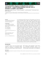
Fig. 3. Sequence alignment of a-glucosidases of known substrate spe-
cificityintheconsensusregionII.Asp residue of the c atalytic nucleo-
phile is labeled with an arrow and the next residue is highlighted in
bold. Shown are: Sce D-346, S. cerevisiae isomaltase (this study); Bt h,
B. thermoglucosidasius oligo-1,6-glucosidase [27]; Bce, B. ce reus suc-
rase-isomaltase [28]; Bco, B. coagulans sucrase-isomaltase [29]; Bsp1,
Bacillus sp. D G0303 a-glucosidase [30] , Bsp2, Basillus sp . F5 sucrase -
isomaltase [31]; Bfl, B. flav ocaldarius oligo-1,6-glucosidase [32]; Spn,
Streptococcus pneumoniae a-1,6-glucosidase [33]; Bsu, B. subtilis suc-
rase-isomaltase-maltase [34]; B sp3, Bacillus sp. a-glucosidase [35], T cu,
Thermomonospora curvata a-glucosidase [36] ; Sce727–14C, S. cerevis-
iae maltase (this study); Sca, S. c arlsbergensis maltase [20]; C al,
C. albicans maltase [37]; H po, Hansenula polymorpha maltase [38].
3418 K. Yamamoto et al.(Eur. J. Biochem. 271) Ó FEBS 2004
S218G) exhibited a change in the h ydrolyzing ratio o f
maltose/isomaltoseto5:1,3:1,and10:1,respectively.
These facts indicate that the three residues in consensus
region II, particularly Val, plays an importan t role in
distinguishing between the a-glucosidic linkages of a-1,4
and a-1,6.
McCarter and Withers [26] indicated that Asp214 on
the consensus region II of maltase is the catalytic
nucleophile. Because the Asp214 of maltase is equivalent
to the Asp215 of isomaltase, a mutant with the residue
altered to Ala was tested for its activity on a-pNPG.
None of the mutants including D215A had activity on
a-pNPG and a-mg although the proteins were detected
with antiserum against isomaltase by immunoblotting
(data not shown). T hus, the Asp215 of isomaltase is one
of three active acidic residues which are completely
conserved in a-glucosidase group.
Amino acid sequence alignment
Figure 3 shows the amino acid sequence alignment of the
consensusregionIIofa-glucosidases o f known substrate
specificity. In the case of a-glucosidases hydrolyzing the
a-1,6-glucosidic linkage, the amino acid residue following
the catalytic nucleophile is Val. On the other hand, the
corresponding residue of a-glucosidases which acting on
the a-1,4-glucosidic linkage but does not a-1,6-linkage is
Thr. X-ray crystallographic analysis of B. cereus oligo-
1,6-glucosidase revealed t hat Val200 following t he cata-
lytic nucleophile Asp199 locates on the long loop region
followed by Nb4, and the side chain of Val200 faces
toward the inside of the c atalytic cleft [39]. Figure 4 shows
the h ypothetical structure of the active site of S. cerevisiae
isomaltase in complex with isomaltose or maltose using
the crystal structure of B. cereus oligo-1,6-glucosidase [39]
as the starting model. In the case of wild-type isomaltase,
isomaltose fit to the active site, whereas maltose cannot
bind to the active site because the side chain of Val216
interfere with binding of a 4-linked glucose. The CG1 of
Val216 is too close to the O3¢ of maltose. On the other
hand, both isomaltose a nd maltose can bind to th e V216T
mutant because the steric hindrance between OG1 of
Thr216 and O3¢ of maltose is canceled by the rotation of
the s ide c hain of Thr216. The results indicate that the
amino acid residue just after the catalytic nucleophile in
consensus region II must b e involved in the recognition of
a-glucosidic linkages.
In conclusion, this work was successful in identifying the
region and residue important in the determination of the
substrate specificity of a-glucosidases. The identification of
V216T and doubly and triply mutated enzymes altered in
substrate specificity w ill serve as a bas is for p rogress toward
further understanding the structure-function relationship of
family 13 a-glucosidases.
References
1. Henrissat, B. (1991) A c lassifocation of glycosyl hydrolases based
on ami n o acid s equence similarities. Biochem. J. 28 0 , 309–316.
2. Henrisaat, B. (1996) Updating the sequence-based c lassification of
glucosyl hydrolases. Bioc hem. J. 316, 695–696.
3. Matsuura, Y., Kusunoki. M., Harada, W. & Kakudo, M. (19 84)
Structure and possible catalytic residues of Taka-amylase A.
J. Bi ochem. 95, 697 –702.
4. Nakajima, R., Imanaka, T. & Aiba, S. (1986) Comparison of
amino acid s equenc es of eleven d ifferent a-amylases. Appl.
Microbiol. Bio technol. 23, 355–360.
5. Svensson, B. (1988) Regional distant sequence homology between
amylases, a-glucosidases, and transglucanosylases. FEBS Lett.
230, 7 2–76.
6. Machius, M., Wiegand, G. & H uber, R . (1995) Crystal s tructure
of calcium-depleted Bacillus licheniformis a-amylase at 2.2 A
˚
Resolution. J. Mol. Biol. 246 , 545–559.
7. Brayer, G .D., Luo, Y. & Withers, S.G. (1995) The structure of
human pancreatic a-amylase at 1.8 A
˚
resolution an d c omparisons
with related en zymes. Protein Sci. 4, 1730 –1742.
8. Jespersen,H.M.,MacGregor,E.A.,Sierks,M.R.&Svensson,B.
(1991) Comparison of the domain-level o rganization of starch
hydrolases and related e nzymes. Biochem. J. 28 0, 51–55.
9. Jespersen, H.M., MacGregor, E.A., Henrissat, B., Sierkes, M.R.
& Svensson, B. (1993) Starch and glycogen-debranching and
Fig. 4. Hypothetical model structure o f the ac tive site of isomaltase in complex with i somaltose or m altose. (A) W ild type. (B) V216T m utant. The
models were co nstructed by the use o f the program
HOMOLOGY
from the Insight II (Accelrys Inc., San D iego,CA,USA).Thecrystalstructureof
B. cereus oligo-1,6-glucosidase was used as the starting model. Isomaltose and maltose a re colored dark gray and pale gray, respectively. F igures
were produced with
MOLSCRIPT
[40] and
RENDER
from the Raster3D package [ 41].
Ó FEBS 2004 Substrate specificity of a-glucosidase (Eur. J. Biochem. 271) 3419
branching enz ymes: P redic tion o f s t ructural f eatures of the cata-
lytic (b/a)
8
-barrel domain a nd evoluti onary rela tionship to other
amylolytic e nzymes. J. Prote in Chem. 12 , 791–805.
10. Svensson, B. (1994) Regional sequence alignment for extentions
from b-strands 4 a nd 7 of catalytic (b/a)
8
-barreldomaininglyc-
osyl hydrolase s family 13. Plant Mol . Biol. 25, 141–157.
11. Takata, H., Kuriki, T ., Okuda, S., Takesada, Y., I izuka, M.,
Mimamiura, N. & Imanaka, T. (1992) Action of neopullulanase
catalyzes both hydrolysis and transglycosilation at a-(1-4)- and
a-(1-6)-glucosidic linkages. J. Biol. Chem. 267, 18447–18452.
12. Ibuka, A., T onozuka, T., Matsuzawa, H. & Sakai, H. (1998)
Conversion of neopullulanase-a-amylase from Thermoactinomy-
ces vulgaris R-74 into an amylopullulanase-type enzyme. J. Bio-
chem. 123, 275–282.
13. MacGregor, E.A., Janecek, S. & Svensson, B. (2001) Relationship
of sequence and structure to specificity in the a-amylase family of
enzymes. Biochim. Biop hys. Acta 1546, 1–20.
14. Carlson, M. (1987) Regulation of sugar utilization in Saccharo-
myces species. J. Bacteriol. 169, 4873–4877.
15.Vanoni,M.,Sollitti,P.,Goldenthal,M.&Marmur,J.(1989)
Structure and regulation of the multigene family controlling
maltose fermentation in budding yeast. Prog. Nucleic Acid Res.
Mol. Biol. 37, 281–322.
16. Johnson, M. & Carlson, M. (1992) Regulation of carbon and
phosphate utilization. In The Molecular and C elullar Biology of
Yeast Saccharomyces: Gene Expression (Johns, E .W ., Prngle, J.R.
& Broach, J., eds), pp. 193–281. Cold Spring Harbor Laboratory
Press, Cold Spring Harbor, NY.
17. Khan, N.A. & Eaton, N.R. (1 967) Purification and characteriza-
tion of maltase and a-methyl glucosidase from yeast. Biochim.
Biophys. A cta 146, 173–180.
18. Needleman, R.B., F ede roff, H.J., Eccleshall, T.R., Buchferer, B. &
Marmur, J . (1978) Purific ation and c haracterization of a-glucosi-
dase from Sacchromyces carlsbergensis. Biochemistry 37, 4567–
4661.
19.Goffeau,A.,Aert,M.L.,Agostini-Carbone,A.,Ahmed,M.,
Aigle, L., Alberghina, K., Albermann, M., Albers, M., Aldea, D.,
Alexandraki, G., et al. (1997) Th e yeast g eno me directory . Nature
387 (Suppl ), 1–105.
20. Hong, H.S. & Marmur, J. (1986) Primary s equence o f t he m altase
gene of th e MAL6 l o cus of Saccharomyces carlsbergensis. Gene 41,
75–84.
21. Miki, T., Yasukochi, T., Nagatani, H., Furuno, M., Orita, T.,
Yamada, H., Imoto, T. & Horiuchi, T. (1987) Construction of
a plasmid vector for the regulatable high level expression of
eukaryotic ge nes in Escherichia c oli: an application to over-
production of chicken lysozyme. Protein Eng. 1, 327 –332.
22. Tabata, S., Ide, T., Umemura, Y. & Trii, K. ( 1984) Purification
and c h aracterization of a-glucosidasesproducedbySaccharomy-
ces in response to three d istinct maltose genes. Biochim. Biophys.
Acta 797, 231–238.
23.Kunst,A.,Draeger,B.&Ziegenhorn,J.(1984)Methods of
Enzymatic Analysis, Vol. VI, 3rd edn. (Bergmeyer, H.U., ed), pp.
163–172. Ve rlag Chemie, Weinheim.
24. Chomczynski, P. & Sacchi, N. (1977) Single-step method of RNA
isolation by acid guanidinium thiocyanate–phenol–ch loroform
extraction. Anal. B iochem. 162, 256– 159.
25. Sanger, F., Nicolen, S. & C oulson, A.R. (1977) DN A sequencing
with chain-t erminating inhibitors. Pro c. Natl A cad. Sci . USA 74,
5463–5467.
26. McCarter, J .D. & Withers, S.G. (1996) Unequivocal identification
of Asp-214 as the catalytic nucleophile of Sac charomyces cerevisiae
a-glucosidase using 5-fluoro glyco syl fl uorides. J. Bi ol. Chem. 271,
6889–6894.
27. Watanabe,K.,Chiishiro,K.,Kitamura,K.&Suzuki,Y.(1991)
Proline residues responsible for t hermostability o ccur with high
frequency in the loop regions of an extremely thermostable oligo-
1,6-glucosidase from Bacillus thermoglucosidasius KP1006. J. Bio l.
Chem. 266, 24287–24294.
28. Suzuki, Y., Aoki, R. & Hayashi, H. (1982) Assignment of a
p-nitrophenyl-a-
D
-glucopyranoside-hydro lyzing a-glucosidase of
Bacillus cereus ATCC 7064 to an exo-oligo-1,6-glucosidase.
Biochim. Bi ophys. Acta 704, 476–483.
29. Suzuki, Y . & Tomura, Y. (1986) Purification and characterization
of Bacillus c oagulans oligo-1,6-glucosidase. Eur. J. Bioc hem. 15 8,
77–83.
30. Lee, Y E. (2000) Cloning and characterization of a-glucosidase
gene from thermophilic Bacillus sp. DG0303. J. Microbiol. Bio-
technol. 10, 244–250.
31. Yamamoto, M. & H orikoshi, K. (1990) Nucleotide sequence of
alkalophilic Bacillus olig o-1,6-glucosidase geneandtheproperties
ofthegeneproductinEscherichia c oli HB101. Denpun Kag aku 37,
137–144.
32. Kashiwabara, S., M atsuki, Y ., Kishimoto, T. & Suzuki, Y. ( 1998)
Clustered proline r esid ues a roun d t he active-site cleft in therm o-
stable oligo-1,6 glucosidase of Bacillus flavocaldarius KP1228.
Biosci. B iotechnol. B iochem. 62 , 1093–1102.
33. Coffey, T.J., Enright, M .C., Daniels, M., M orona, J.K., Morona,
R., H ryniewicz, W., Paton, J.C. & Spratt, B.G . (1998)
Recombinational exchanges at the capsular polysaccharide bio-
synthetic locus lead to frequent serotype changes among natural
isolates of Stre ptococcus pne umoniae. Mol. Microbiol. 27 , 73–83.
34. Scho
¨
nert, S., Buder, T. & D ahl, M.K. (1998) Identification and
enzymatic characterization of the maltose-inducible a-glucosidase
MalL (sucras e-isomaltase-maltase) of Bacillus subtilis. J. Bacteriol.
180, 2 574–2578.
35. Nakao, M., N akayama, T., Kakudo, A., Inohara, M., Harada,
M., O mura, F. & Shibano, Y. (1994) Struc ture and expression of a
gene coding for t hermostable a-glucosidase with a broad substrate
specificity from Bacillus sp. SAM1606. Eur. J. Biochem. 220, 293–
300.
36. Janda, L., Pavelka, P., Tichy, P., Spizek, J. & P etricek, M. (1997)
Production and properties of a-glucosidase from the t hermotol-
erant bacterium Thermomonospora curvata. J. Appl. Micobiol. 83,
470–476.
37. Geber, A., Williamson, P.R., R ex, J.H., Sweeney, E.C. & B ennett,
J.E. (1992) Cloning and characterization of a Candida albicans
maltase gene involve d in s ucrose utilization. J. Ba cteriol. 174,
6992–6996.
38. Liiv, L., Pa
¨
rn, P. & Alama
¨
e, T. (2001) Cloning of maltase gene
from a m ethylotrophic yeast, Hansenula polymorpha. Gene 265,
77–85.
39. Watanabe, K., Hata, Y., Kizaki, H., Katsub e, Y. & Suzuki, Y.
(1997) The r efined crystal structure of Bacillus cereus oligo-
1,6-glucosidase at 2.0 A
˚
resolution: structural characterization of
proline-substitution sites for prote in t h ermostab ilizatio n. J. Mo l.
Biol. 269 , 142–153.
40. Kuraulis, P.J. (1991)
MOLSCRIPT
: a pro gram to produce both
detailed and s chemat ic plots o f p roteins. J. Appl . Crystallog. 23,
946–950.
41. Merritt, E.A. & Murphy, M .E.P. (1994)
RASTER
3
D
, a program for
photorealistic molecular graphics, Version 2.0. Acta Crystallog.
Sect. D 50, 869–873.
3420 K. Yamamoto et al.(Eur. J. Biochem. 271) Ó FEBS 2004