Báo cáo khoa học: Characterization of a cathepsin L-associated protein in Artemia and its relationship to the FAS-I family of cell adhesion proteins pot
Bạn đang xem bản rút gọn của tài liệu. Xem và tải ngay bản đầy đủ của tài liệu tại đây (888.77 KB, 12 trang )
Characterization of a cathepsin L-associated protein in
Artemia
and its relationship to the FAS-I family of cell adhesion proteins
Alden H. Warner
1
, Ervin Pullumbi
1
, Reinout Amons
2
and Liqian Liu
1
1
Department of Biological Sciences, University of Windsor, Windsor, Ontario, Canada;
2
Department of Molecular Cell Biology,
Sylvius Laboratory, Leiden, the Netherlands
We reported previously that the major cysteine protease in
embryos and larvae of the brine shrimp, Artemia franciscana,
is a heterodimeric protein consisting of a catalytic subunit
(28.5 kDa) with a high degree of homology with cathep-
sin L, and a noncatalytic subunit (31.5 kDa) of unknown
function. In the study reported here the noncatalytic subunit,
or cathepsin L-associated protein (CLAP), was separated
from cathepsin L by chromatography on Mono S and
found to contain multiple isoforms with pIs ranging from 5.9
to 6.1. Heterodimeric and monomeric cathepsin L showed
similar activity between pH 5 and 6.5, while the heterodimer
was about twice as active a s m onomeric cathepsin L below
pH 5. The heterodimer w as more stable than the m onomer
between pH 6 a nd 7.4 and at 30–50 °C. Artemia CLAP and
cathepsin L are p resent in nearly equimolar amounts at all
stages in the life cycle and most abundant in encysted eggs
andembyros.Moreover,CLAP,eitherfreeorasacomplex
with cathepsin L, was resistant to hydrolysis by cathepsin L.
Two clones coding for CLAP were isolated from an Artemia
embryo cDNA library and sequenced. Both clones have
nearly identical open reading frames, but show differences at
the 5¢-and3¢-termini. Each cDNA clone has an extensive
3¢-untranslated region containing 70–72% A+T. The
deduced amino acid sequence of CLAP cDNA revealed two
domains which were very similar to domains in fasciclin I
and other cell adhesion proteins. The nucleotide sequences of
clones 1 and 2 have been entered into the NCBI database
(AY307377 and AY462276). This study supports the view
that the noncatalytic subunit of the heterodimeric cysteine
protease in Artemia stabilizes cathepsin L at various pH and
temperatures normally inconsistent with cathepsin L from
other organisms, and that CLAP serves as a docking
mechanism for cathepsin L at nonlysosomal sites in Artemia
embryos.
Keywords: Artemia; cathepsin L; cell adhesion proteins;
fasciclins.
Cathepsin L (CL) is a ubiquitous cysteine protease in
eukaryotes and essential for development in several organ-
isms including Xenop us laevis [1], Caenorhabditis elegans [2],
and Artemia franciscana [3]. Inhibition of CL activity in
these o rganisms, o r deletion of t he CL gene, l eads to severe
abnormalities and even death. Developmental e vents
dependent on cysteine protease activity are numerous and
include yolk utilization [3–5], activation of latent enzymes
[6], gastrulation [1], differentiation [7–9], tissue r emodelling
[10], i mplantation [ 11], a nd molting [3,12,13]. In developing
embryos, cysteine proteases are often found in the cyto-
plasm a nd extracellular matrix where they may have
regulatory functions, unlike in somatic cells of multicellular
organisms where these enzymes are primarily lysosomal
and thought to play a role i n intracellular protein turno ver
and degradation [14,15]. In mammals, cysteine proteases
may function in transcription factor regulation [16], in
antigen processing [17], and in several parasitic organisms
cysteine proteases are considered to be virulence factors
because they are secreted at the site of invasion [18,19].
Over-expression and sec retion of c ysteine proteases is also
common in various pathological conditions [20–22].
In embryos and larvae of the brine shrimp, A. franciscana,
the major protease is a heterodimeric cathepsin L-like
protease (CLP) consisting of a catalytic subunit (CL) of
28.5 kDa and noncatalytic subunit of 3 1.5 kDa with a total
molecular mass of 60 kDa [23,24]. The catalytic subunit of
the complex has a high degree of homology with cathepsin L
from several sources [24]. T he noncatalytic subunit (cathep-
sin L-associated protein; CLAP) has, in vitro, a high affinity
for monomeric CL, and together, they form a heterodimeric
protease which has been resolved into seven isoforms with pI
values ranging from 4.6 to 6.2 [24]. Both subunits of CLP are
glycosylated; t he catalytic subunit contains O -linked carbo-
hydrates and the noncatalytic subunit contains N-linked
carbohydrate [24]. Cell fractionation and immunocyto-
chemical studies of Artemia embryos and larvae indicate
that about 85% of the protease is nonlysosomal with
considerable antibody stain a ppearing at the surface of y olk
platelets and in the extracellular m atrix [ 3,25].
cDNAs encoding the CL subunit of Artemia CLP
have been isolated and sequenced and their amino acid
Correspondence to A. H. Warner, Department of Biological Sciences,
University of Windsor, Windsor, Ontario, N9B 3P4, Canada.
Fax: + 519 971 3609, E-mail:
Abbreviations: CL, cathepsin L, catalytic subunit, monomer; CLP,
cathepsin L-like protease, dimer; CLAP, cathepsin L-associated
protein; PI-PLC, phosphatidylinositol-specific phospholipase C;
GPI, glycosyl-phosphatidylinositol; CNBr, cyanogen bromide;
TNBS, trinitrobenzenesulfonic acid.
(Received 2 3 April 2004, revised 1 9 July 2004,
accepted 19 August 2004 )
Eur. J. Biochem. 271, 4014–4025 (2004) Ó FEBS 2004 doi:10.1111/j.1432-1033.2004.04338.x
composition d educed [24]. At t he amino acid level, Artemia
CL has 73.9% identity with Drosophila CL and 68.7%
identity with human CL. Despite the high degree of
similarity with Drosophila, human and o ther cathepsin Ls,
Artemia CL appears to function as a heterodimer (i.e., CLP)
of 60 kDa and not as a monomeric protein like in other
eukaryotes. Until now the noncatalytic subunit of CLP (i.e.,
CLAP) has received little attention.
This report f ocuses mainly on characterization of CLAP
and its potential role in the function of CL. Herein, we
present e videnc e that C LAP enhances t he stability o f CL t o
temperatures and pH normally inconsistent with CL
activity. Primary sequence analysis of CLAP and cDNA
clones coding for CLAP s how it to be a cell adhesion
protein and member of the fasciclin I family of proteins.
These results support the hypothesis that CL in Artemia
embryos i s unique and functions outside lysosomes, in the
cytoplasm and extrace llular matrix, unlike CL in many
other higher eukaryotes.
Materials and methods
Purification of cathepsin L-like protease
The c athepsin L-like protease (CLP) in embryos o f the
brine shrimp, A. franciscana was purified using a m odifica-
tion of a published method [24]. Fifty grams of fully
hydrated Artemia cysts were homogenized in ice-cold
homogenization buffer (50 m
M
Tris/HCl, pH 7.2, 5 m
M
KCl, 1 m
M
dithiothreitol and 10 m
M
MgCl
2
)usinga
motorized mortar and pestle (Torsion Balance Co, Clinton,
NJ, U SA). Following centrifugation to remove nuclei, yolk
platelets, mitochondria (10 000 g, 20 min) and ribosomes
(105 000 g, 2.5 h), the soluble material was treated with
solid ammonium sulfate t o obtain t he 35–75% a mmonium
sulfate insoluble material. The latter was collected by
centrifugation, d issolved in Buffer A [15 m
M
potassium
phosphate, pH 6.8, 25 m
M
KCl and 10% (w/v) glycerol],
then desalted on a column of Sephadex G-25 using Buffer A
as the eluent. The protease was purified to near homo-
geneity b y s equential chromatography on DEAE–Seph-
arose, Concanavalin A–Sepharose, Superos e 12 and Mono
Q [23,24]. The major isoforms of Artemia CLP that eluted
from the Mono Q column were combined and concentrated
to about 1 mL u sing Centricon 10 filters ( Amicon Canad a,
Oakville, ON, Canada). All chromatographic media were
from Amersham Pharmacia Biotech (Baie d’Urfe, QC,
Canada).
Protein and protease assays
The p rotein content o f all column fractions was determined
by the B io-Rad microassay [26] or bicinchoninic a cid a ssay
[27] using BSA as the protein standard. Cysteine protease
activity of column fractions was determined using protamine
sulfate as substrate and the trinitrobenzene sulfonic acid
(TNBS) method [23]. One unit of protease activity was
defined as the release of 1 micromole of a mino peptide per
minute from the substrate at pH 4.0 and 40 °C. CL assays
were carried out using a modified method of Barrett &
Kirschke [28]. All reaction vessels contained the following:
0.2 m
M
Cbz-Phe-Arg-4 -methoxy-b-naphthylamide, 83 m
M
potassium phosphate, pH 5.0, 0.67 m
M
EDTA, 0.5 m
M
dithiothreitol, and 35–100 pmol of enzyme. The reaction
also contained dimethylsulfate (1.0–1.5%) in which the
substrate was dissolved. At the desired incubation time an
aliquot of the reaction mixture was added to an equal volume
of coupling buffer [5 m
M
mersalyl a cid, 30 m
M
NaOH, 2%
(v/v) Brij a nd 0.81 m
M
EDTA, adjusted to p H 4.0 with 1
M
HCl] to which w as added a n a dditional volume of coupling
buffer containing 0.5 mgÆmL
)1
Fast Garnet (Sigma, Mis-
sissauga, ON, Canada). After 15 min incubation at room
temperature, the complex wasextracted with 1 mL n-butanol
and the color intensity determined by analysis at 520 nm.
The number of pmoles of cathepsin L were determined b y
titration of t he acti ve site with E-64 as described previously
[29]. The concentration of heterodimeric cathepsin L was
64–65% of that calculated from the protein concentration,
while monomeric cathepsin L was 60–61% of the calculated
value based on protein content. Rate constants were
calculated as pmol b-naphthylamine released per minute
per pmol of active protease at p H 5.0 and temperature indi-
cated. Artemia p26 protein was a gift of T. MacRae
(Dalhousie University, Halifax, N S, Canada), while the
protein artemin was prepared f rom Artemia cysts [30].
Isoelectric focussing and sodium dodecylsulfate
polyacrylamide gel electrophoresis
Isoelectric focussing (IEF) was performed in glass tubes
(0.5 · 12 cm) containing 6% (w/v) acrylamide, 2% (v/v)
4/6 ampholytes (Bio-Lyte; Bio-Rad, Mississauga, ON,
Canada), 1% (v/v) 3/10 ampholytes (Bio-Lyte), and 12.5%
(v/v) glycerol using a Haake–Buchler unit (Baxter, McG-
raw Park, IL, USA). The protein samples contained 10%
(v/v) glycerol, 0.1% (v/v) 3/10 ampholyte, 0.002% (w/v)
bromphenol blue and either CLAP or IEF standards (pI
4.45–9.6) i n a final volume of 0.1 mL. The top buf fer
(catholyte) was 100 m
M
NaOH and the bottom buffer
(anolyte) was 3 m
M
indole-acetic acid. Isoelectric focussing
was initiated at 350 V a nd 1.5 mA per gel c olumn, and the
focussing was completed by 18 h at 4 °C. The ampholytes
and IEF standards were from Bio-Rad. Following electro-
phoresis, t he gels were soaked in several c hanges of distilled
water f or about 1 0 min then stained with the B io-Rad
silver reagent as recommended by the supplier. A control
gel containing buffer in p lace of protein was washed briefly
in distilled water, then 0.5 cm sections were placed in
1.0 mL distilled water for pH measurement. Gels contain-
ing the IEF standards and buffer only g ave identical linear
responses with gel length. In a separate experiment, CLAP
was treated with phosphatidylinositol-specific phospho-
lipase C (PI-PLC) (Sigma) prior to analysis by IEF to test
for glycosyl-phosphatidylinositol (GPI) units in the protein
[31].
SDS/PAGE was performed in 12% (w/v) acrylamide gels
[32]. Following electrophoresis, gels were stained for 1 h
with 0.1% (v/v) Coomassie blue R-250 in 40% (v/v)
methanol and 10% (v/v) acetic acid then destained
overnight in 5% (v/v) me thanol and 7.5% (v/v) acetic acid.
Acrylamide gels containing various preparations of CLP
and its subunits were also stained with Pro-Q Diamond
phosphoprotein stain (Molecular Probes, Eugene, OR,
USA) according to the manufacturer’s instructions.
Ó FEBS 2004 Cathepsin L and cell adhesion protein in Artemia (Eur. J. Biochem. 271) 4015
Cysteine protease analysis at different stages
in the
Artemia
life cycle
Harvested organisms were reared in the laboratory to the
desired stage in their life cycle [3,33]. At the desired stage,
intact organisms were washed with distilled water, blotted of
excess water then frozen by immersion in liquid nitrogen.
Ovisacs from adult females containing encysted embryos or
nonencysted embryos were removed with a scalpel while
frozen in liquid N
2
. Gravid f emales from which the ovisacs
had been removed were saved for analysis. Immature,
nongravid females containing no visible signs of eggs, and
adult males, w ere collected, washed a nd frozen in liquid N
2
.
All tissues were stored at ) 70 °C until needed. The frozen
tissues were homogenized in a buffer containing 50 m
M
sodium phosphate, pH 7.4, 1 m
M
EDTA and 5% (w/v)
SDS (at 70 °C) using small glass homogenizers. The
insoluble material was removed by centrifugation, and
aliquots were taken for protein measurement and analysis in
7–18% SDS/PAGE gels. The amounts of catalytic and
noncatalytic subunits of CLP in each tissue extract were
determined by densitometry as described previously [25].
Amino acid sequencing of CLAP and CLAP fragments
Mono S purified and untreated CLAP was subjected to
Edman sequencing on a Hewlett–Packard G1005A pro-
tein sequencer. A cyanogen bromide (CNBr) generated
peptide of CLAP of about 25 kDa was purified by SDS/
PAGE, transferred to a poly(vinylidene difluoride) mem-
brane and sequenced by Edman degradation along with
five peptides obtained by Lys-C treatment of CLAP
(Eastern Quebec Peptide Sequencing Facility, Ste-Foy,
QC, Canada). In addition, pool sequencing, i.e. sequen-
cing of the complete m ixture of CNBr-generated peptides,
was also performed.
Isolation and sequencing of cDNA clones encoding CLAP
A c DNA library prepared from cysts of A. franciscana was
a g ift from T. MacRae ( Dalhousie University, Halifax, NS,
Canada) prepared originally by L. Sastre (Instituto de
Investigaciones Biome
´
dicas, CSIC/UAM, Madrid, Spain).
The library was constructed in phage kZAP II ( Stratagene,
La Jolla, C A, USA) with the cDNAs w ere inserted between
the EcoRI and XhoI sites in the multiple cloning region of
the vector. The phage were amplified in XL1-Blue-MRF¢
(Stratagene) then probed with a
32
P-labeled 564 bp PCR
product generated using primers constructed from amino
acid sequence data of CLAP, and c loned i nto p CR2.1
(Invitrogen, Burlington, ON, Canada). Approximately
2 · 10
6
plaques w ere screened using standard protoco ls
[34], and six plaques, identi fied by h ybridization to the
564 bp P CR product, were chosen for f urther analysis.
The isolated phage were converted to Bluescript phage-
mids using ExAssist h elper phage and a protocol provided
by the supplier (Stratagene). Six cDNA clones were grown
overnight in the presence of ampicillin (50 lgÆmL
)1
)andthe
DNA was isolated using the Wizard M iniprep Kit (Promega,
Madison, WI, U SA). All c lones showed identical restriction
maps, and two were sequenced by cycle sequencing using
primers c onstructed f rom the original 564 bp PCR product
and from information in the Bluescript phagemid. Sequen-
cing was performed on a Visible Genetics (Suwanee, GA,
USA) instrument using the Thermo Sequenase Cy5.5
Terminator Cycle S equencing Kit (Amersham Pharmacia).
Results
Separation of
Artemia
CLP subunits by HPLC on Mono S
Various fractionation methods have been attempted to
separate the catalytic and noncatalytic subunits of Artemia
CLP without loss of protease activity, but none has been
successful except cation-exchange chromatography. How-
ever, partial separation of Artemia CLP subunits was
achieved by chromatography on Mono S following pre-
incubation of the complex at pH 5 for at least 2.5 h at
0–4 °C, including dialysis (Fig. 1A). This step resulted in
two, partially separated, fractions of CLAP (e and f)which
could not be resolved completely b y re-chromatography on
MonoS(Fig.1B,C).
Fig. 1. Fractionation of Artemia cathepsin L sub units by HPLC on a
Mono S column. Prior to chro matography on M ono S 0.9 mg of
purified heterodimeric Artemia cathepsin L was adjusted to pH 5.0
with 1
M
sodium acetate, pH 4, incubated for 1 h at 0 °C, then dia-
lyzed against Buffer A. (A) Elution profile of the Artemia cathepsin L
subunits monitored a t 280 nm and expressed in mV o n the y axis.
Column fractions in th e region of a–f were concentrated for protease
assays and subunit analysis by SDS/PAGE (Fig. 2A,B). (B,C) Frac-
tions e and f were re-chromatograp hed on the Mono S column under
conditions identical to t hose used initially.
4016 A. H. Warner et al .(Eur. J. Biochem. 271) Ó FEBS 2004
The protein composition of Mono S fractions a–f were
analyzed by SDS/PAGE (Fig. 2). The main protein in
fraction a was the catalytic subunit o f 28 kDa, while peaks
or areas labelled b, c and d contained both subunits.
Column fractions b, c and d probably represent specific
undissociated isoforms of the heterodimeric CLP, as each
contained both subunits of the native protease. Gel lanes e
and f contained mainly CLAP of molecular mass 31.5 kDa.
The residual protease activity in peaks e and f disappeared
during re-chromatography on Mono S (Fig. 2B). Treat-
ment of an SDS/PAGE gel containing CLP and CLAP with
a phosphoprotein stain did not reveal phosphate additions
to these proteins. Only lanes in the gel containing the known
phosphoproteins ovalbumin, b-casein and pepsin gave a
reaction. Thus, while Artemia CLAP fractions e and f are
clearly distinguishable on Mono S, they have identical
molecular masses (31.5 kDa), and they are devoid of
phosphate linked to Ser, Thr and Tyr.
Analysis of
Artemia
CLAP by isoelectric focussing
CLAP fractions e and f (Fig. 1B,C) were analyzed by I EF.
Fractions e and f showed three and four bands, respectively,
on IEF gels with p I v alues r an ging from 5.9 to 6 .1 (Fig. 3).
Fractions e and f have at leas t one unique isoform e ach ( pI
5.9 for e and pI 6 .1 for f), while two bands of pI 5.95 and pI
6.0werecommontoeachofthemajorCLAPfractions,
although this does not mean that these are identical
isoforms. Overall, Artemia CLAP appears to contain four
isoforms in nearly equal a mounts, but these isoforms were
not resolved by chromatography on a C-18 reverse phase
column in which fractions e and f show ed identical elution
characteristics using acetonitrile/trifluoroacetic acid as the
eluent (data not shown and [24]).
Activity of dimer and monomer forms of
Artemia
CLP
at different pH and temperatures
Freshly prepared Artemia CLP (60 kDa, dimer) and CL
(28.5 kDa, monomer) (Fig. 1A, peak a) were assayed for
CL activity in parallel reaction vessels at 30 °C a nd various
pH (Fig. 4A). The monomer showed maximum activity at
pH 5.0, while the dimer showed a slightly different activity
profile with the maximum around pH 4.7. The rate
constants f or CLP ( dimer) and CL (monomer) w ere s imilar
between pH 5.0 a nd 6.5, whereas the dimer had about
A
B
Fig. 2. SDS/PAGE analysis of Artemia cathepsin L fractions from
Mono S c ol umn . (A) App roximat ely 4.5 lg of M ono S f ractions a–f
shown in Fig. 1 were applied t o individual lanes of a 12% polyacry l-
amide gel, and following electrophoresis, the gel was stained with
Coomassieblue.Theproteaseactivityoffractionsa–f was determined
prior to e lectrophoresis using the TNBS assay, and the results (pro-
tease activity p er mg protein) are s hown in b ra ckets below each lane.
The m igration position of CL and CLAP, the catalytic and noncata-
lytic subunits, respectively, of th e protease are s hown on t he right,
while protein standards are shown on the left. (B) Lanes labelled e (1.5
and 3.0 lg) and f (1.5 and 3.0 lg) show the electrophoretic position of
CLAP fractions e and f, respectively, after re-chromatograp hy on
Mono S (Fig. 1B,C). The (0) at the bottom shows the absence of
protease ac tivity in e and f after re-chromatography. M w, molecular
mass ma rker.
Fig. 3. Isoelectric focusing of CLAP. Twenty-fi ve micro grams of
CLAP fractions e and f (Fig. 1 B,C) in a volume of 100 lLwere
applied to the top of separate glass tubes containing 6% acrylamide as
described in M aterials and m ethod s. Tubes containing pI standards
and column buffer only were prepared. After the proteins reached their
equilibrium positions, the gels containing the CLAP e, f, pI standards,
and buffer only were removed from their glass tubes, soaked in distilled
water for 5–10 min then stained with silver reagent. The pI values
assigned to bands in columns e and f were determined from b oth IEF
standards (Std) and buffer control gelruninparallel.Thenumbersat
the right represent the pI values of the major bands in e and f, while the
numbers at the left are the pI values of standard proteins. The arrow at
the right represents the pI value of 6.84 calculated for the unmodified
CLAP p rotein based on its d educed amino acid composition.
Ó FEBS 2004 Cathepsin L and cell adhesion protein in Artemia (Eur. J. Biochem. 271) 4017
2-fold higher activity at pH 4.3–4.7. Preincubation (1 h at
30 °C) of Artemia CL at pH 6.0 and 7.4 resulted in 85%
and 95% loss of cathepsin L activity, respectively, com-
pared to CLP which was less affected by these treatments
(Fig. 4B). Also, the monomer was completely inactivated
after 2 h preincubation at 40 °C and pH 6.8, whereas the
dimer retained about 70% of its initial activity under these
conditions (Fig. 4C). Similar differences in cathepsin L
activity were observed at a ll incubation temperatures
between 40 and 53 °C (data not shown). Overall, the CLP
complex is more stable than CL below pH 5, and between
pH 6.0 and 7.4 at temperatures exceeding that found in
Artemia’s natural environment [6].
Resistance of CLAP to degradation by
Artemia
cathepsin L monomer
EarlyresearchontheArtemia cysteine protease demonstra-
ted that native CLP undergoes autodegradation when
stored below pH 5 irrespective o f temperature [23]. In the
present stu dy we tested the sensitivity of CLAP and BSA,
artemin, and p26 to the Artemia CL. Results showed that
CLAP is resistant to hydrolysis b y CL at 30 °C and pH 5.0,
while BSA a nd two a bundant proteins in Artemia embryo s,
artemin a nd p26, are degraded b y Artemia CL after 60 min
incubation (Fig. 5 ).
Abundance of the catalytic and noncatalytic subunits
of CLP at various stages in the
Artemia
life cycle
Artemia grown i n t he laboratory w ere collected at different
stages i n t he life cycle, and total p rotein isolated from
different tissues or whole a nimals was analyzed for the
catalytic and noncatalytic subunits using Western blotting
after SDS/PAGE separation of the proteins. Ovisacs with
encysted embryos contained the largest amount of both
protease subunits (about 0.15% of total protein) in nearly
equimolar amounts (Fig. 6). Ovisacs containing nonency-
sted embryos contained considerably less of the Artemia
CLP subunits (0.038% of the total protein in the extract),
while somatic tissues in gravid females and immature
females had still smaller amounts of each subunit
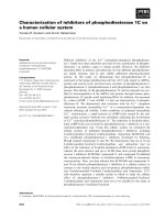
Fig. 4. Activity of the monomeric and dimeric forms of Artemia embryo
cathepsin L at different pH and temperatures. (A) CLP (dimer) and CL
(monomer) were assayed at d ifferent pH f or cathepsin L activity (rate
constants). Each reaction vessel contained 40–60 pmoles of the a ctive
protease. (B) Different forms of the protease (solid bars, CLP; unfilled
bars, CL) were incubated for 1 h at 30 °Cin25m
M
KCl, 10 m
M
sodium phosphate, 10% glycerol and 0.2 mgÆmL
)1
BSA at the pH
indicated, then assayed f or cathepsin L activity at pH 5 .0 a nd 30 °C
and the rate constants determined. The control represents CL
(monomer) a nd CLP ( dimer) maintained at 0 °C and pH 6.8 prior t o
the assay. (C) Incubation vessels were set up to contain 80–100 pmoles
of CL (m on omer) a nd CL P ( d imer) in pH 6.8 buffer as described in
(B). The vessels were incubated at 40 °C and aliquots were removed at
30 min intervals, assayed f or cathepsin L activity at pH 5.0, and their
rate constants d etermined.
Fig. 5. Sensitivity of various proteins to Artemia cathepsin L monomer.
Reaction vessels contained 50 m
M
sodium acetate, pH 5.0, 0.5 m
M
dithiothreitol, 2.4 lg of CL (monomer), and 12–14 lg of CLAP, BSA,
artemin or p26 in a final v olume of 4 0 lL. A fter 0 and 60 m in incu-
bation at 30 °C, 10 lL were t aken from each reaction ve ssel for ana-
lysis by SDS/PAGE on a 12% gel. The numbers above each lane
represent the incubation time of the monomer with proteins shown
above each lane. Left lane (mw) contains molecular mass standard
proteins with their molecular mass (kDa) shown at the left. The
migration position of t he Artemia cathepsin L monomer is shown at
the right (ACL ). Faint bands at 16–18 kDa in th e 6 0 m in lanes rep-
resent CL autodegradation p roducts o bserved in similar experiments
using Western blot ting.
4018 A. H. Warner et al .(Eur. J. Biochem. 271) Ó FEBS 2004
( 0.01%). Adult m ales had the lowest level (< 0.01%) o f
CL and CLAP of any tissue t ested.
Amino acid sequence of cathepsin L-associated protein
Early attempts to sequence CLAP by Edman degradation
yielded no results, suggesting that the N-terminus of the
protein was blocked. However, amino acid sequence was
obtained from a 25 kDa f ragment g enerated by CN Br
treatment of CLAP (DNVIDHEGKFTLFAPTNEAF),
and from a peptide (KSLIFSIK) generated by Lys-C
treatment of CLAP. More recently, we obtained the
sequence EAKNLVDLAESLGLSILVKALE from Edman
degradation of an untreated preparation of CLAP indica-
ting that the N-terminus of the mature protein begins with
a g lutamic acid r esidue. To obtain the full amino acid
sequence of CLAP, an Artemia cDNA library in phage
kZAP II was screened with a PCR derived probe and six
clones potentially coding for the CLAP were isolated.
Following excision of Bluescript phagemid from kZAP II,
two cloned cDNAs were amplified and sequenced (Fig. 7).
Clone 1 contained 1888 nucleotides with two potential start
codons (nucleotides 38–40 and 92–94) and a n open reading
frame of 996 nucleotides. Clone 2 contained 1870 nucleo-
tides with one potential start c odon (nucleotides 24–26) and
an open reading frame spanning 945 nucleotides. Clone 2
differed from clone 1 mainly in that it lacked a 68 nucleotide
sequence at the 5¢ end, including sequence coding for the
first 15 amino acids in clone 1. Also, at position 568 in clone
2 an A was substituted for a G changing the amino acid
from R t o K. B oth cDNA clones have a short 5¢
untranslated region, and extensive 3¢ untranslated regions
rich in A + T, representing nearly 45% of the mature
transcripts. The 3¢ UTR of c lones 1 and 2 are composed of
about 72% A + T and differed f rom each other by 2.1%.
Also, c lone 1 c ontains seven consensus AT-rich m otifs,
while clone 2 contains five AT-rich motifs. Both clones
contain several putative pol y(A) addition signals ( AAT
AAA and ATTAA). The nucleotide sequences of clone 1
and 2 have been entered into t he NCBI database with
accession numbers A Y307377 and A Y462276, r espectively.
Starting from the amino terminus of the mature protein
(E44) (Fig. 7), a calculated molecular mass of 32.3 kDa and
pI of 8.0 were obtained using
EXPASY
(http://www.
expasy.org/) if the m ature protein te rminated at Q332.
While the calculated molecular mass is close to that
observed by SDS/PAGE (31.5 kDa), the pI value is
distinctly different from the values (5.9–6.1) obtained by
IEF for mature CLAP. These observations suggested
further post-translational modifications occur, leading to
mature CLAP. A possibility could be that the protein is also
shortened at its C-terminus, which contains an excess of
basic residues (Fig. 7). Indeed, t runcation of t he C-terminus
by 16–26 residues leads to a predicted IEF for CLAP which
fits the observed data nicely. The conclusion that part of the
C-terminus is indeed missing is also supported by direct
amino acid sequence analysis of CLAP being cleaved by
CNBr because we could follow the sequences of all five
CNBr peptides expected (not shown), beginning with E44,
D70, K124, E264 and Q269, respectively. The C-terminal
CNBr peptide beginning with Q269 could be followed until
V301 (in cycle 33), suggesting that the C-terminus of the
deduced protein has been truncated at or a few residues after
V301 (see below).
A high stringency search of the NCBI Conserved Domain
Database ( revealed two
domains in Artemia CLAP with a h igh degree o f homology
with fasciclin I, an extracellular protein found in numerous
organisms. The first Fas I domain in clone 1 spans 128
amino acids (45–173), while the second Fas domain spans
132 amino acids (177–309) (Fig. 7). A nalysis of C LAP
cDNA (clone 1) using
EXPASY
revealed potential GTP
binding sites at positions 99–202 (DRAG) and 265–272
(GTTMQGKS) [35]. Having recognized CLAP as a mem-
ber o f the fas ciclin family of proteins, w e w ere i nterested t o
know whether the protein – like many o ther fasci clins – has
been modified C-terminally with a GPI moiety [36]. The
presence of such a moiety would possibly account in part for
the protein’s observed heterogeneity when analyzed by IEF.
Moreover, the truncation of the protein chain beyond the
site of GPI modification would be in line with the pool
sequencing r esults, which suggest only a few additional
residues after V301 (see above). In one experiment, treat-
ment of CLAP (fraction f) with PI-PLC altered the band
pattern o n an IEF gel (data not shown) suggesting that at
least one isoform terminated with a GPI unit. Overall, the
combined data indicate that the primary translation product
(prepro-CLAP) is processed at the N-terminus between G43
and E44, and probably at the C-terminus at D306, the latter
being one of the two weak sites i ndicated by the GPI
prediction tool (Discussion). A similar result would be
expected to occur during the processing of CLAP clone 2
translation product. Post-translational processing of pro-
CLAP at both th e N- and C-termini is required to achieve
the properties observed f or mature CLAP.
Fig. 6. Relative abundance of t he catalytic a nd noncatalytic su bunits of
CLP at different stages in the life cycle of Artemia. Protein extracts were
prepared from various tissues or whole organisms at different stages in
the life cycle of Artemia, then 33–135 lg were subjected to SDS/PAGE
and Western blot analyses along with five different concentrations of
purified Artemia cathepsin L in separate lanes. The solid bars represent
the noncatalytic subunit (CLAP), while the u nfilled bars represent t he
catalytic subunit (CL). EE, ovisacs containing e ncysted embryos
(33 lg protein); NEE, ovisacs containing nonencysted embryos (34 lg
protein); GF, gravid females somatic tissue (126 lg protein); NGF,
nongravid adult females (135 lg protein); M, adult males (135 lg
protein).
Ó FEBS 2004 Cathepsin L and cell adhesion protein in Artemia (Eur. J. Biochem. 271) 4019
Higher order structure of CLAP
The secondary structure o f CLAP w as predicted a ccording
to
PREDICTPROTEIN
available at Columbia U niversity Bioin-
formatics Center ( (Fig. 8).
For comparison the same figure i ncludes the secondary
structure of chain A, a fasciclin I domain of Drosophila
melanogaster, derived from its observed spatial structure
[37]. Both prote ins were aligned using the
CLUSTALW
program; they share substantial amounts of secondary
structural elements, and they are clearly related to each other
regarding t heir amino acid s eque nces (Fig. 9). Whe ther this
correspondence points to s imilar roles in extracellular
function of both p roteins r emains to be seen although they
probably h ave a common evo lutionary origin.
Discussion
Previous attempts to obtain Ar temia cathepsin L-associated
protein ( CLAP) in a n undenatured f orm had n ot been
CLAP_1:AATTCGGCACGAGGCAAAAACAAATAAATGCTTAATTATGTTGTATATTATTCCATTATTTCTTATTATTGGCTGCTCAAATGCCATATGGATGTTAAAT 100
CLAP_2:AATTCGGCACGAGG GCCATATGGATGTTAAAT 32
M L Y I I P L F L I I G C S N A I W M L N 21
CLAP_1:TTGAATGCTGTCACCACTGAGCCAGAAGCTAAGCTAGAACATGCTGCTATCCCTATCAAAGATGGTGAGGCAAAAAACCTTGTGGATCTTGCAGAGTCTC 200
CLAP_2:TTGAATGCTGTCACCACTGAGCCAGAAGCTAAGCTAGAACATGCTGCTATCCCTATCAAAGATGGTGAGGCAAAAAACCTTGTGGATCTTGCAGAGTCTC 132
L N A V T T E P E A K L E H A A I P I K D G E A K N L V D L A E S L
55
CLAP_1:TTGGACTGTCCATCCTTGTCAAGGCTCTTGAAGAAACTGGAATGGATAATGTGATTGATCATGAAGGTAAATTTACTTTATTTGCTCCAACTAATGAAGC 300
CLAP_2:TTGGACTGTCCATCCTTGTCAAGGCTCTTGAAGAAACTGGAATGGATAATGTGATTGATCATGAAGGTAAATTTACTTTATTTGCTCCAACTAATGAAGC 232
G L S I L V K A L E E T G M D N V I D H E G K F T L F A P T N E A
88
CLAP_1:ATTTAAAAGAATTCCCGAATGGGCCAAGGATCTTCCATTGAAAGAAGTTTTGAGGTATCACATTGCAAGAGGGTTGTATTATGATAAAGATCTCCAGAAT 400
CLAP_2:ATTTAAAAGAATTCCCGAATGGGCCAAGGATCTTCCATTGAAAGAAGTTTTGAGGTATCACATTGCAAGAGGGTTGTATTATGATAAAGATCTCCAGAAT 332
F K R I P E W A K D L P L K E V L R Y H I A R G L Y Y D K D L Q N 121
CLAP_1:GACATGAAACTGAGAACTCTCCTCACAAAGAGGGACTTGAGGATTAATTTGTATGACAATGGGCAGACAATTCTTGCCGGTGGGAAACGTATAAATGGAT 500
CLAP_2:GACATGAAACTGAGAACTCTCCTCACAAAGAGGGACTTGAGGATTAATTTGTATGACAATGGGCAGACAATTCTTGCCGGTGGGAAACGTATAAATGGAT 432
D M K L R T L L T K R D L R I N L Y D N G Q T I L A G G K R I N G S
155
CLAP_1:CAAATTATGAAGCTCACAATGGTGTTCTGCATCTCCTTGAAGATGTGATTGTCTCTATACCAGCACGACATGGAACAGTGATTCACCAGCTGAGAAGATG 600
CLAP_2:CAAATTATGAAGCTCACAATGGTGTTCTGCATCTCCTTGAAGATGTGATTGTCTCTATACCAGCACGACATGGAACAGTGATTCACCAGCTGAGAAGATG 532
N Y E A H N G V L H L L E D V I V S
I P A R H G T V I H Q L R R C 188
CLAP_1:TCCAGTTTTTTCTGATCTTGTGGAGCTCATTGATAGAGCAGGTCTTGATGAAGCTCTTCAAACCCATGGACCTATTACTTTCTTTGCCCCAAGCAATGAT 700
CLAP_2:TCCAGTTTTTTCTGATCTTGTGGAGCTCATTGATAAAGCAGGTCTTGATGAAGCTCTTCAAACCCATGGACCTATTACTTTCTTTGCCCCAAGCAATGAT 632
P V F S D L V E L I D R A G L D E A L Q T H G P I T F F A P S N D
221
K
CLAP_1:GTCATAAGGAAACTCCCTCCTGATGTGATTAAACACCTTGTTGATGACCCAGCTCTCCTAAAAGAAGTTTTAACCTACCATGTCTTGTCTGGAACCTTCT 800
CLAP_2:GTCATAAGGAAACTCCCTCCTGATGTGATTAAACACCTTGTTGATGACCCAGCTCTCCTAAAAGAAGTTTTAACCTACCATGTCTTGTCTGGAACCTTCT 732
V I R K L P P D V I K H L V D D P A L L K E V L T Y H V L S G T F Y
255
CLAP_1:ATTCTCCTGGCATTAAAGATGGAATGGAGGGAACCACGATGCAAGGAAAGAGTCTCATATTTTCAATCAAAGATGGTGAGGTTATAATCAACAGCAAGAC 900
CLAP_2:ATTCTCCTGGCATTAAAGATGGAATGGAGGGAACCACGATGCAAGGAAAGAGTCTCATATTTTCAATCAAAGATGGTGAGGTTATAATCAACAGCAAGAC 832
S P G I K D G M E G T T M Q G K S L I F S I K D G E V I I N S K T
288
CLAP_1:TAAGGTTACCAGTGCTGATTCCAACGCATCTAATGGTGTAATTCACAGCATTGATAATGTTCTAATTCCACCACAAATTCAAGCTAAGCTGAAGCGTCGA 1000
CLAP_2:TAAGGTTACCAGTGCTGATTCCAACGCATCTAATGGTGTAATTCACAGCATTGATAATGTTCTAATTCCACCACAAATTCAAGCTAAGCTGAAGCGTCGA 932
K V T S A D S N A S N G V I H S I D N V L I P P
Q I Q A K L K R R 321
CLAP_1:ATTCTGAAGAAATCGAGAGCATTTAGCTTCCAGTAG
AAAACGGTGGTTTCGTAGTGCTTTTCTTTTCCATGGGCGTGAATGTTTCTCATTTCTCTGGTGA 1100
CLAP_2:ATTCTGAAGAAATCGAGAGCATTTAGCTTCCAGTAG
AAAACGGTGGTGTCGTAGTGCTTTTCTTTTCCATGGGCGTGAATGTTTCTCATTTCTCTGGTGA 1032
I L K K S R A F S F Q * 332
CLAP_1:AAGTCTGTCGTCAAAATGTTATGAACGTCTCTTGTCATAAAGAAAGATAACCTCTCTTTTTAGTTTGGTTTAGATATTAAGGACAGATCCAAAATATTTG 1200
CLAP_2:AAGTCTGTCGTCAAAATGTTATGAACGTCTCTTGTCATAAAGAAAGAGAACCTCTCTTTTTAGTTTGGTTTAGATATTAAGGACAGATCCAAAATATTTG 1132
*
CLAP_1:AGGACCTTTTATTAGACATTTCAAATATATAATAAACGTTATTTTA
AAATTAGAAAAATTGAAAGACAAGCTAATGAAAGCTTATTGCCGATTGGAAAGT 1300
CLAP_2:AGGACCTTTTATTAGACATTTCAAATATATAATAAACGTTATTTTA
AAATTAGAAAAATTGAAAGACAAGCTAATGAAAGCTTATTGCCGATTGGAAAGT 1232
CLAP_1:TTGCTTGGGGGGAAGACTCGTTACAATTCTTTTTCTTTATTTTCTTTTTAGGTAGCTTCTTTATTTTATTTTTTT-A
TCTCTTTCTTGATTTTCTTTTCT 1399
CLAP_2:TTGCTTGGGTG-AAGACTCGTTACAATTCTTTTTCTTTATTTTCTTTTTAGGTAGCTTCTTTATTTTATTTTTTTTA
TCTCTTTCTTGATTTTCTTTTCT 1331
* * *
CLAP_1:GGCAACTTCTTTATATTTTTCTTATTTCTGTTCTTTATTTCTTTATTTTTTGAATAGTTTCTATTGCTATAGGATTAGCTTGTCTAAGTAAATTCTAAGT 1499
CLAP_2:GGCAGCTTCTTTATATTTTTCTTATTTCTGTTCTTTATTTCTTTATTTTTTGAATAGTTTCTATTGCTATAGGATTAGCTTGTCTAAGTAAATTCTAAGT 1431
*
CLAP_1:TTTTTTTTTTTTTTAATCAGAAAAACACTAGATTTCGTAAGATTAATGTGGGTTTCATGAAAACCTTTTTATTGACATT-TAAATAAATTGGGTTTTGCA 1598
CLAP_2:TTTTTTTTTTTTTAAATCAGAAAAACACTAGATTTCGTAAGATTAATGTGGGTTTCATGAAAACCTTTTTATTGACATTCTAAATAAATTGGGTTTTGCA 1531
* *
CLAP_1:CAAGTTTCTTGGACTTTA-GAAAAGTATGTTTAATTTTTCATAAGAATGTCTAAGGTTTCGTATTTTTTTA
CACAAATACTTCAACCGAGAGGATTCCAT 1697
CLAP_2:CAAGTTTCTTAGACTTTAAGAAAAGTATGTTTAATTTTTCATAAGAATGTCTAAGGTTTCGTAATTGTT-ACACAAATACTTCAATCGAGAGGACTCCGT 1630
* * * * * * * *
CLAP_1:ATTAGTGCTATAGTTTGGGAAATATTTA
GCCCTTGTTTTGTGTGATCTTATAAGATAATATTTGTAGTTTGTGCTTTTATATAATTTAGCTCATTGGATT 1797
CLAP_2:ATTAGTGCTATAGTTTGGGAAATATTTAGTCCTTGTTTTGTGTGATCTTATAAGATAATATTTGTAGTTTGTGCTTTTATATAATTTAGCTCATTGGATT 1730
*
CLAP_1:AA
GATCTTCTGAATGTGATTATATGCGGCTGTGTTTTCTAATAGATTTCTAGATACGAAAAAAAAAAAAAAAAAAAAAAAAAAAAAAAAAA 1888
CLAP_2:AAGATCTTCTGAATGTGATTATATGCGGCTGTGTTTTCTAATAGATTTCTAGATACGAAAAAAAAAAAAAAAAAAAAAAAAAAAAAAAAAAA(A)
48
1870
Fig. 7. Nucleotide and deduced amino acid sequence of two cDNA clones coding for CLAP. Clones 1 and 2 were found to be 95% identical except for
a gap of 68 nucleotides near the 5¢-end of clone 2. Amino acid sequences determined by Edman degradation are shown in bold, and each is a perfect
match with the deduced amino acid sequence. The putative start (ATG) and stop (TAG) translation sites are double-underlined. Two fasciclin I-like
domains are underlined, and two potential N-glycosylation sites are boxed. The double-underlined and bold sequence near the 3¢-end of clones 1
and 2 (ATTAA) are putative poly(A) recognition sites. Two potential GTP binding sites are present at 199–202 (DRAG) and 265–272
(GTTMQGKS) in clones 1 and 2. Putative destabilizing elements ( AU/T-rich) in the 3¢ UTR a re underlined. The arrowheads represent putative
cleavage sites in prepro-CLAP. The asterisks represent sites in the noncoding region of clones 1 and 2 where mismatched nucleotides are present.
4020 A. H. Warner et al .(Eur. J. Biochem. 271) Ó FEBS 2004
successful [6,23,24]. In the p resent study we found that
chromatography of the CLP complex on a high perform-
ance cation matrix (Mono S) yielded both CL and CLAP in
a high s tate of purity. However, dissociation of CLP to its
subunits (CL and CLAP) required incubation of CLP at pH
5 for at least 1 h at 4 °C prior to chromatography on Mono
S. Dissociation of CLP could be blocked by inclusion of
Z-Phe-Ala-CH
2
F, a reversible cysteine protease inhibitor, in
CLP preparations, suggesting that CLAP was modified in
the process of i ts separation from CL. A ttempts to
recombine CL and CLAP into an active CLP c omplex
after purification on Mono S have not been successful. Thus
the mechanism of CLAP binding to the catalytic subunit
resulting i n CL s tabilization appears t o be c omplex and not
yet understood. CLAP might prevent ÔunzippingÕ or
destablization of the active site region of cathepsin L at
higher than normal temperature and pH as suggested for
cathepsin B [38]. The increased stability o f CL in the CLP
complex is consistent with the adaptive nature of Artemia
embryos which have the ability to survive harsh environ-
mental conditions [39].
Analyses of CLAP on IEF gels revealed four isoforms
with pI values ranging from 5.9 to 6.1. Staining for
phosphate adducts on the Artemia CLP ( heterodimer) and
purified CLAP were negative (data not shown). The
presence of N-linked carbohydrates in CLAP dem onstrated
previously could g enerate CLAP i soforms [24], but this idea
needs further experimentation. Another possible r eason f or
the isoforms is that the C-terminus of mature CLAP
contains heterogeneous GPI units resulting from a post-
translational event as discussed below.
As demonstrated in this study the activity of CLP and CL
was similar between pH 5 and 6.5, while CLP showed about
twofold greater activity below pH 5. CLP was also more
stable than CL near neutral p H a nd 30–40 °C. Addition of
purified CLAP, i n equimolar amounts, to reaction mixtures
containing CL did not affect the protease activity at pH 4 , 5
or 6. Reasons for these ob servations are not clear, but they
may be d ue to modifications in CLAP during incubation of
CLP at pH 5 prior to chromatography on Mono S. The fact
that we have not been able to a chieve recombination of CL
and CLAP to form the naturally occurring CLP complex
in vitro is consistent with the latter observation.
A s earch o f the literature has revealed that heterodimeric
CLP in Artemia is functionally similar to a novel cysteine
protease in Entamoeba histolytica known as adhesin [40].
Adhesin is a heterodimer composed of a cathepsin L-like
protease and a protein with an adherence domain contain-
ing four glycosylation sites. Adhesin promotes the binding
of E. histolytica phagocytic trophozoites to target (host)
cells such as erythrocytes, which are then consumed by
phagocytosis and degraded by the associated cathepsin L.
Cysteine proteases such as CL are used frequently by
parasitic organisms to promote i nvasion a nd destruction of
target organisms [19].
From a search of he Conserved Domain Database
(NCBI) the similarity of the t wo fasciclin domains in CLAP
with other fasciclin I containing proteins is clear (Fig. 9).
Using
BLAST
( to
identify related proteins, a putative cell adhesion protein
from the sea anemone Anthopleura elegantissima showed the
highest identity with CLAP. Other proteins of relevance
were HLC-32, a protein secreted into the extra-embryonic
matrix of sea urchins at fertilization [41], and a 30 kDa yolk
granule p rotein in sea urchins [42]. However, the sea urchin
protein self-dimerizes, while CLAP, as a component of the
Fig. 8. Structural co mparisons between
Drosophila fascicl in I and CLAP. The a mino
acid se quence of Drosophila fasciclin chain A
(NCBI database entry 1070), was aligned with
the proposed (mature) translation product, i.e.
with polypeptide 44–306, of clone 1 o f CLAP
using the
CLUSTALW
multiple alignment p ro-
gram. The d eterm ined secondary structure
(alpha helices and beta strands), as b ased on
the spatial structure of t he Drosophila protein
[37], is indicated by single an d double under-
lining, respectively. The predicted secondary
structure of CLAP according to
PREDICT-
PROTEIN
is presented in the same way.
Ó FEBS 2004 Cathepsin L and cell adhesion protein in Artemia (Eur. J. Biochem. 271) 4021
β
β
β
β
Fig. 9. Comparison of the fasciclin I domains in CLAP with selected proteins in the protein database. The Conserved Domain Database of NCBI was
screened with the protein coding sequence of clone 1 of CLAP and the Fas I domains in the protein compared with 10 other fasciclin I-containing
proteins. The most highly conserved sequences (containing more than four amino acids) are boxed, and the number below each highly conserved
sequence indicates the percent identity to Artemia CLAP with the consensus sequence for each region of the fasciclin domains. Because the fasciclin
domains in the Conserved Domain Database are compared with only one of the four domains present in the Drosophila protein [37], its sequence
501–616 appears twice in the figure. In addition, it should be noted that the alignment between Artemia and Drosophila proteins also differs from
Fig. 8, because, in the latter figure, a different computer program (
CLUSTALW
) was used. Details of each sequence above can be found in the NCBI
protein s equ ence database as f o llows: Art-clap1 (Artemia cathepsin L-associated pro tein, c lone 1, AAP69998), Dros-fasI (Drosophila fasciclin I,
NP_732166), Ory-big-H3 (Oryctolagus, rabbit, transforming growth factor-b induced protein precursor, Q95215), Hom o-osf2 (Homo sapiens,
osteoblast s pecific factor 2, S36111), A ntho-cap (Anthopleura e legantissima putative c ell adhesion p rotein Sym32, A AF65308), Scoel-lipo ( Strep-
tomyces coelicolor A3 putative lipoprotein, NP_624948), Rdur-osf2 (Deinococcus radiodurans osteoblast specific factor-2 related protein,
NP_294122), Lyt-30kDaYP (Lytechinus va riegatu s 30 kDa yolk granule protein, AAG02421), Mus-tgf-bi(Mus musculus transforming growth
factor-b induced protein IG-H3 precursor, Q95215), Mus-osf2 (Mus musculus osteoblast specific factor-2 pending protein, AAH31449), Smel-ind-
pr (Sinorhizobium meliloti Nex 18 symbiotically induced conserved protein, NP_435828), and Syn-hypo-pp (Synechocystis sp. hypothetical protein
s111483 pre cursor, P74615).
4022 A. H. Warner et al .(Eur. J. Biochem. 271) Ó FEBS 2004
heterodimeric CLP at the surface of yolk platelets [3],
appears to dimerize (in vivo ) only with CL.
The function of the Fas I domains in CLAP is unknown,
but generally Fas I domains are thought to represent ancient
cell adhesion domains [37]. Of importance to understanding
the structure and function of CLAP, is that most proteins
containing Fas I domains are anchored to cell membranes
through a GPI unit at the C-terminus of the protein [36].
Thus, while the GPI Predictor tool (.
univie.ac.at/sat/gpi/gpi_server.html) did not show a GPI
attachment site near the C-terminus of pro-CLAP, t he
possibility exists that mature CLAP is terminated with a
GPI unit a t N299 o r D306, w eak sites identified b y the GPI
Predictor tool. The observation that PI-PLC produced an
altered band pattern in CLAP suggests that a GPI unit is
present at the C-terminus. Addition of GPI, if it occurred,
would b e accompanied by cleavage o f the highly basic
peptide chain behind the modified residue [43]. Such a
modification of pro-CLAP would result in a predicted
molecular mass closer to that observed for mature CLAP by
SDS/PAGE (31.5 kDa), and a n i soelectric point in the
range of values observed by IEF (p I 5 .9–6.1). Processing of
the pro-CLAP C-terminus is essential to lower the mole-
cular mass and pI of the protein to values observed by SDS/
PAGE and IEF. Interestingly, pool sequencing of the
mixture o f CNBr pep tides generated from CLAP revealed
in the C-terminal CNBr peptides, the presence of N299,
G300, and V 301 i n s equence c ycles 31–3 3, with V301 being
the last visible residue of this peptide. Thus, because N299 is
observed in the C-terminus in an internal position, we infer
that the other candidate, D306, is the target for GPI
modification and site of C-terminus truncation.
In some systems, the addition of GPI to the C-terminus of
a protein is an energy dependent process requiring ATP
and/or GTP [ 43]. T he f act t hat C LAP contains an intrinsic
ATP/GTP binding site near its C-terminus might support
this type of post-translational modification. The presence o f
GPI at the C-terminus of CLAP provides a m echanism to
anchor heterodimeric C LP at various sites w here it is found
in embryos and newly hatched larvae [3].
Previous analysis of cDNA clones coding for cathep-
sin L, and the sequence of clone 1 coding for CLAP from
the first AUG codon onward, indicate that the prepro-
form of both CL and CLAP have well defined h ydro-
phobic signals in their N-terminus (Fig. 7) [24]. Thus
prepro-CL a nd p repro-CLAP p robably enter the e ndo-
plasmic r eticulum where post-translational modifications
occur. The assembly of CL and CLAP to form the
heterodimer could also occur in the ER, although the
bonds or motifs linking the two subunits have not been
determined. Modifications to the predicted amino acid
sequence of prepro-CLAP, including removal of N- and
C-terminal peptides, w ould probably be a chieved a long the
ER/Golgi pathway. Alternatively, prepro-CLAP could
avoid the ER by using an a lternate start codon in the
mRNA (positions 92–94, clone 1), but this is unlikely as
both the N- and C-termini of prepro-CLAP must undergo
post-translational modifications that are normally accom-
plished a long the ER/Golgi pathway. H owever, t ranslation
of clone 2 from t he firs t start codon (positions 22–24)
would result in a pro-CLAP that would avoid trafficking
through the ER/Golgi complex.
Immunocytochemical and cell fractionation methods
demonstrated that considerable amounts of CLP reside at
the surface of yolk platelets in Artemia, but that the pathway
that CLAP , CL or CLP takes to the surface of platelets is
unknown. While we can only speculate at this time about
the mechanism of C LP attachment to yolk platelets, neither
lysosomes nor transport v esicles are visible at t he surface of
mature platelets [44]. However, electron microscopy has
shown that yolk platelets acquire a vesiculated periphery
during vitellogenesis which may represent the uptake of
vesicles containing CLP derived from the ER/Golgi path-
way [ 3,44]. T he fact that yolk platelets i n s ea urchin possess
a 30 kDa fasciclin-co ntaining p rotein with a high d egree o f
homology with Artemia CLAP is noteworthy [42].
Considerable CLP has been detected in extracellular
regions of embryos a nd in tissues of l arvae, especially in the
developing gut [3]. T ransport of CLP to extracellular sites
probably requires molecular signals different from those
that direct transport of C LP to th e surface o f yolk platelets.
How this m ight occur is speculative, but it should be n oted
that the C-terminus of Artemia CL contains a secretion
signal (ASYPLV) nearly identical to signals that promote
CL secretion in mammalian tissues [24,45] and parasitic
nematodes [ 2]. Localization o f CLP in the e xtracellular
matrix could occur through the Fas I domains or putative
GPI unit, if one exists in CLAP as it does in Drosophila
fasciclin I and many other fasciclin-containing proteins
[36,46]. Fas I domains in proteins are a lmost always found
in the e xtracellular compartment of tissues, where they are
believed to promote intermolecular and homotypic adhe-
sion. Thus CLAP, through i ts Fas I domains, may promote
docking and stabilization of CL at various extracellular
sites. The r esistance o f mature C LAP t o d estruction by CL
and serine proteases appearing in third and fourth instar
larvae of Artemia suggests that CLAP plays an important
role in CL stability and localization outside lysosomes
during embryonic and early larval development [12].
Analysis of the cDNA c lones coding for C LAP indicated
that each clone has an extensive, but slightly different 3¢
UTRs rich in AT-residues, representing AU-rich r egions in
CLAP mRNA. AU-rich sequences in eukaryotic mRNA
are thought to represent destabilizing elements leading to
rapid deadenylation and messenger breakdown [47,48].
Thus, while CLAP appears to be somewhat refractory to
protease degr adation, its mRNAs m ay b e degraded rapidly
due to AU-rich s equences in their 3 ¢-UTR. Preliminary
evidence from our laboratory on CLAP mRNA levels in
developing embryos and larvae of Artemia supports the
view that CLAP mRNA is unstable during development. In
addition, we have obser ved that the 3¢ UTR of CLAP
mRNA contains over 125 transcription factor binding sites
as determined by the molecular tool
MATINSPECTOR
(http://
www.genomatrix.de/) [49]. Whether these sites participate
in formation of a functional promoter for transcription of
the CLAP gene or in transcription regulation of down-
stream genes remains to be determined.
Finally, we have not yet investigated the potential
importance of the nucleotide binding domain in CLAP,
but the p resence of this domain suggests a n energy-
dependent mechanism for CLP translocation to various
sites in embryos and l arvae or for C-terminus modification
[43]. Fas I containing proteins generally lack nucleotide
Ó FEBS 2004 Cathepsin L and cell adhesion protein in Artemia (Eur. J. Biochem. 271) 4023
binding domains, so the presence of a GTP/ATP binding
region in CLAP may indicate an energy dependent mech-
anism for pro-CLAP processing or cathepsin L docking
and stabilization not found in other organisms.
Acknowledgements
The authors wish to thank the Natural Sciences and Engineering
Research Co uncil of Canada for the ir financial support of t his s tudy.
We also wish to thank Dr T homas MacRae of Dalhousie University
and Dr Dora Cavallo-Medved of Wayne State University for their
critical reading a nd comments of an ea rlier version of this work.
References
1. Miyata, S. & K ubo, T. (1997) Inhibition of gastrulation in
Xenopus embryos by an antibody against cathepsin L-like pro-
tease. Dev. Growth Differ. 39, 111–115.
2. Britton, C. & Murray, L. (2002) A c athepsin L p rotease essential
for Caenorhabditis elegans embryogenesis is functionally
conserved in parasitic n ematode s. Mol. Biochem. Parasitol. 122,
21–33.
3. Warner, A.H., Perz, M .J., Osahan, J .K. & Ziel inski, B.S. (1995)
Potential ro le i n development of the major cysteine protease in
larvae of the brine shrimp Artemia franciscana. Cell Tissue Res.
282, 2 1–31.
4. Fagotto, F. (1990) Yolk degradationintickeggs.II.Evidencethat
cathepsin L-like proteinase is stored as a latent, acid-activable
proenzyme. Arch. Insect Biochem. Physiol. 14, 237–252.
5. Wood, A.W. & Van Der Kraat, G. (2003) Yolk proteolysis in
rainbow trout oocytes afterserum-free culture: evidence for a novel
biochemical m ec hanism of atresia in oviparous vertebrate s. Mo l.
Reprod. D ev. 65, 219–227.
6. Warner, A.H. (1987) The role o f proteases and their control in
Artemia development. In Artemia Research & its Applications.
Physiology, Biochemistry, Molecular Biology. (Decleir, W., Moens,
L., S legers, H., Ja spers, E & Sorgeloos, P., eds), Vol. 2. pp. 203 –
219. Uni versa Press, Wetteren, Belgium.
7. Matsumoto, I., Watanabe, H., Abe, K., Arai, S. & Emori, Y.
(1995) A putative digestive cysteine proteinase from Drosophila
melanogaster is predominantly expressed in the embryonic and
larval midgut. Eur. J. Biochem. 22 7, 582–587.
8. Homma, K., Kurata, S. & Natori, S. (1994) Purification, char-
acterization, a nd cDNA cloning o f procathepsin L from the cul-
ture medium of NIH-Sape-4, an embryonic cell line of Sarcophaga
peregrina (flesh fly), and its involveme nt in the d ifferentiation o f
imaginal d iscs. J. Biol. Chem. 26 9 , 15258–15264.
9. Tobin, D.J., Foitzik, K., Reinbeckel, T., Mecklenburg, L.,
Botchkarev, F.A., Peters, C. & Paus, R. (2002) The lysosomal
protease cathepsin L is an important regulator of keratinocyte and
melanocyte differentiation during hai r follicle morphogenesis a nd
cycling. Am. J . Pathol. 160, 1807–1821.
10. Vogel, A.M. & Gerster, T. (1997) Expression of a zebrafish
cathepsin L gene in anterior mese ndode rm and h atching gland.
Dev. Gen es Evol. 20 6 , 477–479.
11. Cheon, Y.P., DeMayo, F.J ., Bagchi, M.K. & Bagchi, I.C. (2003)
Induction of cytotoxic T-lymphocyte antigen-2 beta, a cysteine
protease inhibitor in deciduas: a potential regulator of embryo
implantation. J. Biol. Chem. 144, 5623–5630.
12. Warner, A.H. & Matheson, C. ( 1998) Re lease of p roteases from
larvae of the brine shrimp Artemia franciscana and their potential
role during the molting process. Comp.Biochem.Physiol.119B,
255–263.
13. Hashmi, S., Britton, C., Lie, J., Guiliano, D.B., Oksov, Y. &
Lustigman, S. (2002) Cathepsin L is essential for embryogenesis
and development of Caenorhabditis elegans. J.Biol. Chem. 277,
3477–3486.
14. Kirschke, H. (1998) Cathepsin L. In Handbook of Proteolytic
Enzymes (Barrett, A.J., Rawlings, N.D. & Woessner, J.F., eds),
pp. 617– 624. Academic P ress, San D iego.
15. Chauhan, S.S., Popesco, N.C., Ray, D., Fleischmann, R., Got-
tesman, M.M. & Troen, B.R. (1993) Cloning, genomic organiza-
tion, and chromosomal localization of human cathepsin L. J. Biol.
Chem. 268, 1039–1045.
16. Goulet, B., Baruch, A., Moon, N S., Poirier, M., Sansregret, L.L.,
Erickson, A., Bogyo, M. & Nepveu, A. (2004) A cathepsin L
isoform that is devoid of a signal peptide localizes to the nucleus in
S phase and processes the CDP/Cux transcription factor. Mol.
Cell 14, 2 07–219.
17. Lennon-Dumenil, A.M., Roberts, R.A., Valentijn, K., Driessen,
C.,Overkleeft,H.S.,Erickson,A.,Peters,P.J.,Bikoff,E.,Ploegh,
H.L. & W olf, B.P. (2001) The p41 isoform of inv ariant chain is a
chaperone for cathepsin L. EMBO 20 , 4055–4064.
18. Que, X. & Reed, S.L. (2000) Cysteine proteinases and the
pathogenesis of amebiasis. Clin. M icrobiol. Rev. 13 , 196–206.
19. Mundodi, V., Somanna, A., Farrell, P.J. & Gedamu, L. (2002)
Genomic o rgan ization and functional expression of differentially
regulated cysteine protease genes of Leishmania donovani complex.
Gene 282 , 257–265.
20. Deval, C., Mordier, S., Obled, C.,Bechet,D.,Combaret,L.,
Attaix, D. & Ferrara, M. (2001) Identification of cathepsin L as a
differentially expressed message associated with skeletal muscle
wasting. Biochem. J. 360, 143–150.
21. Bervar, A., Zajc, I., Sever, N., Katunuma, N., Sloane, B.F. & Lah,
T.T. (2003) Invasiveness of transformed human breast epithelial
cell lines i s r elated to cathepsin B a nd i nhibit ed by c ystein e p ro-
teinase inhibitors. Biol. C hem. 384, 447–455.
22. Stypmann, J., Glaser, K., Roth , W ., Tobin, D.J., P etermann , I.,
Matthias, R., Monnig, G., Haverkamp, W., Breithardt, G.,
Schmahl, W., Peters, C. & Reinheckel, T. (2002) Dilated
cardiomyopathy in mice de ficient for the lysosomal cysteine
peptidase cathepsin L. Pr oc. Natl A cad. Sci. U SA 99, 6 234–6239.
23. Warner, A.H. & Shridhar, V. (1985) Purification and character-
ization of a cytosol protease from dormant cysts of the brine
shrimp Artemia. J. Biol. Chem. 260, 7008–7014.
24. Butler, A.M., Aiton, A.L. & Warner, A .H. (2001) Characteriza-
tion of a novel heterodimeric cathepsin 1-like protease and cDNA
encoding the catalytic subunit of the protease in embryos of
Artemia f ranciscana. Biochem. Cell Biol. 79, 43–56.
25. Lu, J. & Warner, A.H. (1991) Immunodetection of thiol protei-
nase levels in various populations of Artemia cysts and during
development. Biochem. Ce ll Biol. 69 , 96–101.
26. Bradford, M . (1976) A rapid and sensitive method for the quan-
titation of microgram quantities of protein utilizing the principle
of pr otein-dye binding. Anal. Biochem. 72 , 248–254.
27. Smith, P.K., Krohn, R.J., Hermanson, G.T., Mallia, A.K.,
Gartner, F.H., Provenzano, M.D., Fujimoto, E.K., Goeke, N.M.,
Olson, B.J. & Klenk, D.C. (1985) Measurement of p rotein using
bicinchoninic a cid. Anal . Biochem. 150, 76–85.
28. Barrett, A.J. & Kirschke, H. (1981) Cathepsin B, cathepsin H and
cathepsin L. Metho ds Enzymol. 80 , 535–561.
29. Barrett, A.J., K embhavi, A.A., Brown, M .A., Kirschke, H.,
Knight, C .G., Tam a i, M. & &Hanada, K . (1982) 1 -trans-Epoxy-
succinyl-leucylamido (4-guanidino) butane ( E-64) and its analo-
gues as inhibitors of cysteine proteinases including cathepsins B, H
and L. Biochem. J. 201, 1 89–198.
30. Warner, A.H., Brunet, R.T., MacRae, T.H. & Clegg, J.S.
(2004)ArteminisanRNA-bindingproteinwithhighthermal
stability and potential RNA chaperone activity. Arch. Biophys.
424, 1 89–200.
4024 A. H. Warner et al .(Eur. J. Biochem. 271) Ó FEBS 2004
31. Takos, A.M., D ry, I .B. & Soole, K.L. (2000) G lycosyl-phospha-
tidylinositol-anchor addition signals are processed in Nicotiana
tabacum. Plant J. 21, 4 3–52.
32. Laemmli, U.K. (1970) Cleavage of structural proteins during
assembly of the h ead of b acteriophage T4. Nature 227, 6 80–685.
33. Warner, A.H. & McCle an, D.K. (1968) Studies o n the biosynth-
esis and role of diguanosine tetraphosphate during growth and
development of Artemia s alina. Dev. Bio l. 18, 278–293.
34. Sambrook, J., Fritsch, E.F. & Maniatis, T. (1989) Molecular
Cloning: a Laboratory Manual, 2nd edn. Cold Spring Harbor
Laboratory, Cold Spring Harbor, NY.
35. Sprang, S.R. (1997) G protein mechanisms: insights from s truc-
tural a nalysis. Annu. Rev. Biochem. 66, 6 39–678.
36. Wang, W.C., Zinn, K. & Bjorkman, P.J. (1993) Expression and
structural studies of fasciclin I, an insect cell adhesion molecule.
J. Biol. Chem. 268, 1448–1455.
37. Clout, N .J., Tisi, D. & Hohenester, E. (2003) N ovel fold revealed
by the structure of a FAS 1 domain pair from the insect cell
adhesion molecule fasciclin I. Structure 11 , 197–203.
38. Turk,B.,Dolene,I.,Zerovnik,E.,Turk,D.,Gubensek,F.&
Turk, V. ( 1994) Hu man cathepsin B is a metastable enzy me sta-
bilized by specific ionic interactions associated with the active site.
Biochemistry 33 , 14800–14806.
39. Clegg, J.S. & Trotman, C.N.A. (2002) Physiological and bio-
chemical aspects of Artemia ecology. In Artemia. Bas ic and
Applied Biology (Abatzopoulos, T.J., Beardmore, J.A., Clegg, J.S.
& Sorgeloos, P ., eds), pp. 129–170. Kluwer, Dordrecht, t he
Netherlands.
40. Garcia-Riv e ra, G. , R od rigu ez, M.A., Oc adi z, R ., M ar tin ez-
Lopez,M.C.,Arroyo,R.,Gonzalez-Robles,A.&Orozco,E.
(1999) Entamoeba histolytica: a novel cysteine protease and an
adhesin form the 112 kDa surface protein. Mol. Microbiol. 33,
556–568.
41. Brennan, C. & Robinson, J.J. (1994) Cloning and characterization
of HLC-32, a 32-kDa protein component of the sea urchin
extraembryonic matrix, the hyaline layer. Dev. Biol. 165, 556–565.
42. Wessel, G.M., Zaydfredim, V., Hsu, Y.I., Laidlaw, M. & Brooks,
J.M. (2000) Direct molecular interaction of a conserved yolk
granule p rotein in sea urchins. De v. Growth Diffe r. 42, 507–517.
43. Amthauer,R.,Kodukula,K.,Brink,L.&Udenfriend,S.(1992)
Phosphatidylinositol-glycan (PI-G)-anchored membrane proteins:
requirement of ATP and GTP for translation-indepen dent
COOH-terminal processing. Proc. Natl A cad. Sci. USA 89,
6124–6128.
44. Warner, A.H., Chu, P.P.Y., Shaw, M.F. & C riel, G. (2002) Yolk
platelets in Artemia embryos: are they really storage sites of
immature mitochondria? Comp.Biochem.Physiol.B132, 491–450.
45. Chauhan, S.S., Ray, D., Kane, S.E., Willingham, M.C. &
Gottesman, M.M. (1998) Involvement of carboxy-terminal amino
acids in secretion of human l ysoso mal protease c athepsin L. Bio-
chemistry 37, 8584–8594.
46. McAllister, L., Goodman, C.S. & Zinn, K. (1992) Dynamic
expression of the cell adhesion molecule fasciclin I during em-
bryonic development in Drosophila. Development 115 , 267–276.
47. Chen, C Y.A. & Shyu, A B. (1995) AU rich elements: char-
acterization and i m portance i n m RNA d egradation . Trends B io-
chem. Sci. 20 , 465–470.
48. Neininger, A., Kontoyiannis, D.,Kotlyarov,A.,Winzen,R.,
Eckert,R.,Volk,H D.,Holtmann,H.,Kollias,G.&Gaestel,M.
(2002) MK2 targets AU-rich e lementsandregulatesbiosynthesis
of tumor necrosis factor and int erluk in-6 independently at differ-
ent post-transcriptional l evels. J. Biol. C he m. 277, 3 065–3068.
49. Quandt, K., Frec h, K. , Karas, H., Winge n der, E. & W errner, T.
(1995) MatInd and MatInspector – new fast and versatile tools for
detection of consensus matches in nucleotide sequence data.
Nucleic A cids Res. 23, 4878–4884.
Ó FEBS 2004 Cathepsin L and cell adhesion protein in Artemia (Eur. J. Biochem. 271) 4025