Báo cáo khoa học: Testosterone 1b-hydroxylation by human cytochrome P450 3A4 pdf
Bạn đang xem bản rút gọn của tài liệu. Xem và tải ngay bản đầy đủ của tài liệu tại đây (346.47 KB, 8 trang )
Testosterone 1b-hydroxylation by human cytochrome P450 3A4
Joel A. Krauser
1
, Markus Voehler
2
, Li-Hong Tseng
3
, Alexandre B. Schefer
3
, Markus Godejohann
3
and F. Peter Guengerich
1
1
Department of Biochemistry and Center in Molecular Toxicology, Vanderbilt University School of Medicine and
2
Department of
Chemistry and Center in Molecular Toxicology, Vanderbilt University, Nashville, TN, USA;
3
Bruker Bio-Spin GmbH, Rheinstetten,
Germany
Human c ytochrome P450 3A4 forms a series o f minor
testosterone hydroxylation products in addition to 6b-hy-
droxytestosterone, the major product. One of these, formed
at the next highest rate after the 6 b-and2b-hydroxy prod-
ucts, was identified as 1b-hydroxytestosterone. This p roduct
was characterized from a mixture of testosterone oxidation
products using an HPLC-solid phase extraction-cryoprobe
NMR/time-of-flight mass spectrometry system, with an
estimated total of 6 lg of t his product. Mass spectrometry
established the formula as C
19
H
29
O
3
(MH
+
305.2080). The
1-position of the added hydroxyl group was established by
correlated s pectroscopy and heteronuclear spin quantum
correlation experiments, and the b-stereochemistry of the
added hydroxyl group was assigned with a nuclear Over-
hauser correlated spectroscopy experiment (1a-H). Of
several human P450s examined, only P450 3A4 formed this
product. The p roduct was also formed in human liver
microsomes.
Keywords: cytochrome P450; NMR spectroscopy; HPLC-
NMR combinations; testosterone.
Cytochrome P450 (P450; also termed heme-thiolate P450
[1]) enzymes h ave long been of interest because of their roles
in steroid metabolism [2,3]. These oxidations are most
critical in steroidogenic tissues, and a set of 12 P450s are
most important [4,5]. The hepatic P450s have also been
studied extensively in the context of their abilities to
hydroxylate steroids, even though few of the o xidations
involve the generation of products with distinctive biological
activities. Seminal in this area is the work o f C onney a nd his
associates, who studied the hydroxylation of testosterone in
rat liver systems a nd develope d the hypothesis t hat d ifferent
hydroxylations are catalyzed by individual P450 enzymes
[6]. Subsequently testosterone hydroxylation patterns have
been utilized extensively as probes of the presence and
function of individual rat liver P450s [7–11].
Testosterone hydroxylation has also been studied exten-
sively with human liver microsomal P450s. Early work with
liver microsomes resulted in reports of hydroxylation at the
2b,6a,6b,7a,15b,16a, and 17 positions (17-hydroxylation
yields the ketone androstenedione) [12–17]. A human liver
P450 was isolated that w as shown to b e t he maj or
6b-hydroxylase [18]; this P450 was originally termed nifedi-
pine oxidase (P450
NF
) and subsequently named P450 3A4.
Other work confirmed the role of P450 3A4 as the major
enzyme involved in testosterone 6b-hydroxylation [19].
Other P450 3A subfamily enzymes (3A5, 3A7, 3A43) can
also catalyze this reaction [20,21].
Testosterone hydroxylation is i n general use tod ay as one
of the characteristic assays of P450 3A4, which was
subsequently shown to be the most abundant P450 in
human liver and small intestine [ 22] and involved in t he
oxidation of approximately one-half t he drugs used today
[23]. The major product is 6b-hydroxytestosterone [18,19]
but several other hydroxylations occur, including those at
the 2b and 1 5b positions [18,19] (Fig. 1). Not a ll of the
products have been identified, however. In t he course of our
investigations we noted that a peak (X) formed at a rate in
the o rder 6b >2b >X 15b (hydroxylation) had not been
characterized and did not correspond to any of our
standards available in our set, including 2a-, 2 b-, 6a-, 6b-,
11b-, 15b-,16a-, or 16b-hydroxytestosterone or androsten-
edione. We utilized an HPLC-solid phase extraction (SPE)-
cryoprobe NMR/MS system and now provide a full
spectral characterization o f t his p roduct f rom 6 lgof
the p roduct i njected. The product is 1 b-hydroxytestosterone
(Scheme 1) and is formed only by P450 3A4, of the set of
human P450s examined.
Experimental procedures
Chemicals
GC-grade acetonitrile (CH
3
CN) and HPLC-grade H
2
Ofor
the separation (combined HPLC-MS-NMR) was from
Merck (Darmstadt, Germany). CD
3
CN and CD
3
OD (both
99.8% deuterium-enriched) were from Deutero GmbH
Correspondence to F. P. Guengerich, Department of Biochemistry and
Center in Molecular T oxicology Vanderbilt Un iversity School of
Medicine Nashville, Tennessee 37232–01 46, USA.
Fax: +1 615 3223141, Tel.: +1 615 3222261,
E-mail:
Abbreviations: P450, cytochrome P450 (a lso termed h eme-thiolate
P450, substrate, reduced flavoprotein: oxygen o xidoreductase);
HSQC, heteronuclear spin quantum correlation; SPE, solid phase
extraction.
Enzymes: P450, substrate, reduced flavoprotein:oxygen
oxidoreductase (EC 1.14.14.1).
(Received 16 July 2004, accepted 19 August 2004)
Eur. J. Biochem. 271, 3962–3969 (2004) Ó FEBS 2004 doi:10.1111/j.1432-1033.2004.04339.x
(Kastellaun, Germany). T estosterone (Sigma -Aldrich, St.
Louis, MO, USA) was used without further purification.
Hydroxytestosterone standards were purch ased from
Stearoaloids (Newport, RI, USA).
Enzymes
Microsomes were prepared [24] from a h uman liver sample
(denoted HL 97), which had been used in some previous
investigations in this laboratory [25]. Recombinant
P450 3A4 was used either in the form of Escherichia coli
membranes in w hich both P450 3A4 and NADPH-P450
reductase were coexpressed [26] (termed Ôbicistronic mem-
braneÕ system) o r m icrosomes prepared from insect cells
infected with a baculovirus vector and e xpressing NADPH-
P450 reductase in excess of P450 3A4 (PanVera, Madison,
WI, U SA). Other human P450s were expressed together
with NADPH-P450 reductase in the bicistronic membrane
systems (E. coli membranes) for use [26].
Testosterone hydroxylation assays
Incubations (0.5 mL total volume) were carried out with
bacterial m embranes (from E. coli, bicistronic e xpression
vectors, see above) containing P450 (100 pmol of P450 1A1,
1A2, 1B1, 2C9, or 2D6, or 40 pmol of P450 3A4) human
liver microsomes containing 100 pmol total P450, or
microsomes from baculovirus-infected insect cells contain-
ing 4 pmol of P450 3A4. (Varying amounts of P450 were
used because of differences in rates of the systems, in order
to maintain linearity of product formation vs. time.) A
typical system contained the NADPH-P450 reductase (see
above), a n N ADPH-generating s ystem [ 24], a nd varying
concentrations of testosterone, and was incubated for
8–10 min at 37 °C [27].
HPLC-UV
HPLC-UV assays were used to quantify the rates of
formation of i ndividual testosterone hyd roxylation prod-
ucts. T he dichloromethane extract from each incubation
was taken to dryness under a stream of N
2
. Aliquots were
dissolved in 30 lL of methanol, injected into a 20-lL loop,
and separated on a 4 .6 · 150 mm Phenomenex Prodigy
ODS o ctadecylsilane HP LC column (C18, 3-lmparticle
size, Phenomenex, Torrence, CA, U SA) with a gradient
formed from solvent A (95% CH
3
CN, 5% H
2
O, v/v) and
solvent B (H
2
O), u sing the schedule as follows: 0–5.5 min,
75% (v/v) solvent B; 5.5–12 min, 75% to 64% solvent B;
12–24 min, 64% (v/v) solvent B; 25–26 min, 64% to 75%
(v/v) solvent B; and 26–30 min, hold at 7 5% solvent B. The
pumping system was a Hitachi-L-7100 single pump ternary
apparatus (Hitachi High Technologies America, San Jose,
CA, USA). A
244
measurements were used, with a UV3000
rapid scanning detector (ThermoSeparations, Piscataway,
NJ, U SA), and integration was done using t he software
supplied by the manufacturer.
HPLC-MS-NMR
Sample preparation. A preparative incubation was done
with the P450 3A4 bicistronic membrane preparation
(200 pmol P450 3A4, total volume 5 mL) containing
500 l
M
testosterone a nd an NADPH-generating system
[24] for 12 min at 37 °C. The reaction was extracted with
dichloromethane (15 mL) an d the organic phase was washed
with brine, dried over magnesium sulfate, filtered, and
concentrated to dryness. The r esulting solid was dissolved in
300 lLofCD
3
OD and filtered prior to injection.
HPLC (including UV)
The H PLC s ystem c onsisted of an A gilent 1100 Syst em
including a vacuum degasser, quaternary HPLC pump, an
autosampler, and a diode array detector.
Chromatographic separation was carried out on a
Phenomenex Prodigy ODS3 (5-lm particle size,
4.6 · 250 mm, Phenomenex, Torrence, CA, USA). The
chromatographic conditions were as follows: solvent A,
CH
3
CN; solvent B, H
2
O; initial conditions 5% A/95% B
(v/v), followed by a linear gradient to 95% A/5% B (v/v)
over 30 min; 10-min linear gradient to 1 00% A and held for
5 min; back t o initial conditions in 0.1 min; re-equilibration
for 10 min at a flow rate of 0.8 mLÆmin
)1
. The peaks were
detected at a wavelength o f 244 nm using a diode array
detector.
MS (time-of-flight)
An aliquot (5%, v/v) of the eluent from the HPLC column
was s plit to the mass spectrometer u sing a splitte r from
LCPackings (Amsterdam, the Netherlands). The split ratio
was guided to a MicroTOF mass spectrometer (Bruker
Daltonic, Bremen, Germany) equipped with an orthogonal
electrospray ion source. Measurements were carried out in
the positive mode with a scan range from 20 to 800 mass-
to-charge ratio (m/z). The capillary was set to 4500 V w ith
an end-plate offset of )400 V. The nebulizer was operated
Scheme 1. 1b-H ydroxytestosterone.
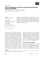
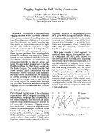
Fig. 1. HPLC of testosterone ox idatio n product s.
Ó FEBS 2004 1b-Hydroxytestosterone (Eur. J. Biochem. 271) 3963
at 1.3 bar and the dry gas was set to 4.3 LÆh
)1
at a
temperature of 200 °C. The capillary e xit was to 120 V
with a skimmer voltage of 40 V. The h exapole RF was set
to 50 Vpp (volts peak to peak) to enable the detection of
smaller masses.
Solid phase extraction of peaks
After detection of peaks with the diode array detector, H
2
O
was added using a Knauer K120 pump operated at a flow
rate of 1.6 mLÆmin
)1
. The flow was guided to a modified
Prospekt 2 solid phase extraction unit from Bruker/Spark
(Bruker Biospin, Rheinstetten, Germany/Spark Holland,
Emmen, the Netherlands). Peaks were automatically
trapped o n 2 · 10 mm SPE cartridges filled with Hysphere
GP, a cross-linked polystyrene-divinylbenzyl copolymer
(Spark Holland).
After the trapping step the cartridges were automatically
dried for 30 min under a stream of N
2
gas and eluted into
the NMR flow cell with CD
3
OD.
Cryo-NMR
An Avance spectrometer equipped with a Dual Inverse
1
H/
13
C30-lL Cryofit Probe operated at 600.13 MHz f rom
Bruker BioSpin (Rheinstetten, Germany) was used for
NMR investigation. The data was obtained after threefold
trapping of the peak on G P cartridges a nd elution with
subsequent on-line NMR analysis. The analyses were
performed with three 20-lL injections, each containing
420 lg o f t otal m aterial (substrate plus other products). The
chromatographic separation is shown below (Fig. 1). The
spectra of testosterone and 6b-hydroxytestosterone were
recorded but are not presented here. Sp ectra of the
previously unidentified o xidation product were recorded
eluting at 12.1 min, which was subsequently identified as
1b-hydroxytestosterone using the LC-NMR data discussed
below.
The trapped product was eluted in a mixture of
d
6
-methanol and D
2
O with small amounts of residual
CH
3
CN present. The temperature was controlled at
25 ± 0.1 °C. Chemical shifts were referenced to the water
resonance a t 4 .88 p.p.m. at 25 °C . The 1D s pectrum u tilized
double presaturation to minimize any residual water and
methanol signals. A total of 65 536 complex data points
were recorded with a sweep width of 5531 Hz and 32 scans.
The data was processed with a line broadening of 0.3 Hz.
Two-dimensional techniques (
1
H-
1
HCOSY,
1
H-
1
H
NOESY, and
1
H-
13
C HSQC) were also used for the
structure elucidation of the trapped compounds. The
parameters for the phase sen sitive (States-TPPI mode)
1
H-
1
H COSY spectra with water suppression were: spectral
width, 5531 Hz, 4096 complex data points, relaxation
delay 2 s, and eight scans for each of the 512 increments.
The same parameters were used for phase sensitive
1
H-
1
H
NOESY with H
2
O suppression on the water signal except
for the number of scans (32), the number of data points
(2 k) and number of increments ( 256). T he mix ing t ime
was 500 ms. The
1
H-
13
C HSQC experiment was acquired
in the phase sensitive mode with sensitivity enhancement,
echo/anti-echo-TPPI gradient selection and adiabatic car-
bon decoupling during evolution and acquisition [28–30].
Further parameters were: spectral width of 5531 Hz, 4096
data points in the
1
H dimension, 25 000 Hz with 256 data
points in the
13
C dimension and a relaxation delay of 2 s.
The data was processed using Bruker
XWINNMR
software
on an SGI workstation (Silicon Graphics, Mountain View,
CA, USA). The d ata was zero-filled in the acquisition
dimension and linear prediction was applied in the indirect
dimension.
Results
HPLC of testosterone oxidation products
The chromatogram acquired at 244 nm (Fig. 1) showed a
number of UV absorbing peaks eluting at shorter retention
times when compared with the major peak (t
R
¼ 23 min),
which can be easily assigned to the substrate testosterone.
This indicates the presence of more polar components at
much lower concentrations. Several of these were known
because of their coelution with standards in this and
previous work. However, the peak eluted at 12.1 min did
not correspond to any o f the available s tandards in our
collection (2a-, 2b-, 6a-, 6b-, 11b-,15b-,16a-or16b-
hydroxytestosterone or androstenedione), and the sample
was submitted for HPLC-solid phase extraction-cryoprobe
NMR/time-of-flight MS analysis.
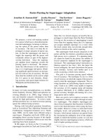
Mass spectrometry
Preliminary HPLC-electrospray MS experiments indicated
an [M + H]
+
ion at m/z 305, corresponding to a mono-
hydroxylated testosterone product. The r esult was con-
firmed in the HPLC-MS-NMR w ork with the MicroTOF
instrument, yielding MH
+
at m/z 305.2080 (theoretical
m/z for C
19
H
29
O
3
305.2111) (Fig. 2).
NMR
The total amount of the product estimated to have been
collected for the analysis is 6 lg. The 1D
1
Hspectrumwas
devoid of impurities (Fig. 3). The carbinol peak of interest
was noted at d 3.95 p.p.m., observed as a multiplet.
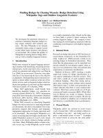
The COSY spectrum (Fig. 4) was very informative. The
carbinol proton of interest (d 3.95 p.p.m.) was coupled to
two protons in the d 2.5 p.p.m. region, indicating that the
hydroxylation w as at either C-1 or C -7, i.e. the carbinol is
coupled to either an H-2 or H-6 proton. The lack o f
coupling t o the H-8 proton ( d 1.69 p.p.m.) indicates that the
proton can only be at C-1.
The HSQC spectrum (Fig. 5) allowed complete assign-
ment of all proton-attached
13
C s ignals, confirming the basic
structure. The resulting information i s presented in Table 1.
The NOESY spectrum (Fig. 6) clearly indicates that
the added hydroxyl g roup at C-1 m ust be b.TheH-1
carbinol proton clearly shows correlation peaks with
protons established as C-2 (d 2.53, 2.46 p.p.m.), C-9
(d 1.15 p.p.m.) and the equatorial positioned proton
C-11 (d 2.07 p .p.m.) (but not with the C-19 methyl
group). Thus, the carbinol proton must be in the
a-p osition. If the proton were b, it would b e e xpected
to show a strong interaction with the C-19 methyl, as
indicatedinFigs5and6.
3964 J. A. Krauser et al. (Eur. J. Biochem. 271) Ó FEBS 2004
One synt hesis of 1b-hydroxytesto sterone was found in the
literature (seven steps from dihydrotestosterone benzoate)
[31]. The chemical shifts presented in that paper (1, 2, 4, 17,
18 and 19 protons assigned) are simila r to ours. However,
the J
1a,2a
and J
1a,2b
values differ. Our assignments are also
consistent with those reported for 1a-hydroxytestosterone,
except for the differences at and near C-1 (http://www.
unibas.ch/mdpi/ecsoc-4/a0099/a0099.htm).
Formation of 1b-hydroxytestosterone by recombinant
P450 3A4 systems
The 1b-hyroxylation of testosterone was observed in both
bacterial- and baculovirus-based P450 3A4 expression sys-
tems (Table 2). Rates of formation of the products were
similar. The 1b-hydroxy product accounts for 5% of all
testosterone products formed in both systems.
The formation of 1b-hydroxytestosterone was also
observed in human liver microsomes. The ratio of 1b-to
6b-hydroxylation was less than that measured with the
recombinant P 450 3A4 systems due to contribution of s ome
other P450s to 6b-hydroxylation (Table 3; see below also).
The liver sample used (HL 97) had previously been shown
to have a concentration of P450 3A4 intermediate between
that of high and low individuals [25].
Testosterone hydroxylation by other human P450s
Several human P450s were examined for the ability to form
the individual hydroxylated testosterone products, at a
Fig. 2. MS of previously unidentified tes to-
sterone oxidation product. (A) Experimental
spectrum. (B) Theoretical. The molecular ion
(MH
+
305.2080) corresponds to the formula
C
19
H
29
O
3
(theoretical 305.2111).
Fig. 3. COSY (
1
H) NMR spectrum of testo-
sterone oxidation product. Eight scans,
4096 · 512 acquisition matrix, 2-s relaxation
delay, with water s uppression. See Table 1 for
assignments.
Ó FEBS 2004 1b-Hydroxytestosterone (Eur. J. Biochem. 271) 3965
single substrate concentration of 100 l
M
(Table 3). All of
the P450s examined produced some products, but only
P450 3A4 formed 1b-hydroxytestosterone.
Discussion
The use of a combined HPLC-MS-NMR system facilitated
the c haracterization of one of the minor hydroxylation
products of testosterone, with an estimated total amount of
6 lg. Spectroscopy alone yielded an unequivocal assign-
ment of the product. Traces of a product designated
1a,b-hydroxytestosterone had been reported previously in
rat and mouse liver systems but only on the basis of the
expected t
R
[11,32,33].
The 1(b)-hydroxylation of androgens has been reported
previously. Dodson et al. [34] r eported that microbial
ppm
20
30
40
50
60
70
80
4.0 3.5 3.0
2.5 1.5 1.0 ppm
ppm
ppm
125
130
6.0 5.8
2.0
Fig. 4. HSQC NMR spectrum of testo-
sterone oxidation product. Sixty-four scans,
4096 · 256 acquisition matrix, 2-s relaxation
delay, with PFG coherence selection. The inset
shows a cross-peak out of the range of the rest
of the scale.
Fig. 5. NOESY (
1
H)NMR spectrum of tes-
tosterone oxidation product. Thirty-two scans,
2048 · 256 acquisition matrix, 2-s relaxation
delay, with water suppression. H-1
a
cross-
peaks are boxed.
3966 J. A. Krauser et al. (Eur. J. Biochem. 271) Ó FEBS 2004
(Xylaria sp.) oxidation of androstenedione yield ed a product
identified as the 1b-hydroxy derivative. The assignment was
based largely on chemical conversion to D
1,2
-dehydrotesto-
serone and the optical rotation [34]. This compound was
reduced to 1b-hydroxytestosterone, which has been used as
a standard o r substrate in m ost subsequent work, e ither
directly or by indirect comparisons. Gustafsson’s group
reported 1b-hydroxylation of testosterone by human fetal
liver micorosomes, using the Xylaria-derived product as a
standard [35,36]. On the basis of our own s tudy, it may be
speculated t hat the e nzyme re spo nsible i s P450 3A7, an
enzyme closely r elated to P450 3A4 and fetal-specific
(P450 3A4 is not expressed until after birth) [37].
Other work with the 1b-hydroxytestosterone derived
from Xyleria oxidations [34] has yielded reports that it is a
weak inhibitor of human placental aromatase (P450 19A1),
the enzyme that oxidizes testosterone to 17b-estradiol, with
an IC
50
value of 1m
M
[38]. Another report i ndicated that
human placental microsomes used 1b-hydroxytestosterone
30% as efficiently as testosterone or antrostenedione [35],
but apparently has not been confirmed.
Very recent work on possible functions of 1b-hydroxy-
testosterone has appeared in a paper published after our
own work was submitted [39]. Porcine gonadal P450 19A1
(aromatase) converted testosterone to significant amounts
of 1b-hydroxytestosterone, as well as 19-hydroxy- and
19-oxotestosterone and 17b-estradiol [39]. The assignment
of the structure was based on (a) comparison of an MS
fragmentation pattern w ith an earlier literature s pectrum
[35] (going back to the original Xylaria product [34]), and
(b) labilization of
3
Hfrom[1b-
3
H]-testosterone [39]. Corbin
Table 1. NMR shifts (see Figs 3–5). N/A, no protons attached; Ô–Õ indicates that the sh ift was not ide ntified. 1b-OH, 1b-hydroxyt estosteron e;
2b-OH, 2b-hydroxytestosterone.
Atom
1b-OH 2b-OH Testosterone
1
H
d (p.p.m.)
Multiplicity
and coupling, J (Hz)
13
C
d (p.p.m.)
1
H
d (p.p.m.)
13
C
d (p.p.m.)
1
H
d (p.p.m.)
13
C
d (p.p.m.)
1a 3.95 dd 1H, J ¼ 10.2 Hz, H
2a
, 4.6 Hz, H
2b
74.3 2.31 40.46 1.64 36.22
1b – – – 1.48 40.46 2.02 36.22
2a 2.46 dd 1H, J ¼ 4.6 Hz, H
1
, 16.1 Hz, H
2b
44.05 4.11 69.06 2.18 34.33
2b 2.53 dd 1H, J ¼ 10.2 Hz, H
1
, 16.1 Hz, H
2b
44.1 N/A N/A 2.41 34.33
3 N/A N/A – N/A – N/A –
4 5.75 – 123.55 5.70 119.33 5.64 123.84
5 N/A N/A – N/A – N/A –
6a 2.31 m 1H 34.3 2.20 33.18 2.23 33.12
6b 2.54 m 1H 34.3 2.51 33.18 2.40 33.12
7a 1.00 m 1H 33.7 1.09 37.08 1.01 37.17
7b 1.90 m 1H 33.7 1.77 37.08 1.77 37.17
8 1.69 m 1H 37.35 1.90 30.55 1.59 36.19
9 1.15 ddd 1H, J ¼ 4.2 Hz, 10.3Hz, 16.2 Hz 55.65 1.39 50.90 0.90 54.76
10 N/A N/A – N/A – N/A –
11a 2.07 m 1H 24.20 1.71 22.89 1.56 21.30
11b 1.55 m 1H 24.20 1.15 22.89 1.43 21.30
12 1.09 dd 1H, J ¼ 3.9 Hz, H
11
, 13.1 Hz, H
12
37.8 0.94 34.96 0.94 32.32
12b 1.84 m 1H 37.8 1.92 34.96 1.81 32.32
13 N/A N/A – N/A – N/A –
14 0.98 m 1H 51.65 0.96 51.03 0.93 51.09
15a 1.60 m 1H 24.1 1.51 23.57 1.56 23.78
15b 1.31 m 1H 24.1 1.27 23.57 1.26 23.78
16a 1.98 m 1H 30.2 1.88 30.60 1.91 30.48
16b 1.47 m 1H 30.2 1.36 30.60 1.38 30.48
17 3.56 dd 1H, J ¼ 8.5 Hz 82.2 3.49 81.45 3.50 81.54
18 0.78 s 3H 11.4 0.71 11.36 0.72 11.39
19 1.24 s 3H 13.25 1.15 22.88 1.18 17.48
Fig. 6. Space-filling model of 1b-hydroxytestosterone. The model was
produced with the program
CHEM
3
DPRO
v.5, CambridgeSoft Corp.
(Cambridge, MA, USA). Black denotes oxygen, medium gray denotes
carbon, and light gray denotes hydrogen atoms.
Ó FEBS 2004 1b-Hydroxytestosterone (Eur. J. Biochem. 271) 3967
et al. [39] postulated physiological activity of 1b-hydroxy-
testosterone and showed activation of the androgen recep-
tor in tw o different cell lines. 1b-Hydroxytestosterone was
not (enzymatically) reduced to the D
4,5
derivative. Interest-
ingly, the 1b-hydroxylation reaction was not catalyzed by
human P450 19A1 or by any other (tissue-specific) form of
porcine P450 19A1. Although some biological activity has
been demonstrated, the relevance o f 1b-hydroxytestosterone
to human physiology is not clear at this point.
The biological properties of 1b-hydroxytestosterone,
although speculated (see above), a re currently unknown.
It is of interest to note that almost all of the P450 3A4-
catalyzed hydroxylations of testosterone are on the b face.
This information is of interest in considerations of the
steroselectivity of P450 3A4 and general considerations
about the juxtaposition of the substrate in the active site,
particularly in predicting sites and rates of P450 3A4
reactions deals with models based on chemical reactivity.
The concept has often been proposed that P450 3A4 has a
relatively open a ctive site and that reactions are influenced
largely by the chemical lability of C-H bonds [40,41].
However, the striking stereochemical selectivity at each of
the several hydroxylation positions would appear to argue
against this and in favor of a relatively large but organized
active site.
Acknowledgements
The authors t hank M. V. Martin and C . G. T u rvy for preparing
bacterial membranes. This work was supported i n part by United States
Public Health Service grants R01 CA90426, P30 ES00267, and T32
ES07028.
References
1. Pa lmer, G. & R e edijk, J. (1992) Nomenclature of electron-transfer
proteins: recommendation s 1989. J. Biol. Chem. 267, 665–677.
2. Dorfman, R., Cook, J.W. & H amilton, J.B. (1939) Conversion by
the human of the testis hormone, testosteron e, into the urinary
androgen, androsterone. J. Biol. Chem. 130, 285–295.
3. Ryan, K.J. (1958) Conversion of androstenedione to estrone by
placental microsomes. Biochim. Biophys. Acta 27, 658–662.
4. Kagawa, N. & Waterman, M.R. (1995) Regulation of steroido-
genic a nd related P 450s. In Cytochrome P4 50: Structure,
Mechanism, and Biochemistry, 2nd edn. (Ortiz de Montellano,
P.R., ed.), pp. 419–442. Plenum Press, New York.
5. Guengerich, F.P. (2003) Cytochrome P450s, drugs, and diseases.
Mol. Interventions 3, 8–18.
6. Conney,A.H.,Levin,W.,Jacobsson,M.&Kuntzman,R.(1969)
Specificity in the regulation of the 6b,7a,and16a-hydroxylation
of testosterone by rat live r microsomes. In Microsomes and Drug
Oxidations (Gi llette, J.R., ed.), pp. 279–301. Academic Press, New
York.
7. Wood, A.W., Ryan, D.E., Thomas, P.E. & Levin, W. (1983)
Regio- an d stereoselective met abolism o f two C19 steroids by five
highly purified and reconstituted rat hepatic cytochrome P-450
isozymes. J. Biol. Chem. 258, 8839–8847.
8. van der Hoeven, T. (1984) Assay of hepatic microsomal testo-
sterone hydroxylases by high-performance liquid chromatogra-
phy. Anal. Biochem. 138, 57–65.
9. Dutton, D.R., McMillen, S.K., Sonderfan, A.J., Thomas, P .E. &
Parkinson, A. (1987) Studies on the rate-determining factor in
testosterone hydroxylation by rat liver microsomal cytochrome
P-450: evidence against cytochrome P-450 isozyme: isozyme
interaction. Arch. Biochem. Biophys. 255, 316–328.
10. Sond erfan, A.J., A rlotto, M.P. & Parkinson, A. (1989) Identifi-
cation of the cytochrome P -450 isozyme s respon sible for te sto-
sterone oxidation in rat lung, kidney, and testis: evidence that
cytochrome P-450a (P450IIA1) is the physiologically important
testosterone 7a-hydroxylase in rat testis. Endocrinology 125,
857–866.
11. Purdon, M.P. & Lehman-McKeeman, L.D. (1997) Im proved
high-performance liquid chromatographic procedure for the
separation and quantification o f hydroxytestosterone metabolites.
J. Pharmacol. Toxicol. Methods 37, 67–73.
12. Gustafsson, J.A. & Lisboa, B.P. (1968) Biosynthesis of 6-beta-
hydroxytestosterone from testosterone by human fetal liver
microsomes. Steroids 11, 555–563.
13. Lisboa, B.P. & Gustafsson, J.A. (1969) S tudies on the metabolism
of ster oids in th e foetus: bi osynthesis of 6a-hydroxytestosterone in
the human foetal liver. Biochem. J. 115, 5 83–586.
14. Yaffe,S.J.,Rane,A.,Sjoqvist,F.,Boreus,L.O.&Orrenius,S.
(1970) The presence of a monooxygenase system in human fetal
liver microsomes. Life Sci. II, 1189–1200.
Table 2. Hydroxylation of testosterone by rec ombinant human P450 3A4 systems and hu man liver microsomes. The range of substrate concen tratio ns
used in most cases was 25–400 l
M
.
Product
E. coli membranes Baculovirus microsomes Human liver microsomes
k
cat
(min
)1
) K
m
(l
M
) k
cat
/K
m
k
cat
(min
)1
) K
m
(l
M
) k
cat
/K
m
k
cat
(min
)1
)
a
K
m
(l
M
) k
cat
/K
m
1b-OH 4.1 ± 0.1 10 ± 2 0.40 7.1 ± 0.3 17 ± 2 0.41 1.9 ± 0.1 55 ± 9 0.035
2b-OH 11 ± 1 49 ± 6 0.23 14 ± 4 44 ± 4 0.30 12 ± 1 170 ± 40 0.072
6b-OH 78 ± 2 26 ± 3 3.0 78 ± 3 23 ± 2 3.4 88 ± 5 90 ± 10 0.98
15b-OH 3.0 ± 0.2 41 ± 12 0.072 7.1 ± 0.2 32 ± 3 0.22 8.4 ± 0.8 81 ± 20 0.10
a
Based on total P450.
Table 3. Testosterone hydroxylation by various human P450 enzymes.
Assays were done (in triplicate) with bacterial membranes (Ôbicistro-
nicÕ) containing P450 a nd NADPH-P450 reductase [26] using a single
testosterone concentration of 100 l
M
. Ô–Õ Indic ates rate < 0.1 min
)1
.
P450
Rate (min
)1
)
1b-OH 2b-OH 6b-OH 15b-OH
1A1 – – 1.1 ± 0.1 –
1A2 – – 6.7 ± 0.3 –
1B1 – – 7.7 ± 0.6 –
2C9 – – 0.93 ± 0.03 –
2D6 – 0.31 ± 0.01 0.91 ± 0.01 –
3A4 4.8 ± 0.1 11 ± 0.1 83 ± 1 5.0 ± 0.1
3968 J. A. Krauser et al. (Eur. J. Biochem. 271) Ó FEBS 2004
15. Krem ers, P., Beaune, P., C resteil, T., de Graeve, J., Co lumelli, S.,
Leroux,J.P.&Gielen,J.E.(1981)CytochromeP-450mono-
oxygenase activities in human and rat liver microsomes. Eur. J.
Biochem. 118, 599–606.
16. Cresteil,T.,Beaune,P.,Kremers,P.,Flinois,J.P.&Leroux,J.P.
(1982) Drug-metabolizing enzymes in human foetal liver: partial
resolution of multiple cytochromes P450. Pediatr. Pharma col.
(New York) 2, 199–207.
17. Distlerath, L.M. & Guengerich, F.P. (1987) Enzymology of
human liver cytochrome s P-450. In Mammalian C ytochromes
P-450, Vol. 1 (Guengerich, F.P., ed.), pp. 133–198. CRC Press,
Boca Raton, Florida.
18. Guengerich, F .P., Martin, M.V., Beaune, P.H., Kremers, P.,
Wolff,T.&Waxman,D.J.(1986)Characterizationofratand
human liver microsomal cytochrome P-450 forms involved in
nifedipine oxidation, a prototype for genetic polymorphism in
oxidative drug metabolism. J. Biol. Chem. 261, 5051–5060.
19. Waxman, D.J., At tisano, C., Guengerich, F.P. & La penson, D.P.
(1988) Cytochrome P-450 steroid hormone metabolism catalyzed
by human liver microsomes. Arch. Biochem. Biophys. 263,424–
436.
20. Kitada, M., Kamataki, T., Itahashi, K., Rikihisa, T. & Kanakubo,
Y. (1987) Significance of cytochrome P-450 (P-450 HFLa) of
human fetal livers in the steroid and drug oxidations. Biochem.
Pharmacol. 36, 453–456.
21. Wrighton, S.A., Brian, W.R., Sari, M.A., Iwasaki, M., Guenge-
rich, F.P., Raucy, J.L., Molowa, D.T. & VandenBranden, M.
(1990) Studies on the expression and metabolic capabilities of
human liver cytochrome P450 IIIA5 ( HLp3). Mol. Pharmacol. 38,
207–213.
22. Guengerich, F.P. (1995) Human cytochrome P450 enzymes. In
Cytochrome P450: S tructure, Mechanism, a nd Bioc hemsitry (Orti z
de Montell ano, P.R., ed.), pp. 473–535. Plenum Pre ss, New York.
23. Evans, W.E. & Relling, M.V. (1999) Pharmacogenomics: trans-
lating function genomics into rational therapeutics. Science 286,
487–491.
24. Guengerich, F.P. (2001) Analysis and characterization o f enzymes
and nucleic acid s. In Principles and M ethods of Toxicology (Hayes,
A.W., ed.), pp. 1625–1687. Taylor & Francis, Philadelphia.
25. Guengerich, F.P. (1988) Oxidation o f 17a-ethynylestradiol by
human liver cytochrome P-450. Mol. Pharmacol. 33, 500–508.
26. Parikh, A ., Gillam, E.M.J. & Guengerich, F.P. (1997) Drug
metabolism by Escherichia coli expressing human cytochromes
P450. Nat. Biotechnol. 15, 784–788.
27. Yamaza ki, H., Nakano, M., Imai, Y., Ueng, Y F., Guengerich,
F.P. & Shimada, T. (1996) Roles of cytochrome b
5
in the oxidation
of testosterone and n ifedipine by recombinant cytochrome P450
3A4 and by human liver microsomes. Arch. Biochem. Biophys.
325, 174–182.
28. Palmer, A.G., 3rd, Cavanagh, J., Wright, P.E. & Rance, M. (1991)
Sensitivity improvement in proton-detected two-dimensional
heteronuclear correlation NMR spectroscopy. J. Mag. Res. 93,
151–170.
29. Kay, L.E., Keifer, P. & S aarinen, T. (1992) P ure absorption
gradient enhanced heteronuclear single quantum correlation
spectroscopy with improved sensitivity. J. Am. Chem. Soc. 114,
10663–10665.
30. Sc hleuch er, J., Schwendinger, M., Sattler, M., Schmidt, P.,
Schedletzky, O., Glaser, S.J., Sorensen, O.W. & Griesinger, C.
(1994) A general enhancement scheme in heteronuc lear multi-
dimensional NMR e mploying pulsed field gradients. J. Biomol.
NMR 4, 301–306.
31. Sharma, P.K. & Akhila, A. (1991) A facile synthesis of 1b-hy-
droxytestosteron e. Indian J. Chem., Sect. B 30B, 554–556.
32. Ford, H.C., Wheeler, R. & Engel, L.L. (1975) Hydroxylation of
testosteroneatcarbons1,2,6,7,15and16bythehepatic
microsomal fraction from adult female C57BL/6J mice. Eur. J.
Biochem. 57, 9–14.
33. Halvorson, M., Greenway, D., Eberhart, D., Fitzgerald, K. &
Parkinson, A. (1990) Reconstitution of testosterone oxidation by
purified rat cytochrome P450p (IIIA1). Arch. Biochem. Biophys.
277, 166–180.
34. Dodson,R.M.,Kraychy,S.,Nicholson,R.T.&Mizuba,S.(1962)
Microbiological transformations. IX. The 1b-hydroxylation of
androstenedione. J. Org. Chem. 27, 3159–3164.
35. Lisboa, B.P. & Gustafsson, J.A. (1968) Biosynthesis of two new
steroids in the human foetal liver, 1b-and2b-hydroxytestosterone.
Eur. J. Biochem. 6, 419–424.
36. Ingelman-Sundberg, M., Rane, A. & Gustafasson, J.A. (1975)
Properties of hydroxylase systems in the human fetal liver active
on free and sulfoconjugated steroids. Biochemistry 14, 429–437.
37. Komori, M., Nishio, K., Kitada, M., Shiramatsu, K., Muroya, K.,
Soma,M.,Nagashima,K.&Kamataki,T.(1990)Fetus-specific
expression of a form of cytoc hrome P -450 in human livers.
Biochemistry 29, 4430–4433.
38. Schwarzel, W.C., Kruggel, W.G. & Brodie, H .J. (1973) Studies on
the mechanism of estrogen biosynthesis.8.Thedevelopmentof
inhibitors of the enzym e system in human placenta. Endo crinology
92, 866–880.
39. Corbin, C.J., Mapes, S.M., Marcos, J., Shackleton, C.H., Mor-
row, D., Safe, S., Wise, T., Ford, J.J. & Conley, A.J. (2004)
Paralogues of porcine aromatase cytochrome P450: a novel
hydroxylase a ctivity is associated with the survival of a duplicated
gene. Endocrinology 145, 2157–2164.
40. Smith, D.A. & Jones, B.C. (1992) Speculations on the substrate
structure–activity relationship (SSAR) of cytochrome P450
enzymes. Biochem. Pharmacol. 44, 2089–2098.
41. Singh, S.B., Shen, L.Q., Walker, M.J. & Sheridan, R.P. (2003) A
model for predicting likely sites of CYP3A4-mediated metabolism
on drug-like molecules. J. Med. Chem. 46, 1330–1336.
Ó FEBS 2004 1b-Hydroxytestosterone (Eur. J. Biochem. 271) 3969