Báo cáo khoa học: Structural and functional analysis of the interaction of the AAA-peroxins Pex1p and Pex6p pptx
Bạn đang xem bản rút gọn của tài liệu. Xem và tải ngay bản đầy đủ của tài liệu tại đây (842.9 KB, 12 trang )
Structural and functional analysis of the interaction
of the AAA-peroxins Pex1p and Pex6p
Ingvild Birschmann
1,
*
,
†, Katja Rosenkranz
2,
†, Ralf Erdmann
2
and Wolf-H Kunau
1
1 Abteilung fu
¨
r Zellbiochemie, Medizinische Fakulta
¨
t der Ruhr-Universita
¨
t Bochum, Germany
2 Abteilung fu
¨
r Systembiochemie, Medizinische Fakulta
¨
t der Ruhr-Universita
¨
t Bochum, Germany
Peroxisomes are ubiquitous, single membrane-bound
organelles involved in many metabolic pathways [1].
Thirty-two proteins required for peroxisome biogenesis
have been described [2–6]. They are encoded by PEX
genes and are collectively called peroxins. Most of the
peroxins are directly involved in the import process for
peroxisomal matrix and membrane proteins while oth-
ers are required for proliferation and inheritance of the
organelles [2]. The current model of peroxisome bio-
genesis suggests that peroxisomal proteins are synthes-
ized on free ribosomes and post-translationally
targeted to the organelle [7–9]. Targeting and trans-
location of newly synthesized peroxisomal matrix pro-
teins requires ATP and depends on peroxisomal
targeting signals [10], PTS1 and PTS2, and their
cognate receptors Pex5p and Pex7p, respectively
[11,12]. These two peroxins bind their cargo in the
cytoplasm, transport them to (and possibly across) the
peroxisomal membrane and return to the cytoplasm
for the next round of import. The membrane-bound
steps of this receptor cycle are currently under inten-
sive investigation [13,14].
PEX1 and PEX6 genes encode members of the
AAA family of ATPases, a large superfamily of pro-
teins involved in the ATP-dependent rearrangement of
protein complexes [15–17]. AAA-proteins are found in
all organisms and are essential for many activities, e.g.
cell cycle function, vesicular transport, mitochondrial
Keywords
AAA-proteins, peroxisomal biogenesis,
Pex1p, Pex6p, peroxin
Correspondence
Dr Ralf Erdmann, Institut fu
¨
r
Physiologische Chemie, Abteilung fu
¨
r
Systembiochemie, Medizinische Fakulta
¨
t der
Ruhr-Universita
¨
t Bochum, D-44780 Bochum,
Germany. Tel: +49 234322 4943
Fax: +49 234321 4266
E-mail:
*Present address
Institut fu
¨
r Klinische Biochemie und Patho-
biochemie, Medizinische Universita
¨
tsklinik,
D-97078 Wu
¨
rzburg, Germany
†Both authors contributed equally to this
manuscript
(Received 21 July 2004, accepted 25 August
2004)
doi:10.1111/j.1432-1033.2004.04393.x
The AAA-peroxins Pex1p and Pex6p play a critical role in peroxisome bio-
genesis but their precise function remains to be established. These two
peroxins consist of three distinct regions (N, D1, D2), two of which (D1,
D2) contain a conserved 230 amino acid cassette, which is common to
all ATPases associated with various cellular activities (AAA). Here we
show that Pex1p and Pex6p from Saccharomyces cerevisiae do interact
in vivo. We assigned their corresponding binding sites and elucidated the
importance of ATP-binding and -hydrolysis of Pex1p and Pex6p for their
interaction. We show that the interaction of Pex1p and Pex6p involves
their first AAA-cassettes and demonstrate that ATP-binding but not
ATP-hydrolysis in the second AAA-cassette (D2) of Pex1p is required for
the Pex1p–Pex6p interaction. Furthermore, we could prove that the second
AAA-cassettes (D2) of both Pex1p and Pex6p were essential for peroxi-
somal biogenesis and thus probably comprise the overall activity of the
proteins.
Abbreviations
AAA, ATPases associated with various cellular activities; NSF, N-ethylmaleimide-sensitive fusion protein.
FEBS Journal 272 (2005) 47–58 ª 2004 FEBS 47
functions and proteolysis [17,18]. AAA-proteins share
the presence of one or two AAA-cassettes, comprising
about 230 amino acids and are characterized by Wal-
kerA and B motifs for ATP-binding and ATP-hydro-
lysis [19,20]. Pex1p and Pex6p are functionally
nonredundant AAA-proteins required for the biogen-
esis of peroxisomes. A direct interaction of Pex1p and
Pex6p has been demonstrated in some organisms [21–
24] indicating that the two peroxins cooperate in per-
oxisome assembly. Sequence comparison of Pex1p and
Pex6p with other AAA members indicates that the two
AAA-peroxins belong to an AAA-protein subfamily
characterized by a tripartite structure. These proteins
possess large N-terminal regions followed by two AAA-
cassettes. In N-ethylmaleimide-sensitive fusion protein
(NSF), the best characterized AAA-protein, distinct
functions could be assigned to its three parts [25,26].
For yeast and human Pex6p it has been reported that
the N-terminal region of this protein functionally inter-
acts with the yeast peroxin Pex15p or human Pex26p,
respectively [3,27]. However, despite these findings and
the fact that both AAA-peroxins are conserved from
yeast to humans their function in peroxisome biogenesis
remains unknown.
In this study we mapped the mutual binding sites
and assayed the effects of deletion and point mutations
in Pex1p and Pex6p for the interaction of the proteins
and for their overall function in peroxisome biogenesis.
Our results demonstrate a different role of the two
AAA-cassettes for the Pex1p–Pex6p interaction and
their functional role in peroxisome biogenesis.
Results
The contribution of Pex1p and Pex6p to peroxisomal
biogenesis is well established on the basis of the pheno-
types of the corresponding null mutants. Both pex1D
[24,28] and pex6D [29,30] mutants show a characteristic
pex phenotype with only residual, ghost-like peroxi-
somal structures and mislocalization of peroxisomal
matrix proteins to the cytosol. Here we confirm and
extend earlier studies of these AAA-peroxins and give
a further detailed functional analysis of their cassette
structure and interaction.
The interaction of Pex1p and Pex6p involves
their first AAA-cassettes (D1)
Pex1p and Pex6p have been shown to interact in Pichia
pastoris, Hansenula polymorpha and human [21–24]. To
further limit the corresponding binding regions, we first
confirmed the interaction of the two proteins in bakers
yeast. For this purpose, we constructed plasmids carry-
ing PEX1 fused to the coding sequence of the activation
domain of Gal4p (PEX1-GAL4-AD) or PEX6 linked to
the coding region of the DNA binding domain of Gal4p
(PEX6-GAL4-BD). The two-hybrid reporter strain
PCY2 was cotransformed with the two plasmids. Acti-
vation of the reporter gene lacZ, indicated by blue
colonies on X-Gal medium was observed, when PEX6-
GAL4-BD was coexpressed with PEX1-GAL4-AD
(Fig. 1A,B). Transformation of either of these two plas-
mids alone did not lead to an activation of the reporter
gene. The same results were obtained when the reporter
strain HF7c was used to assay for histidine prototrophy
(data not shown). These data demonstrate that ScPex1p
interacts with ScPex6p. The two-hybrid studies did not
provide an indication for homo-oligomerization of the
two AAA-peroxins when PEX1-GAL4-BD was coex-
pressed with PEX1-GAL4-AD or PEX6-GAL4-BD was
coexpressed with PEX6-GAL4-AD (data not shown).
This finding in turn is in agreement with the assumption
that these two AAA-peroxins fulfill their function in
peroxisome biogenesis as a transient or stable hetero-
meric protein complex. To investigate the influence of
the recent published interaction between Pex6p and
Pex15p [27], we carried out the two-hybrid assay in the
absence of Pex15p (PCY2 pex15D). Pex1p and Pex6p
still showed interaction in pex15D indicating that the
association of the two proteins does not depend on
Pex15p (data not shown).
To identify the regions in Pex1p and Pex6p that are
responsible for their interaction, truncated versions of
the proteins were tested for interaction in the yeast
two-hybrid system. According to the predicted domain
structure, the Pex1p sequence was divided into three
parts: the N-terminal region (N, aa1–400), the first
AAA-cassette (D1, aa394–681) and the second AAA-
cassette (D2, aa669–1043). DNA fragments encoding
these parts of Pex1p were fused to the GAL4-AD and
coexpressed with PEX6-GAL4-BD in PCY2.As
judged by the lack of lacZ gene expression, none of
these cassettes alone can mediate binding to Pex6p
(Fig. 1A). However, a Pex1p fragment consisting of
the N-terminal part and the first AAA-cassette
(N + D1, aa1–681) gave rise to a low but significant
lacZ gene activation (Fig. 1A). The fragment compri-
sing both AAA-cassettes (D1 + D2), however, led to
a b-galactosidase activity comparable to that of the
full-length Pex1p (Fig. 1A). These findings indicate
that binding of Pex6p is best in the presence of both
AAA-cassettes of Pex1p. However, the data also indi-
cate that the first AAA-cassette together with the
N-terminal region or the second cassette of Pex1p is
already sufficient for interaction of the two AAA-
peroxins. Consequently, these results demonstrate the
Domain function of Pex1p and Pex6p I. Birschmann et al.
48 FEBS Journal 272 (2005) 47–58 ª 2004 FEBS
critical role of the first AAA-cassette of Pex1p for the
binding of Pex6p. Our data suggest that the binding
site for Pex6p is comprised within the first AAA-
cassette (D1) of Pex1p. Binding efficiency can be
increased by the additional presence of either the
N-terminal fragment or more drastically by the pres-
ence of the second AAA-cassette. This is consistent
with the observation that a mutation in the beginning
of Pex1pD2 (G843D in human, corresponding to
G700 in yeast) attenuates the interaction of the two
AAA-peroxins [23].
To identify the Pex6p binding site for Pex1p, we tes-
ted the N-terminal region (N, aa1–428), the first AAA-
cassette (D1, aa421–716) and the second AAA-cassette
(D2, aa704–1030) and all possible combinations for
interaction with Pex1p in the yeast two-hybrid system.
The corresponding PEX6 fragments were fused to
GAL4-BD and coexpressed with PEX1–GAL4-AD.
The results shown in Fig. 1B demonstrate that an acti-
vation of the lacZ reporter gene occurred only in dou-
ble transformants carrying PEX1 –GAL4-AD and the
construct coding for the Pex6p region comprising the
N-terminal fragment and the first AAA-cassette of
PEX6 (PEX6N + D1–GAL4-BD). Neither of the cas-
settes alone nor the two AAA-cassettes together or the
N-terminal part together with the second cassette led
to an activation of the reporter gene and thus is suffi-
cient to maintain the interaction with Pex1p. These
findings demonstrate the importance of the first AAA-
cassette together with the N-terminal region of Pex6p
for the described interaction.
To confirm these results, we carried out coimmuno-
precipitation with different genomic integrated con-
structs: Pex1pN–ProteinA, Pex1pN + D1–ProteinA,
Pex1p–ProteinA, Pex6pN–ProteinA, Pex6pN + D1–
ProteinA, Pex6p–ProteinA. For this purpose, we
generated strains that express the proteins of interest,
C-terminally fused to two IgG-binding domains derived
from Staphylococcus aureus protein A (ProtA). An
advantage of these fusion proteins was the possibility
to detect even the truncated constructs via the ProtA-
tag. Moreover, as expression of the constructs was
under the control of the endogenous promoters,
overexpession was avoided, which has been shown to
A
BD
C
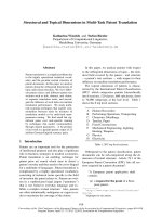
Fig. 1. Coimmunoprecipitation and two-hybrid interaction of Pex1p and Pex6p. A schematic representation of the Pex1p and Pex6p con-
structs which were analyzed in the two-hybrid assay (A,B) or by coimmunoprecipitation (C,D) is shown on the left. Analysis of PCY2 trans-
formants expressing the indicated fusion proteins of (A) Pex1p truncations and full-length Pex6p or (B) Pex6p truncations and full-length
Pex1p. The interactions were analyzed for b-galactosidase activity by filter assays with X-gal as the substrate. Three independent double-
transformations are shown. Extracts of oleic acid-induced wild-type cells expressing (C) Pex1p and Pex1p-truncations or (D) Pex6p and
Pex6p-truncations fused to ProteinA were immunoprecipitated with anti-IgG and immunoblotted with the same antisera (C,D), or antibodies
to Pex1p (D) or Pex6p (C). As a control, wild-type cells expressing no ProteinA fusion protein were treated equally (C,D; lane 4).
I. Birschmann et al. Domain function of Pex1p and Pex6p
FEBS Journal 272 (2005) 47–58 ª 2004 FEBS 49
affect peroxisome biogenesis [31–33]. To ascertain
whether the ProtA fusion proteins were functional or
not, we investigated the ability of the strains to grow
on medium containing oleic acid as the sole carbon
source and monitored for the correct proliferation of
peroxisomes via the analysis of cell morphology by
electron microscopy. We observed that both full
length proteins Pex1p and Pex6p fused to ProtA
were functional in vivo, whereas the four deleted
strains (Pex1pN– ProtA, Pex 1pN + D1–ProtA, Pex6p N–
ProtA, Pex6pN + D1–ProtA) showed neither growth
on oleic acid nor morphologically detectable peroxi-
somes (data not shown).
ProtA fusion proteins in the eluates were detected
by immunoblot analysis with anti-IgG (Fig. 1C,D).
The different migration behaviour reflects the different
sizes of the fusion proteins. Immunological analysis
revealed the presence of Pex6p in the precipitate of
full-length Pex1p–ProtA and also the presence of
Pex1p in the full-length Pex6p–ProtA-precipitate
(Fig. 1C,D). These data confirm the in vivo interaction
of the two proteins. We tested two truncations for
their interaction behaviour, the N-terminal region
and the N-terminal region together with the first AAA-
cassette. Attempts to express ProtA fusion constructs
comprising the first and the second cassette were not
successful. Neither Pex1p nor Pex6p was present in the
precipitates of the N-terminal region of Pex6, or
Pex1p, respectively (Fig. 1C,D). These data indicate
that the N-terminal fragment alone does not interact
with the corresponding binding partner. However, a
significant amount of Pex1p and Pex6p was detected in
the precipitates of the N + D1 fusions (Fig. 1C,D).
These data support the two-hybrid results and indicate
that D1 of both Pex1p and Pex6p contributes to the
binding of the two proteins. Truncation of either pro-
tein, however, did result in a significant decrease in the
coimmunoprecipitation of the binding partner. These
data might indicate that the contact sites between the
two proteins are not limited to the first cassette but
also comprise regions of the N-terminal fragment or
the second cassette, or that the binding site is com-
prised by D1 but its binding capability is significantly
enhanced in the presence of the N-terminal fragment
or D2.
ATP-binding but not ATP-hydrolysis in the
second AAA-cassette (D2) of Pex1p is required
for the Pex1p–Pex6p interaction
Pex1p and Pex6p both contain two AAA-cassettes and
thus two consensus ATP-binding sites. Typically,
Walker-type nucleotide-binding sites consist of two
conserved motifs. The WalkerA motif is essential for
nucleotide-binding, while the WalkerB motif is
required for hydrolysis of the nucleotide. To investi-
gate the influence of the binding and ⁄ or hydrolysis of
ATP on the interaction of the AAA-peroxins, Pex1p
and Pex6p carrying mutated ATP-binding sites were
tested for interaction with the binding partner in the
yeast two-hybrid system. In the first set of mutants,
the lysine residue in the GXXGXGKT sequence of
either one of the two WalkerA motifs was replaced by
a glutamate or alanine residue which led to Pex1pA1-
(K467E), Pex1pA2(K744E), Pex6pA1(K489A) and
Pex6pA2(K778A). These invariant lysine residues have
been shown to be essential for the biological activity of
a number of ATP- and GTP-binding proteins and their
replacement yields proteins with significantly reduced
ATP-binding capacity [34]. Such an impairment of the
biological activity has also been reported for Pex1p
and NSF [19,35,36]. Similarly, we also introduced
point mutations of amino acids in the ATP-hydrolysis
sites (conserved sequence is four hydrophobic amino
acid D ⁄ E) to investigate their influence on the des-
cribed interaction. We changed the conserved aspartate
of the WalkerB motifs of Pex1p and Pex6p into gluta-
mine leading to Pex1pB1(D525Q), Pex1pB2(D797Q)
and Pex6pB2(D831Q) [37]. It was not necessary to cre-
ate a B1 point mutation because wild-type Pex6p
already contains an alanine instead of the critical
aspartate (aa548), strongly suggesting that D1 of
Pex6p can bind but not hydrolyse ATP.
DNA fragments encoding the entire Pex1p or Pex6p
harboring the different mutations in the AAA-cassettes
were fused to GAL4-AD ⁄ GAL4-BD and coexpressed
with PEX6–GAL4-BD or PEX1–GAL4-AD, respect-
ively (Fig. 2). Compared to wild-type Pex1p or Pex6p,
only the Pex1pA2 mutation resulted in a significantly
less efficient interaction with Pex6p as judged by the
decreased activation of the lacZ gene (Fig. 2A). Neither
a mutation in Pex1pA1 nor in Pex6pA1 or Pex6pA2 had
any influence on the Pex1p–Pex6p interaction (Fig. 2).
Moreover, the mutations of the WalkerB motif did not
affect the interaction of the AAA-peroxins (Fig. 2). All
constructs used for two-hybrid analyses were over-
expressed and showed the same protein level, demon-
strating that the described effects were not a result of
different expression levels (data not shown).
These results give rise to the notion that ATP-bind-
ing to D1 of Pex1p and to D1 and D2 of Pex6p as
well as the capability for ATP-hydrolysis in general is
dispensible for the Pex1p–Pex6p interaction. However,
the results also clearly demonstrate that the ability of
the second AAA-cassette of Pex1p to bind ATP is
required for the interaction of Pex1p and Pex6p.
Domain function of Pex1p and Pex6p I. Birschmann et al.
50 FEBS Journal 272 (2005) 47–58 ª 2004 FEBS
The second AAA-cassettes (D2) of Pex1p and
Pex6p are essential for peroxisomal biogenesis
To investigate the effects of the described WalkerA
and WalkerB mutants of the ATP-binding sites for the
function of Pex1p and Pex6p in peroxisomal biogen-
esis, we analyzed the functional and morphological
phenotypes of the transformed mutants in further
detail.
Cells deficient in Pex1p or Pex6p are characterized
by the inability to grow on oleic acid as the single car-
bon source, mislocalization of matrix proteins to the
cytosol and the absence of morphologically detectable
matrix-filled peroxisomes [21,24,28,30,35,38]. First, we
tested different mutant constructs for their ability to
complement the oleic acid-growth phenotype of the
pex1 or pex6 null mutant. As demonstrated in Fig. 3,
null mutants expressing Pex1p mutated at the first
ATP-binding site (Pex1pA1, Pex1pB1) showed the
wild-type phenotype with respect to growth on oleic
acid medium. The same has been shown for Pex6p
(Pex6pA1) [27]. Complementation of the mutant
strains is indicated by growth on oleic acid medium
(YNO)-agar plates, which gives rise to a typical halo
reflecting the consumption of oleic acid. Interestingly,
the null mutants expressing Pex1p or Pex6p mutated
at WalkerA or WalkerB of the second ATP-binding
site were not able to grow on oleic acid as sole carbon
source (Fig. 3; [27,35]), indicating that both ATP-bind-
ing and ATP-hydrolysis at the conserved D2 is
required for Pex1p and Pex6p function in peroxisomal
biogenesis. This is also supported by the ultrastructural
appearance of the corresponding mutants (Figs 4 and
5). Oleic acid-induced pex1D (Fig. 4) or pex6D (Fig. 5)
mutant cells transformed with plasmids encoding
Pex1pA2 (Fig. 4E), Pex1pB2 (Fig. 4F), Pex6pA2
(Fig. 5E), or Pex6pB2 (Fig. 5F) are characterized by
the absence of morphologically detectable peroxisomes
and thus exhibit the same phenotype of the corres-
ponding pex1 (Fig. 4B) and pex6 (Fig. 5B) null
mutants, indicative of no complementation. Peroxi-
somes reappear, however, upon complementation of
pex1D or pex6D strains with the wild-type Pex1p
(Fig. 4A) or Pex6p (Fig. 5C) proteins or proteins har-
boring mutations in the first AAA-cassette [Pex1pA1
(Fig. 4C), Pex1pB1 (Fig. 4D), Pex6pA1 (Fig. 5D)].
These results are consistent with an essential role of
the conserved AAA-cassette for Pex1p and Pex6p func-
tion in peroxsiome biogenesis. The results also demon-
strate that ATP-binding and ATP-hydrolysis at the
conserved AAA-cassette is required for the biological
function of Pex1p and Pex6p.
A
B
Fig. 2. Two-hybrid interaction of Pex1p and Pex6p harboring point
mutations of the WalkerA and B motifs of their ATP-binding sites.
A schematic representation of the Pex1p and Pex6p mutants which
were tested for two-hybrid interaction is shown on the left. PCY2
transformants expressed the indicated fusion protein combinations
of (A) Pex1p mutations and full-length Pex6p or (B) Pex6p muta-
tions and full-length Pex1p. The interactions were analyzed for b-ga-
lactosidase activity by filter assays with X-gal as substrate and
three independent double-transformations are shown.
Fig. 3. Effects of point mutation of the WalkerA and B motifs of
the ATP-binding sites on the complementation activity of Pex1p.
Growth behaviour on oleic acid medium (YNO) was analyzed for
wild-type, pex1D and pex1D expressing genes encoding wild-type
or indicated point mutated Pex1p. Complementation is indicated by
growth on YNO-agar plates, which gives rise to a typical halo
reflecting the consumption of oleic acid.
I. Birschmann et al. Domain function of Pex1p and Pex6p
FEBS Journal 272 (2005) 47–58 ª 2004 FEBS 51
Discussion
In this report, we demonstrate that the two AAA-
peroxins Pex1p and Pex6p of Saccharomyces cerevisiae
interact in vivo. We analyzed this interaction in more
detail paying special attention to the cassette structure
of the proteins. We assigned the binding sites of these
two AAA-proteins to their different protein regions
and elucidated the importance of ATP-binding to the
two AAA-cassettes of Pex1p and Pex6p for their inter-
action.
The amino acid sequences of both peroxins sug-
gest an architecture with three different regions: an
N-terminal fragment (N) followed by two AAA-cas-
settes (D1 and D2) [20]. Sequence comparison with
other members of the large AAA-protein family shows
that in both AAA-peroxins D2 is well conserved, while
D1 exhibits much less sequence similarity. We have
reported previously that ATP-binding by D2 but not
D1 is strictly required for the overall function of Pex1p
in peroxisome biogenesis. This conclusion was based
on the analysis of functional consequences of muta-
tions of the conserved lysine in WalkerA of both
AAA-cassettes [35]. Our further analyses demonstrated
that the same is true for Pex6p [27]. Additionally, point
mutations in WalkerB of D2 in both AAA-peroxins
AB
DC
EF
Fig. 4. Effects of point mutation of the
WalkerA and B motifs of the AAA-cassettes
of Pex1p on the morphological appearance of
peroxisomes. Morphological appearance of
oleic acid-induced pex1D (B), and pex1D-
cells, expressing genes coding for wild-type
(A), Pex1pA1 (C), Pex1pB1 (D), Pex1pA2 (E)
or Pex1pB2 (F). Bar, 1 lm; p, peroxisome;
n, nucleus.
Domain function of Pex1p and Pex6p I. Birschmann et al.
52 FEBS Journal 272 (2005) 47–58 ª 2004 FEBS
indicate that not only ATP-binding but also ATP-
hydrolysis catalyzed by the conserved AAA-cassettes
of Pex1p as well as Pex6p is strictly required for peroxi-
some biogenesis.
The data presented here demonstrate the inverse
importance of D1 and D2 in Pex1p and Pex6p for the
interaction of these two proteins. While a fragment of
Pex6p consisting of N and D1 alone exhibits signifi-
cant binding properties for Pex1p, all three individual
fragments of Pex6p (as well as any other combination
of these) failed to do so. Moreover, our mapping
experiments also underscored the importance of D1 in
Pex1p for the binding of both AAA-peroxins. Also in
this case none of the three fragments of Pex1p alone
reacted with full length Pex6p. However both combi-
nations, N + D1 and D1 + D2, which did interact
with Pex6p, contained the less well conserved AAA-
cassette (D1). These data together with coimmuno-
precipitation analysis strongly suggest that in both
AAA-peroxins, the actual binding is performed by D1.
However two possibilities account for the observations:
first, that the contact site between the two proteins is
not limited to the first cassette but also comprises parts
of the N-terminal fragment and ⁄ or the second cassette;
AB
DC
EF
Fig. 5. Effects of point mutation of the
WalkerA and B motifs of the AAA-cassettes
of Pex6p on the morphological appearance
of peroxisomes. Wild-type (A), pex6D (B),
and pex6D-cells, expressing genes coding
for wild-type (C), Pex6pA1 (D), Pex6pA2 (E)
or Pex6pB2 (F) are shown. Bar, 1 lm;
p, peroxisome; n, nucleus.
I. Birschmann et al. Domain function of Pex1p and Pex6p
FEBS Journal 272 (2005) 47–58 ª 2004 FEBS 53
or second, that the contact site is contained within D1
but binding is enhanced via conformational changes in
the presence of N or D2. In analogy to NSF (see
below), the latter possibility seems to be more likely.
The importance of the first cassette for the interaction
is also supported by coimmunoprecipitation analysis
with HsPex1p (from fibroblasts of individuals with
wild-type PEX1 and of patients carrying PEX1 alleles
mutated in D1) and HsPex6p, which show a lowered
Pex6p binding to Pex1p mutated in D1 [39]. The
strong contribution of the well conserved AAA-cas-
sette (D2) of Pex1p to the strength of the interaction
between Pex1p and Pex6p is also demonstrated by the
fact that of all point mutations in either WalkerA or
WalkerB motifs of the two AAA-peroxins, only the
mutation A2 in Pex1p weakened this interaction. This
in turn is in line with the previous observation that a
distinct point mutation in the vicinity of the WalkerA
motif in Pex1pD2 attenuates the interaction of both
AAA-peroxins [23]. How can these different functional
roles of D1 and D2 of both AAA-peroxins for peroxi-
some biogenesis and interaction of the two proteins be
explained? Our data demonstrate at the molecular level
that Pex1p and Pex6p, despite their structural similar-
ity, exhibit different functions. One possibility is that
the binding of Pex1p to Pex6p is only one and may be
the first step in the overall function of both peroxins
in peroxisome biogenesis. This assumption is suppor-
ted by structure–function analysis of NSF, another
member of the AAA-protein family and within this
family that most closely related to the two AAA-
peroxins. NSF is an ATPase whose hydrolytic activity
is essential for membrane fusion [19,40,41]. Similar to the
two AAA-peroxins, NSF contains two AAA-cassettes
of different degrees of conservation but compared with
the AAA-peroxins in reverse order. In NSF, D2 is less
conserved than D1, which is the well conserved cas-
sette. NSF has been shown to form cylindrical homo-
oligomeric complexes with a stacked ring structure
[42]. Its less conserved cassette (D2) mediates oligo-
merization [19,40]. D2 cassettes of NSF have a high
affinity for each other and for ATP and form the sta-
ble core of the NSF oligomer. In contrast, the well
conserved cassette (D1) of NSF accounts for the
majority if not all of the ATPase activity of the NSF
oligomer and is critical for NSF function in the vesicu-
lar transport process [40]. ATP-binding to D1 appears
to induce a conformational change in NSF so that it
is able to bind with its N-terminal region to the
SNAP–SNARE complex [42]. As the ADP-bound
form of NSF is unable to interact with this protein
complex, ATP-hydrolysis in D1 leads to the disassem-
bly of the NSF–SNAP–SNARE complex [40,43].
If one assumes that the two AAA-peroxins, in con-
trast to NSF, do not form homo-oligomers but instead
interact with each other, there is a functional analogy
between these proteins. For both, NSF and Pex1p ⁄
Pex6p, the less conserved AAA-cassettes are crucial for
oligomerization but not for overall function. The lat-
ter, in turn, requires ATP-binding and ATPase activity
of the well conserved cassettes.
Moreover, just as in NSF, the N-terminal fragment
of Pex6p apparently has a privotal role in converting
the energy of ATP-hydrolysis into conformational
changes. Recently, we showed that the N-terminal
fragment of Pex6p binds in an ATP-dependent manner
to Pex15p, a hitherto functionally uncharacterized
peroxin of the peroxisomal membrane [27]. In analogy,
it is tempting to speculate that a functionally import-
ant binding partner should also exist for the N-ter-
minal fragment of Pex1p. As a defect in the interaction
of Pex1p and Pex6p is the most common cause for the
peroxisomal biogenesis disorders [23,44] our data will
contribute to the pathogenesis of these fatal diseases.
Experimental procedures
Strains and culture conditions
The yeast strains used in this study were S. cerevisiae wild-
type UTL-7A (MATa, ura3–52, trp1, leu2–3112) (W. Duntze,
Ruhr University, Bochum, Germany) and its derivates pex1D
[35] and pex6D [27]. Yeast strains used for two-hybrid experi-
ments were PCY2 (MATa, gal4D, gal80D, URA3::GAL1-
lacZ, lys2–801
amber
,his3-D200, trp1-D63, leu2 ade2–101
ochre
)
[45] and its derivative pex15D (this study, primers
KU718 ⁄ KU719). Deletion mutants were constructed using
the kanMX marker according to [46]. Complete and minimal
media used for yeast culturing have been described elsewhere
[47]. YNDO medium contained 0.1% (w ⁄ v) oleic acid, 0.1%
(w ⁄ v) glucose, 0.05% (v ⁄ v) Tween 40, 0.1% (w ⁄ v) yeast
extract and 0.67% (w ⁄ v) yeast nitrogen base without amino
acids, adjusted to pH 6.0. YNO medium contained the same
components without glucose. The bacterial strain used for
cloning was DH5a (recA, hsdR, supE, endA, gyrA96, thi-1,
relA1, lacZ).
Plasmids
The primers used are listed in Table 1.
The PEX1 open reading frame comprising the 5¢- and 3¢-
flanking regions was subcloned from psk100 (HindIII ⁄ XhoI
fragment from pRC111 [28]) into pRS416 using HindIII
and XhoI as the restriction enzymes (pIB1 ⁄ 1). PEX1B1 was
constructed by overlapping PCR of pIB1 ⁄ 1 with primers
KU531 and KU532. The PCR product was digested with
EcoRV ⁄ SpeI and ligated with EcoRV ⁄ XhoI and XhoI ⁄ SpeI
Domain function of Pex1p and Pex6p I. Birschmann et al.
54 FEBS Journal 272 (2005) 47–58 ª 2004 FEBS
fragments of pIB1 ⁄ 8 (construct of PEX1 in pET21d) to
plasmid pIB1 ⁄ 7. A SauI ⁄ KpnI digested fragment from this
plasmid containing the mutation and two fragments of
pIB1 ⁄ 1(XhoI ⁄ KpnI and XhoI ⁄ SauI) were ligated to yield
the final construct pIB1 ⁄ 10. PEX1B2 was introduced by
overlapping PCR with primers KU473 and KU474 and
digestion with XbaI and PstI. After ligation with Bam-
HI ⁄ XbaI and PstI ⁄ BamHI fragments of psk100 and subse-
quent digestion with BglI, it was cloned into pIB1 ⁄ 1,
replacing the corresponding wild-type fragment. PEX1A1
(mut-1) and PEX1A2 (mut-2) have been described previ-
ously by Krause et al. [35].
For two-hybrid studies, the PEX1 ORF was cloned into
the transcription activation domain-containing plasmid
pPC86 (pIB1 ⁄ 31) and the DNA binding domain-containing
pPC97 (pBM5) [45]. The constructs were obtained by PCR
using primers KU500 and KU143 (template pIB1 ⁄ 1) and
cloned into vectors by primer-derived SalI ⁄ SacI sites. For
construction of PEX1A1 and PEX1A2, SpeI ⁄ PstI fragments
from plasmid mut-1 and mut-2 were used to replace the
corresponding fragments of plasmid pIB1 ⁄ 31, resulting in
plasmid pIB1 ⁄ 16 and pIB1 ⁄ 14. PEX1B1 (pIB1 ⁄ 12) was
constructed by ligation of fragments derived from pIB1 ⁄ 7
(NsiI ⁄ EcoRI) and pIB1 ⁄ 31 (SalI ⁄ NsiI and SalI ⁄ EcoRI).
For PEX1B2 (pIB1 ⁄ 15) construction, the ClaI ⁄ BglII frag-
ment of plasmid pIB1 ⁄ 5 was used and ligated with SalI ⁄
ClaI and SalI ⁄ BamHI fragments of pIB1 ⁄ 31. Truncated
versions of PEX1 were created as follows. The first cassette
(D1, aa394–681) was amplified from pIB1 ⁄ 1 by PCR (pri-
mers KU1234 and KU617) and subcloned into pPC97 by
using the primer-derived SalI ⁄ NotI sites (pIB1 ⁄ 26). The
corresponding construct in pPC86 was designed using
restriction sites SalI ⁄ SacI (pIB1 ⁄ 32). The N-terminus
(pIB1 ⁄ 25) (N, aa1–400) and the second cassette (pIB1 ⁄ 27)
(D2, aa669–1043) of PEX1 were amplified from pIB1 ⁄ 1
using primers KU500 ⁄ KU1233 and KU1235 ⁄ KU143,
respectively. Three further truncated versions of PEX1 were
generated by combining the N-terminus and first cassette
(pIB1 ⁄ 28), the N-terminus and second cassette (pIB1 ⁄ 29)
and the first and second cassette (pIB1 ⁄ 30). Plasmid
pIB1 ⁄ 28 was obtained by SpeI ⁄ BamHI digestion of plasmid
pIB1 ⁄ 32 and ligation with the corresponding fragment of
plasmid pIB1 ⁄ 31. For the construction of plasmid pIB1 ⁄ 29,
the N-terminal part of PEX1 was amplified using primers
KU500 and KU1236. The coding sequence for the second
cassette was amplified using primers KU1237 and KU143.
Subcloning of the two parts into the SalI ⁄ SpeI digested
pPC86 was performed using SalI ⁄ BamHI (N-terminus) and
BamHI ⁄ SpeI (D2). For plasmid pIB1 ⁄ 30 construction, the
fragment of PEX1 was amplified using primers KU1234
and KU475 and ligated after digestion with SalI ⁄ PstIin
pIB1 ⁄ 31. Pex1pD1-His
6
constructed for antibody produc-
tion was obtained by PCR using primers KU616 and
KU617 (template pIB1 ⁄ 1) and cloned into pET9dHis by
NcoI ⁄ NotI sites derived from the primers. PEX6 ORF and
PEX6 fragments have been described previously [27].
Strains in which the genomic copies of genes express pro-
teins fused to ProteinA (ProtA) were produced by trans-
forming haploid yeast cells with the PCR products
according to Knop et al. [48]. Primers used were KU1009 ⁄
KU1010 for Pex1p-ProtA and KU1011 ⁄ KU1012 for
Pex6p-ProtA. Constructs with Pex1pN(aa1–412)-ProtA,
Pex1pN + D1(aa1–681)-ProtA, Pex6pN(aa1–428)-ProtA
and Pex6pN + D1(aa1–716)-ProtA were amplified using
primers KU1132 ⁄ KU1010, KU1133 ⁄ KU1010, KU1130 ⁄
KU1012 and KU1131 ⁄ KU1012, respectively.
All point mutations generated were confirmed by DNA
sequencing. Recombinant DNA techniques, including enzy-
matic modification of DNA, fragment purification, bacterial
Table 1 Primers used
Primer Sequence (5¢)3¢)
KU143 TCTAAGCTTGAGCTCTTTCACATAAGGGAGAGTCG
KU473 CTGTATTCTATTTTTTCAAGAGTTCGATTCTATTG
KU474 CAATAGAATCGAACTCTTGAAAAAATAGAATACAG
KU475 CTTCTCTACGCCCGTGCTC
KU500 GGAGGATCCGTCGACCATGACGACGACCAAGAG
KU531 CCTTCTTTGATTGTGCTGCAGAACGTTGAGGCTCTATT
TGG
KU532 CCAAATAGAGCCTCAACGTTCTGCAGCACAATCAAAGA
AGG
KU616 CGGATCCATGGTTAAAGATATTATCGAAAGGCATTTGC
KU617 CCGCATGCGGCCGCTCAAGATGGTGTAAACGCACTAA
GCG
KU718 CACTAGCAGAACGTACTACGGTGTGGTTTATACAAGAG
GGTTCCAGCTGAAGCTTCGTACGCT
KU719 GATGAAAAACCATTATGTATGTCATTAAAATAAGTAG
GTAGGATAGGCCACTAGTGGATCTG
KU1233 5¢-GGCGCCGAGCTCTCATTTACCCATTTGGGTTAC
TTTC-3¢
KU1234 5¢-CCATGGAGGTCGACCGTAACCCAAATGGGTAAA
GAAG-3¢
KU1235 5¢-CCATGGAGGTCGACCCTTTTTTCGAAGTCGCTT-3¢
KU1236 TCTAGAGGATCCGGTTACTTTCCATTGAACGG
KU1237 GAATTCGGATCCCTTTTTTCGAAGTCGC
KU1009 GCCCAATGGTGAGAATTCCATCGACATTGGTAGC
CGACTCTCCCTTATGCGTACGCTGCAGGTCGAC
KU1010 GCCCTTTAAAGGGAAACGCGCTTTGTTCTTTTCTTCT
TCCTTTATCGATGAATTCGAGCTCG
KU1011 GAATCATTATGAAGCGGTGAGAGCTAATTTTGAAGG
TGCTCGTACGCTGCAGGTCGAC
KU1012 TATTTACAAATTTACCTATACGCTCTGAGTTGATATTAC
ATCGATGAATTCGAGCTCG
KU1130 GCTAATAACAACTAATATCACTAACAGAAGACCATTAC
CCCGTACGCTGCAGGTCGAC
KU1131 GGAGGATTTATCAAAAGCTACTTCGAAAGCTAGGAACG
AACGTACGCTGCAGGTCGAC
KU1132 GGGTAAAGAAGAAGTTAAAGATATTATCGAAAGGCAT
TTGCCTCGTACGCTGCAGGTCGAC
KU1133 GGGAACTTTTTTCGAAGTCGCTTAGTGCGTTTACACCAT
CTCGTACGCTGCAGGTCGAC
I. Birschmann et al. Domain function of Pex1p and Pex6p
FEBS Journal 272 (2005) 47–58 ª 2004 FEBS 55
transformation and plasmid isolation were performed as
described previously [49,50].
Antibodies ⁄ Immunoblots
Western blot analyses were performed according to stand-
ard protocols [51]. Anti-rabbit and anti-(mouse IgG)-
coupled HRP (Sigma-Aldrich, Germany) were used as
secondary antibodies and blots were developed using the
ECL system (Amersham Buchler GmbH & Co KG,
Braunschweig, Germany).
Rabbit polyclonal antibodies to Pex1pD1-His
6
were pro-
duced by Eurogentec (Seraing, Belgium) according to
standard methods [51]. Further polyclonal antibodies used
were anti-Pex6p Ig [27], anti-GAL4-DBD Ig and anti-
GAL4-TA Ig (Santa Cruz Biotechnology, Santa Cruz, CA,
USA) and anti-(rabbit IgG) Ig fraction (Sigma-Aldrich,
Taufkirchen, Germany).
Two-hybrid analysis
The applied two-hybrid assay was based on the described
method by [52]. Cotransformation of two-hybrid vectors
into the strain PCY2 was performed according to [53].
Transformed yeast cells were plated onto SD synthetic med-
ium without tryptophane and leucine. b-Galactosidase filter
assays were performed as described elsewhere [54].
Coimmunoprecipitation
For immunoprecipitations, S. cerevisiae cells grown on
YNBO were lysed according to Lamb et al. [55] using glass
beads and lysis buffer (50 mm Tris ⁄ HCl pH 7.5, 50 mm
NaCl) containing protease inhibitors (240 lgÆmL
)1
phenyl-
methanesulfonyl fluoride, 2 lgÆmL
)1
aprotinin, 0.35 lgÆmL
)1
bestatin, 1 lgÆmL
)1
pepstatin, 2.5 lgÆmL
)1
leupeptin, 0.1 6 mgÆ
mL
)1
benzamidin, 5 lg Æ mL
)1
antipain, 0.21 mgÆmL
)1
NaF,
6 lgÆmL
)1
chymostatin). Cell debris was sedimented, and
the supernatant was centrifuged at 100 000 g at 4 °C for
1 h (Hitachi, himac CP 56 GII rotor RP45AT). The
derived sediment was resuspended in solubilization buffer
[lysis buffer containing 1% (w ⁄ v) Digitonin (Calbiochem,
Merck Biosciences, Darmstadt, Germany)] and incubated
at 4 °C for 30 min. Solubilized extracts normalized for pro-
tein and volume were added to magnetic beads (Dyna-
beadsÒM-280, Dynal Hamburg, Germany), covered with
anti-(rabbit IgG fraction) Ig and incubated at 4 °C for 1 h.
After a repeated washing step with solubilization buffer,
bound proteins were released by cooking the beads in SDS
sample buffer.
The immunoprecipitated proteins were analyzed for the
presence of Pex1p and Pex6p by SDS ⁄ PAGE and immuno-
blot analysis with antibodies against Pex1p, Pex6p and
anti-(rabbit IgG).
Microscopy
Potassium permanganate fixation and preparation of intact
yeast cells for electron microscopy were performed accord-
ing to [47].
Acknowledgements
We are indebted to Sigrid Wu
¨
thrich for technical help.
This work was supported by the Deutsche Forschung-
sgemeinschaft, SFB 642 (Teilprojekt A13).
References
1 van den Bosch H, Schutgens RB, Wanders RJ & Tager
JM (1992) Biochemistry of peroxisomes. Annu Rev Bio-
chem 61, 157–197.
2 Eckert JH & Erdmann R (2003) Peroxisome biogenesis.
Rev Physiol Biochem Pharmacol 147, 75–121.
3 Matsumoto N, Tamura S & Fujiki Y (2003) The patho-
genic peroxin Pex26p recruits the Pex1p-Pex6p AAA
ATPase complexes to peroxisomes. Nat Cell Biol 5,
454–460.
4 Rottensteiner H, Stein K, Sonnenhol E & Erdmann R
(2003) Conserved function of pex11p and the novel
pex25p and pex27p in peroxisome biogenesis. Mol Biol
Cell 14, 4316–4328.
5 Vizeacoumar FJ, Torres-Guzman JC, Tam YY, Aitchi-
son JD & Rachubinski RA (2003) YHR150w and
YDR479c encode peroxisomal integral membrane pro-
teins involved in the regulation of peroxisome number,
size, and distribution in Saccharomyces cerevisiae. J Cell
Biol 161, 321–332.
6 Vizeacoumar FJ, Torres-Guzman JC, Bouard D, Aitch-
ison JD & Rachubinski RA (2004) Pex30p, Pex31p,
and Pex32p form a family of peroxisomal integral
membrane proteins regulating peroxisome size and
number in Saccharomyces cerevisiae. Mol Biol Cell 15,
665–677.
7 Lazarow PB & Fujiki Y (1985) Biogenesis of peroxi-
somes. Ann Rev Cell Biol 1, 489–530.
8 Titorenko VI & Rachubinski RA (2001) Dynamics of
peroxisome assembly and function. Trends Cell Biol 11,
22–29.
9 Subramani S (2002) Hitchhiking fads en route to peroxi-
somes. J Cell Biol 156, 415–417.
10 Dodt G & Gould SJ (1996) Multiple PEX genes are
required for proper subcellular distribution and stability
of Pex5p, the PTS1 receptor: Evidence that PTS1 pro-
tein import is mediated by a cycling receptor. J Cell Biol
135, 1763–1774.
11 Gould SJ & Valle D (2000) Peroxisome biogenesis
disorders: genetics and cell biology. Trends Genet 16,
340–345.
Domain function of Pex1p and Pex6p I. Birschmann et al.
56 FEBS Journal 272 (2005) 47–58 ª 2004 FEBS
12 Lazarow PB (2003) Peroxisome biogenesis: advances
and conundrums. Curr Opin Cell Biol 15, 489–497.
13 Hazra PP, Suriapranata I, Snyder WB & Subramani S
(2002) Peroxisome remnants in pex3delta cells and the
requirement of Pex3p for interactions between the per-
oxisomal docking and translocation subcomplexes.
Traffic 3, 560–574.
14 Agne B, Meindl NM, Niederhoff K, Einwachter H,
Rehling P, Sickmann A, Meyer HE, Girzalsky W &
Kunau WH (2003) Pex8p: an intraperoxisomal organi-
zer of the peroxisomal import machinery. Mol Cell 11,
635–646.
15 Dalal S & Hanson PI (2001) Membrane traffic: what
drives the AAA motor? Cell 104, 5–8.
16 Ogura T & Wilkinson AJ (2001) AAA+ superfamily
ATPases: common structure – diverse function. Genes
Cells 6, 575–597.
17 Lupas AN & Martin J (2002) AAA proteins. Curr Opin
Struct Biol 12, 746–753.
18 Dougan DA, Mogk A, Zeth K, Turgay K & Bukau B
(2002) AAA+ proteins and substrate recognition, it
all depends on their partner in crime. FEBS Lett 529,
6–10.
19 Whiteheart SW, Rossnagel K, Buhrow SA, Brunner M,
Jaenicke R. & Rothman JE (1994) N-ethymaleimide-
sensitive fusion protein: a trimeric ATPase whose
hydrolysis of ATP is required for membrane fusion.
J Cell Biol 126 , 945–954.
20 Beyer A (1997) Sequence analysis of the AAA protein
family. Protein Sci 6, 2043–2058.
21 Faber KN, Heyman JA & Subramani S (1998) Two
AAA family peroxins, PpPex1p and PpPex6p, interact
with each other in an ATP-dependent manner and are
associated with different subcellular membranous struc-
tures distinct from peroxisomes. Mol Cell Biol 18, 936–
943.
22 Tamura S, Shimozawa N, Suzuki Y, Tsukamoto T,
Osumi T & Fujiki Y (1998) A cytoplasmic AAA family
peroxin, Pex1p, interacts with Pex6p. Biochem Biophys
Res Commun 245, 883–886.
23 Geisbrecht BV, Collins CS, Reuber BE & Gould SJ
(1998) Disruption of a PEX1–PEX6 interaction is the
most common cause of the neurologic disorders Zellwe-
ger syndrome, neonatal adrenoleukodystrophy, and
infantile Refsum disease. Proc Natl Acad Sci USA 95,
8630–8635.
24 Kiel JA, Hilbrands RE, van der Klei IJ, Rasmussen
SW, Salomons FA, van der Heide M, Faber KN,
Cregg JM & Veenhuis M (1999) Hansenula polymorpha
Pex1p and Pex6p are peroxisome-associated AAA
proteins that functionally and physically interact. Yeast
15, 1059–1078.
25 Whiteheart SW & Kubalek EW (1995) SNAPs and
NSF: general members of the fusion apparatus. Trends
Cell Biol 5, 64–68.
26 May AP, Whiteheart SW & Weis WI (2001) Unraveling
the mechanism of the vesicle transport ATPase NSF,
the N-ethylmaleimide-sensitive factor. J Biol Chem 276,
21991–21994.
27 Birschmann I, Stroobants AK, van den Berg M, Schafer
A, Rosenkranz K, Kunau WH & Tabak HF (2003)
Pex15p of Saccharomyces cerevisiae provides a mole-
cular basis for recruitment of the AAA peroxin Pex6p to
peroxisomal membranes. Mol Biol Cell 14, 2226–2236.
28 Erdmann R., Wiebel FF, Flessau A, Rytka J, Beyer A,
Fro
¨
hlich KU & Kunau W-H (1991) PAS1, a yeast gene
required for peroxisome biogenesis, encodes a member
of a novel family of putative ATPases. Cell 64, 499–510.
29 Hettema EH, Distel B & Tabak HF (1999) Import of
proteins into peroxisomes. Biochim Biophys Acta 1451,
17–34.
30 Hashiguchi N, Kojidani T, Imanaka T, Haraguchi T,
Hiraoka Y, Baumgart E, Yokota S, Tsukamoto T &
Osumi T (2002) Peroxisomes are formed from complex
membrane structures in PEX6-deficient CHO cells upon
genetic complementation. Mol Biol Cell 13, 711–722.
31 Komori M, Rasmussen SW, Kiel JAKW, Baerends
RJS, Cregg JM, van der Klei IJ & Veenhuis M (1997)
The Hansenula polymorpha PEX14 gene encodes a novel
peroxisomal membrane protein essential for peroxisome
biogenesis. EMBO J 16, 44–53.
32 Bottger G, Barnett P, Klein AT, Kragt A, Tabak HF &
Distel B (2000) Saccharomyces cerevisiae PTS1 Receptor
Pex5p Interacts with the SH3 domain of the peroxiso-
mal membrane protein Pex13p in an unconventional,
non-PXXP-related manner. Mol Biol Cell 11, 3963–
3976.
33 Albertini M, Girzalsky W, Veenhuis M & Kunau WH
(2001) Pex12p of Saccharomyces cerevisiae is a compo-
nent of a multi-protein complex essential for peroxiso-
mal matrix protein import. Eur J Cell Biol 80, 257–270.
34 Walker JE, Saraste M, Runswick MJ & Gay NJ (1982)
Distantly related sequences in the alpha- and beta-subu-
nits of ATP synthase, myosin, kinases and other ATP-
requiring enzymes and a common nucleotide binding
fold. EMBO J 1, 945–951.
35 Krause T, Kunau W-H & Erdmann R. (1994) Effect of
site-directed mutagenesis of conserved lysin residues
upon Pas1-protein funtion in peroxisome biogenesis.
Yeast 10, 1613–1620.
36 Matveeva EA, He P & Whiteheart SW (1997) N-ethyl-
maleimide-sensitive fusion protein contains high and
low affinity ATP-binding sites that are functionally dis-
tinct. J Biol Chem 272, 26413–26418.
37 Linder P, Lasko PF, Ashburner M, Leroy P, Nielsen
PJ, Nishi K, Schnier J & Slonimski PP (1989) Birth of
the D-E-A-D box. Nature 337, 121–122.
38 Hettema EH, Girzalsky W, van Den Berg M, Erdmann
R & Distel B (2000) Saccharomyces cerevisiae Pex3p
and Pex19p are required for proper localization and
I. Birschmann et al. Domain function of Pex1p and Pex6p
FEBS Journal 272 (2005) 47–58 ª 2004 FEBS 57
stability of peroxisomal membrane proteins. EMBO J
19, 223–233.
39 Tamura S, Matsumoto N, Imamura A, Shimozawa N,
Suzuki Y, Kondo N & Fujiki Y (2001) Phenotype-geno-
type relationships in peroxisome biogenesis disorders of
PEX1-defective complementation group 1 are defined by
Pex1p–Pex6p interaction. Biochem J 357, 417–426.
40 Nagiec EE, Bernstein A & Whiteheart SW (1995) Each
domain of the N-ethylmaleimide-sensitive fusion protein
contributes to its transport activity. J Biol Chem 270,
29182–29188.
41 Hay JC & Scheller RH (1997) SNAREs and NSF in
targeted membrane fusion. Curr Opin Cell Biol 9, 505–
512.
42 Hanson PI, Roth R., Morisaki H, Jahn R. & Heuser JE
(1997) Structure and conformational chages in NSF
and its membrane receptor complexes visualized by
quick-freeze ⁄ deep-etch eletron mycroscopy. Cell 90,
523–553.
43 Steel GJ & Morgan A (1998) Selective stimulation of
the D1 ATPase domain of N-ethylmaleimide-sensitive
fusion protein (NSF) by soluble NSF attachment pro-
teins. FEBS Lett 423, 113–116.
44 Poll-The BT, Gootjes J, Duran M, De Klerk JB, Wen-
niger-Prick LJ, Admiraal RJ, Waterham HR, Wanders
RJ & Barth PG (2004) Peroxisome biogenesis disorders
with prolonged survival: phenotypic expression in a
cohort of 31 patients. Am J Med Genet 126A, 333–338.
45 Chevray PM & Nathans D (1992) Protein interaction
cloning in yeast: identification of mammalian proteins
that react with the leucine zipper of Jun. Proc Natl Acad
Sci USA 89, 5789–5793.
46 Gu
¨
ldener U, Heck S, Fiedler T, Beinhauer J & Hege-
mann JH (1996) A new efficient gene disruption cassette
for repeated use in budding yeast. Nucleic Acids Res 24 ,
2519–2524.
47 Erdmann R., Veenhuis M, Mertens D & Kunau W-H
(1989) Isolation of peroxisome-deficient mutants of Sac-
charomyces cerevisiae. Proc Natl Acad Sci USA 86,
5419–5423.
48 Knop M, Siegers K, Pereira G, Zachariae W, Winsor B,
Nasmyth K & Schiebel E (1999) Epitope tagging of
yeast genes using a PCR-based strategy: more tags and
improved practical routines. Yeast 15, 963–972.
49 Maniatis T, Fritsch EF & Sambrook J (1982) Molecular
cloning: A laboratory manual. Cold Spring Harbor
Laboratory Press, Cold Spring Harbor, New York.
50 Ausubel FJ, Brent R, Kingston RE, Moore DD, Seid-
man JG, Smith JA & Struhl K (1992) Current Protocols
in Molecular Biology. Greene Publishing Associates,
New York.
51 Harlow E & Lane D (1988) Antibodies – A Laboratory
Manual. Cold Spring Harbor Laboratory Press, Cold
Spring Harbor, New York.
52 Fields S & Song OK (1989) A novel genetic system to
detect protein–protein interactions. Nature 340, 245–246.
53 Gietz RD & Woods RA (1994) High efficiency transfor-
mation in yeast. In Molecular Genetics of Yeast: Practi-
cal Approaches (Johnston JA, ed), 121–134. Oxford
University Press, Oxford.
54 Rehling P, Marzioch M, Niesen F, Wittke E, Veenhuis
M & Kunau W-H (1996) The import receptor for the
peroxisomal targeting signal 2 (PTS2) in Saccharomyces
cerevisiae is encoded by the PAS7 gene. EMBO J 15,
2901–2913.
55 Lamb JR, Michaud WA, Sikorski RS & Hieter PA
(1994) Cdc16p, Cdc23p and Cdc27p form a complex
essential for mitosis. EMBO J 13, 4321–4328.
Domain function of Pex1p and Pex6p I. Birschmann et al.
58 FEBS Journal 272 (2005) 47–58 ª 2004 FEBS