Báo cáo khoa học: Competition between neighboring topogenic signals during membrane protein insertion into the ER pptx
Bạn đang xem bản rút gọn của tài liệu. Xem và tải ngay bản đầy đủ của tài liệu tại đây (367.6 KB, 9 trang )
Competition between neighboring topogenic signals
during membrane protein insertion into the ER
Magnus Monne
´
*, Tara Hessa, Laura Thissen and Gunnar von Heijne
Department of Biochemistry and Biophysics, Stockholm University, Sweden
The topology of integral membrane proteins is nor-
mally determined at the time of insertion into a target
membrane. In both eukaryotic and prokaryotic cells,
most membrane proteins are inserted initially into the
endoplasmic reticulum (ER) or inner bacterial mem-
branes by homologous translocation machineries: the
Sec61p complex in eukaryotes and the SecYEG com-
plex in prokaryotes [1,2]. Although the sequence deter-
minants that control the final topology are fairly well
understood [3,4], very little is known about the kinetics
of the insertion process and whether this has any bear-
ing on the topology. A widely accepted model is that
insertion of successive transmembrane segments pro-
ceeds sequentially from the N- to the C-terminus [5,6],
but detailed studies on the topology adopted by var-
ious engineered model proteins have suggested the pos-
sibility of nonsequential insertion mechanisms, where
interactions between neighboring transmembrane seg-
ments or re-orientation of transmembrane segments
during the insertion process determine the final topol-
ogy [7–12].
Using the modification kinetics of engineered glyco-
sylation sites as a measure of translocation rate, we
now show that the rate of translocation of an N-ter-
minal lumenal tail is influenced strongly by the pres-
ence of charged amino acids in the tail, and that the
size of the loop separating two transmembrane seg-
ments can affect the final topology adopted by the pro-
tein. There is a strong correlation between the timing
of the translocation of the N-tail relative to a down-
stream lumenal domain and the final topology adopted
by the protein, suggesting that different parts of the
protein with different topological preferences may
compete within the translocon.
Keywords
endoplasmic reticulum; kinetics; membrane
protein; positive inside rule; topology
Correspondence
G. von Heijne, Department of Biochemistry
and Biophysics, Stockholm University,
SE-106 91 Stockholm, Sweden
Fax: +46 8 153679
Tel: +46 8 162590
E-mail:
*Present address
Medical Research Council, Dunn Human
Nutrition Unit, Hills Road, Wellcome
Trust ⁄ MRC Building, Cambridge CB2 2XY,
UK
(Received 1 July 2004, revised 3 August
2004, accepted 11 August 2004)
doi:10.1111/j.1432-1033.2004.04394.x
To better define the mechanism of membrane protein insertion into the
membrane of the endoplasmic reticulum, we measured the kinetics of
translocation across microsomal membranes of the N-terminal lumenal tail
and the lumenal domain following the second transmembrane segment
(TM2) in the multispanning mouse protein Cig30. In the wild-type protein,
the N-terminal tail translocates across the membrane before the down-
stream lumenal domain. Addition of positively charged residues to the
N-terminal tail dramatically slows down its translocation and allows the
downstream lumenal domain to translocate at the same time as or even
before the N-tail. When TM2 is deleted, or when the loop between TM1
and TM2 is lengthened, addition of positively charged residues to the
N-terminal tail causes TM1 to adopt an orientation with its N-terminal
end in the cytoplasm. We suggest that the topology of the TM1-TM2
region of Cig30 depends on a competition between TM1 and TM2 such
that the transmembrane segment that inserts first into the ER membrane
determines the final topology.
Abbreviations
OST, oligosaccharide transferase enzyme; TM, transmembrane segment; RM, rough microsomes.
28 FEBS Journal 272 (2005) 28–36 ª 2004 FEBS
Results
Translocation of lumenal domains in Cig30 N-tail
Arg mutants
In a previous study [8], we showed that efficient trans-
location across the ER membrane of a mutated form
of the polytopic murine Cig30 protein (four Arg resi-
dues added to the 35-residues long lumenal N-tail) [13]
requires the presence of at least two of the five pre-
dicted transmembrane segments, strongly suggesting
that membrane insertion may not always be strictly
N-to-C-terminal. Similar results were also obtained
with ProW, another multispanning membrane protein
with a translocated N-tail [7].
In order to directly characterize the timing of trans-
location of different lumenal parts of Cig30, we engin-
eered constructs where a region containing the two
most N-terminal transmembrane segments (residues
1–100) from Cig30 and Cig30(4R), a mutant with four
Args added between positions 9 and 10 in the N-tail
[8], is fused to the P2 reporter domain from the
Escherichia coli Lep protein, Fig. 1A. The insertion of
the constructs into dog pancreas rough microsomes
(RMs) was followed in an in vitro translation ⁄ translo-
cation system by determining the kinetics of glycosyla-
tion of acceptor sites with the Asn located either in
position 6 in the N-tail (G1 site, Asn-Phe-Ser), in posi-
tion 118 the P2 domain (G2 site, Asn-Ser-Thr), or in
both. The kinetic assay is based on the fact that
N-linked glycosylation can only be performed by the
lumenally oriented oligosaccharide transferase enzyme
(OST) in intact microsomes and not when the micro-
somes have been dissolved by detergent. The transla-
A
B
C
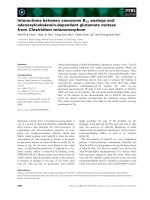
Fig. 1. Initiation of N-tail translocation is delayed in Cig30(1–100)(4R)-P2. (A) Schematic representation of the Cig30(1–100)(wt)-P2 and
Cig30(1–100)(4R)-P2 constructs. G1 and G2 indicate acceptor sites for N-linked glycosylation. (B) In vitro translation of mRNAs encoding
Cig30(1–100)(wt)-P2 (left) and Cig30(1–100)(4R)-P2 (right) constructs carrying both the G1 and G2 glycosylation acceptor sites (top), only the
G1 site (middle), or only the G2 site (bottom). The translation initiation inhibitor aurintricarboxylic acid was added to the translation mix
1.5 min after addition of the mRNA, and Triton X-100 was then added at the indicated times. The total translation time was 60 min. s,Un-
glycosylated molecules; d, singly glycosylated molecules; dd, doubly glycosylated molecules. (C) Quantification of the data for Cig30(1–100)
(wt)-P2 and Cig30(1–100)(4R)-P2 constructs carrying either the G1 or the G2 glycosylation acceptor site (middle and bottom panels in Fig. 1B).
n
, Cig30(1–100)(wt)-P2 (G1); h, Cig30(1–100)(wt)-P2 (G2); d, Cig30(1–100)(4R)-P2 (G1); s, Cig30(1–100)(4R)-P2 (G2); m, Cig30(1–100)(4R-6aa)-
P2 (G1). The final glycosylation levels after a 60 min incubation are shown by arrows for Cig30(1–100)(wt)-P2 (G1), Cig30(1–100)(4R)-P2 (G1),
and Cig30(1–100)(4R-6aa)-P2 (G1). The maximum level of glycosylation obtained in the in vitro system is around 80%.
M. Monne
´
et al. Kinetics of membrane protein insertion
FEBS Journal 272 (2005) 28–36 ª 2004 FEBS 29
tion reaction, in contrast, is insensitive to the presence
of detergent, and the glycosylation status of nascent
polypeptide chains can thus be determined as a func-
tion of translation time by adding detergent at differ-
ent times after chain initiation and then allowing
translation to proceed to completion [14,15].
As seen in Fig. 1B (left panel), for Cig30(1–100)(wt)-
P2, the G1 acceptor site in the N-tail is glycosylated
more rapidly than the G2 acceptor site in the P2
domain. For the G2 site to become glycosylated, an
additional 65 residues beyond this site must be poly-
merized to bridge the distance between the OST active
site and the ribosomal P-site [16], corresponding to a
total chain length of 183 residues. From Fig. 1C, the
t
1 ⁄ 2
for glycosylation of G2 is 6 min, corresponding to
a translation rate of 183 ⁄ 360 ¼ 0.5 residuesÆs
)1
, com-
parable to previously published values [14,15,17]. As
an independent measure of the average translation
rate, we also determined the t
1 ⁄ 2
necessary for the
appearance of a 190-residues long, truncated form
of Cig30 in the translation reaction to be 5.5 min,
corresponding to an average translation rate of
0.6 residuesÆs
)1
(data not shown).
Assuming that TM1 (residues 34–56) has to emerge
from the ribosome before the protein can be targeted
to the translocon, and given that the nascent-chain
conducting tunnel in the ribosome covers some 30–40
residues of the nascent chain, the calculated t
1 ⁄ 2
for
glycosylation of the G1 site in the N-tail is 90⁄ 0.5 ¼
180 s, in good agreement with the observed kinetics,
Fig. 1C. We conclude that glycosylation of the G1 and
G2 sites happen as soon as they become exposed to
the lumen of the microsome, and that the kinetics of
glycosylation is a good measure of the timing of trans-
location of the corresponding segment of the protein.
Compared to Cig30(1–100)(wt)-P2, the behavior of
the Cig30(1–100)(4R)-P2 construct is strikingly differ-
ent, Fig. 1B (right panel). The G2 site is glycosy-
lated with indistinguishable kinetics compared to
Cig30(1–100)(wt)-P2, while the modification of the G1
site is now dramatically delayed and is initiated con-
comitantly with or even after the modification of the
G2 site in the P2 domain, Fig. 1C. It is also notewor-
thy that there is an initial, fast phase of glycosylation
of the G1 site (up to a level of 20% glycosylation)
followed by a much slower until the final level of 42%
is reached (arrows). A similar slow phase has been seen
recently for glycosylation of Asn-X-Thr acceptor sites
located close to a protein’s C-terminus [17]. A possible
explanation could be that N-tails are so rapidly trans-
located across the membrane that some chains pass
OST too fast to be glycosylated and only become
modified in a slower, post-translational process.
The final level of glycosylation of the G1 site in
Cig30(1–100)(4R)-P2 is lower than in Cig30(1–100)(wt)-
P2 (42% vs. 69%); however, this seems to be due
mainly to a partial blocking of the Asn-Phe-Ser accep-
tor site by the nearby Arg residues, as the addition
of a six residues long spacer (VGAGVG) between
the G1 site and the four Arg residues [construct
Cig30(1–100)(4R-6aa)-P2] leads to an increase in the
final glycosylation level to 60% without appreciably
affecting the kinetics of the modification of the G1 site
(Fig. 1C and data not shown). A similar increase in
glycosylation efficiency (from 53% to 69%) was seen
previously when the 4R insertion was moved from
position 9 to position 28 in the N-tail of full-length
Cig30(4R) [8], again consistent with a partial blocking
effect of the 4R mutation when present next to the G1
glycosylation site.
We also followed the kinetics of glycosylation of the
G1 site for the full-length Cig30 protein fused to the
P2 reporter domain, and for a series of mutants with
increasing numbers of Arg residues in the N-tail
(Fig. 2). The initial delay increases with the number of
Arg residues in the N-tail. The kinetics of modification
of the G1 site in Cig30(wt)-P2 and Cig30(4R)-P2 are
indistinguishable from the corresponding kinetics for
the Cig30(1–100)(wt)-P2 and Cig30(1–100)(4R)-P2 con-
structs, respectively. Again, the glycosylation of the G1
site in the 4R and 5R mutants proceeds through a
rapid phase up to 30% glycosylation followed by a
much slower phase leading to a final level of 53%
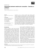
Fig. 2. Initiation of N-tail translocation is progressively delayed in
Cig30(wt)-P2 when increasing number of Arg residues is added to
the N-tail. Experiments were performed and quantified as in as in
Fig. 1B, but using the Cig30(wt)-P2 fusion protein with 0, 2, 4, and
five Arg residues added between residues 9 and 10 in the N-tail as
indicated. Only the G1 glycosylation acceptor site in the N-tail is
present in these constructs.
n
, Cig30(wt)-P2 (G1); h, Cig30(2R)-P2
(G1); d, Cig30(4R)-P2 (G1); s, Cig30(5R)-P2 (G1). The final glycosy-
lation levels after a 60 min incubation is shown by arrows for
Cig30(4R)-P2 (G1) and Cig30(5R)-P2 (G1).
Kinetics of membrane protein insertion M. Monne
´
et al.
30 FEBS Journal 272 (2005) 28–36 ª 2004 FEBS
glycosylation for Cig30(4R)-P2 (63% when the six resi-
dues long spacer mentioned above is inserted into the
N-tail) and to 39% for Cig30(5R)-P2.
We conclude that there is an initial delay in N-tail
translocation, as measured by glycosylation of the G1
site, in both truncated and full-length Cig30 mutants
with extra Arg residues in the N-tail compared to the
wild-type protein, and that translocation of the lume-
nal P2 domain following the second transmembrane
segment in the Cig30(1–100)(4R)-P2 construct, as
measured by glycosylation of the G2 site, is initiated
concomitant with or even before N-tail translocation.
Asp residues in the N-tail have a minor kinetic
effect on translocation
We also tested the effect of placing four Asp residues
in the Cig30 N-tail, both in the context of the full-
length Cig30(wt)-P2 fusion and in Cig30(1–100)(wt)-P2.
The G1 site in the N-tail of Cig30(4D)-P2 is glycosyl-
ated with slightly delayed kinetics compared to Cig30-
P2, and the final level of glycosylation is the same for
both constructs ( 75%) (Fig. 3). A slight delay was
also seen for Cig30(1–100)(4D)-P2. Thus, the 4D
mutation has a weaker but still detectable effect on the
translocation kinetics of the N-tail.
Competition between neighboring
transmembrane segments
The delayed glycosylation of the G1 site in
Cig30(1–100)(4R)-P2 suggested to us that the intrinsic
topological preference of the N-tail ⁄ TM1 region in this
construct may be N
cyt
–C
lum
, but that TM1 is either
prevented from inserting with this orientation by the
more rapid membrane insertion and translocation of
the neighboring TM2 ⁄ P2 domain, or that it initially
inserts in the N
cyt
–C
lum
orientation but then re-orients
as a result of the insertion of the TM2 ⁄ P2 domain
[9,11,18].
To gain further insight into this phenomenon, we
compared two constructs where only the N-tail ⁄ TM1
region of Cig30 (residues 1–70) is fused to the P2
domain: one with the wild-type N-tail [Cig30(1–
70)(wt)-P2], and one with the 4R mutation in the
N-tail (Cig30(1–70)(4R)-P2), Fig. 4A. As shown previ-
ously [8], the G1 site in Cig30(1–70)(wt)-P2 was effi-
ciently glycosylated, and only the G2 site in Cig30(1–
70)(4R)-P2 was glycosylated to a final level of 74%
(data not shown), indicating that the Cig30(1–70)(4R)-
P2 is oriented with the opposite topology compared to
the corresponding wt construct. The glycosylation kin-
etics of the G1 site in Cig30(1–70)-P2 and the G2 site
in Cig30(1–70)(4R)-P2 were as expected from the posi-
tions of the respective glycosylation sites and the aver-
age translation rate (0.5 residuesÆs
)1
), Fig. 4B.
We also tested the related construct Cig30(1–70)(4D)-
P2 with four Asp residues in the N-tail. As noted
above, glycosylation of the N-tail is somewhat delayed
Fig. 3. Asp residues in the N-tail have a minor kinetic effect on trans-
location. Experiments were performed and quantified as in as
in Fig. 1B, but using the Cig30(wt)-P2 (denoted FL) and
Cig30(1–100)(wt)-P2 (denoted TM1-2) fusion proteins with or without
four Asp residues added between residues 9 and 10 in the N-tail as
indicated. Only the G1 glycosylation acceptor site in the N-tail is pre-
sent in these constructs.
n
, Cig30(wt)-P2 (G1); h, Cig30(4D)-P2
(G1); d, Cig30(1–100)(wt)-P2 (G1); s, Cig30(1–100)(4D)-P2 (G1).
A
B
Fig. 4. Arg and Asp residues in the N-tail of the Cig30(1–70)(4R)-P2
construct promote the N
cyt
orientation. (A) Schematic representa-
tion of the Cig30(1–70)-P2 constructs. (B) Experiments were
performed and quantified as in as in Fig. 1B but using the
Cig30(1–70)-P2 fusion protein (denoted TM1) with or without four
Arg or four Asp residues added between residues 9 and 10 in the
N-tail as indicated. Results for constructs containing either the G1
or the G2 glycosylation acceptor sites are shown.
n
, Cig30(1–
70)(wt)-P2 (G1); h, Cig30(1–70)(4R)-P2 (G2); d, Cig30(1–70)(4D)-P2
(G1); s, Cig30(1–70)(4D)-P2 (G2). In vitro translations of Cig30(1–
70)(4D)-P2 constructs carrying either the G1 site in the N-tail or the
G2 site in the P2 domain are also shown (left). s, Unglycosylated
molecules; d, glycosylated molecules.
M. Monne
´
et al. Kinetics of membrane protein insertion
FEBS Journal 272 (2005) 28–36 ª 2004 FEBS 31
in the longer Cig30(1–100)(4D)-P2 and Cig30(4D)-P2
constructs compared to Cig30(1–100)(wt)-P2 and
Cig30(wt)-P2. Interestingly, the 4D mutation has a clear
effect on the topology of Cig30(1–70)(4D)-P2. This con-
struct is glycosylated with about equal efficiencies on
the G1 and G2 sites, Fig. 4B, and hence has a mixed
topology with only about half the molecules in the
N
lum
–C
cyt
orientation. Glycosylation of the N-tail is
delayed in Cig30(1–70)(4D)-P2 compared to Cig30(1–
70)(wt)-P2, and the G1 site is glycosylated only slightly
before the G2 site in this construct (Fig. 4B).
The finding that TM1 adopts the N
cyt
–C
lum
orienta-
tion in Cig30(1–70)(4R)-P2 but the N
lum
–C
cyt
orienta-
tion in Cig30(1–100)(4R)-P2 indicated that the length
of the loop between TM1 and TM2 may be a critical
topological determinant, such that a longer loop
might allow the intrinsic topological preference of the
N-tail ⁄ TM1 region to become more dominant. We
therefore made a series of constructs based on full-
length Cig30(4R)-P2 where the length of the loop
between TM1 and TM2 was increased from 11 to 72
residues (Fig. 5). Consistent with our expectations, the
level of N-tail glycosylation decreased from 53% for
the shortest loop to background levels (8%) for the
longest loop. In the latter construct, when a glyco-
sylation acceptor site was inserted in the TM1-TM2
loop it was efficiently modified (71% glycosylation;
black circle, Fig. 5), showing that TM1 indeed has a
N
cy
–C
lum
orientation in this case.
We conclude that the Cig30(4R) N-tail ⁄ TM1 region
intrinsically prefers the N
cyt
–C
lum
orientation, and sug-
gest that the translocation of the downstream segment
is initiated very soon after the TM1 segment enters the
translocon when TM2 is absent or when the separation
between TM1 and TM2 is sufficiently long. When the
TM1-TM2 loop is short, however, TM1 adopts its less
preferred N
lum
–C
cyt
orientation.
For Cig30(1–70)(4D)-P2, the N
lum
–C
cyt
and N
cyt
–
C
lum
orientations are roughly equally preferred, and
the G1 and G2 sites are modified with similar kinetics.
In Cig30(1–100)(4D)-P2 and Cig30(4D)-P2, in con-
trast, the N-tail is efficiently translocated in essentially
100% of the molecules.
Discussion
So far, not much is known about the kinetics of mem-
brane protein insertion into the ER membrane and
whether membrane protein topology is in some sense
under kinetic control. Studies in vivo using an engin-
eered phosphorylation site as a reporter for the trans-
location of an N-tail across the ER membrane have
shown that targeting to the ER is rapid, and that the
factors determining the overall rate of N-tail transloca-
tion are the time it takes for the ER targeting signal to
become exposed outside the ribosome and the rate of
the ensuing N-tail translocation reaction, which has
been estimated to be 1.6 times faster than the trans-
lation rate on a per-residue basis [19]. A study using
engineered glycosylation sites has further shown that
N-tails are translocated in a C-to-N-terminal direction,
starting from the N-terminal transmembrane segment
[7]. Finally, the extracytoplasmic segments in the
multispanning membrane protein bacterioopsin have
been shown to become exposed on the extracellular
surface cotranslationally and in a sequential order
starting with the N-tail when the protein is expressed
in Halobacterium salinarium [6]. Neighboring trans-
membrane segments may also affect each other’s orien-
tation, suggesting a rather complex process of topology
determination in the ER translocon [7–9,11,20].
Here, we have used engineered glycosylation sites in
fusions between the full-length mouse Cig30 protein,
the Cig30 N-tail ⁄ TM1 region (residues 1–70), and the
N-tail ⁄ TM1 ⁄ TM2 region (residues 1–100) and a repor-
ter domain (P2) from the E. coli Lep protein to follow
the translocation of the N-tail and the P2 domain
across microsomal membranes in vitro. As estimated
from the average translation rate in the in vitro system,
the engineered glycosylation sites become modified as
soon as they enter the ER lumen. The glycosylation
kinetics of a given acceptor site can thus be used to
track the translocation of the corresponding domain in
the model protein.
For Cig30(1–100)(wt)-P2, we find a sequential trans-
location process where the N-tail is translocated as
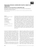
Fig. 5. Increased separation between TM1 and TM2 in Cig30(4R)-
P2 favors a cytoplasmic location of the N-tail. Cig30(4R)-P2-derived
constructs with increasingly long loops between TM1 and TM2
were translated in the presence of microsomes. s, Percentage of
molecules glycosylated on the G1 site in the N-tail; d level of gly-
cosylation of a glycosylation acceptor site engineered into in the
loop between TM1 and TM2 in the construct with a 72 residues
long loop.
Kinetics of membrane protein insertion M. Monne
´
et al.
32 FEBS Journal 272 (2005) 28–36 ª 2004 FEBS
soon as TM1 enters the translocon, followed by trans-
location of the P2 domain initiated by TM2, Fig. 1.
In sharp contrast, however, the kinetics of N-tail
translocation is strongly delayed in the Cig30(1–
100)(4R)-P2 mutant (where four Arg residues have
been introduced into the N-tail), and is initiated at
about the same time or even after translocation of the
P2 domain (Fig. 1). Similarly, addition of two to five
Arg residues to the N-tail of the full-length Cig30-P2
fusion progressively delays the onset of N-tail translo-
cation (Fig. 2). Interestingly, addition of four Asp resi-
dues to the N-tail of full-length Cig30(wt)-P2 and to
Cig30(1–100)-P2 also causes a somewhat delayed onset
of translocation (Fig. 3).
The delayed translocation of the N-tail in
Cig30(4R)-P2 and Cig30(1–100)(4R)-P2 suggests that
the N-tail ⁄ TM1 region in these constructs has an
intrinsic preference for inserting into the ER with
N
cyt
–C
lum
orientation – in keeping with the so-called
positive-inside rule [21] – rather than the N
lum
–C
cyt
orientation adopted in the presence of TM2. This is
indeed the case. When the Cig30(4R) N-tail ⁄ TM1
region is fused directly to the P2 reporter domain [con-
struct Cig30(1–70)(4R)-P2], P2 is translocated rapidly
across the ER membrane (Fig. 4). In contrast, the
N-tail is translocated rapidly in Cig30(1–70)(wt)-P2.
Finally, Cig30(1–70)(4D)-P2 adopts a mixed topology
with roughly equal amounts of N
cyt
–C
lum
and N
lum
–
C
cyt
oriented molecules, and with almost identical
translocation kinetics for the N-tail and the P2
domain. For constructs such as Cig30(1–100)(4R)-P2
where the N-tail ⁄ TM1 region has an intrinsic prefer-
ence for the N
cyt
–C
lum
orientation, more of the N
cyt
–
C
lum
orientation is observed when the loop between
TM1 and TM2 is progressively lengthened (Fig. 5).
These results show a strong correlation between the
relative timing of translocation initiated from the TM1
and TM2 transmembrane segments and the final topol-
ogy of the protein, suggesting that some topological
signals may dominate over others within the translo-
con. A possible mechanism of topology formation sug-
gested by these results, Fig. 6, is that TM1 enters the
translocon first and thus gets a head start over TM2.
If the N-tail is not highly charged, it translocates rap-
idly, fixing TM1 in a N
lum
–C
cyt
orientation. TM2 then
initiates translocation of the P2 domain. In contrast, if
the N-tail contains a sufficient number of positively
charged residues it prefers a N
cyt
–C
lum
orientation.
This is the orientation obtained when only TM1 is pre-
sent, or when the connecting loop between TM1 and
TM2 is sufficiently long. If the connecting loop is
short, however, either of two things may happen:
(a) TM1 inserts initially in its preferred N
cyt
–C
lum
orientation but is then somehow forced to re-orient to
the N
lum
–C
cyt
orientation when TM2 inserts [9,11], or
(b) TM1 does not have time to insert before TM2 initi-
ates rapid translocation of the P2 domain, giving TM1
no option but to translocate the N-tail. A somewhat
more complicated variation on (a), suggested by some
recent work [9,18], is that TM1 inserts initially in the
N
lum
–C
cyt
orientation, but then re-orients (before it
has time to become glycosylated) if the N-tail contains
many charged residues, unless the rapid insertion of
TM2 prevents re-orientation.
Regardless of the exact mechanism, however, our
results strongly suggest that there is a critical period
from about the time when TM1 enters the translocon
during which the appearance of TM2 in the translocon
Fig. 6. Model for the membrane insertion of Cig30(1–70)-P2 (panel
A) and Cig30(1–100)-P2 (panel B) derived constructs into the micro-
somal membrane. The top cartoon in each panel shows constructs
such as Cig30(1–100)(wt)-P2 that have no additional charge resi-
dues in their N-tail, while the bottom cartoons are for constructs
such as Cig30(1–100)(4R)-P2 with a positively charged N-tail. TM1
is white and TM2 is striped. The G1 and G2 glycosylation acceptor
sites are indicated; unfilled symbols represent nonglycosylated
sites, filled symbols represent glycosylated sites.
M. Monne
´
et al. Kinetics of membrane protein insertion
FEBS Journal 272 (2005) 28–36 ª 2004 FEBS 33
can affect the final orientation of TM1. One attractive
possibility is that the end of this critical period corres-
ponds to the point where TM1 exits the translocation
channel and becomes lodged in the surrounding lipid
bilayer [22–24].
In summary, we have shown that the introduction
of charged residues in the lumenal N-tail of Cig30 cau-
ses a strong delay in the translocation of the N-tail
that goes in parallel with an increased intrinsic prefer-
ence for a N
cyt
–C
lum
orientation of the isolated
N-tail ⁄ TM1 domain. This intrinsic preference can be
overridden by the following TM2 segment (that also
has a preference for the N
cyt
–C
lum
orientation), provi-
ded that the loop between TM1 and TM2 is short.
The final topology of the protein thus seems to result
from a finely tuned competition between neighboring
topogenic signals (the TM segments and their immedi-
ate flanking regions) that is influenced both by the
presence or absence of charged residues (where posi-
tively charged residues are more potent topological
determinants than negatively charged ones), possibly
by the hydrophobicity of the transmembrane segments
[25], and by the length of the loops separating them
[9]. Finally, our results suggest that the ‘positive inside’
rule for membrane protein topology [21] may at least
in part be explained by a reduction in the rate of mem-
brane translocation of segments of the nascent poly-
peptide chain with a high content of positively charged
residues.
Experimental procedures
Enzymes and chemicals
Unless otherwise stated, all enzymes were from Promega
(Madison, WI, USA). Ribonucleotides, deoxyribonucleo-
tides, dideoxyribonucleotides, the cap analog m7G(5¢)-
ppp(5¢)G, T7 DNA polymerase, and [
35
S]methionine were
from Amersham–Pharmacia Biotech (Uppsala, Sweden).
Plasmid pGEM1, dithiothreitol, BSA, RNasin and rabbit
reticulocyte lysate were from Promega (Madison). Spermi-
dine, aurintricarboxylic acid, and Triton X-100 were from
Sigma–Aldrich Inc. (St. Louis, MO, USA). Oligonucleo-
tides were from Cybergene (Stockholm, Sweden).
DNA techniques
SalI and XbaI restriction sites were introduced by PCR at
the 5¢ and 3¢ ends of the cig30 gene, respectively. The PCR
fragment was cloned into phage M13mp18, and into a
pGEM1-derived plasmid after a modified upstream region
of the lepB gene containing a Kozak consensus sequence
[26] for efficient ribosome binding. Site-directed mutagen-
esis was performed according to Kunkel [27,28] to intro-
duce four Arg codons between the 9th and 10th codon in
the cig30 coding region. In some constructs, the natural G1
glycosylation site of Cig30 was silenced by mutating
Asn6fiThr, and in others a six residue spacer (VGAGVG)
was inserted after the 9th codon using the QuickChange kit
(Stratagene).
Cig30 fusions with the P2 domain of Lep were made by
introducing a NdeI site in codon 70 (i.e. before TM2) or in
codon 100 (after TM2) by PCR amplification using primers
encoding the two flanking restriction sites. The SalI-NdeI
restricted PCR fragments were cloned into a pGEM1-
derived vector containing the P2 domain (codons 81–323)
of Lep preceded by a NdeI site. The natural glycosylation
site in P2 was silenced by mutating Asn214 fi Gln and two
version of P2, with and without the G2 consensus glycosy-
lation site [NST(96–98)], were used.
For the elongation of the loop between TM1 and TM2,
parts of the P2 domain of Lep were cloned into the first
cytoplasmic loop in Cig30 utilizing engineered EcoRV and
NdeI restriction sites. The 11 residues in the Cig30 TM1–
TM2 loop were retained both in the N- and C-terminal
parts of the elongated loops. In one construct, an Asn-Ser-
Thr glycosylation acceptor site was introduced in the mid-
dle of the 72 residues long loop. The three resulting loops
had the following lengths and sequences (Lep-derived seg-
ments are underlined; the three residues LIG in the 72 resi-
dues long loop that were exchanged to NST in the
glycosylation mutant are in bold; numbers refer to residue
positions in the Cig30 and Lep proteins): 40 residues,
QRP(67)
Y(81)EPFQIPSGSMMPTLLI(97)DI R(57)trk; 55
residues, QRP(67)
Y(81)EPFQIPSGSMMPTLLIGDFILVE
KFAYGIKD(112)DIR(57)trk; 72 residues, QRP(67)Y(81)
EPFQIPSGSMMPTLLIGDFILVEKFAYGIKDPIYQKTL
IENGHPKRG(128)DIR(57)TRK.
All constructs were confirmed by sequencing of plasmid
DNA using T7 DNA polymerase.
In vitro expression
Constructs in pGEM1 were amplified using a 5¢ primer
hybridizing upstream of the SP6 promoter and the 3¢ pri-
mers described above. The PCR products were transcribed
by SP6 RNA polymerase for 1 h at 37 °C. The transcription
mixture was as follows: 1–5 lg DNA template, 5 lL10·
SP6 H-buffer [400 mm Hepes ⁄ KOH (pH 7.4), 60 mm Mg
acetate, 20 mm spermidine ⁄ HCl], 5 lL BSA (1 mgÆmL
)1
),
5 lL m7G(5¢)ppp(5¢)G (10 mm), 5 lL dithiothreitol (50 mm),
5 lL rNTP mix (10 mm ATP, 10 mm CTP, 10 mm
UTP, 5 mm GTP), 18.5 lLH
2
O, 1.5 lL RNase inhibitor
(50 units), 0.5 lL SP6 RNA polymerase (20 units).
Translation of 1 lL mRNA in 9 l L nuclease-treated reti-
culocyte lysate, 1 lL RNase inhibitor (40 unitsÆlL
)1
), 1 lL
[
35
S]Met (10 lCiÆlL
)1
), 1 lL amino acids mix (1 mm of
each amino acid except Met), 1 lL dog pancreas rough
Kinetics of membrane protein insertion M. Monne
´
et al.
34 FEBS Journal 272 (2005) 28–36 ª 2004 FEBS
microsomes was performed as described [29] at 30 °C for
60 min.
For the kinetic studies, the translation mix was preincuba-
ted 4 min before [
35
S]Met and mRNA was added, and the
translation initiation inhibitor aurintricarboxylic acid was
added to a final concentration of 0.075 mm after an addi-
tional 1.5 min [14]. Samples were removed at different time
points and were incubated further at 30 °C in the presence of
1% (v ⁄ v) Triton X-100 until a total translation time of
60 min.
Translation products were analyzed by SDS ⁄ PAGE
and gels were quantified on a Fuji FLA-3000 phosphor-
imager using the image reader 8.1j software. The glyco-
sylation efficiency was calculated as the quotient between
the intensity of the glycosylated band divided by the
summed intensities of the glycosylated and nonglycosyla-
ted bands. In general, the glycosylation efficiency varied
by no more than ±5% between different experiments (at
least two independent measurements were made for all
constructs).
Acknowledgements
This work was supported by grants from the Swedish
Cancer Foundation and the Swedish Research Council
to G.vH. Dog pancreas microsomes were a kind gift
from Dr M. Sakaguchi, University of Hyogo, Japan.
References
1 Johnson AE & van Waes MA (1999) The translocon: a
dynamic gateway at the ER membrane. Annu Rev Cell
Dev Biol 15, 799–842.
2 de Gier JW & Luirink J (2001) Biogenesis of inner
membrane proteins in Escherichia coli. Mol Microbiol
40, 314–322.
3 Goder V & Spiess M (2001) Topogenesis of membrane
proteins: determinants and dynamics. FEBS Lett 504,
87–93.
4 von Heijne G (2000) Recent advances in the under-
standing of membrane protein assembly and structure.
Quart Rev Biophys 32, 285–307.
5 Blobel G (1980) Intracellular protein topogenesis. Proc
Natl Acad Sci USA 77, 1496–1500.
6 Dale H, Angevine CM & Krebs MP (2000) Ordered
membrane insertion of an archaeal opsin in vivo. Proc
Natl Acad Sci USA 97, 7847–7852.
7 Nilsson I, Witt S, Kiefer H, Mingarro I & von Heijne
G (2000) Distant downstream sequence determinants
can control N-tail translocation during protein insertion
into the endoplasmic reticulum membrane. J Biol Chem
275, 6207–6213.
8 Monne
´
M, Gafvelin G, Nilsson R. & von Heijne G
(1999) N-tail translocation in a eukaryotic polytopic
membrane protein – Synergy between neighboring trans-
membrane segments. Eur J Biochem 263, 264–269.
9 Goder V, Bieri C & Spiess M (1999) Glycosylation can
influence topogenesis of membrane proteins and reveals
dynamic reorientation of nascent polypeptides within
the translocon. J Cell Biol 147, 257–266.
10 Anthony V & Skach WR (2002) Molecular mechanism
of P-glycoprotein assembly into cellular membranes.
Curr Protein Pept Sci 3, 485–501.
11 Lu Y, Turnbull IR, Bragin A, Carveth K, Verkman AS
& Skach WR (2000) Reorientation of aquaporin-1
topology during maturation in the endoplasmic retic-
ulum. Mol Biol Cell 11, 2973–2985.
12 Carveth K, Buck T, Anthony V & Skach WR (2002)
Cooperativity and flexibility of cystic fibrosis transmem-
brane conductance regulator transmembrane segments
participate in membrane localization of a charged
residue. J Biol Chem 277, 39507–39514.
13 Tvrdik P, Asadi A, Kozak L, Nedergaard J, Cannon B
& Jacobsson A (1997) Cig30, a mouse member of a
novel membrane protein gene family is involved in the
recruitement of brown adipose tissue. J Biol Chem 272,
31738–31746.
14 Garoff H, Huylebroeck D, Robinson A, Tillman U,
Liljetro
¨
m & P (1990) The signal sequence of the p62-
protein of Semliki Forest virus is involved in initiation
but not in completing chain translocation. J Cell Biol
111, 867–876.
15 Rothman J & Lodish H (1977) Synchronised transmem-
brane insertion and glycosylation of a nascent mem-
brane protein. Nature 269, 775–780.
16 Whitley P, Nilsson IM & von Heijne G (1996) A nas-
cent secretory protein may traverse the ribosome ⁄ ER
translocase complex as an extended chain. J Biol Chem
271, 6241–6244.
17 Hessa T, Monne
´
, M & von Heijne G (2003) Stop-trans-
fer efficiency of marginally hydrophobic stop-transfer
segments depends on the length of the C-terminal tail.
EMBO Report 4, 178–183.
18 Goder V & Spiess M (2003) Molecular mechanism of
signal sequence orientation in the endoplasmic reticu-
lum. EMBO J 22, 3645–3653.
19 Goder V, Crottet P & Spiess M (2000) In vivo kinetics
of protein targeting to the endoplasmic reticulum deter-
mined by site-specific phosphorylation. EMBO J 19,
6704–6712.
20 Ota K, Sakaguchi M, von Heijne G, Hamasaki N &
Mihara K (1998) Forced transmembrane orientation of
hydrophilic polypeptide segments in multispanning
membrane proteins. Mol Cell 2, 495–503.
21 von Heijne G (1986) The distribution of positively
charged residues in bacterial inner membrane proteins
correlates with the trans-membrane topology. EMBO J
5, 3021–3027.
M. Monne
´
et al. Kinetics of membrane protein insertion
FEBS Journal 272 (2005) 28–36 ª 2004 FEBS 35
22 Mothes W, Heinrich S, Graf R., Nilsson I, von Heijne
G, Brunner J & Rapoport T (1997) Molecular mechan-
isms of membrane protein integration into the endoplas-
mic reticulum. Cell 89, 523–533.
23 Heinrich S, Mothes W, Brunner J & Rapoport T (2000)
The Sec61p complex mediates the integration of a mem-
brane protein by allowing lipid partitioning of the trans-
membrane domain. Cell 102, 233–244.
24 McCormick PJ, Miao Y, Shao Y, Lin J & Johnson AE
(2003) Cotranslational protein integration into the ER
membrane Is mediated by the binding of nascent chains
to translocon proteins. Mol Cell 12, 329–341.
25 Wahlberg JM & Spiess M (1997) Multiple determinants
direct the orientation of signal-anchor proteins: the
topogenic role of the hydrophobic signal domain. J Cell
Biol. 137, 555–562.
26 Kozak M (1999) Initiation of translation in prokaryotes
and eukaryotes. Gene 234, 187–208.
27 Kunkel TA (1987) Rapid and efficient site-specific muta-
genesis without phenotypic selection. Methods Enzymol
154, 367–382.
28 Geisselsoder J, Witney F & Yuckenberg P (1987) Effi-
cient site-directed in vitro mutagenesis. Biotechniques 5,
786–791.
29 Liljestro
¨
m P & Garoff H (1991) Internally located clea-
vable signal sequences direct the formation of Semliki
Forest virus membrane proteins from a polyprotein
precursor. J Virol 65, 147–154.
Kinetics of membrane protein insertion M. Monne
´
et al.
36 FEBS Journal 272 (2005) 28–36 ª 2004 FEBS