Báo cáo khoa học: Solution structure of the active-centre mutant I14A of the histidinecontaining phosphocarrier protein from Staphylococcus carnosus ppt
Bạn đang xem bản rút gọn của tài liệu. Xem và tải ngay bản đầy đủ của tài liệu tại đây (704.33 KB, 10 trang )
Solution structure of the active-centre mutant I14A of the histidine-
containing phosphocarrier protein from
Staphylococcus carnosus
Andreas Mo¨ glich
1,
*, Brigitte Koch
2
, Wolfram Gronwald
1
, Wolfgang Hengstenberg
2
, Eike Brunner
1
and Hans Robert Kalbitzer
1
1
Institute of Biophysics and Physical Biochemistry, University of Regensburg, Germany;
2
SG Physiology of Microorganisms,
Ruhr-University of Bochum, Germany
High-pressure NMR experiments performed on the histi-
dine-containing phosphocar rier protein (HPr) from Sta-
phylococcus carnosus have shown that residue Ile14, which is
located in t he active-centre loop, exhibits a peculiarly small
pressure response. In contrast, the rest of the loop shows
strong pre ssure effects a s i s e xpected for t ypical protein
interaction sites. To elucidate the structural role of t his
residue, the mutant protein HPr(I14A), in which Ile14 is
replaced by Ala, was produced and s tudied by solution
NMR spectroscopy. On the basis of 1406 structural
restraints including 20 directly detected hydrogen bonds, 49
1
H
N
-
15
N, and 25
1
H
N
-
1
H
a
residual dipolar couplings, a well
resolved three-dimensional structure could be determined.
The o verall fold of the protein is not influenced by the
mutation but characteristic conformational changes are
introduced into the active-centre loop. They lead to a dis-
placement of the ring system of His15 and a distortion of the
N-terminus of the first helix, which supports the histidine
ring. In ad dition, the C -terminal helix is bent because the side
chain of Leu86 located at the end of this helix partly fills the
hydrophobic cavity created by the mutation. Xenon, which
is known to occupy hydrophobic cavities, causes a partial
reversal of the mutation-induced structural effects. The
observed structural changes explain the reduced phospho-
carrier activity of the mutant and agree well with the earlier
suggestion that Ile14 r epresents an anchoring point stabil-
izing the active-centre loop in its correct conformation.
Keywords: histidine-containing phosphocarrier protein
(HPr); mutant protein; nuclear magnetic resonance (NMR);
protein structure.
Histidine-containing phosphocarrier protein (HPr) is a
central part of the bacterial carbohydrate/phosphoenolpyru-
vate phosphotransfer system (PTS) first described in Escheri-
chia coli [1]. The PTS catalyses the phosphorylation of a
metabolite and its concomitant transport across the plasma
membrane into the cytosol (PTS reviewed in [2,3]). During
the transport p rocess, the phosphoryl group of phos-
phoenolpyruvate is transferred first to enzyme I (EI) and
then to His15 of HPr. The phosphoryl group is transiently
bound to the N
d1
atom of the imidazole ring of His15. Via
enzymes IIA, IIB and IIC/D, the group is finally transferred
to the imported metabolite. Compared to conventional
substrate import and consecutive phosphorylation, the
import v ia the PTS is energetically favorable. From the
residue His15 of HPr, the phosphoryl group can also be
transferred t o transcription factors containing PTS regula-
tion domains (PRDs). Depending on their phosphorylation
state, these proteins control the activity of operons mainly
responsible for catabolism [3,4]. The activity of HPr from
Gram-positive bacteria is regulated by the bifunctional
enzyme HPr kinase/phosphorylase, which controls the
phosphorylation state of the HPr residue Ser46 [5]. When
phosphorylated at residue Ser46, HPr interacts with cata-
bolite control protein A (CcpA), which regulates the activity
of genes involved in carbon and nitrogen metabolism [6,7].
To exert its various biological functions, the HPr
molecule must be able to interact with different proteins
and ligands in a tightly regulated manner mainly depending
upon the nutritional state of the bacterium. Probably these
different interactions are mediated by conformational
changes of HPr, particularly in the active site region.
Structural studies have contributed significantly to a
detailed understanding of the bacterial PTS.
The three-dimensional structu res of HPr molecules from
different organisms have been determined both by NMR
spectroscopy and X-ray crystallography (e.g. [8–12]). Signi-
ficant structural changes in the active site region of the HPr
were observed upon phosphorylation at Ser46 [13,14]. The
solution structure of HPr from Staphylococcus carnosus,a
small protein with a molecular mass of 9511 Da, has been
determined by Go
¨
rler et al. [15]. It shows the open-faced
b-sandwich fold common to all HPr structu res known so f ar.
It consists of a four-stranded antiparallel b-sheet, one short
Correspondence to H. R. Kalbitzer, Institute of Biophysics and Phys-
ical Biochemistry, University of Regensburg, Regensburg, Germany.
Fax: +49 941943 2479, Tel.: +49 941943 2594,
E-mail:
Abbreviations: CHAPSO, 3-( cholamidopropyl)-dimethylammonio
2-hydroxyl-1-propane sulfonate; DIODPC, 1,2-di-O-dodecyl-sn-glyc-
ero-3-phosphocholine; HPr, histidine-containing phosphocarrier pro-
tein; PRD, PTS regulation domain; PTS, phosphoenolpyruvate-
dependentphosphotransferasesystem;RDC,residualdipolarcoupling.
*Present address:DepartmentofBiophysicalChemistry,Biozentrum,
University of Basel, Switz erland.
(Received 28 July 2004, revised 13 October 2004,
accepted 21 October 2004)
Eur. J. Biochem. 271, 4815–4824 (2004) Ó FEBS 2004 doi:10.1111/j.1432-1033.2004.04447.x
and t wo long a-helice s. K albitzer et al.[16]usedhigh-
pressure
1
Hand
15
N N MR measurements to study the ability
of HPr to adopt different conformations. In general,
dynamic regions in proteins are expected to be capable of
undergoing large conformational changes. This should
result in strong effects induced by the variation of external
conditions such as pressure. While the core region of HPr
from S. carnosus, w hich mainly consists of the b-sheet,
showed only little or m oderate variation, large pressure-
induced changes o f chemical shifts w ere observed i n the
active site region encompassing residues 12–18. In contrast to
its surrounding residues, Ile14 displayed only a s mall
pressure-induced change of chemical shift. Kalbitzer et al.
[16] suggested that this r esidue, which is not strictly conserved
between different species, serves as an anchoring point for the
active site loop. The isoleucine side chain would stabilize the
loop but still allow it to adopt the different conformations
necessary for the interaction with diverse proteins andligands.
To evaluate this hypothesis, a mutant form of HPr from
S. carnosus was produced in which the isoleucine at position
14 is replaced by an alanine residue, referred to as
HPr(I14A). In this paper we report the solution structure
of this protein as determined by NMR spectroscopy.
Materials and methods
Protein purification and sample preparation
The gene for the I14A mutant of HPr was constructed using
the PCR-Megaprimer method [17] and cloned into the
pET11 vector. T he plasmid was overexpress ed in E. coli
strain BL21 DE3. HPr(I14A) was isolated as described
previously [18]. Uniformly
15
Nand
15
N/
13
C isotope-labelled
protein was obtained accordingly. HPr(I14A) was studied
under the same conditions as used for the structure
determination of the wild-type protein [15]. Lyophilized
HPr protein was dissolved in a buffer solution containing
20 m
M
potassium phosphate, 100 m
M
KCl, 0.1 m
M
EDTA,
1m
M
NaN
3
,1 l
M
pepstatin, 1 l
M
leupeptin, 0.1 l
M
bovine
pancreatic trypsin inhibitor and, as an internal reference,
0.1 m
M
2,2-dimethyl-2-silapentanesulfonic acid. The pH
was adjusted to 7.14 by addition of KOH. The fi nal protein
concentrationwasbetween1.4and1.7m
M
depending on
the NMR experiment. For the determination of residual
dipolar couplings (RDCs), partial molecular orientation of
the HPr sample was obtained by the addition of 7.5% or
4.0% (w/v) of the bicelle forming lipid mixture 1,2-di-O-
dodecyl-sn-glycero-3-phosphocholine (DIODPC)/3-(cho-
lamidopropyl)-dimethylammonio 2-hydroxyl-1-propane
sulfonate (CHAPSO) at a 4.3 : 1 ratio [19].
NMR spectroscopy
NMR spectra were recorded on Bruker (Karlsruhe, Ger-
many) DMX-500, DMX-600 and DMX-800 spectromete rs
with
1
H resonance frequencies of 500, 600 and 800 MHz,
respectively. All measurements were carried out at a
temperature of 2 98 K. Time-domain NMR data were
processed using the
XWINNMR
package (Bruker). P roton
chemical shifts were assigned on the basis of 2D TOCSY
and HCCH-TOCSY spectra measured a t 500 and
600 M Hz, respectively. Nitrogen (
15
N) and carbon (
13
C)
resonances could be d etermined from HSQC, HNCA,
HNCO and CBCA(CO)NH spectra recorded at 600 MHz.
Distance restraints were derived from homonuclear 2D
NOESY spectra in
1
H
2
Oand
2
H
2
O and from a
13
Cresolved
NOESY spectrum, measured at 800, 500 and 600 MHz,
respectively. The assignment of NOE signals was facilitated
by using a homology structure of the HPr(I14A) protein
which was generated by the computer program
PERMOL
(A.
Mo
¨
glich, D. Weinfurtner, T. Maurer, W. Gronwald, H. R.
Kalbitzer, unpublished data). From this structure and the
assigned resonance frequencies, 2D NOESY spectra were
calculated using the computer program
RELAX
[20]. These
calculated spectra w ere c ompared with the experimental
data. The
1
H chemical shifts were referenced relative to 2,2-
dimethyl-2-silapentane-5-sulfonic acid.
15
Nand
13
Creso-
nances were referenced indirectly [21]. Spectral visualization
and volume i ntegration of NOE signals was carried out
using the computer program
AUREMOL
[22].
Determination of dihedral angles and hydrogen bonds
Three-bond coupling c onstants between H
N
and H
d
atoms,
3
J
HN-Ha
, were measured by MOCCA-SIAM experiments
[23,24]. T he values of the coupling constants were determined
using the procedure described by Titman and Keeler [25].
Structural restraints for the main chain dihedral angles F
were calculated according to the Karplus equation [26]
employing the parameters determined by Vuister and Bax
[27]. Hydrogen bonds between main chain amide protons
and carbonyl oxygen atoms were directly detected in
H(N)CO experiments as described by Cordier et al. [28,29].
Residual dipolar couplings
The measurement of residual d ipolar couplings requires
partial molecular a lignment of the sample molecules, which
was obtained by t he addition of 7.5% (w/v) of the DIODPC/
CHAPSO lipid mixture to the sample solution. RDCs
[30–32], were measured for the
1
H
N
-
15
N amide bond using
both conventional nondecoupled
1
H-
15
N-HSQC and IPAP-
[
1
H-
15
N]-HSQC experiments [33]. R DCs were de termined as
the difference between the coupling constants observed in
isotropic and anisotropic solution. Using the computer
programs
SVD
[34],
DIPOCOUP
[35] and
PALES
[36], the
molecular alignment tensor could be determined from the
measured residual dipolar couplings and a structure model
that was calculated from all experimental restraints except
for the residual dipolar couplings. The eigenvalues of the
tensor were found to be S
zz
¼ 0.000491, S
yy
¼ )0.000313,
S
xx
¼ )0.000178. From the experimental residual dipolar
couplings and the alignment tensor, the quality factor Q can
be calculated [37]. A value of 0.2880 was obtained, indicating
good agreement between the RDCs and the other structural
restraints derived from NMR experiments.
MOCCA-SIAM experiments in isotropic and anisotropic
solution were used to measure residual dipolar couplings for
the
1
H
N
-
1
H
a
-coupling. In this case the anisotropic solution
contained only 4.0% (w/v) of the above lipid mixture. The
values for the residual dipolar couplings were again
determined as the difference of the coupling constants in
isotropic and anisotropic solution. At pH values above 6.0,
Cavagnero et al. [19] reported severe line broadening of
4816 A. Mo
¨
glich et al.(Eur. J. Biochem. 271) Ó FEBS 2004
resonance lines in solutions containing the DIODPC/
CHAPSO lipid mixture, which was ascribed to aggregation
of the lipid bicelles. Due to this effect (which also occurred
in this study) only a limited number of
1
H
N
-
1
H
a
-RDCs
could be determined with sufficient accuracy.
Structure calculation
Structures were determined by simulated annealing employ-
ing version 1.0 of the computer program
CNS
[38,39]. A total
number of 1406 non redundant structural restraints derived
from NMR experiments were used in the calculations
(Table 1). This corresponds to a ratio of about 16 restraints
per residue. Approximate distances between
1
Hatomswere
derived from NOE cross-peak intensities in two- and three-
dimensional spectra. The standard simulated a nnealing
protocol supplied with
CNS
was modified to allow two
different classes of residual dipolar couplings to be used as
restraints. Apart from this, all oth er p arameters correspon-
ded to the standard values. Of 300 calculated structures, the
ensemble of the 10 structures with the lowest pseudoenergies
was further refined in explicit solvent [40,41]. To facilitate
comparison with the wild-type HPr from S. carnosus its
structure was recalculated employing exactly the same
protocol as for the mutant protein. Mean structures of the
mutant and wild-type HPr proteins were calculated with the
computer program
MOLMOL
[42] by fitting the positions of
the backbone atoms C
a
,C¢ andN.Structuralimageswere
prepared with the computer program
MOLMOL
and rendered
with
POVRAY
().
Database deposition
Chemical shift values for
1
H,
13
Cand
15
N atoms have been
deposited in the BioMagRes database (entry number 6254),
and the atomic coordinates of the structure in the Protein
Data Bank under PDB accession code 1TXE.
Calculation of combined chemical shift changes
HPr(I14A) was investigated in the presence [43] and absence
(this study) of xenon. The combined chemical shift changes
Dd
tot
between two states a and b are calculated from the
amide proton shifts, d
H
, and the amide nitrogen shifts, d
N
,in
these two states according to Eqn (1 ):
Dd
tot
¼
ffiffiffiffiffiffiffiffiffiffiffiffiffiffiffiffiffiffiffiffiffiffiffiffiffiffiffiffiffiffiffiffiffiffiffiffiffiffiffiffiffiffiffiffiffiffiffiffiffiffiffiffiffiffiffiffi
w
2
ðd
a
H
À d
b
H
Þ
2
þðd
a
N
À d
b
N
Þ
2
q
ð1Þ
Here, w denotes a weight factor that accounts for the
different s ensitivities of the c hemical s hifts of the amide
proton and the amide nitrogen towards structural changes
and xenon, respectively. Following Gro
¨
ger et al .[43],w was
computed as the ratio of the s tandard deviations of the
chemical shifts of the amide nitrogen and p roton nuclei.
Results
Determination of the three-dimensional structure
of HPr(I14A)
To allow the comparison of the structures of HPr(I14A) and
the wild-type protein the same experimental conditions were
used as in the study of Go
¨
rler et al. [15]. Spectral assign-
ments of
1
H,
13
Cand
15
N resonance lines have been
obtained from conventional homonuclear and h etero-
nuclear NMR e xperiments. A list of the restraints used in
Table 1. Structural statistics of H Pr(I14A). NMR experiments were
conducted at 298 K and pH 7.14. Structures were calculated with
CNS
using the standard simulated annealing pro tocol including the use of
two different classes of residual dipolar couplings [38,39], followed by a
refinement in explicit solvent [40,41]. The quality of the 10 lowest
energy structures was assessed using
PROCHECK
-
NMR
[46].
Type of restraint
Number of
restraints
NOE contacts 1268
Intraresidual (i, i) 637
Short and intermediate
distance (i, i + j; 1 £ j £ 4)
365
Long distance (i, i + j; j ‡ 5) 266
F dihedral angles from
3
J couplings
(MOCCA-SIAM)
44
Hydrogen bonds from H(N)CO experiment 20
RDCs 74
1
H
N
-
15
N RDC from
IPAP/HSQC experiments
49
1
H
N
-
1
H
a
RDC from
MOCCA-SIAM experiments
25
Total 1406
Quality factors for the residual dipolar couplings Q
1
H
N
-
15
N residual dipolar couplings 0.2880
1
H
N
-
1
H
a
´
RDC residual dipolar couplings 0.4089
Restraint violations in the 10 lowest-energy
structures number
NOE violations > 0.05 nm 0
J-coupling violations > 1.7 Hz 6
RMSD values for the
10 lowest-energy structures RMSD (nm)
Core region (residues 2–9, 16–27, 32–37,
40–43, 47–53, 59–84),
backbone atoms C
a
,C¢,N
0.066
Core region (residues 2–9, 16–27, 32–37,
40–43, 47–53, 59–84), heavy atoms
0.102
All residues, backbone atoms C
a
,C¢, N 0.084
All residues, heavy atoms 0.121
Ramachandran plot
(except glycine and proline residues) Incidence
Most favored regions 83.7%
Additional allowed regions 13.5%
Generously allowed regions 1.7%
Disallowed regions 1.1%
Energies of the 10 selected
structures after refinement in water E/kJÆmol
)1
E
total
)13534 ± 306
E
NOE
137 ± 11
Ó FEBS 2004 Solution structure of the I14A mutant of HPr (Eur. J. Biochem. 271) 4817
the structure calculation of H Pr(I14A) is given in Table 1.
Interatomic distance restraints have been derived from
homonuclear and
13
C-resolved NOESY spectra. Structural
restraints for the backbone dihedral angle F were calculated
from three bond J-couplings between H
N
and H
a
atoms
measured in MOCCA-SIAM spectra [24]. Twenty hydro-
gen bonds could be directly detected in H(N)CO experi-
ments [28,29].
Using nondecoupled
1
H-
15
N-HSQC and IPAP-[
1
H-
15
N]-
HSQC experiments [33], RDCs for the
1
H
N
-
15
N bond
vector were determined. Remarkably, these RDCs showed a
bimodal frequency distribution in contrast to the unimodal
distribution expected for isotropically distributed bond
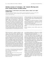
vectors [31]. A comparison of the sequence dependence of
the observed RDCs with a prediction of secondary structure
based upon chemical shift values [44] shows that the size of
the c oupling is strongly dependent upon the secondary
structure ( Fig. 1). Residues in a-helices mainly display
positive
1
H
N
-
15
N RDCs while those located in b-sheets
usually show negative values. This observation can be
accounted for by the orientation of the principal axis system
of the molecular alignment tensor in the HPr molecule
(Fig. 2). The z-axis, which denotes the direction o f largest
partial molecular orientation, is arranged almost parallel to
the a-helices and the b-sheet of the protein. As the
1
H
N
-
15
N
bond vectors in the a-helices are therefore almost parallel to
the z-axis, their size is mainly determined by the positive
eigenvalue S
zz
of the tensor [31]. In contrast, the
1
H
N
-
15
N
bond vectors in the b-sheets are almost perpendicular to
the z-axis and, therefo re are determined by the negative
eigenvalues S
yy
and S
xx
. A similar dependence of the
magnitude and sign of residual dipolar couplings on the
secondary structure was also reported for the F48W mutant
of HPr from E. coli [45]. Residual dipolar couplings for the
vectors connecting the H
N
and H
a
atoms have been
determined in MOCCA-SIAM experiments.
Structures were calculated by simulated annealing fol-
lowed by further refinement in water. A total of 1406
structural restraints was used, corresponding to a ratio of
approximately 1 6 r estraints p er amino acid r esidue
(Table 1). A n average st ructure was c alcu lated from the
final 10 models (Fig. 3). Figure 2 shows a schematic
representation of the secondary structure elements of
HPr(I14A). Overall, the structure is well defined but the
precision varies in different regions according to the number
of experimental restraints, which is indicated by the colour
code in Fig. 3. For the heavy (nonhydrogen) atoms and the
backbone atoms (C
a
,C¢, N) located in the core region of the
molecule comprising th e canonical secondary structure
elements, RMSD values of 0.102 nm and 0.066 nm were
obtained, respectively (Table 1). Larger variations can be
seen in the region of the loops L1 con necting strand A with
helix a, and L5 joining helix b with strand D. HPr(I14A)
shows the open-faced b-sandwich fold which was also
observed for the wild-type protein and other HPr molecules.
The analysis of the 10 lowest energy structures with the
Fig. 1. Dependen ce of
1
H
N
-
15
N residual dipolar couplings on secondary
structure. Thesizeofthe
1
H
N
-
15
N residual dipolar couplings is strongly
correlated with secondary structure. A prediction of sec ondary struc-
ture elements by the program
CSI
[44] based upon the chemical shift
values of the H
a
,C
a
,C
b
and C¢ atoms is shown in grey. The observed
1
H
N
-
15
N residual dipolar couplings are plotted as a function of amino
acid number, shown in white. A clear correlation between secondary
structure and the magnitude of the coupling values can be seen with
residues in a-helical regions predominantly showing positive residual
dipolar couplings and residues in b-sheet regions having negative
values. Note that a residual dipolar coupling of )23 Hz has been
measured for residue 60, which is located in a loop region. For sake of
clarity the ordinate of th e figure has bee n restricted t o the regio n of
)15 to 15 Hz .
Fig. 2. Three-dimensional structure of HPr(I14A) relative to molecular
alignment t ensor. The 3D structure of HPr(I14A) is shown relative to
the principal axis system of the molecular alignment tensor d etermined
for the
1
H
N
-
15
N residual dipolar couplings. The se condary structural
elements are indicated by labels. Note that the z-axis denoting the
direction of largest partial alignment is oriented nearly parallel to the
b-sheet and the a-helices. The eigenvalues of the tensor are S
zz
¼
0.000491, S
yy
¼ )0.000313, S
xx
¼ )0.000178.
4818 A. Mo
¨
glich et al.(Eur. J. Biochem. 271) Ó FEBS 2004
program
PROCHECK
-
NMR
[46] recognizes a central antipar-
allel b-sheet consisting of strands A (residues 2–7), B
(31–37), C (40–43) and D (60–67), two relatively long
a-helices a (16–27) and c (70–83), as well as the short a-he lix
b (47–50). The analysis of chemical shifts [44] predicts
essentially the same secondary structure elements at slightly
different positions (b-strands: strand A, 1–9; strand B,
32–36; strand C, 39–42; strand D, 59–66; a-helices: helix a,
12–26; helix b, 47–51; helix c, 69–82). The active site of the
HPr molecule containing the residue His15 is formed by
loop L1.
Recalculation of the 3D-structure of wild-type HPr
and comparison with the mutant protein
It is known that the NMR structures obtained from a given
set of experimental restraints also depend on the programs
used for the structural calculations. Even when using the
same program, they depend on the specific protocol used for
the calculations. Therefore, we recalculated the structure of
the w ild-type protein on the b asis of the restraints used
previously [15] with the same protocol used here for the
mutant protein. Compared to the wild-type structure of
HPr from S. carnosus stored in the PDB (entry 1QR5) no
significant structural changes w ere observed. However, the
extended water refinement protocol led to a significant
improvement of the general geometry. The structural
statistics and the
PROCHECK
-
NMR
analysis are summarized
in Table 2.
The wild-type protein and the mutant form studied in this
paper show the same global fold with essentially identical
secondary structure elements. However, compared to
the wild-type p rotein, h elix b is significantly shorter in the
mutant protein and distorted at its C-terminal end. In
the core r egion of the protein, which encompasses the
canonical secondary structural elements, the average struc-
tures of the wild-type and the mutant protein molecule agree
reasonably well with an RMSD value for the backbone
atoms (C
a
,C¢, N) of 0.119 nm. When all backbone atoms of
the proteins are taken i nto account, this value increase s to
0.155 nm.
Significant deviations between t he two proteins are seen
in the active site region where the mutation has been
introduced. The replacement of I le14 by Ala causes a slight
longitudinal compression of the mutant protein (Fig. 4). At
its N-terminal end, helix a displays a kink towards the
interior of the protein. The space that in the wild-type
molecule is occupied by the large hydrophobic side chain of
Fig. 3. Structure ensemble of HPr(I14A). The average structure of the
10 lowest- energy structures out of 300 calculated with
CNS
is shown.
TheradiusofthesplinereflectstheRMSDvaluesoftheC
a
atom
positions. Th e s cale bar indicates a length of 0.2 nm correspo nding to a
RMSD value of 0.1 nm. Residues are colour-coded according to the
number of restraints used in the stru cture calculations for this amino
acid. Light grey indicates 10 or fewer, yellow 11–20, orange 21–40, red
more than 40 restraints per residue.
Table 2. Structural statistics of wild-type HPr. The structures were
recalculated from the data from Go
¨
rler et al. [15] with the same pro-
tocol used for the mutant. The NMR data have been recorded at
298 K and pH 7.14. In total 1301 NOE, 78 dihedral angle, and 39
hydrogen bond restraints were used. The quality of the 10 lowest
energy structures was assessed using
PROCHECK
-
NMR
[46].
Restraint violations in the
10 lowest-energy structures Number
NOE violations > 0.05 nm 9
J-coupling violations > 1.7 Hz 28
RMSD values for the
10 lowest-energy structures RMSD (nm)
Core region (residues 2–9, 16–27, 32–37,
40–43, 47–53, 59–84), backbone atoms
C
a
,C¢,N
0.071
Core region (residues 2–9, 16–27, 32–37,
40–43, 47–53, 59–84), heavy atoms
0.112
All residues, backbone atoms C
a
,C¢, N 0.088
All residues, heavy atoms 0.130
Ramachandran plot
(except glycine and proline residues) Incidence (%)
Most favored regions 77.5
Additional allowed regions 16.5
Generously allowed regions 4.7
Disallowed regions 1.3
Energies of the 10 selected structures
after refinement in water E/kJÆmol
)1
E
total
) 11752 ± 456
E
NOE
485 ± 21.3
Ó FEBS 2004 Solution structure of the I14A mutant of HPr (Eur. J. Biochem. 271) 4819
the isoleucine residue is instead partly filled by the backbone
and s ide chain atoms of Ala19. In addition, the C-terminus
folds back onto the core of the protein thereby allowing the
side chain of Leu86 to partly fill the hole created by the
removal of Ile14. Due to these changes other alterations are
induced in the HPr(I14A) molecule. The catalytically active
residue His15 is moved closer to the p rotein interior and its
orientation relative to the protein core is changed. The loops
L1 and L5 show a significantly different conformation.
Helix b is distorted at its C-terminal end and the loop L4 at
its N-terminal end is bent into another d irection than in the
wild-type protein. To allow the hydrophobic side chain of
Leu86 to project into the protein core, the orientation of
helix c is slightly changed in the mutant form. These
changes observed in the mutant protein are also supported
by other NMR parameters. For example, NOE contacts
between the side chain protons of Leu86 and protons of
amino acids Ala14, Val55 and Leu81 are observed, none of
which are seen for the wild-type protein. Analysis of the
backbone dihedral angles F and Y of mutant and wild-type
protein also s upports the observed structural differences
(Fig. 5 ). Significant changes in dihedral angles between the
two proteins were observed for almost all regions of the
molecules. In Fig. 5 the residues for which the difference in
dihedral angles exceeds the sum of the errors are indicated
by black dots. Particularly for residues 13 and 14 of the
active-centre loop, residues 38 and 39 of loop L3 and for
residue 54 located in loop L5, distinctly different confor-
mations are f ound. In ad dition, the dihedral angles of
residue 84 are changed in the mutant protein allowing the
C-terminus to bend to the protein core.
Effect of xenon-binding on the mutation-induced
structural changes
It has been shown previously [43] that a xenon atom
binds into the hydrophobic cavity of HPr(I14A) that i s
created by the replacement of t he bulky Ile14 by an
alanine (Fig. 6). P otentially, the binding of xenon inside
this cavity could lead to a reversal of the structural
changes induced by the m utation because the size of an
isoleucine side chain almost exactly corresponds to that of
a xenon atom. As
1
H
N
and
15
N chemical shifts provide a
sensitive measure for the local structural environment of
the amide bond,
1
H
N
-
15
N-HSQC spectra were recorded
for wild-type and mutant HPr. Following Gro
¨
ger et al.
[43], combined chemical shift changes were calculated for
the amide groups according to Eqn (1). The changes
induced by the mutation of the wild-type protein were
compared with the combined chemical s hift changes
observed in the I14A m utant upon xenon-binding
(Fig. 7). While on average the total cha nges in chemical
shifts due to the introduction of the mutation are about
four times as large as those i nduced by xenon-binding,
they show a similar dependence on the amino acid
sequence. Note that not only the magnitudes of the
individual shift changes but also that their signs closely
correspond. Thus, for most residues the chemical shift
changes caused by the mutation were at least partly
compensated by the binding of xenon.
Discussion
Structural basis of the strongly reduced pressure
response at position 14 in HPr
During the phosphoryl group transfer from enzyme EI to
enzyme EII or other proteins, the active centre loop L1 of
wild-type HPr has to adapt to different functional states.
High-pressure NMR spectroscopy studies have revealed
that protein regions, which are able to exist in different
conformational (sub)states, often show large, n onlinear
pressure reponses [47]. In agreement with these findings such
a pressure response was also experimentally observed for
loop L1 of wild-type HPr [16]. The sole exception was
residue Ile14, which is adjacent to the His15 involved in
phosphoryl transfer, and shows only a very small pressure
response indicating that its position is stabilized in some
way. The NMR structure shows the side chain of this amino
acid to be located in a hydrophobic cavity, which might
possibly stabilize the conformation of this residue as well as
that of the entire loop L1.
Fig. 4. Comparison of wild-type and mutant
HPr. Comparison of the three-dimensional
structures of the mutant (left) and wild-type
HPr (right). T he sid e chains o f the catalytically
active histidine residue 15 and of residue 14
(isoleucine to alanine) are shown in blue and
yellow, respectively. Residues Ala19 and
Leu86 are indicated in red. The removal of the
isoleucine side c hain in the mutant protein
leads to significant structural rearrangements
(see text).
4820 A. Mo
¨
glich et al.(Eur. J. Biochem. 271) Ó FEBS 2004
Fig. 5. Dihedral angle analysis of HPr(WT) and HPr(I14A). Structural differences between the wild -type and mutan t form of HPr are visualized by
a comparison of t he co rrespondin g backbo ne dihedral angles. Values for the wild-type and the mutant protein are indicated by w hite and g rey bars,
respectively. The corresponding standard deviations are indicated by error bars. Significant variations between the two proteins are marked by
black dots and indicate residues for which the absolute value of the difference in dihedral angles exceeds the sum of the errors.
Ó FEBS 2004 Solution structure of the I14A mutant of HPr (Eur. J. Biochem. 271) 4821
Our data provide the experimental evidence of this
hypothesis. After removing the hydrophobic isoleucine side
chain by mutating residue Ile14 to a lanine, the conforma-
tion of loop L1 is strongly changed due to a kink in helix a
(Figs 4 and 5). Particularly the relative position of the
catalytically active histidine i s clearly different an d less
accessible to the solvent c ompared to the wild-type protein.
These structural changes should also have a profound effect
on the biological activity of H Pr(I14A). In agreement with
this assumption we have found a much reduced phospho-
transferase activity of the mutant compared to the wild-type
protein in the standard complementation a ssay [48].
Reversal of the mutation-induced changes
by xenon-binding
One might reasonably assume that the removal of the bulky
sidechain of an isoleucine residue via mutation to alanine
simply leads to t he creation of a h ydrophobic c avity of
corresponding size and shape. Our structural studies
clearly show that this is not the case f or the mutant
HPr(I14A). Although the general fold of t he protein is
conserved, the overall conformation is changed leading to
distinctly different structures for wild-type a nd mutant
HPr with a RMSD of 0.155 nm for the backbone atoms
(C
a
,C¢, N). The replacement of t he large hydrophobic
side chain of isoleucine with the much smaller one of
alanine causes a collapse of the protein in that region. The
resulting hydrophobic cavity is partly filled by side chain s
of other hydrophobic residues. Helix a bends towards the
protein interior to partially fill the void left by the removal
of the isoleucine. Moreover, Leu86 undergoes a pro-
nounced rearrangement of its side chain which also
protrudes into t he space o ccupied by I le14 in the wild-
type HPr. These structural changes induce further distor-
tions of the c onformation of the m utant protein.
However, despite all these structural rearrangements the
surface map of HPr(I14A) shows that a small hydropho-
bic cav ity remains (Fig. 6).
The existence of this cavity was recently confirmed by
xenon-binding studies [43]. Xenon atoms are known to bind
preferentially into hydrophobic pockets of proteins [49–51].
Further, the difference in volume between the sidechains of
isoleucine and alanine closely corresponds to the volume
of a xenon atom, which has a van d er Waals radius of
0.217 n m. Most of the larger xenon-induced changes i n
chemical shift were observed near the site of the mutation,
which could readily be accounted for by the existence of a
hydrophobic cavity [43]. In contrast, it was hard to
rationalize why large changes were also observed for the
C-terminal residues and why throughout the whole protein
the xenon-induced shift changes were considerably larger
than in the wild-type.
An explanation for these findings is provided by this
study. The chemical shift changes induced by xenon binding
to the hydrophobic cavity of HPr(I14A) are strongly
correlated with the corresponding differences of chemical
Fig. 6. Hydrophobic cavity of HPr(I14A). The solvent-accessible sur-
face of the HPr(I14A) molecule is shown. Residues 14, 15, 19 and 86
are coloured as in Fig. 4. A cavity in the reg ion where the m utation has
been introduced is marked by the arrows. The existence of this cavity
was confirmed by xenon-binding studies.
Fig. 7. Changes in chemical shift ca used by xenon-binding and the
Ile14Ala mutation. The normalized changes of the combined chemical
shifts Dd
tot
/<Dd
tot
> of the amide groups are plotted as a function of
their position in the sequ ence. T he comb ined ch emical sh ift cha nges
Dd
tot
have been calculated according to Eqn (1). <Dd
tot
> represents
the average value of the corresponding chemical shift values and is
indicated by the broken line. Chemical shift changes were determined
in
1
H-
15
N-HSQC spectra for t he wild-type protein and the mutant
protein both in the absence and the p resence of xenon [43]. Xenon-
induced chemical shift changes in HPr(I14A) (blue); chemical shift
changes induced by the mutation in the absence of xenon (red).
4822 A. Mo
¨
glich et al.(Eur. J. Biochem. 271) Ó FEBS 2004
shifts between wild-type and mutant protein (Fig. 7). As
detailed above the removal of the hydrophob ic sidechain of
isoleucine effects profound structural rearrangements in
HPr(I14A). Apart from two regions in the direct vicinity
of the mutation site, strong structural differences are also
observed for the C-terminus, most notably for Leu86.
Furthermore, the whole structure displays a subtly different
conformation (Fig. 4). All of these structural distortions are
closely reflected in the xenon-induced chemical sh ift chan-
ges. Large shift changes are mainly observed in the same
two regions close to the hydrophobic cavity introduced by
the m utation and near the C-terminus. In the other regions
of the mutant protein smaller xenon-induced chemical shift
changes, which are still significantly larger than those for
wild-type HPr, are seen and are indicative of global if yet
small conformational changes. Taken together, these find-
ings imply t hat xenon-binding leads to a reversal o f the
structural changes caused by the mutation. By binding to
the h ydrophobic cavity, xenon shifts the conformational
equilibrium of HPr(I14A) towards species closer resembling
the wild-type struct ure.
The smaller size o f the chemical shift changes caused by
xenon-binding compared to the mutation-induced effects
could be due to two reasons. On the one hand, xenon atoms
bound to the protein could rapidly exchange with the bulk
water [52]. Saturation could not be obtained with the pres-
sures possible in our experimental setup. Therefore the
observed shifts represent an average of the bound and the
free state w ith the chemical shift changes being scaled down
accordingly. On the other hand, xenon-binding does not
necessarily reverse the mutation-induced effects completely.
Conclusion
The work presented here further supports the idea that high-
pressure NMR studies are generally suitable to identify
residues important for the stability and the function of
proteins. Pressure changes could be used t o shift the
equilibrium between different protein conformations [53].
In this way, it is possible to populate species only present to a
small extent at atmospheric pressure. NMR spectroscopy is
a convenient technique to monitor such changes with atomic
resolution. Both structurally flexible residues, which m ight
mediate the interaction with different ligands, and residues
that stabilize the protein can be identified by this method. It
would be interesting to see the influence of the mutation
upon the pressure response of HPr. Currently, work is in
progress to address this question. The data also show that
with its affinity to hydrophobic cavities xenon can influence
conformational e quilibria a nd thus can possibly restore
function by stabilizing the active conformation of a protein.
Acknowledgements
The authors thank Dr Mich ael Wenzler, Dr Rolf Do
¨
ker, Jochen
Trenner and Dr Bernhard Ganslmeier for helpful discussions, and
Christian Gro
¨
ger for recording
1
H-
15
N-HSQC sp ectra. F inancial
support by the Deutsche Forschungsgemeinschaft (Br 1278/9–1, SFB
521 projects A6, C6), the Fonds der chemischen Industrie and the EU
(FP6, SPIN E-consortium) is gratefully ac knowledged. Thanks are
further due to Ms Ingrid Cuno for carefully proofreading the
manuscript.
References
1. Kundig, W., Gosh, S. & Roseman, S. (1964) Phosphate bound to
histidine in a protein as as intermediate in a novel phospho-
transferase system. Proc. Natl. Acad. Sci. USA 52, 1067–1074.
2. Postm a, P.W., Lengeler, J.W. & Jacobson, G.R. (1993) Phos-
phoenolpyruvate: carbohydrate p hospho transf erase systems of
bacteria. Microbiol. Rev. 57, 543–594.
3. Stu
¨
lke, J. & Hillen, W. (2000) Regulation of carbon catabolism in
Bacillus species. Annu. Rev. Microbiol. 54, 849–880.
4. Stu
¨
lke, J., Arnaud, M., Rapoport, G. & Martin-Verstraete, I.
(1998) PRD – a p rotein domain involved in PTS-dependent
induction and carbon catabo lite repressio n of catabolic op erons in
bacteria. Mol. Microbiol. 28, 865–874.
5. Deutscher, J. & Saier, M.H. (1983) ATP-dependent protein kinase-
catalyzed phosphorylation of a seryl residue in HPr, a phosphate
carrier protein of the phosphotransferase system in Streptococcus
pyogenes. Proc. Natl. Acad. Sci. USA 80, 6790–6794.
6. He nkin , T.M., Grundy, F.J., Nicholson, W.L. & Chambliss, G.H.
(1991) Catabolite repression of alpha-amylase gene expression in
Bacillus subtilis involves a trans-acting gene product homologous
to the Escherichia coli lacl and galR repressors. Mol. Microbiol. 5,
575–584.
7. Deutscher, J., Ku
¨
ster, E., Bergstedt, U., Charrier, V. & Hillen, W.
(1995) Protein kinase-depende nt HPr/Ccp A interaction lin ks
glycolytic activity to carbon catabolite repression in Gram-positive
bacteria. Mol. Microbiol. 15, 1049–1053.
8. Kalbitzer, H.R. & H engstenberg, W. (1993) The solution structure
of the histidine-containing protein (HPr) from Staphylococcus
aureus as determined by two-dime nsional 1H-NMR s pectrosc opy.
Eur. J. Biochem. 21 6 , 205–214.
9. van Nuland, N.A., H angyi, I.W., van Schaik, R.C., Bere ndsen,
H.J., van Gunsteren, W.F., Scheek, R.M. & Robillard, G.T.
(1994) The high-resolution structure of the histidine -containing
phosphocarrier protein HPr from E scherich ia coli determined by
restrained molecular dynamics from nuclear magnetic resonance
nuclear Overhauser effect data. J. Mol. Biol. 237, 544–559.
10. Hahmann, M., Maurer, T., Lorenz, M., Hengstenberg, W.,
Glaser, S. & Kalbitzer, H.R. (1998) Structural studies of histidine-
containing phosphocarrier prote in from Enterococcus faecalis.
Eur. J. Biochem. 25 2 , 51–58.
11. Maurer, T ., Do
¨
ker, R., Go
¨
rler, A., Hengstenberg, W. & Kalbitzer,
H.R. (2001) Th ree-dime nsional structure of th e histidine-con-
taining phosphocarrier protein (HPr) from Enterococcus faecalis
in solution. Eur. J. Biochem. 268, 635–644.
12. Maurer, T., Meier, S., Kachel, N. , Munte, C.E., Hasenbein, S.,
Koch, B., Hengste nberg, W. & Kalbitzer, H.R. (2004) High
Resolution Structure of the Histidine Containing Phosphocarrier
Protein (HPr) from Staphylococcus aureus and Cha ract erisatio n of
its Interaction with the bifunctional HprKinase/Phosphorylase.
J. Bacteriol. 186, 5906–5918.
13. van Nuland, N.A., Boelens, R., Scheek, R.M. & Robillard, G.T.
(1995) High-resolution structure of the phosphorylated form
of the histidine-containing phosphocarrier protein HPr from
Escherichia coli determined by restrained molecular dynamics
from NMR-NOE data. J. Mol. Biol. 246, 180–193.
14. Jones, B.E., Rajagopal, P. & Klevit, R.E. (1997) Phosphorylation
on histidine is accompanied by localized structural changes in the
phosphocarrier protein HPr fro m Bacillus subtilis. Prot. Sci. 6,
2107–2119.
15. Go
¨
rler, A., Hengstenberg, W., Kravanja, M., Beneicke, W.,
Maurer, T. & Kalbitzer, H.R. (1999) Solution structure of histi-
dine-containing phosphocarrier protein from Staphylococcus car-
nosus. Appl. Magn. Reson. 17, 465–480.
16. Kalbitzer, H.R., Go
¨
rler, A., Li, H., von Dubovskii, P., Hengsten-
berg, W., K owolik, C., Yamada, H . & Akasaka, K. (2000) 15N and
Ó FEBS 2004 Solution structure of the I14A mutant of HPr (Eur. J. Biochem. 271) 4823
1H NMR study of histidine containing protein (HPr) from
Staphylococcus carnosus at high pressure. Prot. Sci. 9, 693–703.
17. Landt, O., Grunert, H.P. & Hahn, U. (1990) A general method for
rapid site-directed mutagenesis using the polymerase chain reac-
tion. Gene 96, 125–128.
18. Kruse, R., Hengstenberg, W., Beneicke, W. & Kalbitzer, H.R .
(1993) Involvement of various amino- and carboxyl-terminal
residues in the active site of t he histidine-containing protein HPr of
the phosp hoenolpyruvate-de pendent phosphotransferase system
of Staphylococcus c arnosus: site-directed mutagenesis with t h e
ptsH gene, biochemical characterization and NMR studies of the
mutant proteins. Protein Eng. 6, 417–423.
19. Cavagnero, S., Dyson, J .H. & Wright, P.E. (1999) Improved low
pH bicelle system for orienting macromolecules over a wide tem-
perature range. J. Biomol. NMR 13, 387–391.
20. Go
¨
rler, A. & Kalbitzer, H.R. (1997) Relax, a flexible program for
the b ack calculation o f NOESY spectra based on complete-
relaxation-matrix formalism. J. Magn. Reson. 124, 177–188.
21. Wishart, D.S., Bigam, C.G., Yao, J., Abildgaard, F., Dyson, H.J.,
Oldfield, E., Markley, J.L. & Sykes, B.D. (1995) 1H, 13C and 15N
chemical sh ift re ferencin g in bio molecu lar NM R. J. Biomol. NMR
6, 135–140.
22. Gron wald, W. & Kalbitzer, H.R. (2004) Automated structure
determin at io n of proteins by NMR spectroscopy. Prog. NMR
Spectrosc. 44, 33–96.
23. Prasch, T., Gro
¨
schke, P. & Glaser, S.J. (1998) SIAM, a novel
NMR experiment for the determination of homonuclear coupling
constants. Angew. Chem. Int. Ed. 37, 802–806.
24. Mo
¨
glich,A.,Wenzler,M.,Kramer,F.,Glaser,S.J.&Brunner,E.
(2002) Determination o f residual dipolar couplings in homo-
nuclear MOCCA-SIAM experiments. J. Biomol. NMR 23, 211–
219.
25.Titman,J.&Keeler,J.(1990)Measurementofhomonuclear
coupling constants f rom NMR c orrelation spectra. J. Magn.
Reson. 89, 640–646.
26. Karplus, M. (1959) Contact electron-spin coupling of nuclear
magnetic moments. J. Chem. Phys. 30, 11–15.
27. Vuister, G.W. & Bax, A. (1993) Quantitative J correlation: a new
approach for measuring homonuclear three-bond J (HNH.alpha.)
coupling constants in 15N-enriched proteins. J. Am. Chem . So c.
115, 7772–7777.
28. Cordier, F. & Grzesiek, S. (1999) Direct observation of hydrogen
bonds in proteins by interresidue
3h
J
NC¢
scalar couplings. J. Am.
Chem. Soc. 121, 1601–1602.
29. Cordier, F., Rogowski, M., Grzesiek, S. & Bax, A. (1999)
Observation of through-hydrogen-bond
2h
J
HC¢
in a perdeuterated
protein. J. Magn. Reson. 140, 510–512.
30. Tjandra, N. & Bax, A. (1997) Direct measurement of distances
and angles in b iomolecules by NMR in a dilute liquid crystalline
medium. Science 278, 1111–1114.
31. Brun ner, E. (2001) Residual dipolar couplings in protein NMR.
Concepts Magn. Reson. 13, 238–259.
32. Bax, A. (2003) Weak alignment offers new NMR opportunities to
study protein structure and dynamics. Prot. Sci. 12, 1–16.
33. Ottiger, M., Delaglio, F. & Bax, A. (1998) Measurement of J and
dipolar couplings from simplified two-dimensional NMR spectra.
J. Magn. Reson. 131, 373–378.
34. Losconzi, J.A., Andrec, M., Fischer, M.W.F. & Prestegard, J.H.
(1999)Ordermatrixanalysisofresidual dipolar couplings using
singular value decomposition. J. Magn. Reson. 138, 334–342.
35. Meiler, J., Peti, W. & Griesinger, C. (2000) DipoCoup: a versatile
program for 3D-structure homology comparison based on
residual dipolar couplings and pseudocontact shifts. J. Biomol.
NMR 17, 283–294.
36. Zweckstetter, M. & Bax, A. (2000) Prediction of sterically induced
alignment in a dilute liquid crystalline phase: aid to protein
structure determination by NMR. J. Am. Chem. Soc. 122, 3791–
3792.
37. Cornilescu,G.,Marquardt,J.L.,Ottiger,M.&Bax,A.(1998)
Validation of Protein Structure from Anisotropic Carbonyl Che-
mical Shifts in a Dilute Liquid Crystalline Phase. J. Am. Chem.
Soc. 120, 6836–6837.
38. Nilges, M., Gronenborn, A.M., Bru
¨
nger, A.T. & Clore, G.M.
(1988) Determ ination of three-dimensional structures o f proteins
by simulated annealing with interproton distance restraints.
Application to crambin, potato carboxypeptidase inhibitor and
barley serine proteinase inhibitor 2. Prot. Eng. 2, 27–38.
39. Bru
¨
nger, A.T., Adams, P.D., Clore, G.M., Delano, W.L., Gros,
P., Grosse-Kunstleve, R.W., Jiang, J.S., Kuszweski, J., Nilges, M.,
Pannu, N.S., Read, R.J., Rice, L.M., S imonson, T. & Warren,
G.L. (1998) Crystallography & NMR system: a new software suite
for macromolecular structure determination. Acta Cryst. D 54,
905–921.
40. Nabu urs, S.B., Nederveen, A .J., Vranken, W., Doreleijers, J.F.,
Bonvin, A.M.J.J., Vuister, G.W., Vriend, G. & Spronk, C.A.E.M.
(2004) DRESS: a database of refined solution NMR structures.
Proteins 55, 483–486.
41. Linge, J.P., Williams, M.A., Spronk, C.A.E.M., Bonvin, A.M.J.J.
& Nilges, M. (2003) Refinement of protein structures in explicit
solvent. Proteins 50, 496–506.
42. Koradi, R., Billeter, M. & Wu
¨
thrich, K. (1996) MOLMOL: a
program for d isplay and analysis of macrom olecular structures.
J. Mol. Graphics 14, 51–55.
43. Gro
¨
ger, C., Mo
¨
glich, A., P ons, M., Koch, B., Hengstenberg, W.,
Kalbitzer, H.R. & Brunner, E. (2003) NMR-spectroscopic map-
ping of an e ngineered cavity in the I14A mutant of HPr from
Staphylococcus carnosus using xenon. J. Am. Chem. Soc. 125,
8726–8727.
44. Wishart, D.S. & Sykes, B.D. (1994) The 13C chemical-shift index: a
simple method fo r the identification o f protein secondary structure
using 13C chemical-shift data. J. Biomol. NMR 4, 171–180.
45. van Lune, F., Manning, L., D ijkstra, K., Be rend sen, H.J.C. &
Scheek, R.M. (2002) Ord er-parameter tensor description of
HPr in a medium of oriented bicelles. J. Biomol. NMR 23, 169–
179.
46. Laskowski, R.A., Rullmann, J.A.C., MacArthur, M.W., Kaptein,
R. & Thornton, J.M. (1996) AQUA and PROCHECK-NMR:
programs for checking the quality of protein structures solved by
NMR. J. Biomol. NMR 8, 477–486.
47. Akasaka, K. (2003) Highly fluctuating protein structures revealed
by variable-pressure nuclear magnetic resonance. Biochemistry 42,
10875–10885.
48. Hengstenberg, W., Penberthy, W.K., Hill, K.L. & Morse, M.L.
(1969) Phosp hotransferase system of Staphylococcus aureus:its
requirement for the ac cumulatio n and metabolism of galacto sid es.
J. Bacteriol. 99, 383–388.
49. Schoenborn, B.P., Watson, H.C. & Kendrew, J.C. (1965) Binding
of xenon to sperm whale myoglobin. Nature 207, 28–30.
50. Montet, Y., Amara, P., Volbeda, A ., Verneda, X., Hatchikian,
E.C. & Field, M.J. (1997) Gas a ccess to the active site of Ni-Fe
hydrogenases probed by X-ray crystallography and molecular
dynamics. Nat. Struct. Biol. 4, 523–526.
51. Prange
´
, T., Schiltz, M., Pernot, L., Colloc’h, N., Longhi, S.,
Bourguet, W. & Fourme, R. (1998) E xploring hydrophobic sites in
proteins with xenon or krypton. Proteins 30, 61–73.
52. Rubin,S.M.,Lee,S Y.,Ruiz,E.J.,Pines,A.&Wemmer,D.
(2002) D etection and characterization of xenon-binding s ites
in proteins by
129
Xe NMR sSpectroscopy. J. Mol. Biol. 322, 425–
440.
53. Akasaka, K. & Yamada, H. (2001) On-line cell high-pressure
nuclear magnetic resonance t echnique: a pp lication to protein
studies. Methods Enzymol. 338, 134–158.
4824 A. Mo
¨
glich et al.(Eur. J. Biochem. 271) Ó FEBS 2004