Báo cáo khoa học: Stimulation of p-nitrophenylphosphatase activity of Na+ ⁄ K+-ATPase by NaCl with oligomycin or ATP docx
Bạn đang xem bản rút gọn của tài liệu. Xem và tải ngay bản đầy đủ của tài liệu tại đây (306.94 KB, 12 trang )
Stimulation of p-nitrophenylphosphatase activity of
Na+ ⁄ K+-ATPase by NaCl with oligomycin or ATP
Haruo Homareda1 and Makoto Ushimaru2
1 Department of Biochemistry, Kyorin University School of Medicine, Mitaka, Tokyo, Japan
2 Department of Chemistry, Kyorin University School of Medicine, Mitaka, Tokyo, Japan
Keywords
diprotomer; Na+ ⁄ K+-ATPase; oligomycin;
p-nitrophenylphosphate (pNPP);
p-nitrophenylphosphatase (pNPPase)
Correspondence
H. Homareda, Department of Biochemistry,
Kyorin University School of Medicine,
Mitaka, Tokyo 181-8611, Japan
Fax & Tel: +81 422 76 7651
E-mail:
(Received 5 July 2004, revised 24 October
2004, accepted 19 November 2004)
doi:10.1111/j.1742-4658.2004.04496.x
It is known that the addition of NaCl with oligomycin or ATP stimulates
ouabain-sensitive and K+-dependent p-nitrophenylphosphatase (pNPPase)
activity of Na+ ⁄ K+-ATPase. We investigated the mechanism of the stimulation. The combination of oligomycin and NaCl increased the affinity of
pNPPase activity for K+. When the ratio of Na+ to Rb+ was 10 in the
presence of oligomycin, Rb+-binding and pNPPase activity reached a maximal level and Na+ was occluded. Phosphorylation of Na+ ⁄ K+-ATPase by
p-nitrophenylphosphate (pNPP) was not affected by oligomycin. Because
oligomycin stabilizes the Na+-occluded E1 state of Na+ ⁄ K+-ATPase, it
seemed that the Na+-occluded E1 state increased the affinity of the phosphoenzyme formed from pNPP for K+. On the other hand, the combination of ATP and NaCl also increased the affinity of pNPPase for K+ and
activated ATPase activity. Both activities were affected by the ligand conditions. Oligomycin noncompetitively affected the activation of pNPPase by
NaCl and ATP. Nonhydrolyzable ATP analogues could not substitute for
ATP. As NaE1P, which is the high-energy phosphoenzyme formed from
ATP with Na+, is also the Na+-occluded E1 state, it is suggested that the
Na+-occluded E1 state increases the affinity of the phosphoenzyme from
pNPP for K+ through the interaction between a subunits. Therefore, membrane-bound Na+ ⁄ K+-ATPase would function as at least an (ab)2-diprotomer with interacting a subunits at the phosphorylation step.
Na+ ⁄ K+-ATPase (Na+ ⁄ K+-exchanging ATPase;
EC 3.6.3.9) is a membrane-integrated protein that actively transports Na+ from the inside of cells to the
outside and transports K+ in the reverse direction,
coupled with ATP hydrolysis. Na+ ⁄ K+-ATPase has
two conformations, the E1 conformational state and
the E2 conformational state. Na+ and ATP bind to
E1. The NaE1ATP formed is phosphorylated to the
high-energy phosphoenzyme with Na+ (NaE1P) and
then transformed to the low-energy phosphoenzyme
(E2P), accompanied by Na+ release. E2P is K+dependently dephosphorylated to E2 + Pi [1]. Because
Na+ ⁄ K+-ATPase can hydrolyze p-nitrophenylphos-
phate (pNPP), a reaction that is ouabain-sensitive and
K+-dependent, the pNPPase activity is presumed to be
a partial reaction of Na+ ⁄ K+-ATPase [2–5]. About
30 years ago, it was reported that NaCl with oligomycin or ATP stimulated K+-dependent pNPPase
activity, although NaCl and ATP individually inhibited
the activity and oligomycin had little effect [6–11].
Oligomycin and ATP are an inhibitor and a substrate
for Na+ ⁄ K+-ATPase, respectively [2–5]. Because it
remains unclear why both the inhibitor and the substrate activate the K+-dependent pNPPase activity in
the presence of Na+ [2], we investigated this question
to clarify the activation mechanism.
Abbreviations
AMPPCP, adenylyl-(b,c-methylene)-diphosphonate; E1P, high-energy phosphoenzyme; E2P, low-energy phosphoenzyme; EP,
phosphoenzyme; K0.5, concentration giving half-maximal activation; pNPP, p-nitrophenylphosphate; pNPPase, p-nitrophenylphosphatase.
FEBS Journal 272 (2005) 673–684 ª 2005 FEBS
673
Stimulation of pNPPase activity of Na+ ⁄ K+-ATPase
In the first part of this paper, we describe the effect
of oligomycin with NaCl on pNPPase activity. The ligand combination induced high-affinity K+-dependent
pNPPase activity. When the ratio of Na ⁄ Rb in the
presence of oligomycin was 10, the binding of Rb+, a
congener of K+, and pNPPase activity reached a maximal level and Na+ remained occluded. Today, it is
well known that oligomycin occludes Na+ within the
Na+ ⁄ K+-ATPase molecule, so that this antibiotic
inhibits Na+ transport and Na+ ⁄ K+-ATPase activity
but not K+-dependent pNPPase activity [12–19].
Therefore, the present data suggest that the Na+occluded E1 state increased the affinity of pNPPase
for K+.
In the second part, the effect of ATP with NaCl on
pNPPase activity is described. The ligand combination
induced high-affinity K+-dependent pNPPase activity
and ATPase activity at the same time. Variation of the
ligand conditions affected both activities. ADP and
adenylyl (b,c-methylene)diphosphonate (AMPPCP)
could not substitute for ATP. Because NaE1P, which
is formed from ATP with Na+, is also the Na+-occluded E1 state [20], the present results suggest that the
Na+-occluded E1 state increases the affinity of the
phosphoenzyme (EP) from pNPP for K+. We explain
the present data using a model in which membranebound Na+ ⁄ K+-ATPase functions as an (ab)2-diprotomer with interacting a subunits.
H. Homareda and M. Ushimaru
pNPP at pH 8.4 and 37 °C. The K0.5 was similar to
that at pH 7.4 (data not shown).
PNPPase activity in the presence of oligomycin
and NaCl
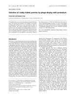
The effect of oligomycin with NaCl on pNPPase activity was observed in the presence of 0–3 mm KCl
(Fig. 1). In the presence of 3 mm KCl without oligomycin, NaCl gradually inhibited pNPPase activity
(Fig. 1A). The addition of 10 lm oligomycin, which
appears to be the maximal concentration in an aqueous solution including 1% (v ⁄ v) ethanol [19], strengthened the inhibitory effect of NaCl on pNPPase
activity in the presence of NaCl up to 20 mm. In the
presence of NaCl at concentrations of more than
20 mm, however, the pNPPase activity with oligomycin
was higher than that without the antibiotic. In the
presence of 1 mm KCl, pNPPase activity with oligomycin was higher than that without the antibiotic in
the presence of NaCl at concentrations of more than
4 mm (Fig. 1B). In the presence of 0.3 mm KCl, the
inhibitory effect of Na+ was absent in the absence of
oligomycin (Fig. 1C). The addition of oligomycin
stimulated the activity fivefold in the presence of
10–30 mm NaCl. These results show that, when the
Results
Effect of pH on ouabain-insensitive pNPPase
activity
Although the ouabain-insensitive ATPase activity of
the Na+ ⁄ K+-ATPase preparation used in this study
was less than 5% in the presence of 0.1 mm ouabain,
16% of the total pNPPase activity was ouabain-insensitive at pH 7.4. To find experimental conditions in
which the ouabain-insensitive pNPPase activity was
minimized, pNPPase activity was measured at pH 6.4,
7.4 and 8.4. The ouabain-insensitive activity was 32%,
16% and 10% at pH 6.4, 7.4 and 8.4, respectively, as
shown by Nagai et al. [21]. In addition to the change
in pH, the ouabain concentration was increased from
0.1 to 1 mm to completely depress the increase in the
ouabain-insensitive activity due to increasing the KCl
concentration. On the other hand, increasing the Na+
concentration had no effect on the ouabain-insensitivity of pNPPase activity. The concentration giving halfmaximal activation (K0.5) of pNPPase for K+ was
1 mm, and the Vmax was 2.8 lmolỈmin)1Ỉmg)1 in the
presence of 10 mm KCl, 5 mm MgCl2 and 2.5 mm
674
Fig. 1. Effect of oligomycin on pNPPase activity in the presence of
NaCl and KCl. Ouabain-sensitive pNPPase activity in the absence
(s) or presence (n) of 10 lM oligomycin was assayed in a mixture
containing 5 lg (A) or 10 lg (B–D) Na+ ⁄ K+-ATPase, the standard ligands [5 mM MgCl2, 50 mM Tris ⁄ Tes (pH 8.4 at 23 °C), 2.5 mM
pNPP, 1 mM EDTA, and with or without 1 mM ouabain], 1% ethanol, 0–300 mM NaCl, and (A) 3 mM KCl, (B) 1 mM KCl, (C) 0.3 mM
KCl, or (D) 0 mM KCl. Data represent the means of two independent experiments.
FEBS Journal 272 (2005) 673–684 ª 2005 FEBS
H. Homareda and M. Ushimaru
ratio of Na ⁄ K was higher than 4, oligomycin activated
pNPPase. In the absence of KCl, oligomycin slightly
enhanced pNPPase activity in the presence of NaCl at
concentrations of more than 10 mm (Fig. 1D).
Figure 2 shows the activation of pNPPase by KCl in
the presence of NaCl with or without oligomycin. In
the absence of oligomycin, increasing the NaCl concentration decreased pNPPase activity (Fig. 2A). In
the presence of oligomycin, increasing the NaCl concentration obviously increased the affinity of pNPPase
for K+ (Fig. 2B). In the presence of 10 lm oligomycin
and 30 mm NaCl, the K0.5 for K+ was 0.3 mm. The
combination of 10 lm oligomycin and 10 mm NaCl
started to demonstrate an activation of pNPP hydrolysis with two phases. This activation was clearly confirmed by the combination of 30 mm NaCl and 10 lm
oligomycin (Fig. 2B). The K0.5 and Vmax were 0.3 mm
and 0.8 lmolỈmin)1Ỉmg)1 for the high-affinity K+dependent pNPPase activity, and were 5 mm and
1.6 lmolỈmin)1Ỉmg)1 for the low-affinity K+-dependent pNPPase activity, respectively (Fig. 3).
Fig. 2. Activation of pNPPase activity by KCl in the presence of
NaCl with or without oligomycin. The pNPPase activity in the
absence (A) or presence (B) of 10 lM oligomycin was assayed in a
mixture containing 10 lg Na+ ⁄ K+-ATPase, the standard ligands, 1%
ethanol, 0–3 mM KCl and 0 (s), 1 mM (n), 3 mM (h), 10 mM (b),
30 mM (d), 100 mM (m) or 300 mM NaCl (j). Data represent the
means of two determinations.
FEBS Journal 272 (2005) 673–684 ª 2005 FEBS
Stimulation of pNPPase activity of Na+ ⁄ K+-ATPase
Fig. 3. Activation of pNPPase by KCl in the presence of NaCl and
oligomycin. The pNPPase activity was assayed in a mixture containing 5 lg Na+ ⁄ K+-ATPase, the standard ligands, 1% ethanol,
0–30 mM KCl, and 10 lM oligomycin (m) or 30 mM NaCl plus 10 lM
oligomycin (n). Plots and bars represent the means ± SD from
three determinations.
The relation between pNPPase activity and
ion-binding
pNPPase activity and ion binding were measured in
reaction mixtures containing 0.1 mm RbCl, 0.5 mm
pNPP and 0–10 mm NaCl (Fig. 4). In this experiment,
the MgCl2 concentration was reduced to 1 mm, and
the reaction temperature lowered to 0 °C because a
high Mg2+ concentration inhibited the binding of
Na+ and K+ [14,15] and a low temperature increased
the affinities of Na+ ⁄ K+-ATPase for Na+ and K+
(H. Homareda, unpublished data). Specific (ouabain-sensitive) Na+ binding was very low in the
absence of oligomycin, which increased the affinity of
Na+ ⁄ K+-ATPase for Na+ [14]. The resultant increase
in Na+ binding was regarded as Na+ occlusion.
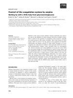
In the absence of oligomycin, NaCl gradually inhibited pNPPase activity and Rb+ binding (Fig. 4A,B). In
the presence of oligomycin, the binding curve of Rb+
and the activation curve of pNPPase showed a convex
shape. The peak of both Rb+ binding and pNPPase
activity occurred at 1 mm NaCl. A plausible explanation
is that the activation of pNPPase by oligomycin with
NaCl is due to the increase in K+ affinity. On the other
hand, Na+ occlusion was preserved under the ligand
conditions used (Fig. 4C). Therefore, the Na+-occluded
E1 state of Na+ ⁄ K+-ATPase seemed to induce the
high-affinity K+-dependent pNPPase activity.
Phosphorylation from pNPP
We examined whether the phosphorylation by pNPP
was affected by oligomycin or other ligands in the
675
Stimulation of pNPPase activity of Na+ ⁄ K+-ATPase
H. Homareda and M. Ushimaru
Table 1. Phosphorylation of Na+ ⁄ K+-ATPase from pNPP, Pi and
ATP. For phosphorylation by pNPP or Pi, Na+ ⁄ K+-ATPase was incubated for 10 min at 37 °C in the presence of 5 mM MgCl2, 0.5 mM
ouabain, 50 mM Tris ⁄ Tes (pH 8.4 at 23 °C), 1 mM [32P]pNPP or
0.3 mM 32Pi with or without the ligands shown. For phosphorylation
by ATP, Na+ ⁄ K+-ATPase was incubated for 30 s at 0 °C in the presence of 5 mM MgCl2, 50 mM Tris ⁄ Tes (pH 8.4 at 23 °C), 0.1 mM
[32P]ATP with or without 2.5 mM pNPP. Data are presented as the
means ± SD from three to six determinations.
nmolỈmg)1
1 mM [32P]pNPP + 0.5 mM ouabain
+ 10 lM oligomycin
+ 10 lM oligomycin + 10 mM NaCl
+ 10 lM oligomycin + 10 mM NaCl
+ 0.3 mM KCl
+ 0.3 mM ATP
+ 0.3 mM Pi
0.3 mM 32Pi + 0.5 mM ouabain
+ 1 mM pNPP
0.1 mM [32P]ATP + 10 mM NaCl
+ 2.5 mM pNPP
Fig. 4. pNPPase activity, Rb+ binding and Na+ occlusion in the
same ligand condition. (A) The pNPPase activity in the absence (s)
or presence (n) of 10 lM oligomycin was assayed in a mixture containing 20 lg Na+ ⁄ K+-ATPase, 1 mM MgCl2, 50 mM Tris ⁄ Tes
(pH 8.4 at 23 °C), 0.5 mM pNPP, 0–10 mM NaCl, 0.1 mM RbCl, 1%
ethanol, with or without 1 mM ouabain. The reaction was started
by the addition of pNPP and followed for 90 min at 0 °C. (B) Ouabain-sensitive 86Rb+ binding in the absence (s) or presence (n) of
10 lM oligomycin was assayed in a mixture containing 30 lg
Na+ ⁄ K+-ATPase, 1 mM MgCl2, 50 mM Tris ⁄ Tes (pH 8.4 at 23 °C),
0.5 mM pNPP, 0–10 mM NaCl, 0.1 mM 86RbCl, 1% ethanol, with or
without 0.1 mM ouabain. After the addition of pNPP, the mixture
was centrifuged. (C) Oligomycin-stimulated 22Na+ binding was
assayed in a mixture containing 30 lg Na+ ⁄ K+-ATPase, 1 mM
MgCl2, 50 mM Tris ⁄ Tes (pH 8.4 at 23 °C), 0.5 mM pNPP, 0.3, 1 or
3 mM 22NaCl, 0.1 mM RbCl, 1% ethanol, with or without 10 lM
oligomycin. After the addition of pNPP, the mixture was centrifuged. The detailed procedure is described in Experimental procedures. Plots and bars in (A), (B) and (C) represent the means ± SD
from three determinations.
presence of ouabain (Table 1). The amounts of EP
were not affected by 10 lm oligomycin, 10 mm NaCl,
0.3 mm KCl or 0.3 mm ATP. To determine whether
the phosphorylation by [32P]pNPP was affected by
contaminating 32Pi, which is nonenzymatically released
from [32P]pNPP, or was unreactive 32Pi in the reaction
mixture for [32P]pNPP synthesis, 0.3 mm nonradioactive
676
%
1.86
1.85
1.83
1.88
±
±
±
±
0.05
0.03
0.04
0.02
100
99
98
101
1.63
1.21
1.81
0.65
1.05
1.00
±
±
±
±
±
±
0.03
0.09
0.05
0.02
0.05
0.05
88
65
100
36
100
95
Pi was added to the reaction mixture. If the phosphorylation was due to the contamination by 32Pi, EP
must be significantly decreased by the addition of nonradioactive Pi. The result showed that EP from 1 mm
[32P]pNPP was decreased to 65% by 0.3 mm Pi,
whereas EP from 0.3 mm 32Pi was decreased to 36%
by 1 mm pNPP. pNPPase activity decreased by only
15% in the presence of 0.3 mm Pi. Therefore, the EP
formed was not due to 32Pi.
The amount of EP from pNPP was 1.9 times greater
than that from ATP. This value was consistent with the
ratio of EP from Pi or pNPP to EP from ATP [22–24].
pNPPase activity in the presence of NaCl, KCl
and ATP
The effect of ATP with NaCl on pNPPase activity was
observed in the presence of 1 mm KCl (Fig. 5A). In
the absence of NaCl, increasing the ATP concentration
from 0 to 1 mm decreased pNPPase activity. Increasing
the NaCl concentration without ATP inhibited pNPPase activity. However, simultaneous addition of NaCl
and ATP induced convex-shaped activation curves of
pNPPase. The combination of 10 mm NaCl and
0.1 mm ATP maximally activated pNPPase.
In the presence of ATP and KCl, increasing the
NaCl concentration induced convex-shaped activation
curves of pNPPase (Fig. 5B). When 10 mm NaCl and
0.1 mm ATP were present, KCl at 3, 1 and 0.3 mm
activated pNPPase by 2.4-fold, 6.4-fold and 20-fold
over reactions without NaCl, respectively. Consequently, the combination of 10 mm NaCl, 0.3 mm KCl
FEBS Journal 272 (2005) 673–684 ª 2005 FEBS
H. Homareda and M. Ushimaru
Stimulation of pNPPase activity of Na+ ⁄ K+-ATPase
Fig. 6. K+-dependent activation curves for pNPPase in the presence
of NaCl with or without ATP. pNPPase activity in the absence (s)
or presence (n) of 0.1 mM ATP was assayed in a mixture containing 5 lg Na+ ⁄ K+-ATPase, the standard ligands, 10 mM NaCl and
0–30 mM KCl. Plots and bars represent the means ± SD from three
determinations.
Fig. 5. Effects of ATP and KCl on pNPPase activity in the presence
of NaCl. The pNPPase activity was assayed in a mixture containing
5 lg Na+ ⁄ K+-ATPase, the standard ligands, 0–100 mM NaCl, and
(A) 1 mM KCl with 0 (s), 0.01 mM (d), 0.1 mM (n) or 1 mM ATP
(m), or (B) 0.1 mM ATP with 3 mM (s), 1 mM (d), 0.3 mM (j) or
0 mM KCl (h). In (B), b and c represent 0.1 mM AMPPCP and
0.1 mM ADP in the presence of 0.3 mM KCl, respectively. Plots and
bars represent the means ± SD from three determinations.
and 0.1 mm ATP maximally activated pNPPase. The
combination of 3 mm NaCl and 0.1 mm ATP slightly
activated pNPPase even in the absence of KCl. ADP
and AMPPCP could not substitute for ATP.
Figure 6 shows that the combination of 0.1 mm
ATP and 10 mm NaCl increases the affinity of pNPPase for K+, as shown by oligomycin with NaCl
(Figs 2 and 3). The combination decreased the K0.5 for
K+ from 2 to 0.2 mm, which was one-fifth of the K0.5,
1 mm, under the usual conditions.
Competition between oligomycin and ATP
Figure 7 shows the competition between oligomycin
and ATP. Oligomycin decreased the Vmax without
affecting the K0.5, suggesting that oligomycin was a
noncompetitive inhibitor of ATP.
ATPase activity in the presence of NaCl, KCl
and ATP
The ligand combination of 10 mm NaCl, 0.3 mm KCl,
0.1 mm ATP and 2.5 mm pNPP activated ATPase in
FEBS Journal 272 (2005) 673–684 ª 2005 FEBS
Fig. 7. Effect of oligomycin on pNPPase activity in the presence of
NaCl and ATP. pNPPase activity was assayed in a mixture containing 5 lg Na+ ⁄ K+-ATPase, the standard ligands, 10 mM NaCl,
0–30 mM KCl and 0.1 mM ATP (s), 10 lM oligomycin (n) or 0.1 mM
ATP plus 10 lM oligomycin (h). Plots and bars represent the
means ± SD from three determinations.
addition to pNPPase (Fig. 8A). Omission of KCl significantly decreased both activities (Fig. 8B). Increasing
the NaCl concentration inactivated pNPPase more
than ATPase irrespective of the absence and presence
of KCl (Fig. 8A,B).
In the presence of 10 mm NaCl and 0.1 mm ATP,
KCl concentrations up to 1 mm simultaneously activated both activities with a K0.5 of 0.2 mm for K+
(Fig. 9A). KCl concentrations greater than 1 mm
gradually inactivated ATPase. On the other hand,
omission of NaCl completely inactivated ATPase
(Fig. 9B).
677
Stimulation of pNPPase activity of Na+ ⁄ K+-ATPase
Fig. 8. Na+-dependent activation curves for Na+ ⁄ K+-ATPase and
pNPPase in the presence of ATP with or without KCl. Ouabain-sensitive activities of ATPase and pNPPase in the presence (A) or
absence (B) of 0.3 mM KCl were assayed in a mixture containing
5 lg Na+ ⁄ K+-ATPase, the standard ligands, 0–300 mM NaCl and
0.1 mM ATP (n) or 0.1 mM [32P]ATP (m). n and m represent pNPPase and ATPase activity, respectively. Plots and bars represent the
means ± SD from three determinations.
In the presence of 10 mm NaCl, 0.3 mm KCl and
0.1 mm ATP, increasing the pNPP concentration
increased pNPPase activity (Fig. 10A). The K0.5 for
pNPP was 2 mm. This was equivalent to the value under
the usual conditions. ATPase activity was decreased by
high pNPP concentrations, although the decrease was
not more than 50%. On the other hand, increasing the
ATP concentration had a complex effect on pNPPase
(Fig. 10B). ATP concentrations up to 0.3 mm activated
pNPPase, whereas ATP concentrations greater than
0.3 mm completely inhibited it. The activation curve of
ATPase was biphasic. From a double-reciprocal plot
analysis, the K0.5 values for ATP were 0.14 and 2.0 mm
and the Vmax values were 0.7 and 5.0 lmolỈmin)1Ỉmg)1.
Discussion
The affinity of pNPPase for K+ is an order of magnitude lower than that of Na+ ⁄ K+-ATPase for K+
[2,5]. The combination of NaCl and oligomycin
induced the high-affinity K+-dependent pNPPase
678
H. Homareda and M. Ushimaru
Fig. 9. K+-dependent activation curves for Na+ ⁄ K+-ATPase and
pNPPase in the presence or absence of NaCl. The activities of ATPase and pNPPase in the presence (A) or absence (B) of 10 mM
NaCl were assayed in a mixture containing 5 lg Na+ ⁄ K+-ATPase,
the standard ligands, 0–30 mM KCl, and 0.1 mM ATP (n) or 0.1 mM
[32P]ATP (m). n and m represent pNPPase and ATPase activity,
respectively. Plots and bars represent the means ± SD from three
determinations.
activity (Figs 2 and 3). The K0.5 for K+ was 0.3 mm,
which was equivalent to that, 0.2 mm, for Na+ ⁄ K+ATPase activity (Fig. 9A). The increase in K+ affinity
caused by oligomycin with Na+ was supported by the
ion-binding experiment (Fig. 4). When the ratio of
Na ⁄ Rb in the presence of oligomycin was 10, Rb+
binding and pNPPase activity reached a maximal
value, and the occluded Na+ was preserved (Fig. 4).
Because this antibiotic stabilizes the Na+-occluded E1
state of Na+ ⁄ K+-ATPase, it seemed that NaE1–oligomycin increased the affinity of pNPPase for K+. The
NaE1–oligomycin complex is an arrested form [2–5,12].
Therefore, enzyme states other than the complex must
hydrolyze pNPP. Scheiner-Bobis et al. [25–27] and
Linnertz et al. [28] showed that fluorescein isothiocyanate, which blocks the high-affinity binding site for
ATP, affects Na+ ⁄ K+-ATPase activity but not pNPPase activity and showed that Co(NH3)4ATP, which
blocks the low-affinity site for ATP, preserves Na+dependent phosphorylation by ATP but inactivates
pNPPase. They suggested that Na+ ⁄ K+-ATPase
FEBS Journal 272 (2005) 673–684 ª 2005 FEBS
H. Homareda and M. Ushimaru
Stimulation of pNPPase activity of Na+ ⁄ K+-ATPase
the E1 and E2 states coexist depending on the ligand
conditions (Fig. 11). The consistency between the proposed model and the crystal structure is discussed in
the last paragraph. According to our model [model (2)
in Fig. 11], Na+-occluded E1–oligomycin and E2–
oligomycin coexist in the presence of 10–30 mm NaCl
and 10 lm oligomycin, as shown in Figs 2 and 3. E2–
oligomycin is phosphorylated to E2P–oligomycin,
which has low-affinity for K+, by pNPP, as shown in
Table 1. The Na+-occluded E1–oligomycin complex
increases the affinity of E2P–oligomycin for K+
through the interaction between Na+-occluded E1 and
E2P. This assumption is supported by the finding that
Na+ transforms the K+-insensitive E2P, which is
formed from Pi and has low affinity for K+, to the
K+-sensitive E2P, which has high affinity for K+ [38].
Increasing the KCl concentration in the presence of
NaCl and oligomycin induces the activation curve with
two phases (Fig. 3). As K+ binding competes with
Na+ binding [15], it is likely that Na+-occluded E1 is
transformed to KE2 by high K+ concentration, so that
Fig. 10. Effects of pNPP and ATP on pNPPase and ATPase activities. The activities of ATPase and pNPPase were assayed in a mixture containing 5 lg Na+ ⁄ K+-ATPase, 5 mM MgCl2, 50 mM Tris ⁄ Tes
(pH 8.4 at 23 °C), 1 mM EDTA, 10 mM NaCl, 0.3 mM KCl, with or
without 1 mM ouabain and (A) 0–30 mM pNPP with 0.1 mM ATP
(n) or 0.1 mM [32P]ATP (m), or (B) 2.5 mM pNPP with 0–3 mM ATP
(n) or [32P]ATP (m). n and m represent pNPPase and ATPase activity, respectively. Plots and bars represent the means ± SD from
three determinations.
works as a functional (ab)2-diprotomer, in which the
E1 state and the E2 state coexist [29]. The effect of fluorescein isothiocyanate resembles the effect of oligomycin. Furthermore, our data suggest that the
interaction between NaE1 and E2 increased the affinity
of pNPPase for K+. Mimura et al. [30] showed that
octaethylene glycol dodecyl ether-solubilized Na+ ⁄ K+ATPase forms a loosely associated diprotomer in the
E1 state but a tightly associated one in the E2 state.
Nakamura et al. [31] suggested a monomer–dimer
transition model of Ca2+-ATPase, in which the cytoplasmic domains clap like a castanet duet, and Carvalho-Alves et al. [32] proposed dimerization of the
cytoplasmic domain of Ca2+-ATPase. Abe et al. [33]
showed that H+,K+-ATPase functions as an oligomer
in the membrane, although a monomer of these P-type
ATPases has ATPase activity [34–37]. Therefore, we
attempted to explain the present data using the (ab)2diprotomer model with interactive a subunits, in which
FEBS Journal 272 (2005) 673–684 ª 2005 FEBS
Fig. 11. Proposed models. (1) Activation of pNPPase by K+ and
pNPP. (2) Activation of pNPPase by Na+, K+, pNPP and oligomycin.
(3) Activations of pNPPase and ATPase by Na+, K+, pNPP and ATP.
E1 (Na) represents the Na+-occluded E1 state. E2(s)P and E2(i)P
represent the K+-sensitive E2P and K+-insensitive E2P state,
respectively. lK and hK, (Na) and O represent KCl at low and high
concentrations, occluded Na+ and oligomycin, respectively. P(ATP)
and P(pNPP) represent the phosphate transferred from ATP and
pNPP, respectively. M represents membrane. The conformations
of E, E1 (Na) and E2 are referred to the crystal structure of Ca2–E1,
Ca2–E1–AMPPCP and E2–thapsigargin, respectively [49–51]. The
conformation of E2(s) is slightly different from the one of E2(i). The
domains including M5, M7 and M10 face each other in a diprotomer. The upper and lower side of the enzyme represent the external and internal side of cells, respectively. Oligomycin is accessible
to Na+ ⁄ K+-ATPase at the external side [52]. K+ is not transported
by pNPPase activity [53].
679
Stimulation of pNPPase activity of Na+ ⁄ K+-ATPase
KE2 exhibits the low-affinity K+-dependent pNPPase
activity.
The combination of ATP and NaCl also induced the
high-affinity K+-dependent pNPPase activity (Fig. 6).
The K0.5 for K+ decreased from 1 to 0.2 mm, which
was equivalent to that for K+ in Na+ ⁄ K+-ATPase
activity (Fig. 9A). ADP and AMPPCP could not substitute for ATP (Fig. 5B and [8]). The combination of
ATP and NaCl activated both ATPase and pNPPase,
showing that the equilibrium between the E1 and E2
states is dependent on the ligand conditions (Figs 8–
10). According to the Post-Albers scheme [1], Na+
and ATP bind to the E1 state. The NaE1ATP formed
is phosphorylated to NaE1P and then converted to
E2P + Na+. Because NaE1P is the Na+-occluded E1
state [20] and the phosphoenzymes are in closer contact [39], it would be understood that NaE1P increases
the affinity of E2P from pNPP for K+. Nandi et al.
[40,41] proposed a working model of Na+ ⁄ K+-ATPase and H+,K+-ATPase. They postulated that pNPP
is accessible to the pNPP hydrolytic sites at the internal and external surfaces. However, Garrahan et al.
[42] used acetyl phosphate, a membrane-impermeable
substrate, to show that the pNPP hydrolytic site
locates on the internal side of the cell membrane.
Therefore, we presumed in a proposed model [model
(3) in Fig. 11] that the E1 state and the E2 state have a
high-affinity binding site and a low-affinity binding site
for ATP, respectively [25–28] and that pNPP is internally accessible at the low-affinity site for ATP in the
E2 state but less accessible at the high-affinity site for
ATP in the E1 state, because of a much lower affinity
of pNPP for the high-affinity ATP site (Fig. 5 and
Table 1). Our model facilitates understanding of the
results shown in Figs 8–10. In the presence of high
Na+, low K+, moderate pNPP and low ATP concentrations, both pNPPase and ATPase were activated,
and higher Na+ concentrations inhibited pNPPase
activity more than ATPase activity (Fig. 8). In this
case, ATP binds to the E1 state, which is phosphorylated to NaE1P. On the other hand, pNPP accesses the
low-affinity site for ATP in the E2 state, which is phosphorylated to E2P (Table 1). NaE1P increases the affinity of E2P for K+. Consequently, K+ binds to the
high-affinity K+ site on K+-sensitive E2P and accelerates its dephosphorylation (Fig. 4A). An excess of
NaCl may competitively inhibit the effect of K+ on
pNPPase from the external side [43]. The combination
of NaCl with ATP or oligomycin slightly enhanced
pNPPase activity even in the absence of KCl (Figs 1D,
5B and 8B). Nagamune et al. [44] have demonstrated
ouabain-sensitive pNPPase activity in the absence of
KCl, so these combinations may stimulate the activity.
680
H. Homareda and M. Ushimaru
As another possibility, it is likely that NaE1P formed
by pNPP with Na+, as proposed by Yamazaki et al.
[22], and NaE1P formed by ATP with Na+ are spontaneously dephosphorylated. Figure 9A shows that
increasing the KCl concentration activated pNPPase
but gradually inactivated ATPase. Omission of NaCl
activated pNPPase but not ATPase (Fig. 9B). In this
case, KCl concentrations up to 1 mm activate both
pNPPase and ATPase. Increasing the KCl concentration over 1 mm accelerates dephosphorylation of E2P
from pNPP, whereas the high KCl concentration or
the absence of Na+ disturbs the phosphorylation from
ATP. Figure 10A shows that increasing the pNPP concentration activated pNPPase but partly decreased
ATPase activity. In this case, pNPP incompletely
inhibits the ATP binding to the high-affinity ATP site
in the E1 state. Figure 10B shows that increasing the
ATP concentration activated ATPase, whereas pNPPase was activated by low ATP concentrations but
inactivated by high ATP concentrations. Because ATP
concentrations up to 0.3 mm inhibit phosphorylation
from pNPP little (Table 1), both activities are preserved. ATP above 0.3 mm occupies the low-affinity
ATP site in the E2 state, so that Na+ ⁄ K+-ATPase is
activated but pNPPase activity is completely blocked.
Oligomycin noncompetitively affected pNPPase
activity in the presence of Na+ and ATP (Fig. 7). This
antibiotic binds in the N-terminal domain of the a
subunit [18], whereas ATP binds in the domain containing Lys501 of the a subunit [27]. NaE1–oligomycin
is an arrested form, whereas NaE1ATP is an active
form in the ATP hydrolysis reaction. Therefore, the
differences in the binding sites and in the biochemical
properties between oligomycin and ATP should lead to
the noncompetitive effect of oligomycin on ATP.
Toyoshima et al. [45,46] and Sørensen et al. [47]
have solved the crystal structures of the Ca2–E1, Ca2–
nucleotide–E1 and thapsigarigin–E2 states in Ca2+ATPase at a high resolution. The structures are classified into two groups depending on the structure of the
cytoplasmic domain, i.e. an open form and a compact
form. The Ca2–E1 state is an open form [45–47]. Binding of the nucleotide converts it into a compact form
[47]. The compact form induced by binding of the
ADP–AlF4– complex, one of the ATP analogues,
resembles the E1P state and occludes Ca2+ [47]. The
thapsigargin–E2 state is a compact form, although its
conformation is slightly different from that of the Ca2–
nucleotide–E1 state [47]. The crystal structure of
Ca2+-ATPase is suggested to be similar to that of
Na+ ⁄ K+-ATPase [48–50]. Therefore, the conformations of the intermediates that appeared in this study
were referred to the crystal structures of Ca2+-ATPase
FEBS Journal 272 (2005) 673–684 ª 2005 FEBS
H. Homareda and M. Ushimaru
(Fig. 11). The E state is drawn as an open form. This
state may loosely associate with another E state at the
cytoplasmic domain, as shown by Carvalho-Alves et al.
[32]. E2(i)P, which corresponds to K+-insensitive E2P,
is drawn as a compact form. Because the K+-activation curve of pNPPase showed a positive co-operativity
[51], pNPPase probably works as a diprotomer [model
(1) in Fig. 11]. The Na+-occluded E1 state is drawn as
a compact form. E2(s)P, which corresponds to K+-sensitive E2P, may be slightly different from the E2(i)P
state because the affinity for K+ varies dependently
with the configurations of the 4th, 5th and 6th transmembrane segments (M4, M5 and M6) [50]. It is likely
that Na+-occluded E1 associates with E2(s)P through
M5, M7 and M10 because (a) these segments are linearly arranged in the crystal structure [49], (b) M7 and
M10 are almost unmoved by large movement of the
cytoplasmic domain [45–47], and (c) the association
between the transmembrane domains including these
segments is not disturbed by the cytoplasmic domain
in a compact form [45–47]. The conversion of the Ca2–
E1 state into the Ca2+-occluded E1 state accompanies
movement of M1 and M2 [45–47]. Oligomycin binds to
the domain including M1 and M2 of Na+ ⁄ K+-ATPase
a subunit [18]. Therefore, it is conceivable that arrest
of the movement by oligomycin stabilizes the Na+occluded E1 state and inhibits Na+ transport.
Experimental procedures
Materials
ATP(Na)2 and AMPPCP were purchased from RocheDiagnostics (Penzberg, Germany). A portion of the
ATP(Na)2 was converted into the sodium-free ATP form
by passing it through a cation-exchange column. Oligomycin B and ouabain were purchased from Sigma Chemical
Company (St Louis, MO, USA). Oligomycin B was stored
as a 1-mm solution in cold ethanol. pNPP (ditris salt) was
from ICN Biomedicals Inc. (Aurora, OH, USA). 86RbCl
and 22NaCl were obtained from Amersham Pharmacia Biotech (Amersham, Bucks, UK). [32P]ATP[cP] and 32Pi were
obtained from PerkinElmer Life Sciences Japan (Tokyo,
Japan). Other reagents were purchased from Wako Pure
Chemicals Industries, Ltd (Osaka, Japan).
Preparation of Na+ ⁄ K+-ATPase
Microsomes were prepared from canine kidney outer
medulla and treated with sodium deoxycholate, as described
by Hayashi et al. [54]. More than 95% of the total
Na+ ⁄ K+-ATPase activity (about 9 lmol PiỈmin)1Ỉmg)1)
was ouabain-sensitive under the usual conditions.
FEBS Journal 272 (2005) 673–684 ª 2005 FEBS
Stimulation of pNPPase activity of Na+ ⁄ K+-ATPase
Synthesis of [32P]pNPP
[32P]pNPP was synthesized by the method of Guan &
Dixon [55]. 32Pi (33 MBq in 0.02 m HCl) and 0.2 mmol
(20 mg) crystalline phosphoric acid were dissolved in dehydrated acetonitrile to use as starting materials. The amount
of pNPP (cyclohexylamine salt) synthesized was measured
from the absorbance at 310 nm. The amount and the initial
specific radioactivity of [32P]pNPP were 51 lmol and 9000
cpmỈnmol)1, respectively.
Assay of pNPPase activity
The standard reaction mixture (0.5 mL) was composed of
5 lg Na+ ⁄ K+-ATPase, 5 mm MgCl2, 50 mm Tris ⁄ Tes
(pH 8.4 at 23 °C), 1 mm EDTA, 2.5 mm pNPP and NaCl
and KCl at the concentrations indicated in the figure legends,
with or without 0.1 mm ATP, with or without 10 lm oligomycin, and with or without 1 mm ouabain. The control
experiment for the oligomycin effect was performed in the
presence of 1% ethanol without oligomycin. The pNPPase
reaction was started by addition of the enzyme, and the reaction was followed for 5–10 min at 37 °C and stopped by
addition of 2.5 mL 0.1 m NaOH. The p-nitrophenol liberated
was measured from the absorbance at 420 nm. Ouabain-sensitive activity was determined from the difference between
the activities in the presence and absence of 1 mm ouabain.
When pNPPase activity was measured at 0 °C, the
amount of Na+ ⁄ K+-ATPase was changed to 20 lg and the
concentrations of MgCl2 and pNPP to 1 and 0.5 mm,
respectively. The incubation time was extended to 90 min.
Assay of Na+ ⁄ K+-ATPase activity
The same reaction mixture (0.5 mL) as that used for pNPPase activity was prepared, except that [32P]ATP[cP]
(0.1 MBqỈlmol)1) was used instead of nonradioactive ATP.
The Na+ ⁄ K+-ATPase reaction was started by the addition
of the enzyme, followed for 5 or 6 min at 37 °C and
stopped by the addition of 0.1 mL ice-cold 50% (w ⁄ v) trichloroacetic acid containing 2 mm ATP and 2 mm Pi. To
isolate the liberated 32Pi, 0.2 mL 4 m H2SO4 ⁄ 40 mm ammonium molybdate solution and, next, 0.8 mL of an isobutyl
alcohol ⁄ benzene solution was added to the mixture, which
was mixed for 15 min in a vortex mixer [56]. The 32Pi isolated in the organic layer was measured with a liquid scintillation spectrophotometer. Ouabain-sensitive activity was
determined from the difference between the radioactivities
in the presence and absence of ouabain.
Ion-binding assay
86
Rb+ binding and 22Na+ binding to Na+ ⁄ K+-ATPase
were measured using the centrifugation method developed
681
Stimulation of pNPPase activity of Na+ ⁄ K+-ATPase
by Matsui & Homareda [14–18], with slight modifications.
For the 86Rb+ binding, 30 lg Na+ ⁄ K+-ATPase was preincubated with or without 0.01 lmol ouabain at room temperature for 30 min in a 80-lL reaction mixture composed
of 0.1 lmol MgCl2, 5 lmol Tris ⁄ Tes (pH 8.4 at 23 °C),
0–1 lmol NaCl, and 1 lL ethanol with or without 1 nmol
oligomycin. The reaction mixture was kept in ice water.
After 10 lL 1 mm 86RbCl (1 MBqỈlmol)1) and then 10 lL
5 mm pNPP had been added, the reaction mixture (100 lL)
was immediately centrifuged at 368 000 g for 5 min at 2 °C
in an ultracentrifuge (Hitachi himac CS120FX; Tokyo,
Japan). The supernatant was aspirated, and the inside wall
of each tube was wiped carefully to remove any that
remained. Each pellet was dissolved in 0.1 mL 1 m NaOH
by warming at 60 °C for 20 min; then the entire solution
was transferred to a counting vial, neutralized with
0.15 mL 1 m HCl, which was used to wash the inside of the
tube, and then mixed with a scintillator. The radioactivity
was measured using a liquid scintillation spectrophotometer. Ouabain-sensitive binding was calculated from the
difference between the radioactivities of the pellets in the
presence and absence of ouabain.
For the 22Na+ binding, 30 lg Na+ ⁄ K+-ATPase was suspended in a 80-lL reaction mixture composed of 5 lmol
Tris ⁄ Tes (pH 8.4 at 23 °C), 0.03, 0.1 or 0.3 lmol 22NaCl
(0.2 MBqỈlmol)1), 1 lL ethanol with or without 1 nmol
oligomycin. The incubation mixture was kept in ice-cold
water. After 10 lL 1 mm RbCl and 10 lL 5 mm
pNPP ⁄ 10 mm MgCl2 had been added, the reaction mixture
(100 lL) was immediately centrifuged. Oligomycin-stimulated Na+ binding, which is regarded as the occluded Na+
[13], was calculated from the difference between the radioactivities of the pellets in the presence and absence of oligomycin.
H. Homareda and M. Ushimaru
the difference between radioactivities of reaction mixtures
with native and acid-denatured enzymes.
When the phosphorylation by Pi was examined, 0.1 lmol
[32P]pNPP and 0.03 lmol Pi in the reaction mixture were
replaced by 0.1 lmol pNPP and 0.03 lmol 32Pi, respectively. The phosphorylation reaction was started by the
addition of 32Pi with or without pNPP.
The reaction mixture (0.5 mL) for phosphorylation by
ATP was composed of 50 lg Na+ ⁄ K+-ATPase, 5 mm
MgCl2, 50 mm Tris ⁄ Tes (pH 8.4 at 23 °C), 10 mm NaCl,
0.1 mm [32P]ATP[cP] (1 MBqỈlmol)1), with or without
2.5 mm pNPP. The phosphorylation reaction was started by
addition of the enzyme, followed for 30 s at 0 °C and
stopped by the addition of 0.1 mL ice-cold 5% (v ⁄ v) perchloric acid containing 2 mm ATP and 2 mm Pi. The mixture was filtered through a membrane filter with a pore size
of 0.45 lm. The filter was washed three times with 3 mL icecold 5% perchloric acid containing 2 mm ATP and 2 mm Pi,
and the radioactivity on the filter was measured with a
liquid scintillation spectrophotometer. The amount of EP
was calculated from the difference between radioactivities of
reaction mixtures with native and acid-denatured enzymes.
Determination of protein and oligomycin
concentration
Protein and oligomycin concentrations were determined as
described elsewhere [18,19].
Acknowledgements
We thank Drs R. L. Post, Y. Fukushima and Y. Tahara
for critical reading and helpful suggestions, Mr S. Mikkaichi for synthesis of [32P]pNPP, and Ms E. Hagiwara
for technical support.
Assay of phosphorylated intermediate
To measure the phosphorylation by pNPP, 50 lg
Na+ ⁄ K+-ATPase was preincubated with 0.05 lmol ouabain at room temperature for 15–30 min in a 40-lL reaction mixture composed of 0.5 lmol MgCl2 and 5 lmol
Tris ⁄ Tes (pH 8.4 at 23 °C). Then, the ligands indicated in
Table 1 and 1 lL ethanol with or without 1 nmol oligomycin were added. The reaction was started by the addition of
0.1 lmol [32P]pNPP with or without 0.03 lmol Pi (a reaction mixture of 100 lL), followed for 10 min at 37 °C by
the method of Inturrisi & Titus [57] and stopped by addition of 1 mL ice-cold 5% (w ⁄ v) trichloroacetic acid. The
mixture was centrifuged at 14 000 g for 5 min. The precipitate was washed three times with 1 mL ice-cold 5% trichloroacetic acid containing 5 mm pNPP and 5 mm Pi and
dissolved in 0.3 mL 1 m NaOH by incubation at 60 °C for
10 min. After neutralization with HCl, the radioactivity of
the precipitate was measured with a liquid scintillation
spectrophotometer. The amount of EP was calculated from
682
References
1 Post RL (1979) A perspective on sodium and potassium
ion transport adenosine triphosphatase. In Cation Flux
across Biomembranes (Mukohata Y & Packer L, eds),
pp. 3–19. Academic Press, New York.
2 Glynn IM & Karlish SJD (1975) The sodium pump.
Annu Rev Physiol 37, 13–55.
3 Schwartz A, Lindenmayer GE & Allen JC (1975) The
sodium-potassium adenosine triphosphatase: pharmacological, physiological and biochemical aspects. Pharmacol Rev 27, 3–134.
4 Cavieres JD (1977) The sodium pump in human red
cells. In Membrane Transport in Red Cells (Ellory JC &
Lew VL, eds), pp. 1–37. Academic Press, New York.
5 Robinson JD & Flashner MS (1979) The (Na+ + K+)activated ATPase. Enzymatic and transport properties.
Biochim Biophys Acta 549, 145–176.
FEBS Journal 272 (2005) 673–684 ª 2005 FEBS
Stimulation of pNPPase activity of Na+ ⁄ K+-ATPase
H. Homareda and M. Ushimaru
6 Yoshida H, Nagai K, Ohashi T & Nakagawa Y (1969)
K+-dependent phosphatase activity observed in the
presence of both adenosine triphosphate and Na+.
Biochim Biophys Acta 171, 178–185.
7 Askari A & Koyal D (1968) Different oligomycin sensitivities of the Na+ + K+-activated adenosinetriphosphatase and its partial reactions. Biochem Biophys Res
Commun 32, 227–232.
8 Koyal D, Rao SN & Askari A (1971) Studies on the
partial reactions catalyzed by the (Na+ + K+)-activated ATPase. I. Effects of simple anions and nucleoside
triphosphates on the alkali-cation specificity of the
p-nitrophenylphosphatase. Biochim Biophys Acta 225,
11–19.
9 Askari A & Koyal D (1971) Studies on the partial reactions catalyzed by the (Na+ + K+)-activated ATPase.
II. Effects of oligomycin and other inhibitors of the
ATPase on the p-nitrophenylphosphatase. Biochim
Biophys Acta 225, 20–25.
10 Robinson JD (1970) Phosphatase activity stimulated by
Na+ plus K+: implications for the (Na+ plus K+)dependent adenosine triphosphatase. Arch Biochem Biophys 139, 164–171.
11 Skou JC (1974) Effect of ATP on the intermediary steps
of the reaction of the (Na+ + K+)-dependent enzyme
system. III. Effect on the p-nitrophenylphosphatase
activity of the system. Biochim Biophys Acta 339, 258–
273.
12 Garrahan PJ & Glynn IM (1967) The stoichiometry of
the sodium pump. J Physiol 192, 217–235.
13 Esmann M & Skou JC (1985) Occlusion of Na+ by the
Na,K-ATPase in the presence of oligomycin. Biochem
Biophys Res Commun 127, 857–863.
14 Matsui H & Homareda H (1982) Interaction of sodium
and potassium ions with Na+ ⁄ K+-ATPase. I. Ouabainsensitive alternative binding of three Na+ or two K+ to
the enzyme. J Biochem (Tokyo) 92, 193–217.
15 Homareda H & Matsui H (1982) Interaction of sodium
and potassium ions with Na+ ⁄ K+-ATPase. II. General
properties of ouabain-sensitive K+ binding. J Biochem
(Tokyo) 92, 219–231.
16 Homareda H, Nagano Y & Matsui H (1991) Interaction
of sodium and potassium ions with Na+ ⁄ K+-ATPase.
IV. Affinity change for K+ and Na+ of Na+,K+-ATPase in the cycle of the ATP hydrolysis reaction. J Biochem (Tokyo) 109, 70–77.
17 Arato-Oshima T, Matsui H, Wakizaka A & Homareda
H (1996) Mechanism responsible for oligomycin-induced
occlusion of Na+ within Na ⁄ K-ATPase. J Biol Chem
271, 25604–25610.
18 Homareda H, Ishii T & Takeyasu K (2000) Binding
domain of oligomycin on Na+ ⁄ K+-ATPase. Eur J
Pharmacol 400, 177–183.
19 Homareda H (2002) Oligomycin. In Encyclopedia of
Molecular Biology and Molecular Medicine (Campbell
FEBS Journal 272 (2005) 673–684 ª 2005 FEBS
20
21
22
23
24
25
26
27
28
29
30
31
AM, Hammes GG, Krug A, Lehman IR, Schimmel P,
Tooze J, Wada A & Wright P, eds), Online Books
(), Wiley InterScience,
New York.
Glynn IM & Karlish SJD (1990) Occluded cations in
active transport. Annu Rev Biochem 59, 171–205.
Nagai K, Izumi F & Yoshida H (1966) Studies on
potassium dependent phosphatase; its distribution and
properties. J Biochem (Tokyo) 59, 295–303.
Yamazaki A, Kaya S, Tsuda T, Araki Y, Hayashi Y &
Taniguchi K (1994) An extra phosphorylation of
Na+ ⁄ K+-ATPase by paranitrophenylphosphate
(pNPP): evidence for the oligomeric nature of the
enzyme. J Biochem (Tokyo) 116, 1360–1369.
˚
Taniguchi K, Kaya S, Abe K & Mardh S (2001) The
oligomeric nature of Na ⁄ K-transport ATPase. J Biochem (Tokyo) 129, 335–342.
Abe K, Kaya S, Imagawa T & Taniguchi K (2002) Gastric H ⁄ K-ATPase liberates two moles of Pi from one
mole of phosphoenzyme formed from a high-affinity
ATP binding site and one mole of enzyme-bound ATP
at the low-affinity site during cross-talk between catalytic subunits. Biochemistry 41, 2438–2445.
Scheiner-Bobis G, Fahlbusch K & Schoner W (1987)
Demonstration of cooperating a subunits in working
(Na+ + K+)-ATPase by the use of the MgATP complex analogue cobalt tetrammine ATP. Eur J Biochem
168, 123–131.
Scheiner-Bobis G, Esmann M & Schoner W (1989) Shift
to the Na+ form of Na+ ⁄ K+-transporting ATPase due
to modification of the low-affinity ATP-binding site by
Co (NH3)4ATP. Eur J Biochem 183, 173–178.
Scheiner-Bobis G, Antonipillai J & Farley RA (1993)
Simultaneous binding of phosphate and TNP-ADP to
FITC-modified Na+ ⁄ K+-ATPase. Biochemistry 32,
9592–9599.
Linnertz H, Thonges D & Schoner W (1995) Na+ K+ă
ATPase with a blocked E1ATP site still allows backdoor
phosphorylation of the E2ATP site. Eur J Biochem 232,
420–424.
Schoner W, Thonges D, Hamer E, Antolovic R,
ă
Buxbaum E, Willeke M, Serpersu EH & Scheiner-Bobis
G (1994) Is the sodium pump a functional dimer? In
The Sodium Pump (Bamberg E & Schoner W, eds), pp.
332–341. Springer, New York.
Mimura K, Matsui H, Takagi T & Hayashi Y (1993)
Change in oligomeric structure of solubilized Na+ ⁄ K+ATPase induced by octaethylene glycol dodecyl ether,
phosphatidylserine and ATP. Biochim Biophys Acta
1145, 63–74.
Nakamura J, Tajima G, Sato C & Furukohri T (2000)
Oligomer of two types of conformational variants of
sarcoplasmic reticulum Ca2+-ATPase molecules. In
Na ⁄ K-ATPase and Related ATPases (Taniguchi K &
Kaya S, eds), pp. 373–379. Elsevier, Amsterdam.
683
Stimulation of pNPPase activity of Na+ ⁄ K+-ATPase
32 Carvalho-Alves PC, Hering VR, Oliveira JMS, Salinas
RK & Verjovski-Almeida S (2000) Requirement of the
hinge domain for dimerization of Ca2+-ATPase large
cytoplasmic portion expressed in bacteria. Biochim Biophys Acta 1467, 73–84.
33 Abe K, Kaya S, Hayashi Y, Imagawa T, Kikumoto M,
Oiwa K, Katoh T, Yazawa M & Taniguchi K (2003)
Correlation between the activities and the oligomeric
forms of pig gastric H ⁄ K-ATPase. Biochemistry 42,
15132–15138.
34 Martin DW & Sachs JR (1999) Preparation of
Na+ ⁄ K+-ATPase with near maximal specific activity
and phosphorylation capacity: evidence that the reaction
mechanism involves all of the sites. Biochemistry 38,
7485–7497.
35 Ward DG & Cavieres JD (2003) Inactivation of Na,KATPase following Co(NH3)4ATP binding at a low affinity site in the protomeric enzyme unit. J Biol Chem
278, 14688–14697.
36 Møller JV, Lind KE & Andersen JP (1980) Enzyme
kinetics and substrate stabilization of detergent-solubilized and membraneous (Ca2+ + Mg2+)-activated
ATPase from sarcoplasmic reticulum. Effect of protein–
protein interactions. J Biol Chem 255, 1912–1920.
37 Martin DW (1983) Active unit of solubilized sarcoplasmic reticulum calcium adenosinetriphosphatase: an active enzyme centrifugation analysis. Biochemistry 22,
2276–2282.
38 Post RL, Toda G & Rogers FN (1975) Phosphorylation
by inorganic phosphate of sodium plus potassium ion
transport adenosine triphosphatase. Four reactive states.
J Biol Chem 250, 691–701.
39 Ganjeizadeh M, Zolotarjova N, Huang W-H & Askari
A (1995) Interactions of phosphorylation and dimerizing
domains of the a-subunits of Na+ ⁄ K+-ATPase. J Biol
Chem 270, 15707–15710.
40 Nandi J, Das PK, Levine RA & Ray TK (1988) Half of
the (Na+ + K+)-transporting-ATPase-associated K+stimulated p-nitrophenyl phosphatase activity of gastric
epithelial cells is exposed to the surface exterior. Biochem J 252, 29–34.
41 Ray TK & Nandi J (1986) K+-stimulated p-nitrophenyl phosphatase is not a partial reaction of the gastric
(H+ + K+)-transporting ATPase. Biochem J 233,
231–238.
42 Garrahan PJ, Pouchan MI & Rega AF (1969) Potassium activated phosphatase from human red blood cells.
The mechanism of potassium activation. J Physiol 202,
305–327.
43 Rega AF, Garrahan PJ & Pouchan MI (1970) Potassium-activated phosphatase from human red blood cells.
The asymmetrical effects of K+, Na+, Mg++ and
adenosine triphosphate. J Membr Biol 3, 14–25.
684
H. Homareda and M. Ushimaru
44 Nagamune H, Urayama O, Hara Y & Nakao M
(1986) Characterization of ouabain-sensitive phosphatase activity in the absence of potassium ion in purified
pig kidney Na,K-ATPase. J Biochem (Tokyo) 99,
1613–1624.
45 Toyoshima C, Nakasako M, Nomura H & Ogawa H
(2000) Crystal structure of the calcium pump of sarco˚
plasmic reticulum at 2.6 A resolution. Nature 405, 647–
655.
46 Toyoshima C & Nomura H (2002) Structural changes
in the calcium pump accompanying the dissociation of
calcium. Nature 418, 605–611.
47 Sørensen TL-M, Møller JV & Nissen P (2004) Phosphoryl transfer and calcium ion occlusion in the calcium
pump. Science 304, 1672–1675.
48 Rice WJ, Young HS, Martin DW, Sachs JR & Stokes
˚
DL (2001) Structure of Na+ ⁄ K+-ATPase at 11-A resolution: comparison with Ca2+-ATPase in E1 and E2
states. Biophys J 80, 2187–2197.
49 Sweadner KJ & Donnet C (2001) Structural similarities
of Na,K-ATPase and SERCA, the Ca2+-ATPase of the
sarcoplasmic reticulum. Biochem J 356, 685–704.
50 Ogawa H & Toyoshima C (2002) Homology modeling
of the cation binding sites of Na+ ⁄ K+-ATPase. Proc
Natl Acad Sci USA 99, 15977–15982.
51 Gache C, Rossi B & Lazdunski M (1976) (Na+,K+)activated adenosinetriphosphatase of axonal membranes, cooperativity and control: steady-state analysis.
Eur J Biochem 65, 293–306.
52 Cornelius F & Skou JC (1985) Na+-Na+ exchange
mediated by (Na+ + K+)-ATPase reconstituted into
liposomes. Evaluation of pump stoichiometry and
response to ATP and ADP. Biochim Biophys Acta 818,
211–221.
53 Garrahan PJ & Rega AF (1972) Potassium activated
phosphatase from human red blood cells. The effects of
p-nitrophenylphosphate on cation fluxes. J Physiol 223,
595–617.
54 Hayashi Y, Kimimura M, Homareda H & Matsui H
(1977) Purification and characteristics of (Na+,K+)ATPase from canine kidney by zonal centrifugation in
sucrose density gradient. Biochim Biophys Acta 482,
185–196.
55 Guan K & Dixon JE (1991) Evidence for protein-tyrosine-phosphatase catalysis proceeding via a cysteinephosphate intermediate. J Biol Chem 266, 17026–17030.
56 Martin JB & Doty DM (1949) Determination of inorganic phosphate. Modification of isobutyl alcohol procedure. Anal Chem 21, 965–967.
57 Inturrisi CE & Titus E (1970) Ouabain-dependent
incorporation of 32P from p-nitrophenyl phosphate
into a microsomal phosphatase. Mol Pharmacol 6,
99–107.
FEBS Journal 272 (2005) 673–684 ª 2005 FEBS