Báo cáo khoa học: Missense mutations as a cause of metachromatic leukodystrophy Degradation of arylsulfatase A in the endoplasmic reticulum potx
Bạn đang xem bản rút gọn của tài liệu. Xem và tải ngay bản đầy đủ của tài liệu tại đây (418.14 KB, 10 trang )
Missense mutations as a cause of metachromatic
leukodystrophy
Degradation of arylsulfatase A in the endoplasmic reticulum
Peter Poeppel
1
, Matthias Habetha
2
, Ana Marca
˜
o
3
, Heinrich Bu
¨
ssow
4
, Linda Berna
5
and Volkmar Gieselmann
1
1 Institut fu
¨
r Physiologische Chemie, Rheinische-Friedrich-Wilhelms Universita
¨
t Bonn, Germany
2 Zoologisches Institut, Christian-Albrechts-Universita
¨
t zu Kiel, Germany
3 Instituto de Biologia Molecular e Celular, University of Porto, Portugal
4 Institut fu
¨
r Anatomie, Rheinische-Friedrich-Wilhelms Universita
¨
t Bonn, Germany
5 Institute of Inherited Metabolic Disorders, Charles University, Prague, Czech Republic
Lysosomal storage diseases comprise a group of about
40 disorders, in most cases caused by the deficiency of
a lysosomal enzyme involved in the degradation of,
for example, lipids, glycosaminoglycans and oligo-
saccharides. Much effort has been devoted to the
identification of disease causing mutations in these dis-
orders. Thus, a multitude of mutations has been identi-
fied in recent years (for example [1]). Only a fraction
of missense mutations, however, has been analysed
at the biochemical level in order to understand the
Keywords
ERAD; proteasomal degradation;
arylsulfatase A; metachromatic
leukodystrophy
Correspondence
V. Gieselmann, Institut fu
¨
r Physiologische
Chemie, Rheinische-Friedrich-Wilhelms
Universita
¨
t Bonn, Nussallee 11, 53115
Bonn, Germany
Fax: +49 22 873 2416
Tel: +49 22 873 2411
E-mail:
uni-bonn.de
Note
P. Poeppel and M. Habetha contributed
equally to this work.
(Received 29 October 2004, revised 14
December 2004, accepted 4 January 2005)
doi:10.1111/j.1742-4658.2005.04553.x
Metachromatic leukodystrophy is a lysosomal storage disorder caused by a
deficiency of arylsulfatase A (ASA). Biosynthesis studies of ASA with vari-
ous structure-sensitive monoclonal antibodies reveal that some epitopes of
the enzyme form within the first minutes of biosynthesis whereas other epi-
topes form later, between 10 and 25 min. When we investigated 12 various
ASAs, with amino acid substitutions according to the missense mutations
found in metachromatic leukodystrophy patients, immunoprecipitation
with monoclonal antibodies revealed folding deficits in all 12 mutant ASA
enzymes. Eleven of the 12 mutants show partial expression of the early epi-
topes, but only six of these show, in addition, incomplete expression of late
epitopes. In none of the mutant enzymes were the late forming epitopes
found in the absence of early epitopes. Thus, data from the wild-type and
mutant enzymes indicate that the enzyme folds in a sequential manner and
that the folding of early forming epitopes is a prerequisite for maturation
of the late epitopes. All mutant enzymes in which the amino acid substitu-
tion prevents the expression of the late forming epitopes are retained in the
endoplasmic reticulum (ER). In contrast, all mutants in which a single late
epitope is at least partially expressed can leave the ER. Thus, irrespective
of the missense mutation, the expression of epitopes forming late in biosyn-
thesis correlates with the ability of the enzyme to leave the ER. The degra-
dation of ER-retained enzymes can be reduced by inhibitors of the
proteasome and ER a1,2-mannosidase I, indicating that all enzymes are
degraded via the proteasome. Inhibition of degradation did not lead to an
enhanced delivery from the ER for any of the mutant enzymes.
Abbreviations
ASA, arylsulfatase A; ER, endoplasmic reticulum; Lc, lactacystin; Kif, kifunensine; SOV, sodium orthovanadate; PAO, phenylarsine oxide; OA,
okadaic acid; Lp, leupeptin; DNM, deoxynojirimycin; a1-AT, a1-antitrypsin; MLD, metachromatic leukodystrophy; BHK, baby hamster kidney;
DMEM, Dulbecco’s modified essential medium; FBS, foetal bovine serum; LDL-receptor, low density lipoprotein receptor.
FEBS Journal 272 (2005) 1179–1188 ª 2005 FEBS 1179
molecular basis of the enzyme deficiencies in greater
detail. In many cases, missense mutations lead to an
arrest and more rapid degradation of the encoded
enzyme in the endoplasmic reticulum (ER) (for exam-
ple [2]). In this respect lysosomal storage diseases are
not special as this mechanism is responsible for protein
deficiencies in many diseases. In fact, it has been esti-
mated that ER degradation is the most frequent cause
of protein deficiencies such that the term ‘conforma-
tional diseases’ has been suggested [3].
The mechanisms of ER quality control, retention
and degradation have been investigated in recent years
(reviewed in [4]). Newly synthesized secretory, mem-
brane or lysosomal glycoproteins interact sequentially
with a number of membrane-bound or soluble glyco-
sidases and chaperones of the ER. Modifications of
N-linked oligosaccharide side chains play a major role
in this process.
The precursor of N-linked oligosaccharides is a Glc
3
-
Man
9
-GlcNAc
2
dolichol pyrophosphate, from which
the sugars are transferred en bloc to Asn ⁄ X ⁄ Ser(Thr) in
newly synthesized polypeptide chains within the ER.
Trimming of the Glc
3
-Man
9
-GlcNAc
2
side chains
begins shortly after synthesis by the ER membrane-
located glucosidase I to Glc
2
-Man
9
-GlcNAc
2
, followed
by trimming of an ER-localized soluble glucosidase II
to Glc
1
-Man
9
-GlcNac
2
. Glycoproteins bind to the
ER-resident lectins calnexin and calreticulin, via the
Glc
1
-Man
9
-GlcNAc
2
oligosaccharide. Glucosidase II
then removes the remaining terminal glucose with the
consequence that newly synthesized proteins no longer
bind to the lectins and leave the ER. In case a protein
is not folded correctly, it is recognized by the UDP-glu-
cose:glycoprotein glucosyltransferase, which reglucosy-
lates the Man
9
-GlcNAc
2
of misfolded proteins to
Glc
1
-Man
9
-GlcNAc
2
[5]. Consequently the protein can
bind to calnexin ⁄ calreticulin again and remains in the
ER. This loop can be repeated several times and may
enhance the chances of a protein folding correctly.
Finally, a1,2-mannosidase I removes one mannose
[6,7]. This removal of mannose by a1,2-mannosidase I
has been suggested to be a signal for proteasomal de-
gradation [7,8]. The proteasome seems to be the major
pathway by which misfolded proteins are degraded,
although the existence of an as yet poorly characterized
nonproteasomal pathway has been demonstrated [6,7].
Metachromatic leukodystrophy (MLD) is a lysosomal
storage disorder which is caused by the deficiency of
arylsulfatase A (ASA). This enzyme catalyses the first
step in the degradation pathway of the glycosphingo-
lipid 3-O-sulfogalactosylceramide. Deficiency of the
ASA causes lipid accumulation leading progressive
demyelination and various, ultimately lethal neurologi-
cal symptoms (reviewed in [1]). The gene of human ASA
has been cloned and more than 80 mostly missense
mutations were identified. Some of these mutations
were investigated more closely to reveal the effects of
the amino acid substitutions on the mutant enzyme.
According to these results two main mechanisms cause
ASA deficiency. In about half of the examined cases the
mutant enzymes are retained in the ER [2,9,10], in the
other half, enzymes can leave the ER and be degraded
after arrival in the lysosome [10–12]. Whereas the latter
mechanism has been investigated thoroughly in view
of potential therapeutic intralysosomal stabilization,
nothing is known about the ER-associated degradation
as a cause of MLD. Because it has been shown recently
for Fabry disease [13] ) another lysosomal storage
disorder ) the interference with the ER quality control
mechanism can also be a therapeutic option, we decided
to examine more closely these mechanisms of enzyme
deficiency in MLD.
Results
Biosynthesis of wild-type ASA
To examine the early events in ASA biosynthesis in
more detail, baby hamster kidney (BHK) cells were
transiently transfected with a plasmid encoding human
wild-type ASA cDNA. Cells were pulse labelled with
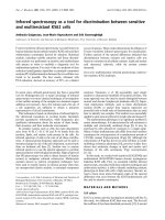
Fig. 1. Early stages of ASA biosynthesis. BHK cells transiently
expressing the human wild-type ASA cDNA were pulse labelled
with 18.5 MBq [
35
S]methionine for 2.5 or 5 min, respectively, and
chased for the times indicated (0, 2.5, 5, 10 and 25 min). Cell homo-
genates were split into eight aliquots and precipitated with preim-
mune serum (I), a polyclonal ASA antiserum (II), or six different
mAbs (A2, A5, B1, C, E and F), which are directed against five dif-
ferent epitopes.
Arylsulfatase A degradation P. Poeppel et al.
1180 FEBS Journal 272 (2005) 1179–1188 ª 2005 FEBS
[
35
S]methionine for 2.5 or 5 min and chased for up to
25 min (Fig. 1). After harvesting, cell homogenates
were divided into eight aliquots, which were immuno-
precipitated with an ASA polyclonal antiserum or six
various mAbs [14]. These mAbs recognize only native
ASA and are directed against different structure-sensi-
tive surface epitopes termed A, B, C, E and F [14].
After a pulse of 2.5 or 5 min, ASA can be readily
detected with the polyclonal antiserum. As this serum
also recognizes denatured ASA, it precipitates ASA
irrespective of the enzyme’s three-dimensional struc-
ture. After 2.5 and 5 min pulse only mAbs A2, A5 and
B1 recognize ASA, whereas no or minute amounts of
ASA are precipitated by the mAbs C, E and F. Epi-
topes recognized by mAbs C, E and F start to develop
slowly within 10 min of chase and have matured after
another 15 min of chase. Thus, in the early stages of
ASA biosynthesis, epitopes recognized by mAbs A2, A5
and B1 appear before those recognized by C, E and F,
demonstrating that ASA folds in a sequential manner.
Recognition of amino acid-substituted ASAs
by mAbs
We have previously identified various missense muta-
tions in the ASA gene and we have examined the
biochemical effects of the corresponding amino acid
substitutions on ASA. In a number of mutants, the
amino acid substitution causes an arrest of ASA in the
ER [2,9,10], whereas others can leave the ER [10–12].
We have expressed these mutant ASAs transiently in
BHK cells. Cells were labelled for 3 h with [
35
S]methi-
onine and after harvesting, cell homogenates were again
divided into eight aliquots, which were immunoprecipi-
tated with the mAbs or polyclonal antiserum (Fig. 2).
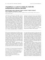
The analysis of 12 amino acid-substituted ASAs reveals
that, according to their reactivity with the mAbs, these
mutants can be divided into three groups. One group
includes mutant ASAs which react weakly with mAbs
A2 and A5 and more strongly with B1 (Gly86Asp,
Tyr201Cys, Pro377Leu, Asp335Val, Pro136Leu,
Asp255His). None of these mutants, however, is recog-
nized by any of the antibodies C, E or F. Substituted
ASAs of the second group (Gly309Ser, Glu312Asp,
Arg84Gln, Arg370Gln, Arg370Trp) react slightly better
with A2, A5 and B1 and react ) although weakly )
with at least one of the mAbs C, E or F. Finally, the
third group has only one member (Thr274Met) which
ASA is not recognized by any of the mAbs.
In a previous publication we located the epitopes
recognized by the various mAbs (Table 3 in [14]).
According to these data amino acid residues 85 and 86
may be part of the epitope recognized by mAbs A2
and A5, and amino acid residues 202–206 by mAb C,
respectively. For this reason the reduced reactivity of
mAbs A2 and A5 with Gly86Asp and Arg84Gln sub-
stituted ASA and mAb C with the Tyr201Cys substi-
tuted ASA, respectively, may reflect changes in the
epitopes rather than conformational alterations. We
could show in the meantime, however, that amino
acids 202–206 are not part of the epitope recognized
by mAb C (P. Poeppel, unpublished data), so that this
cautionary notion does not apply to the immunopre-
cipitation of Tyr201Cys substituted ASA with mAb C.
Degradation of amino acid-substituted ASAs
via the proteasome
In order to investigate the degradation pathway of
amino acid-substituted ASAs in the ER, we used Ltk
–
Fig. 2. Immunoprecipitation of amino acid-substituted ASAs with
structure-sensitive mAbs. Wild-type ASA and 12 amino acid-substi-
tuted ASAs were transiently expressed in BHK cells. Cells were
labelled for 3 h with 1.85 MBq [
35
S]methionine, harvested and
aliquots of cell homogenates were immunoprecipitated as des-
cribed in Fig. 1. + ⁄ – indicates whether or not the mutant enzymes
according to previous publications (references in brackets;
[2,9,10,25–28]) are retained in the ER. Polypeptides of lower appar-
ent molecular mass, which can be seen in some of the experi-
ments are unrelated to ASA.
P. Poeppel et al. Arylsulfatase A degradation
FEBS Journal 272 (2005) 1179–1188 ª 2005 FEBS 1181
cells which stably express the ER-retained ASA
mutant enzymes (Gly86Asp, Tyr201Cys, Pro377Leu,
Asp335Val, Pro136Leu, Asp255His, Thr274Met).
We selected those clones with a medium level of over-
expression and examined them by electron microscopy
for normal ER morphology, in order to exclude the
possibility that enzymes were being unphysiologically
overexpressed. The examined cells showed an ER with
normal morphology (results not shown). Stably trans-
fected Ltk
–
cells were pulse labelled for 2 h and chased
for various time periods to determine the half-life of the
individual enzymes. According to these experiments,
chase times were chosen for the following experiments
so that in most cases about 80–90% of the enzyme was
degraded within the chase periods. Various inhibitors
were added during pulse and ⁄ or chase periods. Inhibits
lactacystin (Lc) irreversibly the 20 S proteasome, leu-
peptin (Lp) is an inhibitor of cysteine and serine prote-
ases, and okadaic acid (OA), phenylarsine oxide (PAO)
and sodium orthovanadate (SOV) are phosphatase
inhibitors. The latter two were used as it has been repor-
ted that misfolded a1-antitrypsin (a1-AT) mutants or
immunoglobulin chains can be stabilized by these com-
pounds [7,8]. Lp has been shown to stabilize some
mutant ASAs, which are degraded in the lysosome [12].
Under the conditions of the experiment Lp should
not inhibit the proteasome and was used as a
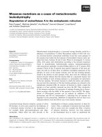
nonproteasomal control inhibitor. Figure 3 shows an
experiment performed with seven different amino
acid-substituted ASAs. The results demonstrate that
all of these mutant ASAs can be partially stabilized
by proteasome inhibition and that the extent of
stabilization varies between the substituted enzymes.
Other inhibitors, in particular phosphatase inhibitors,
showed no effect.
Effects of glycosidase inhibitors on the stability
of amino acid-substituted ASAs
In order to elucidate the role of trimming reactions
of the N-linked oligosaccharide side chains in ER
associated degradation of mutant ASAs, stably trans-
fected Ltk
–
cells were incubated with deoxynojirimy-
cin (DNM), an inhibitor of ER glucosidases I and II
and with kifunensine (Kif), an inhibitor of ER a1,2-
mannosidase I. Cells were pulse labelled for 2 h and
chased for various times, depending on the half-life
of the mutants (Fig. 4). The mutant ASAs were sta-
bilized by Kif, whereas inhibition of glucosidases I
and II causes a more rapid degradation. Thus, all
substituted enzymes showed a uniform pattern of
stabilization or more rapid degradation upon addition
of inhibitors.
Influence of ER a1,2-mannosidase I inhibition
on ER exit of amino acid-substituted ASAs
In order to investigate whether stabilization of mutant
ASAs through inhibition of ER a1,2-mannosidase I
via Kif can lead to an enhanced exit of mutant enzyme
from the ER, stably transfected Ltk
–
cells were pulse-
labelled for 15 h in the presence of Kif and ⁄ or ammo-
nium chloride. After leaving the ER, lysosomal
enzymes including ASA are specifically recognized by
a phosphotransferase in the Golgi apparatus [14]. This
enzyme initiates the phosphorylation of mannose in
the N-linked oligosaccharide side chains of lysosomal
enzymes, yielding mannose-6-phosphate (M6P). In the
trans-Golgi these M6P residues bind to M6P receptors,
which mediate the further vesicular transport of lyso-
somal enzymes from the Golgi to the lysosomes.
Ammonium chloride interferes with this sorting and
causes increased secretion of newly synthesized lyso-
somal enzymes into the medium [15]. Thus, if newly
synthesized lysosomal enzymes appear in secretions in
the presence of ammonium chloride, they must have
left the ER. The addition of ammonium chloride cau-
ses secretion of wild-type ASA to the medium, whereas
it has no effect on ER-retained mutant enzymes.
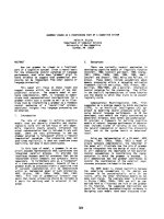
Figure 5 shows Pro377Leu-substituted ASA as an
example. Also the stabilization of amino acid-substi-
tuted ASA with Kif and simultaneous addition of
ammonium chloride does not cause increased secretion
into the medium, indicating that stabilization of
enzymes does not lead to an escape from quality con-
trol mechanisms and increased exit of the ER. Only
after prolonged exposure can minute amounts of
mutant ASAs be detected in the medium, showing a
marginal effect of Kif. We estimate that this accounts
for less than 5% of the enzyme synthesized during the
pulse period.
Discussion
Missense mutations are by far the most frequent type
of mutations in the ASA gene [1]. The effects of these
mutations have been shown to be rather uniform.
Either the amino acid substitutions lead to an arrest of
the mutant enzyme in the ER, or the enzyme is degra-
ded in the lysosome after correct sorting [2,9–12]. Here
we have investigated wild-type and mutant ASAs by
immunoprecipitation with six structure-sensitive mAbs.
These mAbs have recently been shown to recognize
five different ASA epitopes, termed A to F. These epi-
topes, which were recently delimited more closely [14],
depend on the native structure of ASA. Examinations
of the early biosynthetic events reveal that epitope B
Arylsulfatase A degradation P. Poeppel et al.
1182 FEBS Journal 272 (2005) 1179–1188 ª 2005 FEBS
forms rapidly after synthesis. Already after a 2.5-min
pulse, the newly synthesized ASA is efficiently precipi-
tated by mAb B1. At the same time point precipitation
with mAbs A2 and A5 is possible but is less efficient,
indicating that the epitope may be less matured than
the B1 epitope.
Figure 1 shows that the ratio of the signals obtained
with mAb A2 ⁄ A5 and B1 is constant up to 10 min of
chase (densitometric analysis, data not shown), indica-
ting that no further maturation of epitopes A2 ⁄ A5
occurs within this time period. Epitopes C, E and F
are only weakly expressed until 10 min and mature
between 10 and 25 min after synthesis. The maturation
of these late forming epitopes is accompanied by a
further maturation of epitopes A2 and A5. After
25 min of chase, precipitation with mAbs A2 and A5
is almost as efficient as with mAb B1. The location of
epitopes suggests that folding of ASA starts within a
central part of the molecule [14]. This is accompanied
by a partial expression of epitopes in the N-terminal
part. The C-terminal part folds late in biosynthesis,
but its folding is not an isolated event, because epi-
topes A2 and A5 mature concomitantly. Studies on low
density lipoprotein receptor (LDL-receptor) folding
Fig. 3. Effects of protease or phosphatase
inhibitors on the stability of mutant ASAs.
Ltk
–
cells stably expressing the indicated
amino acid-substituted ASAs were incuba-
ted in the presence of various inhibitors
(Lc, Lp, OA, PAO, SOV). Cells were pulse
labelled for 2 h and chased for various times
depending on the half-life of the respective
mutant (Asp335Val, 4.5 h; Gly86Asp,
05.25 hours; Pro377Leu, 4 h; Tyr201Cys,
6 h; Thr274Met, 4.5 h; Asp255His, 4.5 h;
Pro136Leu, 8 h). After the chase ASA was
immunoprecipitated from the homogenates
with the polyclonal ASA antiserum. Precipi-
tated ASA was quantified after SDS ⁄ PAGE
with a bio-imaging analyser (Fujifilm). Col-
umns show mean and SD of arbitrary units
of quadruple experiments. Under each dia-
gram representative immunoprecipitates are
shown.
P. Poeppel et al. Arylsulfatase A degradation
FEBS Journal 272 (2005) 1179–1188 ª 2005 FEBS 1183
have shown recently [16] that its folding does not
proceed in a vectorial, domainwise process from the N
terminus to the C terminus. Instead, folding occurs via
intermediates with disulfide bridges involving distant
parts of the protein. In addition, the N-terminal part
of the LDL-receptor forms late in biosynthesis. Data
on ASA are in agreement with this folding scheme.
Early detectable epitopes are constituted by amino acid
residues between positions 165 and 240 in the central
part of the protein [14]. N-terminal epitopes are also
detectable at an early stage, but do not mature before
the C-terminal part of the protein folds correctly. As
in case of the LDL-receptor, this suggests interactions
of distant parts of ASA during folding.
In addition to wild-type ASA, we also immunopre-
cipitated 12 mutant ASAs, whose underlying missense
mutations were previously found in MLD patients.
Since the mAbs only recognize the native wild-type
enzyme, we reasoned that the reactivity with the mAbs
should provide a measure of the structural integrity of
the substituted enzymes. Surprisingly, the mutant
enzymes did not show an individual reaction pattern
but according to their immunoprecipitation pattern
they can be classified into three groups. One group has
only one member, mutant Thr274Met, which does
not react with any of the mAbs but is readily precipi-
table with the polyclonal antiserum. This reveals
a severe misfolding of this mutant. The second
group of mutants (Gly86Asp, Tyr201Cys, Pro377Leu,
Asp335Val, Pro136Leu, Asp255His) reacts partially
with antibodies recognizing the early epitopes A and B
and not with those recognizing the late epitopes C, E
and F. Interestingly, mutant ASAs of these two groups
are completely retained in the ER. Retention in the
ER due to incorrect folding leads to repetitive regluco-
sylation and binding to the calnexin and calreticulin
chaperones. Finally, a mannose is removed, which is
considered to be a signal for reverse transport out of
the ER into the cytosol. After the transfer of the mis-
folded protein into the cytosol, N-glycans are removed
and the protein is degraded by the proteasome.
The last group is comprised of mutant ASAs which
form, at least partially, one or more of the late epitopes
C, E and F. Thus, except for Thr274Met, all mutant
enzymes express partially the early epitopes whereas
only a fraction expresses the late ones. This suggests
that in general the latter are more sensitive to amino
acid substitutions, irrespective of their localization.
Also none of the mutants expresses the late epitopes
only, or to a larger extent, than the early epitopes. This
Fig. 4. Effects of glycosidase inhibitors on
stability of mutant ASAs. Ltk
–
cells stably
expressing the indicated amino acid-substi-
tuted ASAs were incubated in the presence
of the two inhibitors DNM or Kif. Cells were
pulse labelled for 2 h and chased for various
times depending on the expression and the
half-life of the respective mutants (Asp335V-
al, 4.5 h; Gly86Asp, 5.25 hours; Tyr201Cys,
5 h; Thr274Met, 4.5 h; Asp255His, 4.5 h;
Pro136Leu, 4.5 h). After the chase, ASA
was immunoprecipitated from the homogen-
ates with the polyclonal ASA antiserum.
Precipitated ASA was quantified after
SDS ⁄ PAGE with a bio-imaging analyser
(Fujifilm). Columns show mean, minimal and
maximal deviation of arbitrary units of two
independent experiments. Under each dia-
gram representative immunoprecipitates
are shown.
Arylsulfatase A degradation P. Poeppel et al.
1184 FEBS Journal 272 (2005) 1179–1188 ª 2005 FEBS
indicates that the formation of the epitopes of ASA is
sequential in two aspects: (a) two epitopes (A, B)
form rapidly after translation, whereas others need
several minutes to mature; and (b) formation of early
epitopes is a prerequisite for the maturation of the late
epitopes.
Interestingly, none of the mutants that react with at
least one of the antibodies C, E or F is retained in the
ER (Gly309Ser, Glu312Asp, Arg370Gln, Arg84Gln,
Arg370Trp). Our results suggest that enzymes have
reached a folding state which suffices to pass the ER
quality control, when they express at least epitope C
partially (Arg370Gln and Arg370Trp). In a separate
study we have identified two additional mutations
(Phe219Val, Pro425Thr) that generally also fit into this
pattern ([11] A Marca
˜
o, unpublished data). It should
be mentioned that one of these mutations (Phe219Val)
was found in a patient with an unusual phenotype and
encodes an enzyme that, like Thr274Met, does not
react with any of the mAbs. This mutant ASA, how-
ever, can leave the ER to an extent of about 20% of
the newly synthesized enzyme, the remainder is
retained in the ER.
Recently it was reported that a certain mutant of
a1-AT is retained in the ER and degraded by nonpro-
teasomal pathways [7]. This mutant could be stabilized
by the addition of phosphatase inhibitors PAO and
SOV. The existence of such a pathway is supported by
the fact that phosphatase inhibitors can also inhibit
immunoglobulin chain degradation in the ER [8]. Here
we examined whether different ASA mutants, which
are retained in the ER, show differences in the ER
degradation. For that purpose we have investigated
the influence of various protease and glycosidase inhib-
itors on the stability of the substituted enzymes. All
these mutants are partially stabilized by the protea-
somal inhibitor Lc but not by the serine and cysteine
protease inhibitor Lp, or any of the phosphatase inhib-
itors. All mutant ASAs seem to be uniformly degraded
via the proteasome; there is no indication that different
mutants may use different degradation pathways. It
should also be mentioned, however, that in none of
the cases could we achieve a full stabilization upon
proteasome inhibition. In fact the degree of stabiliza-
tion in some mutants (e.g., Thr274Met, Pro136Leu)
was rather weak. Although the lack of full-scale stabil-
ization was unchanged when we increased the protea-
some inhibitor concentration (data not shown), we
cannot exclude that the proteasome was inhibited only
partially. Nevertheless, the lack of stabilization by the
phosphatase inhibitors indicates that recently detected
nonproteasomal pathways [7,8] do not contribute to
ASA degradation in the cell type used in this examina-
tions.
Proteins may be degraded in an ubiquitin-independ-
ent way by the 20S proteasome. In various experiments
(not shown) we failed to detect ubiquitinylation of the
ASA mutants, suggesting that they may be degraded
in a ubiquitin-independent way by the 20S proteasome
[17].
Glucosidases I and II, as well as ER a1,2-mannosid-
ases, play a role in the targeting of misfolded proteins
in the ER [6–8,18–20]. For this reason we investigated
the influence of glucosidase and mannosidase inhibi-
tion on the mutant ASAs (Gly86Asp, Tyr201Cys,
Asp335Val, Pro136Leu, Asp255His, Thr274Met). In
these experiments all of the mutant ASAs behaved
rather uniformly. They could all be stabilized by Kif,
an ER a1,2-mannosidase I inhibitor. In all cases the
degradation was enhanced when glucosidases I and II
were inhibited by DNM. Increased degradation upon
inhibition of glucosidases and stabilization by inhibi-
tion of mannosidases is a common phenomenon and
has been demonstrated for various misfolded proteins
[6–8,21,22]. The behaviour of ASA mutants in the
ER in the presence of various inhibitors is identical,
Fig. 5. Effects of Kif on secretion of mutant ASAs. Ltk
–
cells stably
expressing wild-type ASA and Pro377Leu substituted ASA were
incubated in the presence of Kif, ammonium chloride (NH
4
Cl) or a
combination of both compounds. Cells were pulse labelled for
16 h. After the labelling ASA was immunoprecipitated from the
homogenates and secretions with the polyclonal ASA antiserum.
The right panel shows an overexposed sample of the immunopre-
cipitates from the secretion of Pro377Leu, which demonstrates
that only low amounts of Kif stabilized ASA appear in the medium.
The same experiment was performed with all ER retained ASAs, all
showed identical results.
P. Poeppel et al. Arylsulfatase A degradation
FEBS Journal 272 (2005) 1179–1188 ª 2005 FEBS 1185
showing that all mutants interact uniformly with com-
ponents of the ER degradation pathway independent
of the underlying mutations.
The interference with ER quality control may open
new therapeutic strategies in the treatment of genetic
diseases. Thus, it has been shown that secretion of an
otherwise ER retained mutant protein, an a1-AT, is
enhanced upon inhibition of ER a1,2-mannosidase I
[23]. For that reason we have examined whether in
principal any of the mutant ASAs can be delivered
from the ER upon inhibition of the degradation path-
way through inhibition of ER a1,2-mann osidase I.
Cells were treated with Kif and ⁄ or ammonium chlor-
ide. The latter interferes with lysosomal enzyme sorting
in the Golgi, so that newly synthesized lysosomal
enzymes appear in the medium. In the case of mutants,
the appearance in the medium is thus an indicator that
the enzyme has left the ER. In none of the analysed
mutants, however, does treatment with Kif lead to a
substantial increase of ASA in the medium. Thus, in
case of ASA, inhibition of the degradation pathway
does not lead to enhanced secretion, which suggests it
will not be a therapeutic option for MLD.
Experimental procedures
Materials, enzymes, chemicals, antibodies
Enzymes used for DNA modification or synthesis were
from New England Biolabs (Frankfurt am Main, Germany)
or Invitrogen (Karlsruhe, Germany). [
35
S]Methionine (spe-
cific activity > 39 TBqÆ mmol
)1
) was from Amersham Bio-
sciences (Buckinghamshire, UK). Oligonucleotides were
from MWG Biotech (Ebersberg, Germany) or Eurogentec
(Seraing, Belgium). The preparation and characterization of
the mAbs has been described previously [14].
Cell culture and transfection
Mouse fibroblast Ltk
–
cells (Ltk
–
) and BHK cells were
maintained in Dulbecco’s modified essential medium
(DMEM) supplemented with 5 or 10% fetal bovine serum
(FBS), penicillin and streptomycin. For transient transfec-
tions, BHK cells were transfected by Lipofectamine
TM
(Gibco, Karlsruhe, Germany). Cells (2 · 10
5
) were plated
onto a 3.5-cm cell-culture dish. Next day, medium was
removed and cells were washed with DMEM devoid of
supplements. Plasmid DNA (2 lg) was mixed with 750 lL
DMEM containing 5 lL Lipofectamine
TM
reagent. After a
30-min incubation, the DNA–Lipofectamine
TM
complexes
were added to the cells in a total volume of 1.5 mL. After a
5-h incubation, the Lipofectamine
TM
-containing medium
was removed and replaced by DMEM ⁄ FBS. Cells were
harvested and analysed for enzyme activity and protein
concentration 48 h after transfection. In case of stable
transfections, 1.2 · 10
6
Ltk
–
cells were plated onto a 6-cm
cell-culture dish. The next day, medium was removed and
1.5 mL DMEM containing 5% FCS, penicillin and strepto-
mycin was added. Plasmid DNA (5 lg) was mixed with
300 lL 150 mm NaCl. After vortexing, 15.5 lL ExGen 500
reagent (Fermentas, St. Leon-Rot, Germany) was added
and incubated for 10 min. This solution was added to the
cells and left for 7 h, after which the ExGen 500-containing
medium was removed and replaced by DMEM in the pres-
ence of 5% FBS, penicillin and streptomycin. In the case of
stable transfections one tenth of the transfected plasmids
was pSV
2
neo carrying a neomycin-resistance gene. Cells
were selected in 800 lgÆmL
)1
G-418 (Invitrogen) and single
colonies were screened for expression of ASA mRNA by
northern blot and protein by western blot analysis. ASA
activity was measured with the artificial substrate 10 mm
p-nitrocatecholsulfate in 170 mm NaCl, 500 mm sodium
acetate pH 5, 0.3% TritonÒ X-100 and 1 mgÆmL
)1
BSA.
200 lL of substrate solution was incubated with 5–50 lg
protein of cell homogenates. Reaction was performed at
37 °C for various time periods and stopped with 500 lLof
1 m NaOH. Absorption was read at 515 nm. To obtain
measurements in the linear range, only samples with an
extinction below 0.7 were included; otherwise the determin-
ation was repeated with shorter incubation times.
Metabolic labelling and immunoprecipitation
Metabolic labelling and immunoprecipitation have been
described in detail elsewhere [24]. In the experiments in
which the degradation pathway of the mutant enzymes
were investigated the following inhibitors and final concen-
trations were used: lactacystin (Lc) 25 lm (Calbiochem,
Bad Soden, Germany), kifunensine (Kif) 100 lm (Calbio-
chem), sodium orthovanadate (SOV) 50 lm (Sigma), phenyl-
arsine oxide (PAO) 800 nm (Sigma, Munich, Germany),
okadaic acid (OA) 100 nm (Calbiochem), leupeptin (Lp)
200 lm (Calbiochem), deoxynojirimycin (DNM) 1 mm
(kindly provided by E. Bause, Institut fu
¨
r Physiologische
Chemie, Rheinische-Friedrich-Wilhelm Universita
¨
t Bonn,
Germany). Lc was present during the pulse and chase peri-
ods, the others only during the chase periods. In the
experiments in which the secretion of newly synthesized
enzymes was enhanced by the addition of NH
4
Cl, the drug
was added to a final concentration of 10 mm and was pre-
sent during labelling periods. When immunoprecipitation
was performed under nondenaturing conditions with the
mAbs, SDS was omitted from all solutions and the immu-
noprecipitation procedure was modified accordingly. In this
case, cells were harvested in 50 mm Tris ⁄ HCl pH 7.0, 0.2%
TritonÒ X-100 containing 25 lgÆmL
)1
leupeptin, 1 mm
phenylmethanesulfonyl fluoride, 5 mm iodoacetamide and
Arylsulfatase A degradation P. Poeppel et al.
1186 FEBS Journal 272 (2005) 1179–1188 ª 2005 FEBS
5mm EDTA. After removing debris by centrifugation at
10 000 g for 10 min the supernatants were adjusted to 5%
BSA, 0.2% TritonÒ X-100, 0.1% sodium deoxycholate
and 150 mm NaCl (buffer A). The adjusted supernatants
were preabsorbed twice for 30 min with 100 lL of a 10%
Staphylococcus aureus (Calbiochem) suspension, which was
removed by centrifugation at 10 000 g for 10 min. mAbs
and antisera were added to the cleared supernatants and
incubation continued for 16 h at 4 °C. Five micrograms of
an anti-mouse IgG, raised in rabbits, was added to the
samples containing the mAbs and incubation proceeded for
another 2 h. ASA–antibody complexes were collected with
25 lL of a 10% S. aureus suspension for 30 min. S. aureus
pellets were washed twice in ice-cold buffer A and once
with NaCl ⁄ P
i
. The quantification of precipitated proteins
was performed after SDS ⁄ PAGE, with a bio-imaging ana-
lyser (Fujifilm, Dusseldorf, Germany).
Acknowledgements
This work was supported by a grant of the Deutsche
Forschungsgemeinschaft. We thank Dr E. Bause for
providing deoxynojirimycin.
References
1 Figura K, Gieselmann V & Jaeken J (2001) Metachro-
matic leukodystrophy. In The Metabolic and Molecular
Bases of Inherited Disease, 8th Edn (Scriver CR,
Beaudet AL, Sly WS, Valle D, Childs B, Kinzler KW
& Vogelstein B, eds), pp. 3695–3724. McGraw-Hill,
New York.
2 Kafert S, Heinisch U, Zlotogora J & Gieselmann V
(1995) A Pro136>Leu substitution in the arylsulfatase
A causes late infantile metachromatic leukodystrophy.
Hum Genet 95, 201–204.
3 Carrell RW & Lomas DA (1997) Conformational dis-
ease. Lancet 350, 134–138.
4 Ellgaard L, Molinari M & Helenius A (1999) Setting
the standards: Quality control in the secretory pathway.
Science 286, 1882–1888.
5 Ritter C & Helenius A (2000) Recognition of local
glycoprotein misfolding by the ER folding sensor UDP-
glucose: glycoprotein glucosyltransferase. Nat Struct
Biol 7, 278–280.
6 Liu Y, Chondhury P, Cabral CM & Sifers RN (1999)
Oligosaccharide modification in the early secretory path-
way directs the selection of a misfolded glycoprotein for
degradation by the proteasome. J Biol Chem 274, 5861–
5867.
7 Cabral CM, Choudhury P, Liu Y & Sifers RN (2000)
Processing by endoplasmic reticulum mannosidases par-
titions a secretion-impaired glycoprotein into distinct
disposal pathways. J Biol Chem 275, 25015–25022.
8 Fagioli C & Sitia R (2001) Glycoprotein quality control
in the endoplasmic reticulum. Mannose trimming by
endoplasmic reticulum mannosidase I times the protea-
somal degradation of unassembled immunoglobulin
subunits. J Biol Chem 276, 12885–12892.
9 Hess B, Kafert S, Heinisch U, Wenger DA, Zlotogora J
& Gieselmann V (1996) Characterization of two arylsul-
fatase A missense mutations Asp335 fi Val and
Thr274 fi Met causing late infantile metachromatic
leukodystrophy. Hum Mutation 7, 311–317.
10 Hermann S, Schestag F, Polten A, Kafert S, Penzien J,
Zlotogora J, Baumann N & Gieselmann V (2002) Char-
acterization of four arylsulfatase A missense mutations
G86D, Y201C, D255H and E312D causing metachro-
matic leukodystrophy. Am J Med Genet 91, 68–73.
11 Marcao A, Simonis H, Schestag FSa, Miranda MC &
Gieselmann V (2003) Biochemical characterization of
two (C300F, P425T) arylsulfatase a missense mutations.
Am J Med Genet 116, 238–242.
12 Von Figura K, Steckel F & Hasilik A (1983) Juvenile
and adult metachromatic leukodystrophy: Partial restau-
ration of arylsulfatase A (cerebroside sulfatase) activity
by inhibitors of thiol proteinases. Proc Natl Acad Sci
USA 80, 6066–6070.
13 Fan JQ, Ishii S, Asano N & Suzuki Y (1999) Acceler-
ated transport and maturation of lysosomal alpha-galac-
tosidase A in Fabry lymphoblasts by an enzyme
inhibitor. Nat Med 5, 112–115.
14 Schierau A, Dietz F, Lange H, Schestag F, Parastar A
& Gieselmann V (1999) Interaction of arylsulfatase A
with UDP-N-acetylglucosamine: lysosomal enzyme-N-
acetylglucosamine-1-phosphotransferase. J Biol Chem
274, 3651–3658.
15 Chang PL & Ameen M., Yu, CZ & Kelly BM (1988)
Effect of ammonium chloride on subcellular distribution
of lysosomal enzymes in human fibroblasts. Exp Cell
Res 176, 258–267.
16 Jansens A, van Duijn E & Braakman I (2002) Coordi-
nated nonvectorial folding in a newly synthesized
multidomain protein. Science 298, 2401–2403.
17 Hoyt MA & Coffino P (2004) Ubiquitin-free routes into
the proteasome. Cell Mol Life Sci 61, 1596–1600.
18 Hosokawa N, Wada I, Hasegawa K, Yorihuzi T,
Tremblay LO, Herscovics A & Nagata K (2001) A
novel ER alpha-mannosidase-like protein accelerates
ER-associated degradation. EMBO J 2, 415–422.
19 Roth J, Zuber C, Guhl B, Fan J & Ziak M (2002) The
importance of trimming reactions on asparagine-linked
oligosaccharides for protein control. Histochem Cell Biol
117, 159–169.
20 Jakob CA, Burda P, Roth J & Aebi M (1998) Degrada-
tion of misfolded endoplasmic reticulum glycoproteins
in Saccharomyces cerevisiae is determined by a specific
oligosaccharide structure. J Cell Biol 142, 1223–1233.
P. Poeppel et al. Arylsulfatase A degradation
FEBS Journal 272 (2005) 1179–1188 ª 2005 FEBS 1187
21 Molinari M, Galli C, Piccaluga V, Pieren M &
Paganetti P (2002) Sequential assistance of molecular
chaperones and transient formation of covalent
complexes during protein degradation from the ER.
J Cell Biol 158, 247–257.
22 Tokunaga F, Brostrom C, Koide T & Arvan P (2000)
Endoplasmic reticulum (ER)-associated degradation of
misfolded N-linked glycoproteins is suppressed upon
inhibition of ER mannosidase I. J Biol Chem 275,
40757–40764.
23 Marcus NY & Perlmutter DH (2000) Glucosidase and
mannosidase inhibitors mediate increased secretion of
mutant alpha1 antitrypsin Z. J Biol Chem 275, 1987–
1992.
24 Gieselmann V, Schmidt B & von Figura K (1992)
In vitro mutagenesis of potential N-glycosylation
sites of arylsulfatase A. J Biol Chem 267, 13262–
13266.
25 Zlotogora J, Bach G, Bosenberg C, Barak Y, von
Figura K & Gieselmann V (1995) Molecular basis of
late infantile metachromatic leukodystrophy in the
Habbanite Jews. Hum Mutat 5, 137–143.
26 Kreysing J, Bohne W, Bosenberg C, Marchesini S,
Turpin JC, Baumann N, von Figura K & Gieselmann V
(1993) High residual arylsulfatase A (ARSA) activity in
a patient with late-infantile metachromatic leukodystro-
phy. Am J Hum Genet 53, 339–346.
27 Kappler J, von Figura K & Gieselmann V (1992)
Late-onset metachromatic leukodystrophy: molecular
pathology in two siblings. Ann Neurol 31, 256–261.
28 Schestag F, Yaghootfam A, Habetha M, Poeppel P,
Dietz F, Klein RA, Zlotogora J & Gieselmann V (2002)
The functional consequences of missense mutations
affecting an intra-molecular salt bridge in arylsulphatase
A. Biochem J 367, 499–504.
Arylsulfatase A degradation P. Poeppel et al.
1188 FEBS Journal 272 (2005) 1179–1188 ª 2005 FEBS