Báo cáo khoa học: Characterization of a prokaryotic haemerythrin from the methanotrophic bacterium Methylococcus capsulatus (Bath) ppt
Bạn đang xem bản rút gọn của tài liệu. Xem và tải ngay bản đầy đủ của tài liệu tại đây (601.11 KB, 13 trang )
Characterization of a prokaryotic haemerythrin from the
methanotrophic bacterium Methylococcus capsulatus
(Bath)
Odd A. Karlsen, Linda Ramsevik, Live J. Bruseth, Øivind Larsen*, Annette Brenner,
Frode S. Berven, Harald B. Jensen and Johan R. Lillehaug
Department of Molecular Biology, University of Bergen, Norway
Haemerythrin proteins comprise a family of O
2
-carrry-
ing proteins mainly found in a few phyla of
marine invertebrates. Members of this family differ
from haemoglobin and haemocyanin in that they con-
tain a nonheme diiron site that reversibly binds one
molecule of O
2
. This oxygen-binding binuclear iron
complex is a characteristic feature of haemerythrins.
The two iron ions are bound to the protein via seven
conserved amino acid residues; five histidines, one
glutamate and one aspartate [1]. All known haemeryth-
rins also share a four-helix bundle fold which surrounds
the diiron site. Furthermore, the haemerythrins are divi-
ded into two subfamilies; the haemerythrins (Hr) and
myohaemerythrins (MHr). Hrs are found in coelomic
cells and typically exist as homopolymers composed
of subunits of 113–117 amino acid residues. MHrs are
Keywords
copper regulated; methanotroph;
Methylococcus capsulatus; prokaryotic
haemerythrin; two-dimensional gel
electrophoresis
Correspondence
O. A. Karlsen, Department of Molecular
Biology, University of Bergen, HIB,
Thormøhlensgt. 55, 5020 Bergen, Norway.
Fax: +47 555 89683
Tel: +47 555 84372
E-mail:
*Present address
Department of Biology, University of
Bergen, Norway
(Received 6 December 2004, revised 25
February 2005, accepted 15 March 2005)
doi:10.1111/j.1742-4658.2005.04663.x
For a long time, the haemerythrin family of proteins was considered to be
restricted to only a few phyla of marine invertebrates. When analysing dif-
ferential protein expression in the methane-oxidizing bacterium, Methylo-
coccus capsulatus (Bath), grown at a high and low copper-to-biomass ratio,
respectively, we identified a putative prokaryotic haemerythrin expressed
in high-copper cultures. Haemerythrins are recognized by a conserved
sequence motif that provides five histidines and two carboxylate ligands
which coordinate two iron atoms. The diiron site is located in a hydropho-
bic pocket and is capable of binding O
2
. We cloned the M. capsulatus
haemerythrin gene and expressed it in Escherichia coli as a fusion protein
with NusA. The haemerythrin protein was purified to homogeneity cleaved
from its fusion partner. Recombinant M. capsulatus haemerythrin (McHr)
was found to fold into a stable protein. Sequence similarity analysis identi-
fied all the candidate residues involved in the binding of diiron (His22,
His58, Glu62, His77, His81, His117, Asp122) and the amino acids forming
the hydrophobic pocket in which O
2
may bind (Ile25, Phe59, Trp113,
Leu114, Ile118). We were also able to model a three-dimensional structure
of McHr maintaining the correct positioning of these residues. Further-
more, UV ⁄ vis spectrophotometric analysis demonstrated the presence of
conjugated diiron atoms in McHr. A comprehensive genomic database
search revealed 21 different prokaryotes containing the haemerythrin signa-
ture (PROSITE 00550), indicating that these putative haemerythrins may
be a conserved prokaryotic subfamily.
Abbreviations
2DE, two-dimensional gel electrophoresis; Hr, haemerythrin; ICP-MS, inductively coupled plasma atomic emission-mass spectrometry;
IPG, immobilized pH gradient; IPTG, isopropyl thio-b-
D-galactoside; McHr, Methylococcus capsulatus haemerythrin; MHr, myohaemerythrin;
MMO, methane monooxygenase; NMS, nitrate mineral salt; pMMO, particulate methane monooxygenase; sMMO, soluble methane
monooxygenase.
2428 FEBS Journal 272 (2005) 2428–2440 ª 2005 FEBS
monomeric proteins of 118–120 amino acid residues,
usually isolated from the muscles of sipunculids [2].
MHrs are very similar to the Hr subunit both in struc-
ture and function.
Until recently, the presence of Hrs or MHrs in
prokaryotes had not been reported. However, Xiong
et al. [3] described a Hr-like domain in the C-terminal
part of the chemotaxis protein Desulfovibrio chemo-
receptor H (DcrH), expressed in the anaerobic, sul-
fate-reducing bacterium Desulfovibrio vulgaris. The
DcrH chemoreceptor was proposed to have a mem-
brane-spanning domain, placing the C-terminally
located Hr domain in the cytoplasm. The biological
function of DcrH and its Hr-like domain is not fully
elucidated.
To our knowledge, we are the first to clone a gene
encoding a prokaryotic haemerythrin and to charac-
terize the encoded protein. The gene was cloned
from the methanotrophic Gram-negative bacterium
Methylococcus capsulatus (Bath) and encodes a pro-
tein of 131 amino acid residues. The prokaryotic
haemerythrin protein contains the haemerythrin sig-
nature typical for members of this family. In vivo
expression of the putative M. capsulatus haemerythrin
(McHr) was increased in cells grown at a high cop-
per-to-biomass ratio, indicating an important physio-
logical role under this growth condition. The latter
observation was also reported by Kao et al. [4].
McHr was expressed in Escherichia coli and ana-
lysed with respect to homology, structure, and metal
binding.
Results
Identification and sequence analysis
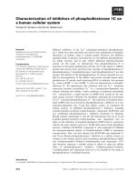
Two-dimensional gel electrophoresis (2DE) analysis of
the soluble fraction of M. capsulatus grown either at
a high or low copper-to-biomass ratio revealed pro-
tein spots significantly affected by the growth condi-
tions (Fig. 1, spots 1–15). In total, 27 protein spots
were identified by MS and N-terminal sequencing
combined with genomic information (Table 1). Fifteen
of these spots were differentially expressed. One of
the polypeptides migrated with an apparent molecular
mass of 14.4 kDa and a pI value of 5.8 (Fig. 1,
spot 15) and was only found in cells cultured at high
(0.8 lm) copper concentrations. MS analysis of the
excised spot resulted in seven matching peptides and
a 56.5% coverage, which in combination with
M. capsulatus genome sequence information identified
the protein as being encoded by the gene MCA0715
(Accession no. AE01782). This gene has previously
been annotated by us as a haemerythrin family pro-
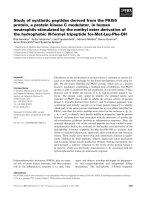
tein [5]. MCA0715 was located to a single transcrip-
tional unit, between the conserved hypothetical
protein MCA0714 and the hypothetical protein
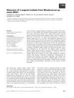
MCA0716 (Fig. 2). After the MS-based gene identifi-
cation, specific PCR primers were designed and the
differential expression in cells cultured at high or low
copper concentrations was verified by northern blot
analysis (Fig. 3).
Sequence analyses of the MCA0715-encoding pro-
tein using scanprosite [6] and conserved domain
search [7,8], identified the conserved H-F-x(2)-[EQ]-
[ENQ]-x(2)-[LMF]-x(4)-[FY]-x(5,6)-H-x(3)-[HR] motif
(PROSITE 00550). This signature pattern is charac-
teristic for members of the haemerythrin family and
it is located in the central region of these proteins.
We therefore named the MCA0715-encoded protein
Methylococcus capsulatus haemerythrin (McHr). The
haemerythrin family motif contains four conserved
iron ligands: the three histidines and the first gluta-
mate ⁄ glutamine of the motif. When using multiple
clustalx [9] alignments of the MCA0715-encoded
protein with members of the Hr (Fig. 4A) and MHr
(Fig. 4B) families, several conserved amino acids were
identified in the McHr protein sequence, as indicated
in Fig. 4. The regions of the McHr that contained
the highest level of identical residues and conserva-
tive replacements corresponded to the known helices
in resolved eukaryotic haemerythrin structures. In
contrast, the loops between helices showed low
degree of identity, thus representing the regions with
more amino acid variation. In the helical segments,
the amino acids H, E, Q, F, D were particularly well
conserved. In addition, all the amino acid residues
that are candidates for conjugating a diiron complex,
and the amino acids forming the O
2
-binding pocket
were found to be conserved (Fig. 4). The putative
M. capsulatus haemerythrin sequence (131 amino
acids) exceeds the sequence length of both haem-
erythrin subfamilies making McHr the largest known
haemerythrin. Thus, assuming that the corresponding
a helices are of equal length in the different haem-
erythrins, the loops (random coils) between the heli-
ces in McHr are longer than those of the eukaryotic
haemerythrins. Furthermore, a clustalx alignment-
based phylogenetic tree branched McHr distantly
to the members of both the eukaryotic Hr and
MHr subfamilies (Fig. 5). The highest similarity
between members of the haemerythrin family and
McHr was observed with the Hr alpha chain of
Lingula reevii, in which the sequence identity and
sequence similarity were calculated to 29 and 42%,
respectively.
O. A. Karlsen et al. Characterization of prokaryotic haemerythrin
FEBS Journal 272 (2005) 2428–2440 ª 2005 FEBS 2429
Phylogenetic distribution
The known phylogenetic distribution of haemerythrins
has long been limited to a relatively small group of
marine invertebrates. However, in light of the many
genomic sequencing projects of prokaryotes, we also
searched the databases SWISSPROT and TrEMBL
for putative prokaryotic proteins containing a haem-
erythrin-like domain (PROSITE PS00550) (Table 2)
[6]. Interestingly, unlike the very restricted distribu-
tion in eukaryotes, the haemerythrin motif seems to
be more widespread in the prokaryotic kingdom, and
to date has been found in 21 different bacteria from
five major genera. A majority of these putative haem-
erythrins are identified from Proteobacteria. Until
now, none of these putative prokaryotic haemeryth-
rins has been characterized. In fact, apart from the
McHr [4], there has been only one report of micro-
bial proteins containing a haemerythrin signature [3].
This haemerythrin-like motif was found in the C-ter-
minal end of DcrH, isolated from D. vulgaris. DcrH
is a member of the dcr gene family which sense and
respond to specific states of its environment. By expres-
sing only this DcrH C-terminal part, Xiong et al.
[3] demonstrated the presence of an oxo-bridged
diiron(III) site whose structure was very similar to that
found in haemerythrins. Most interestingly, a pairwise
alignment of this C-terminal end and McHr revealed a
very strong resemblance, containing 45 identical resi-
dues and 23 conservative replacements (Fig. 6).
Cloning and purification of McHr
Comparative bioinformatics data on the MCA0715
protein suggested that McHr is the first characterized
microbial homologue of the eukaryotic haemerythrins.
A
B
Fig. 1. 2DE patterns of the soluble fraction from low- (A) and high-copper (B) cultured M. capsulatus. The soluble fractions were analysed
using overlapping 18 cm narrow-range IPGs spanning the pH range of 4.0–5.0, 4.5–5.5, 5.0–6.0, 5.5–6.7 and broad range pH 3–10 IPGs
(Amersham). Both (A) and (B) are composite of such individual 2D gels. Numbered arrows refer to N-terminally or MS-identified spots.
Approximate molecular masses and pI values are indicated to the left and at the top of the gels, respectively.
Characterization of prokaryotic haemerythrin O. A. Karlsen et al.
2430 FEBS Journal 272 (2005) 2428–2440 ª 2005 FEBS
Table 1. Summary of identified proteins of M. capsulatus soluble fractions. Spot no. 1–15 represent differentially expressed polypeptides
between cells cultured at high- and low copper-to-biomass ratio. Spot no. 16–27 were found equally expressed in these culture condi-
tions. +, indicates expression in given growth condition. (+), indicate less abundant expression.
Spot number Gene Annotation +Cu –Cu
1, 2, 3, 8 mopE (MCA2589) M. capsulatus outer membrane protein E (MopE) +
12, 13 mmoX (MCA1194) Methane monooxygenase, subunit A, a-chain +
10 mmoY (MCA1195) Methane monooxygenase, subunit A, b-chain +
14 mmoZ (MCA1198) Methane monooxygenase, subunit A, c-chain +
5 mmoB (MCA1196) Methane monooxygenase, subunit B +
4 mmoC (MCA1200) Methane monooxygenase, subunit C +
9 groEL-2 (MCA1202)(mmoG) Chaperonin, 60 kDa subunit (mmoG) +
6, 7 groEL-3 (MCA1704) Chaperonin, 60 kDa subunit (+) +
11 groES-2 (MCA1705) Chaperonin, 10 kDa subunit (+) +
15 MCA0715 Haemerythrin family protein +
16, 17 mxaF (MCA0779) Methanol dehydrogenase, large subunit + +
18 mxaI (MCA0782) Methanol dehydrogenase, small subunit + +
26 MCA0789 MxaD protein, putative + +
24 Ndk (MCA2886) Nucleoside diphosphate kinase + +
21 dapD (MCA1490) 2, 3, 4, 5-Tetrahydropyridine-2,6 dicarboxylate
N-succinyltransferase
++
22 rpe (MCA2582) Ribulose-phosphate 3-epimerase + +
19, 20 MCA1023 Antioxidant, AhpC ⁄ Tsa family protein + +
27 Tkt2 (MCA3046) Transketolase + +
25 Tuf1 ⁄ Tuf2 (MCA1059 ⁄ MCA2374) Translation elongation factor Tu + +
23 atpA-1 (MCA0010) ATP synthase F1, a-subunit + +
Fig. 2. Genomic orientation (A), nucleotide and amino acid
sequence (B) of MCA0715. (A) MCA0715 is located between the
genes MCA0716 and MCA0714, putatively encoding a conserved
hypothetical protein and a hypothetical protein, respectively. (B)
The amino acids are indicated below the nucleotide sequence.
The underlined promoter region is predicted using the Neural
Network Promoter Prediction (itfly.org/seq_tools/
promoter.html) and has a probability of 0.98 (indicated by a red
arrow in A). Enlarged (T) indicates possible transcription start. Putative
ribosomal binding site is enlarged in bold. The predicted termination
loop (black arrows) has a calculated energy of )23.0 kcalÆmol
)1
( (indica-
ted as a hairpin in A).
Fig. 3. Northern blot of MCA0715. Total RNA (5 lg) isolated from
high- (+ Cu) and low-copper (– Cu) cultures of M. capsulatus were
electrophoresed through an agarose ⁄ formaldehyde gel and trans-
ferred to a nylon membrane. The blot was hybridized at 42 °C over-
night, with a radioactive probe made from the PCR fragment
amplified with the primers used in the cloning of MCA0715. The
arrowhead indicates the 540 bp transcript, corresponding to the
predicted length of the MCA0715 transcript. The molecular sizes
are indicated as given by the Invitrogen 0.24–9.5 kb RNA ladder.
O. A. Karlsen et al. Characterization of prokaryotic haemerythrin
FEBS Journal 272 (2005) 2428–2440 ª 2005 FEBS 2431
To study this protein in more detail, the mchr gene
was cloned and expressed in E. coli as a NusA–
(McHr) fusion protein. The fusion protein was puri-
fied, and the putative M. capsulatus haemerythrin was
cleaved from the NusA by TEV-protease and purified
to homogeneity by chromatography (Fig. 7). The rel-
ative molecular mass of the McHr protein was deter-
mined in calibrated gel filtration chromatography to
be 15 kDa (data not shown). This is close to a
molecular mass of 14.7 predicted from its amino acid
Fig. 4. Sequence comparisons between McHr and members of the haemerythrin (A) and myohaemerythrin (B) subfamily with CLUSTALX
software. CLUSTALX was used in secondary structure-based penalty mode. The haemerythrin sequences are obtained from the spinculids
P. gouldii (Pg) [39], T. zostericola (Tz) [40], T. dyscritum (Td) [41], S. cumanense (Sc) [42], and from the brachiopods L. unguis (LuA and LuB)
[43,44], L. reevii (LrA and LrB) [45]. The myohaemerythrin sequences are from the sipunculids P. gouldii (Pg) [46], T. zostericola (Tz) [47], the
achaete annelid T. tessulatum (Tt) [48], and the polychaete annelid N. diversicolor (Nd, MPII) [49,50]. McHr of M. capsulatus is indicated as
Mc in both (A) and (B). The helical regions based on the X-ray crystal structures of P. gouldii [20] (A) and T. zostericola [19] (B) are indicated
with (a) on top of each sequence block (I, II, III and IV). The seven amino acids involved as iron ligands are indicated by (#). (*) indicates the
five hydrophobic pocket-forming amino acids.
Characterization of prokaryotic haemerythrin O. A. Karlsen et al.
2432 FEBS Journal 272 (2005) 2428–2440 ª 2005 FEBS
sequence and indicates that McHr is stable in a
monomeric form under our experimental conditions.
Structure predictions
The tertiary structure of all characterized haemeryth-
rins comprises a left-twisted four a-helix bundle which
provides a hydrophobic pocket where dioxygen binds
the two iron atoms. Bioinformatical secondary struc-
ture predictions using various software programs indi-
cated predominant a-helix content in McHr. We were
able to model a three-dimensional structure of McHr
in swiss-model [10], using the multiple alignments
and the crystallized myohaemerythrin from Themiste
zostericola as a template (Fig. 8A) [11]. Hence, compu-
tational analysis modelled the putative haemerythrin to
a four a-helix bundle, maintaining both the conserved
haemerythrin positioning of the amino acid residues
coordinating the metal ions, and the hydrophobic
pocket in which dioxygen is situated (Fig. 8B,C).
Furthermore, McHr contains two cysteins, Cys88 and
Cys128. In our model, Cys88 was placed near the
Fig. 5. Unrooted phylogenetic tree of the haemerythrin sequences
of eukaryotic members of the haemerythrin family and McHr. A
bootstrapped phylogenetic tree was constructed from the multiple
alignments of Fig. 4. The tree was displayed using
TREEVIEW. The
abbreviations of organism names correspond to those given in
the legend to Fig. 4, and the myohaemerythrins are indicated by
(MHr).
Table 2. Prokaryotes which possess open reading frames encoding putative haemerythrins containing the haemerythrin signature (PROSITE
00550).
Accession no.
(SwissProt and
TrEMBL) Gene name Phylogeny Organism
Amino
acid
length
Q67PL5 STH1393 Actinobacteria Symbiobacterium termophilum 147
O67614 AQ_1719 Aquificae Aquifex aeolicus 131
Q6NE67 ORF13 Proteobacteria (a) Magnetospirillum gryphiswaldense 149
Q62LM6 BMA0605 Proteobacteria (b) Burkholderia mallei 149
Q63SE6 BPSL2377 Proteobacteria (b) Burkholderia pseudomallei 141
Q84IH8 ORF33 Proteobacteria (b) Janthinobacterium sp. J3 149
Q8Y1B3 RSc0777 Proteobacteria (b) Ralstonia solanacearum 137
Q9I352 PA1673 Proteobacteria (c) Pseudomonas aeruginosa 153
Q8GI12 ORF33 Proteobacteria (c) Pseudomonas resinovorans 146
Q9AQM2 ORF33 Proteobacteria (c) Pseudomonas resinovorans 165
Q8P892 XCC2351 Proteobacteria (c) Xanthomonas campestris 153
Q8PJP7 XAC2483 Proteobacteria (c) Xanthomonas axonopodis 153
Q6MHK9 Bd3532 Proteobacteria (d) Bdellovibrio bacteriovorus 136
Q6AMA9 DP1787 Proteobacteria (d) Desulfotalea psychrophila 156
Q748S2 GSU2929 Proteobacteria (d) Geobacter sulfurreducens 131
Q74G47 GSU0402 Proteobacteria (d) Geobacter sulfurreducens 135
Q74GJ0 GSU0256 Proteobacteria (d) Geobacter sulfurreducens 145
Q7MRA7 WS1527 Proteobacteria (e) Wolinella succinogenes 168
Q9PIQ3 Cj0241c Proteobacteria (e) Campylobacter jejuni 133
Q7VK87 HH0005 Proteobacteria (e) Helicobacter hepaticus 187
Q7VK88 HH0004 Proteobacteria (e) Helicobacter hepaticus 184
Q891L2 CTC02359 Firmicutes Clostridium tetani 137
Q97MX1 CAC0069 Firmicutes Clostridium acetobutylicum 129
Q8RCZ5 TTE0259 Firmicutes Thermoanaerobacter tengcongensis 133
Q73N54 TDE1302 Spirochaetes Treponema denticola 143
Q73NY7 TDE1013 Spirochaetes Treponema denticola 137
O. A. Karlsen et al. Characterization of prokaryotic haemerythrin
FEBS Journal 272 (2005) 2428–2440 ª 2005 FEBS 2433
C-terminal end of helix III, pointing towards the inside
of the bundle, and Cys128 was located to the random
coil of the C-terminal end of the protein (C-terminal
to helix IV) (not shown). The long distance between
these two residues in the predicted tertiary structure
would prevent the formation of an intramolecular
disulfide bridge. The major difference in tertiary struc-
ture of this model compared with haemerythrins, is the
increased random coiled structure between helices I
and II, and between helices III and IV (Fig. 8A). How-
ever, because swiss-model transposes only a target
sequence to a model based on a template structure
and does not predict additional regular motifs, it is
interesting that the secondary structure programs
(scratch, phdsec and psipred) utilized predict longer
helices, thereby shortening the modelled loops. To fur-
ther establish the secondary structure of McHr, the
protein was analysed using circular dichroism (CD)
(Fig. 9A). The recorded CD spectrum of McHr
revealed two negative maxima at 208 and 222 nm, and
one positive maximum at 195 nm, typical of proteins
with extensive a-helical secondary structures. The con-
tent of a-helical structures determined by CD (58.0%)
agreed with the values obtained from model the pre-
diction (57%). The stability of the protein was ana-
lysed by the decrease in elipticity at 222 nm for
temperature scans (25–90 °C) (Fig. 9B). The protein
was very stable with an approximate denaturing tem-
perature of 75 °C.
Spectrophotometric analysis and metal binding
In addition to the CD spectrum, UV ⁄ vis spectrophoto-
metric analysis of the purified protein showed a maxi-
mum at 280 nm (Fig. 10). Most importantly, two local
maxima of 330 and 380 nm were also revealed.
These maxima are typical of diiron-centre absorbance,
and thus characteristic for all haemerythrins [12]. This
strongly indicates that our protein preparation con-
tains conjugated iron ions. However, in order to fur-
ther confirm the presence of iron in the purified
protein, McHr was subjected to inductively coupled
plasma atomic emission-mass spectrometry (ICP-MS)
analyses after extensive removal of free metal ions
from the protein solution. The analyses clearly demon-
strated the presence of iron, and we estimated that
33% of the McHr contained two irons ⁄ molecule.
The finding that not all McHr molecules contained
iron was in agreement with previous reports that des-
cribe difficulties in incorporation of iron when over-
expressing heterologous diiron proteins in E. coli [13].
Discussion
Haemerythrins have been considered to be important
O
2
-handling proteins for some marine invertebrates
and for a long time were believed to be restricted to
only a few phyla of such eukaryotes. However, we
identified a putative prokaryotic haemerythrin
expressed in high-copper content cultures of the
methane-oxidizing bacterium M. capsulatus. Further-
more, we found that the haemerythrin motif is wide-
spread in the prokaryotic kingdom, and, to date, we
have located putative haemerythrins in 21 different
bacteria of 174 sequenced bacterial genomes.
Owing to the importance of copper in the biology of
M. capsulatus, it is of great interest to study protein
expression in cells cultured at different copper concen-
trations. Copper ions regulate the expression of
the two types of methane monooxygenase (MMO) of
M. capsulatus [14–16]. At high copper-to-biomass
ratios, the particulate membrane-associated pMMO is
Fig. 6. Sequence alignment of McHr and
DcrH-Hr. Dv: DcrH-Hr fragment of DcrH from
D. vulgaris. Mc: McHr of M. capsulatus.
AB
Fig. 7. SDS ⁄ PAGE analysis of proteins during purification of McHr.
Samples from each step in the purification procedure were collec-
ted and analysed. (A) 10% polyacrylamide gel. Lane 1,
crude extract of E. coli BL21 Star
TM
(DE3)[pETM60] prior to IPTG
induction. Lane 2, as lane 1, after IPTG induction. Lane 3, purified
His-tagged NusA–(McHr) from the induction in lane 2. Lane 4, gel
filtrated His-tagged NusA–(McHr) fusion protein (lane 3). (B) 15%
polyacrylamide gel. Lane 1, NusA–(McHr) before cleavage with
TEV-protease. Lane 2, as lane 1, after TEV treatment. Lane 3, puri-
fied McHr after gel filtration of TEV cleaved NusA–(McHr). Mole-
cular size markers are indicated for both subfigures.
Characterization of prokaryotic haemerythrin O. A. Karlsen et al.
2434 FEBS Journal 272 (2005) 2428–2440 ª 2005 FEBS
produced. pMMO expression is accompanied by the
formation of a complex intracytoplasmic membrane
system, in which the pMMO enzyme activity resides
[17]. At a low copper-to-biomass ratio, the soluble
cytoplasmic version of MMO (sMMO) is expressed.
The underlying mechanisms for these major morpholo-
gical and physiological changes are not known, but
copper-mediated gene activation or repression has been
implicated [18]. Recently, MS technology was used to
study protein expression at different copper concentra-
tions [4].
Our 2DE analysis revealed a strongly copper-regula-
ted protein. Initial sequence analysis indicated that this
protein was a homologue of the eukaryotic haemeryth-
rins. In further sequence analysis, the resolved struc-
tures of Hr and MHr isolated from Phascolopsis
gouldii and T. zostericola [19,20] opened up the possi-
bility of using secondary structure-based penalties in
multiple alignments. Such an approach has been
shown to improve their accuracy and reliability. The
clustalx alignments of McHr vs. Hr and MHr identi-
fied the candidate residues which are known to coordi-
nate two iron atoms, in addition to those forming
the O
2
-binding hydrophobic pocket. The presence of a
corresponding iron-binding motif was also supported
by the UV ⁄ vis spectrum of McHr, which demonstrated
specific iron centre absorbance peaks between 320 and
380 nm, and ICP-MS analyses which confirmed the
presence of iron. In addition, the strong sequence simi-
larity to the bacterial haemerythrin-like domain of
DcrH [3] also supports the presence of an oxo-bridged
diiron(III) site in McHr.
The seven ligands of the diiron site in haemeryth-
rins are amino acid residues each located within the
four helices, and by diiron binding generating an
intramolecular cross-linking of these helices, which
A
B
C
Fig. 8. SWISS-MODEL generated three-dimen-
sional structure of McHr.
SWISS-MODEL was
used in the alignment interface mode. The
multiple alignment of McHr vs. myohaem-
erythrins (Fig. 2B), was subjected for model-
ling using crystallized T. zostericola (PDB
Accession no. 1a7d) as the template and
McHr as the target sequence. The graphical
presentation was prepared in Deep-
View ⁄ Swiss-PdbViewer. (A) 1, X-ray crystal
structure of T. zostericola myohaemerythrin
at 1.8 A
˚
resolution [11]; 2, modelled 3D
structure of McHr. (B) 1, iron-binding ligands
of T. zostericola; 2, candidate iron-binding
residues of McHr; 3, overlay of (1) and (2).
(C) 1, amino acids of T. zostericola forming
the O
2
-binding hydrophobic pocket; 2, candi-
date residues of (1) in McHr; 3, Overlay of
(1) and (2).
O. A. Karlsen et al. Characterization of prokaryotic haemerythrin
FEBS Journal 272 (2005) 2428–2440 ª 2005 FEBS 2435
will stabilize the overall protein structure [21]. The
conservation of all seven ligand amino acid residues
in McHr constitutes strong evidence for a four-helix
bundle structure of this protein. A helical secondary
structure was also determined experimentally by CD
analysis, which estimated 58% a-helices in McHr.
Further, computational analysis modelled McHr to a
four a-helix bundle, maintaining the conserved haem-
erythrin positioning of all the amino acids involved in
the active site. However, the validity of this model
must be explored further by structural analyses as
NMR and X-ray crystallography.
Most haemerythrin proteins have been assigned a
function in handling oxygen, like the multisubunit
haemerythrins found for the haemerythrocytes of sipun-
culids, brachipods and priapulids [2,22]. It is not
unlikely that McHr serves a similar function in
M. capsulatus by reversibly binding oxygen. The con-
servation of both iron-binding ligands and O
2
-binding
pocket residues strongly suggests that interaction with
this molecule is part of its biological function. Sev-
eral enzymes (i.e. oxygenases and oxidases) require a
supply of O
2
and may be recipients for a McHr-bound
dioxygen molecule. Furthermore, at a high copper-to-
biomass ratio, pMMO is the enzyme responsible for
oxidizing methane in M. capsulatus [16]. This enzyme
uses oxygen as an oxidant in the process [23] and
therefore pMMO could also be an important receiver
of McHr delivered O
2
.
In conclusion, we describe the cloning of the first
prokaryotic haemerythrin and demonstrate that the
protein has a high a-helix content and binds iron in its
native form. We also provide novel evidence for the
presence of haemerythrin gene homologues in a large
group of prokaryotes. The exact biological function of
McHr in M. capsulatus is currently not known, but
because its expression is regulated by copper-ion con-
centration in the medium it is suggested that it plays an
important physiological role under high copper-to-bio-
mass growth conditions, possibly in methane oxidation.
Experimental procedures
Growth of M. capsulatus (Bath)
M. capsulatus NCIMB 11132 was grown in batch cultures
at 45 °C while shaking in an atmosphere of CH
4
,CO
2
and
O
2
(45 : 10 : 45, v ⁄ v ⁄ v) in NMS medium as described by
Whittenbury et al. [24]. Cells were grown either at a high
copper-to-biomass ratio by including a final concentration
of 0.8 lm copper in the growth medium, or at a low cop-
per-to-biomass ratio in ‘copper-free’ medium (no copper
added). Low-copper cultures were screened for sMMO
activity using the naphthalene assay described by Brusseau
et al. [25] to ensure that a low copper-to-biomass ratio was
achieved. Batch cultures of M. capsulatus were grown to a
cell density of 10
8
cellsÆmL
)1
before harvesting.
Fig. 10. UV ⁄ vis absorption spectra of recombinant McHr. The spec-
trum was recorded in gel filtration buffer (20 m
M Tris ⁄ HCl, pH 8.0,
400 m
M NaCl, 0.4 mM AEBSF-hydrochloride).
Fig. 9. CD spectra and thermal scan of McHr. (A) Far-UV CD spec-
trum at 25 °C of the purified McHr obtained in 20 m
M KH
2
PO
4
,
pH 8.0. (B) CD-monitored thermal scan following the changes in
ellipticity at 210 nm. The temperature scan was recorded in the gel
filtration buffer (20 m
M Tris ⁄ HCl, pH 8.0, 400 mM NaCl, 0.4 mM
AEBSF-hydrochloride).
Characterization of prokaryotic haemerythrin O. A. Karlsen et al.
2436 FEBS Journal 272 (2005) 2428–2440 ª 2005 FEBS
Preparation of the soluble fraction
Cells were harvested by centrifugation at 5000 g for
10 min. Enriched fractions of the soluble proteins were
obtained as described by Fjellbirkeland et al. [26]. Protein
concentrations were determined by using the Protein
DC-kit from Bio-Rad (Oslo, Norway).
SDS/PAGE
SDS ⁄ PAGE was performed as described by Laemmli et al.
[27] using either 15 or 10% (w ⁄ v) running gels and 3%
(w ⁄ v) stacking gels.
Two-dimensional gel electrophoresis
2DE was performed essentially as described by Rabilloud
et al. [28]. An appropriate volume of sample was precipi-
tated in 80% (v ⁄ v) acetone, and the pellet was solubilized
in an isoelectric focusing buffer containing 7 m urea, 2 m
thiourea, 4% (w ⁄ v) CHAPS, 0.5% (v ⁄ v) Triton X-100,
20 mm dithiothreitol, 0.5% (v ⁄ v) carrier ampholytes
pH 3.5–10 (Amersham, Uppsala, Sweden) and trace
amounts of bromphenol blue. The sample was applied
into an immobilized pH gradient (IPG) strip by ‘in-gel’
rehydration over night, using the Immobiline Drystrip
Reswelling Tray (Amersham). Isoelectric focusing was per-
formed in the Multiphor II system (Amersham) at 20 °C
using the Pharmacia (Uppsala, Sweden) EPS 3500 XL
power supply in gradient mode according to the manu-
facturers’ procedure. Prior to the second dimension
SDS ⁄ PAGE, the IPG strip was equilibrated twice for
15 min in buffer containing 6 m urea, 30% (v ⁄ v) glycerol,
50 mm Tris ⁄ HCl (pH 8.8), 2% SDS, 2.5 mgÆmL
)1
dithio-
threitol and traces of bromophenol blue. In the second
equilibration step, dithiothreitol was omitted and replaced
by 45 mgÆmL
)1
iodoacetamide. The second dimension elec-
trophoresis was carried out in the Protean II XI
(Bio-Rad) apparatus at 20 °C and using a 12.5% poly-
acrylamide gel. The power programme consisted of two
phases; 5 mA per gel for 2 h, followed by 8 mA per gel
until the tracing dye reached the end of the gel. Proteins
were visualized by SYPRO RUBY
TM
fluorescence staining
(Bio-Rad), and gels were scanned with a Fuji (Tokyo,
Japan) FLA-2000 phosphoimager.
Identification of proteins by MS and N-terminal
sequencing
MS analyses were performed at the Mass Spectrometry
Facility at the University of Warwick (Coventry, UK), and
at the PROBE facility at the University of Bergen, Norway.
N-Terminal amino acid sequencing was carried out at the
University of Oslo, Norway.
Cloning, expression and purification of McHr
mchr (MCA0715 of the M. capsulatus genome) was ampli-
fied by PCR (primers: McHr_F_NcoI, 5¢-CCATGGCATT
AATGACGTGG-3¢; McHr_R_XhoI, 5¢-GCTCGAGTTAT
GCGCTCAGG-3¢) and cloned into the pETM60 vector
using XhoI and NcoI restriction sites, thereby fusing mchr
to the nusA gene separated by a linker region [29]. The lin-
ker region contains a His-tag and the sequence for the
TEV-protease cleavage site. Positive clones were verified by
sequencing. Large-scale protein expression was performed
using E. coli BL21 Star
TM
(DE3) containing the pETM60
expression vector grown at 37 °C. Expression was induced
with addition of 0.7 mm isopropyl thio-b-d-galactoside
(IPTG) at A
600
0.6, and cultured for additional 4–5 h.
Pelleted bacteria were resuspended and sonicated in
NaCl ⁄ P
i
, pH 8.0, 0.4 mm AEBSF-hydrochloride (Appli-
chem), 10 mm imidazole. After centrifugation at 39 000 g
for 20 min, the supernatant was filtered (0.22 lm) before
loaded onto a pre-equilibrated HisTrap
TM
chelating col-
umn (Amersham). Bound proteins were eluted by stepwise
addition of increasing concentrations of imidazole in
NaCl ⁄ P
i
. Fractions containing the NusA–(McHr) fusion
protein were pooled and applied onto a Superdex 75 16 ⁄ 60
gel filtration column for removal of imidazole (gel filtra-
tion buffer: 20 mm Tris ⁄ HCl, pH 8.0, 400 mm NaCl,
0.4 mm AEBSF-hydrochloride). Further, TEV-protease
(Invitrogen, Carlsbad, CA, USA) was added to a concen-
tration of 10 units for 20 lg substrate and incubated at
room temperature overnight. His-tagged TEV and NusA
were selectively removed by reloading the solution onto
the HisTrap
TM
chelating column where they were tightly
bound, whereas McHr was recovered in the unbound frac-
tions. A final gel filtration of McHr was performed on a
Superdex 75 16 ⁄ 60 column to remove traces of NusA and
imidazole.
Spectrophotometric analysis
UV ⁄ vis spectrophotometric absorption data was obtained
in 1 cm path-length quartz cuvettes on a UNICAM
UV ⁄ VIS UV2 spectrometer. CD analysis was performed
using a Jasco (Cremella, Italy) J-810 spectropolarimeter
equipped with a Jasco 423S Peltier element for temperature
control. Protein samples were prepared in different buffers
as indicated. The standard analysis program provided with
the instrument was used for analysing the data. Secondary
structure elements were estimated by the cdnn program
that utilizes a neural network procedure [30].
Metal determination
ICP-MS analyses were used to determine the metal content
in McHr, and were carried out at the Department of Earth
O. A. Karlsen et al. Characterization of prokaryotic haemerythrin
FEBS Journal 272 (2005) 2428–2440 ª 2005 FEBS 2437
and Science, University of Bergen, Norway. Prior to analy-
ses, the McHr preparation was passed twice through a
PD-10 desalting column (Amersham) pre-equilibrated with
H
2
O for removal of contaminating metal ions present in
the gel filtration buffer. Furthermore, McHr was diluted
with concentrated nitric acid [6% (v ⁄ v) final concentration]
and digested for 24 h at 110 °C. After cooling, the samples
were diluted with H
2
O to a final concentration of 2% (v ⁄ v)
nitric acid and analysed for iron content.
RNA isolation using the hot phenol extraction
method
M. capsulatus flask cultures were harvested by centrifuga-
tion at 16 300 g for 1 min. Pellets were resuspended in
Tris ⁄ EDTA buffer (0.1 mm Tris ⁄ HCl, 0.1 mm EDTA,
pH 8.0) and submerged directly into liquid nitrogen and
stored at )80 °C until used. RNA isolation and DNase
treatment were carried out as described at http://www.
microarrays.org/pdfs/Total_RNA_from_Ecoli.pdf. The RNA
was dissolved in 50 lL of RNase-free water and the A
260
and A
280
values were determined.
Northern blotting
Blotting and hybridization experiments were performed as
described by Sambrook et al. [31]. The gene probe of mchr
(MCA0715) was generated by PCR amplification using
DyNAzymeEXT polymerase (Finnzymes, Espoo, Finnland)
and the oligonucleotides used in the cloning of mchr.
Bioinformatic tools
Scanprosite [6] and the Conserved Domain Database [7,8]
were used when analysing the McHr primary sequence for
conserved patterns. Multiple alignments were constructed by
clustalx (v. 1.83) [9] using secondary structure-based penal-
ties obtained from the solved haemerythrins of P. gouldii
(Hr, pdb 1i4y) and T. zostericola (MHr, pdb 1a7d), respect-
ively. Standard alignment parameters of clustalx were util-
ized and the resulting multiple alignments were visualized
in genedoc. Bootstrapped phylogenetic trees based on the
multiple alignments were made in clustalx and viewed in
treeview. The 3D model of McHr was built using swiss-
model [10] in multiple alignment mode, using the multiple
alignment of McHr with the myohaemerythrins (Fig. 4B)
and the X-ray structure of T. zostericola MHr (pdb 1a7d) as
a template. The quality of the model was first assessed using
procheck [32,33]. The vast majority of residues (89.7%) fall
into the most favoured regions of the Ramachandran plot,
9.4% in the allowed regions and only one residue (Ala100)
into the disallowed region. These results are consistent with
the values obtained for the template structure (94.3, 5.7, 0%,
respectively). Furthermore, the 3D)1D compatibility was
checked using verify 3d [34] and prosa [35] which both
showed that the model has a plausible overall fold; in parti-
cular, the predicted helical regions have scores in the same
range as the template structure. Secondary structure predic-
tions were made by submitting the McHr sequence to the fol-
lowing prediction programs; scratch (ssPRO) [36], phdsec
[37] and psipred [38].
Acknowledgements
This work was supported by grants from the Nor-
wegian Research Council and the Meltzer Foundation,
University of Bergen. We thank Professor J. C. Murrel
for mass spectrometry analyses performed at the Uni-
versity of Warwick (Coventry, UK) and PROBE at
the University of Bergen for peptide ⁄ protein analyses.
We thank D. Straume for help with the northern blot
analyses, and Rolf-Birger Pedersen and Ole Tumyr at
the Department of Earth and Science at the University
of Bergen, Norway for performing the ICP-MS ana-
lyses. We also thank the Computational Biology Unit
(CBU) at the University of Bergen for assessment of
our structural model.
References
1 Stenkamp RE (1994) Dioxygen and Hemerythrin. Chem
Rev 94, 715–726.
2 Kurtz DM Jr (1992) O
2
carriers in blood and tissues. In
Advances in Comparative and Environmental Physiology
(Mangum CP, ed), pp. 151–171. Springer Verlag,
Heidelberg.
3 Xiong J, Kurtz DM Jr, Ai J & Sanders-Loehr J (2000)
A hemerythrin-like domain in a bacterial chemotaxis
protein. Biochemistry 39, 5117–5125.
4 Kao WC, Chen YR, Yi EC, Lee H, Tian Q, Wu KM,
Tsai SF, Yu SS, Chen YJ, Aebersold R & Chan SI
(2004) Quantitative proteomic analysis of metabolic reg-
ulation by copper ions in methylococcus capsulatus
(Bath). J Biol Chem 279, 51554–51560.
5 Ward N, Larsen E, Sakwa J, Bruseth L, Khouri H,
Durkin AS, Dimitrov G, Jiang L, Scanlan D, Kang KH
et al. (2004) Genomic Insights into Methanotrophy: The
Complete Genome Sequence of Methylococcus capsula-
tus (Bath). Plos Biol 2, E303.
6 Gattiker A, Gasteiger E & Bairoch A (2002) ScanPro-
site: a reference implementation of a PROSITE scan-
ning tool. Appl Bioinformatics 1, 107–108.
7 Marchler-Bauer A, Anderson JB, DeWeese-Scott C,
Fedorova ND, Geer LY, He S, Hurwitz DI, Jackson
JD, Jacobs AR, Lanczycki CJ et al. (2003) CDD: a
curated Entrez database of conserved domain alignments.
Nucleic Acids Res 31 , 383–387.
Characterization of prokaryotic haemerythrin O. A. Karlsen et al.
2438 FEBS Journal 272 (2005) 2428–2440 ª 2005 FEBS
8 Marchler-Bauer A, Panchenko AR, Shoemaker BA,
Thiessen PA, Geer LY & Bryant SH (2002) CDD: a
database of conserved domain alignments with links to
domain three-dimensional structure. Nucleic Acids Res
30, 281–283.
9 Thompson JD, Gibson TJ, Plewniak F, Jeanmougin F
& Higgins DG (1997) The CLUSTAL_X windows inter-
face: flexible strategies for multiple sequence alignment
aided by quality analysis tools. Nucleic Acids Res 25,
4876–4882.
10 Schwede T, Kopp J, Guex N & Peitsch MC (2003)
SWISS-MODEL: An automated protein homology-
modeling server. Nucleic Acids Res 31, 3381–3385.
11 Martins LJ, Hill CP & Ellis WR Jr (1997) Structures of
wild-type chloromet and L103N hydroxomet Themiste
zostericola myohemerythrins at 1.8 A
˚
resolution.
Biochemistry 36, 7044–7049.
12 Zhang JH, Kurtz DM Jr, Xia YM & Debrunner PG
(1991) Reconstitution of the diiron sites in hemery-
thrin and myohemerythrin. Biochemistry 30, 583–
589.
13 Gupta N, Bonomi F, Kurtz DM Jr, Ravi N, Wang DL
& Huynh BH (1995) Recombinant Desulfovibrio vulgaris
rubrerythrin. Isolation and characterization of the diiron
domain. Biochemistry 34, 3310–3318.
14 Nielsen AK, Gerdes K & Murrell JC (1997) Copper-
dependent reciprocal transcriptional regulation of
methane mono-oxygenase genes in Methylococcus capsu-
latus and Methylosinus trichosporium. Mol Microbiol 25,
399–409.
15 Nielsen AK, Gerdes K, Degn H & Murrell JC (1996)
Regulation of bacterial methane oxidation: transcription
of the soluble methane mono-oxygenase operon of
Methylococcus capsulatus (Bath) is repressed by copper
ions. Microbiology 142, 1289–1296.
16 Stanley SH, Prior SD, Leak DJ & Dalton H (1983)
Copper stress underlies the fundamental change in intra-
cellular location of the methane mono-oxygenase in
methane-oxidizing organisms: studies in batch and con-
tinuous cultures. Biotechnol Lett 5, 487–492.
17 Prior SD & Dalton H (1985) The effect of copper ions
on the membrane content and methane mono-oxygenase
activity in methanol-grown Methylococcus capsulatus.
Journal Gen Microbiol 131, 155–163.
18 Murrell JC, McDonald IR & Gilbert B (2000) Regula-
tion of expression of methane mono-oxygenases by
copper ions. Trends Microbiol 8, 221–225.
19 Sheriff S, Hendrickson WA & Smith JL (1987) Struc-
ture of myohemerythrin in the azidomet state at 1.7 ⁄ 1.3
A
˚
resolution. J Mol Biol 197, 273–296.
20 Farmer CS, Kurtz DM Jr, Liu ZJ, Wang BC, Rose J,
Ai J & Sanders-Loehr J (2001) The crystal structures of
Phascolopsis gouldii wild type and L98Y methemery-
thrins: structural and functional alterations of the O
2
binding pocket. J Biol Inorg Chem 6, 418–429.
21 Zhang JH & Kurtz DM Jr (1992) Metal substitutions at
the diiron sites of hemerythrin and myohemerythrin:
contributions of divalent metals to stability of a four-
helix bundle protein. Proc Natl Acad Sci USA 89,
7065–7069.
22 Terwilliger NB (1998) Functional adaptations of
oxygen-transport proteins. J Exp Biol 201, 1085–1098.
23 Chan SI, Chen KH, Yu SS, Chen CL & Kuo SS (2004)
Toward delineating the structure and function of the
particulate methane monooxygenase from methano-
trophic bacteria. Biochemistry 43, 4421–4430.
24 Whittenbury R, Phillips KC & Wilkinson JF (1970)
Enrichment, isolation and some properties of methane-
utilizing bacteria. J General Microbiol 61, 205–218.
25 Brusseau GA, Tsien HC, Hanson RS & Wackett LP
(1990) Optimization of trichloroethylene oxidation by
methanotrophs and the use of a colorimetric assay to
detect soluble methane mono-oxygenase activity. Biode-
gradation 1, 19–29.
26 Fjellbirkeland A, Kleivdal H, Joergensen C, Thestrup H
& Jensen HB (1997) Outer membrane proteins of
Methylococcus capsulatus (Bath). Arch Microbiol 168,
128–135.
27 Laemmli UK (1970) Cleavage of structural proteins
during the assembly of the head of bacteriophage T4.
Nature 227, 680–685.
28 Rabilloud T, Adessi C, Giraudel A & Lunardi J (1997)
Improvement of the solubilization of proteins in two-
dimensional electrophoresis with immobilized pH gradi-
ents. Electrophoresis 18, 307–316.
29 De Marco V, Stier G, Blandin S & de Marco A (2004)
The solubility and stability of recombinant proteins are
increased by their fusion to NusA. Biochem Biophys Res
Commun 322, 766–771.
30 Bohm G, Muhr R & Jaenicke R (1992) Quantitative
analysis of protein far UV circular dichroism spectra by
neural networks. Protein Eng 5, 191–195.
31 Sambrook J, Fritsch EF & Maniatis T (1989) Molecu-
lar Cloning: A Laboratory Manual. Cold Spring Har-
bor Laboratory Press, Cold Spring Harbor, New
York.
32 Morris AL, MacArthur MW, Hutchinson EG & Thorn-
ton JM (1992) Stereochemical quality of protein struc-
ture coordinates. Proteins 12, 345–364.
33 Laskowski R, MacArthur M, Moss D & Thornton J
(1993) PROCHECK: a program to check the stereo-
chemical quality of protein structures. J Appl Cryst 26,
283–291.
34 Luthy R, Bowie JU & Eisenberg D (1992) Assessment
of protein models with three-dimensional profiles.
Nature 356, 83–85.
35 Sippl MJ (1993) Recognition of errors in three-dimen-
sional structures of proteins. Proteins 17, 355–362.
36 Pollastri G, Przybylski D, Rost B & Baldi P (2002)
Improving the prediction of protein secondary
O. A. Karlsen et al. Characterization of prokaryotic haemerythrin
FEBS Journal 272 (2005) 2428–2440 ª 2005 FEBS 2439
structure in three and eight classes using recurrent
neural networks and profiles. Proteins 47, 228–235.
37 Rost B & Sander C (1994) Combining evolutionary
information and neural networks to predict protein
secondary structure. Proteins 19, 55–72.
38 McGuffin LJ, Bryson K & Jones DT (2000) The
PSIPRED protein structure prediction server. Bioinfor-
matics 16, 404–405.
39 Klippenstein GL, Holleman JW & Klotz IM (1968) The
primary structure of Golfingia gouldii hemerythrin. Oder
of peptides in fragments produced by tryptic digestion
of succinylated hemerythrin. Complete amino acid
sequence. Biochemistry 7, 3868–3878.
40 Ferrell RE & Kitto GB (1971) Structural studies on
Dendrostomum pyroides hemerythrin. Biochemistry 10,
2923–2929.
41 Loehr JS, Lammers PJ, Brimhall B & Hermodson MA
(1978) Amino acid sequence of hemerythrin from
Themiste dyscritum. J Biol Chem 253, 5726–5731.
42 Uchida T, Yano H, Satake K, Kubota I & Tsugita A
(1990) The amino acid sequence of hemerythrin from
Siphonosoma cumanense. Protein Seq Data Anal 3,
141–147.
43 Yano H, Satake K, Ueno Y & Tsugita A (1991) The
amino acid sequence of the beta chain of hemerythrin
from Lingula unguis. Protein Seq Data Anal 4, 87–91.
44 Yano H, Satake K, Ueno Y, Kondo K & Tsugita A
(1991) Amino acid sequence of the hemerythrin alpha
subunit from Lingula unguis. J Biochem (Tokyo) 110,
376–380.
45 Negri A, Tedeschi G, Bonomi F, Zhang JH & Kurtz
DM Jr (1994) Amino-acid sequences of the alpha- and
beta-subunits of hemerythrin from Lingula reevii.
Biochim Biophys Acta 1208, 277–285.
46 Long RC, Zhang JH, Kurtz DM Jr, Negri A, Tedeschi
G & Bonomi F (1992) Myohemerythrin from the sipun-
culid, Phascolopsis gouldii: purification, properties and
amino acid sequence. Biochim Biophys Acta 1122,
136–142.
47 Klippenstein GL, Cote JL & Ludlam SE (1976) The
primary structure of myohemerythrin. Biochemistry 15,
1128–1136.
48 Coutte L, Slomianny MC, Malecha J & Baert JL (2001)
Cloning and expression analysis of a cDNA that
encodes a leech hemerythrin. Biochim Biophys Acta
1518, 282–286.
49 Demuynck S, Li KW, Van der Schors R & Dhainaut-
Courtois N (1993) Amino acid sequence of the small cad-
mium-binding protein (MP II) from Nereis diversicolor
(annelida, polychaeta). Evidence for a myohemerythrin
structure. Eur J Biochem 217, 151–156.
50 Takagi T & Cox JA (1991) Primary structure of myo-
hemerythrin from the annelid Nereis diversicolor. FEBS
Lett 285, 25–27.
Supplementary material
The following material is available from http://www.
blackwellpublishing.com/products/journals/suppmat/EJB/
EJB4663/EJB4663sm.htm
Fig. S1. Multiple alignment of McHr and the bacterial
sequences containing the hemerythrin signature (PRO-
SITE 00550) (Table 2). clustalx was used with stand-
ard alignment parameters. The sequences were
obtained by searching with the PROSITE 00550 pat-
tern in the SWISSPROT and TrEMBL databases.
Accession nos. for sequences and abbreviations for
organism names are given in the alignment; Geobacter
sulfurreducens (Gs), Xanthomonas campestris (Xc),
Xanthomonas axonopodis (Xa), Phseudomonas aerugi-
nosa (Pa), Symbiobacterium termophilum (St), Thermo-
anaerobacter tengcongensis (Tt), Treponema denticola
(Td), Helicobacter hepaticus (Hh), Clostridium acetobu-
tylicum (Ca), Clostridium tetani (Ct), Magnetospirillum
gryphiswaldense (Mg), Burkholderia mallei (Bm), Burk-
holderia pseudomallei (Bp), Ralstonia solanacearum
(Rs), Pseudomonas resinovorans (Pr), Janthinobacterium
sp. J3 (Js), Wolinella succinogenes (Ws), Desulfotalea
psychrophila (Dp), Bdellovibrio bacteriovorus (Bb),
Campylobacter jejuni (Cj), Aquifex aeolicus (Aa).
Characterization of prokaryotic haemerythrin O. A. Karlsen et al.
2440 FEBS Journal 272 (2005) 2428–2440 ª 2005 FEBS