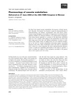
Báo cáo khoa học: Pharmacology of vascular endothelium Delivered on 27 June 2004 at the 29th FEBS Congress in Warsaw pptx
Bạn đang xem bản rút gọn của tài liệu. Xem và tải ngay bản đầy đủ của tài liệu tại đây (297.9 KB, 12 trang )
THE SIR HANS KREBS LECTURE
Pharmacology of vascular endothelium
Delivered on 27 June 2004 at the 29th FEBS Congress in Warsaw
Ryszard J. Gryglewski
Jagiellonian University, Cracow, Poland
Sir Hans Krebs was one of the most versatile biochem-
ists of the twentieth century. Many of his sayings stay
as bright as his science. My favourite quotation is: ‘‘…
we all are committed to correlating biochemical events
to function … the point I want to make is that it is not
always immediately clear what their relevance to func-
tion may be …’’ [1]. Indeed, a burning desire for imme-
diate comprehension, amplified by the abomination of
being just another fact collector may overcome rational
cautiousness. We pharmacologists know this only too
well. Sir John Vane urged his young followers: ‘‘Do
simple experiments and make simple hypotheses – there
are plenty of others who will come along and show
how much more complicated the answer really is …’’
[2]. Keeping in mind the above advice, I present the
vascular endothelium as a newly discovered target for
the pharmacotherapy of arterial hypertension, athero-
thrombosis and diabetic angiopathies.
I intend to focus on the pharmacology of the
endothelial prostacyclin ⁄ nitric oxide radical (PGI
2
⁄
Keywords
ACE-I; ASA; bradykinin; endothelial
dysfunction; nitric oxide; prostacyclin;
statins; thienopyridines
Correspondence
R. J. Gryglewski, Jagiellonian University,
Kasztelan˜ ska 30, 30-116 Cracow, Poland
E-mail:
(Received 10 February 2005, revised
13 April 2005, accepted 20 April 2005)
doi:10.1111/j.1742-4658.2005.04725.x
Sir John Vane named vascular endothelium ‘the maestro of blood circula-
tion’. Recently, ‘the maestro’ has become a target for pharmacotherapy of
atherothrombotic and diabetic vasculopathies with well known cardio-
vascular drugs belonging to the families of Angiotensin Converting Enzyme
inhibitors, HMG CoA reductase inhibitors or b
1
-Adrenoceptor antagonists.
These drugs became upgraded to a position of the pleiotropic endothelial
drugs. It is not a simple verbal change in the nomenclature. It means that
these drugs apart from their well defined mechanisms of action, as indi-
cated in their regular names, in addition they act in an unknown mechan-
ism at the level of vascular endothelium preventing angina, myocardial
infarction and stroke. Many biochemical events take place in endothelial
cells. I chose for a closer inspection the nitric oxide/prostacyclin defensive
system to explain the endothelial pleiotropism of the drugs in question. I
tried to examine the validity of this conception according to the general
rule: in vitro cognitio sed in vivo veritas.
Abbreviations
AA, arachidonic acid; ACE-I, angiotensin converting enzyme (and kininase 2) inhibitors; ADMA, asymmetric dimethylarginine; ASA,
acetylsalicylic acid; BH4, tetrahydrobiopterin; Bk, bradykinin; BPF, bradykinin potentiating factor; CAD, coronary heart disease; CaM,
calmodulin; COX-1, constitutive cyclooxygenase 1; COX-2, inducible cyclooxygenase 2; EDHF, endothelium-derived hyperpolarizing factor;
EDRF, endothelium-derived relaxing factor; EETs, cis-epoxyeicosatrienoic acids; eNOS, constitutive endothelial nitric oxide synthase; FAD,
flavin adenine dinucleotide; FMD, flow mediated dilatation (of brachial artery in humans); FMN, flavin mononucleotide; HMG-CoA,
hydroxymethylglutaryl coenzyme A; HO-1, inducible heme oxygenase; 15-HPAA, 15-hydroperoxyarachidonic acid; HYHC, hyperhomo-
cysteinemia; 6-keto-PGF
1a
, prostaglandin 6-keto-PGF
1a
, a stable product of decomposition of PGI
2
; LDL, low-density lipoproteins; L-NAME,
L-N(G)-nitroarginine methyl ester, a nonselective NOS inhibitor; NOHA, N
’
-hydroxy-Arg; ONOO
–
, peroxynitrite; ox-LDL, oxidized low-density
lipoproteins; PARP, poly ADP ribosyl polymerase; PGE
2
, prostaglandin E
2
; PGHS2, PGH
2
synthase; PGI
2
, prostacyclin; PGIS, prostacyclin
synthase; RNS, reactive nitrogen species; ROS, reactive oxygen species; SDMA, symmetric dimethylarginine; TXA
2
, thromboxane A
2
;
TXAS, thromboxane A
2
synthase; TXB
2
, thromboxane B
2
.
2956 FEBS Journal 272 (2005) 2956–2967 ª 2005 FEBS
NO
•
) defence system. Other aspects of endothelial
biology are reviewed by Nachman and Jaffe [3] with
a special attention being paid to the functioning of
Weibel–Palade bodies and their response to proin-
flammatory or prothrombotic agents as manifested by
the release of von Willebrand factor, P selectin and
interleukin-8. Readers interested in the endothelial
mitochondrion as a propagator of oxidative stress [4]
and the mitochondrion-oriented role of reactive oxy-
gen species (ROS) and hydrogen peroxide [5] are
directed to studies by Keaney and coworkers [4,5].
Mitochondrial oxidases along with NAD(P)H oxidase,
xanthine oxidase and uncoupled constitutive endothel-
ial nitric oxide synthase (eNOS) constitute the source
of endothelial ROS, which may act as modulators of
tone, growth and remodelling of the vascular wall. It
may well be that inflammation plays a primary role
in atherogenesis, whereas oxidative stress is a secon-
dary phenomenon [6]. At low concentrations, ROS
may protect endothelial cells against apoptotic beha-
viour [7]. Long-term treatment with antioxidant vita-
mins does not influence the course of the disease
or correct endothelial dysfunction in patients with
atherosclerosis [8]. The great expectations for the
therapeutic use of antioxidants in patients with athero-
sclerosis need to be re-examined.
Endothelium as the endocrine organ
Why does blood not coagulate within healthy blood
vessels? This question has been addressed for centuries.
The warmth of the body (Plato), the lack of contact
with air (James Hewson) and the vital power of blood
(John Hunter) have all been claimed as reasons. The
truth is that vascular endothelium secretes a bunch of
antithrombotic and thrombolytic mediators that keep
blood fluid within an undamaged circulatory system.
Vascular endothelium is neither a ‘primitive mem-
brane’, as claimed by Rudolph von Virchow, nor a
‘nucleated sheet of cellophane’, as Sir Howard Florey
stated [9]. Sir John Vane named the endothelium ‘the
maestro of blood circulation’ [10], which should be
viewed as a peculiar dissipated endocrine organ (mass
1000 g, surface area 100 m
2
). Among others sub-
stances, endothelium releases into the passing blood –
labile, lipophilic and antithrombotic local hormones
like PGI
2
and NO
•
as well as a peptide – tissue plasmi-
nogen activator. These prevent the build up of thrombi
and disperse any thrombi at an early stage of their for-
mation. This is why blood stays fluid within a healthy
vascular bed. The inherent chemical instability of PGI
2
and NO
•
allows for the immediate transformation of
extravasated blood into a haemostatic plug. Unfortu-
nately, the same transformation may occur locally
inside the circulatory system of patients with athero-
sclerotic plaques or diabetic angiopathies. The endo-
thelium then loses its protective properties and may
even produce proinflammatory and thrombogenic
agents (endothelial dysfunction).
Endothelium generates many biologically active sub-
stances other than PGI
2
or NO
•
, to mention just
four regioisomers of cis-epoxyeicosatrienoic acid (EETs)
produced from AA by CYP2J2 epoxygenases [11].
EETs are vasoprotective vasodilators. Some may be
responsible for the activity of endothelium-derived
hyperpolarizing factor (EDHF) [11], and for prevent-
ing platelet adhesion to endothelium [12]. A potent
vasoconstrictor, endothelin, is also produced [13], as
are a vast number of mediators of haemostasis, growth
factors and cytokines [14]. The outer endothelial layer
of the glycocalyx houses the membrane sensors for
shear stress and various types of endothelial receptors
such as B
2
for bradykinin (Bk), P
2y
subtypes for ADP
from platelets and ATP from erythrocytes, PAF-R for
platelet activating factor (PAF) from leukocytes, and
PAR for thrombin [15]. The membrane-bound endo-
thelial enzymes include kininase 2, also called angio-
tensin 1-converting enzyme (ACE-I).
Prostacyclin
Prostacyclin (PGI
2
) was discovered in 1976 during the
search for biological systems that in addition to blood
platelets might convert prostaglandin endoperoxides
(PGG
2
or PGH
2
) to thromboxane A
2
(TXA
2
) [16,17].
This search was possible because newly discovered
PGG
2
and PGH
2
were kindly offered to John R. Vane
by the discoverer of TXA
2
, Bengt Samuelsson of the
Karolinska Institutet. This search was not successful,
except for the detection of minute amounts of TXA
2
made from PGH
2
by lung and spleen microsomes.
Instead we found that a microsomal fraction of pig
aorta transformed prostaglandin endoperoxides into
an unknown, unstable substance (with a half-life of
4 min at 37 °C) that had vasodilator and platelet-
suppressant properties in vitro. This substance was
later named prostacyclin (PGI
2
). Further studies
revealed that PGI
2
, when administered intravenously,
dissipated platelet-rich thrombi in arterial blood in vivo
[18] and that this effect was augmented by theophyl-
line. This latter finding confirmed that a cyclic nucleo-
tide (in this case cAMP) was the second messenger of
PGI
2
in platelets [19].
The common precursor for prostanoids including
PGI
2
is the four double-bonds 2-carbon fatty acid –
arachidonic acid (AA). It was found that the nonenzy-
R. J. Gryglewski Endothelium and drugs
FEBS Journal 272 (2005) 2956–2967 ª 2005 FEBS 2957
matic product of AA monooxygenation (15-hydro-
peroxy-arachidonic acid; 15-HPAA) and other linear
lipid peroxides are inhibitors of microsomal prosta-
cyclin synthase (PGIS). Therefore, we hypothesized
that PGI
2
deficiency resulted from an excessive non-
enzymatic peroxidation of body lipids might contribute
to development of atherosclerosis [20]. Consequently,
we hoped to use synthetic PGI
2
as a replacement ther-
apy in patients with atherosclerosis.
Actually, I was the first healthy volunteer to receive
an intravenous infusion of synthetic PGI
2
sodium salt.
I lost the noble position of an observer during the last
stage of this experiment. Still, these early trials allowed
us to establish a range of therapeutic doses for PGI
2
and to observe the side effects caused by its overdos-
age [21]. Eventually, PGI
2
was infused into patients
with atherosclerosis of the leg arteries [22]. However,
like most other powerful biological mediators, both
PGI
2
(epoprostenol) [23] and its stable analogues (e.g.
iloprost) [24] never became first-line drugs for the
treatment of atherothrombosis, instead giving way to
drugs that act as releasers of endogenous endothelial
PGI
2
[25]. However, some PGI
2
analogues (e.g. tre-
prostinil) are still used to treat patients with pulmon-
ary arterial hypertension [26], including those with
connective tissue disease [27].
The lung is a rich source of eicosanoids including
leukotrienes. Various prostanoids are generated within
different pulmonary compartments. Tracheal smooth
muscles generate PGE
2
, contractile elements of lung
parenchyma generate TXA
2
[28], whereas pulmonary
endothelium secrets PGI
2
. We hypothesized [29] that
pulmonary endothelium may serve as a source for
circulating PGI
2
[30]. This concept was not well
accepted. What kind of a circulating hormone has a
half-life in blood of 3–4 min? Nonetheless, assuming
PGI
2
is generated continuously by pulmonary endo-
thelium, the stability of PGI
2
might be sufficient for
it to be transported within the blood from the lung
to atherosclerotic coronary or cerebral arteries with
dysfunctional endothelium, and to save them from
being occluded by platelet-rich thrombi. Pulmonary
endothelium might be a good target for new specific
releasers of circulating PGI
2
, although the local gen-
eration of PGI
2
by the endothelium lining the vascu-
lar tree is probably a more important therapeutic
target, at least up to the point when the efficacy of
peripheral endothelium in not seriously disturbed by
an advanced atherothrombosis. Interestingly, overex-
pression of pulmonary PGIS decreases the incidence
of cancerogenesis in murine models of lung cancer [31].
The crude microsomal fraction of aortic homogen-
ates that allowed us to discover biosynthesis the of
PGI
2
from PGH
2
[16,17] contained PGIS. This enzyme
was purified and characterized as a member of cyto-
chrome P450 family (CYP 8A1) [32]. In endothelial
cells it collaborates with a supplier of PGH
2
, i.e. with
PGH
2
synthase (PGHS-2), commonly, but less pre-
cisely, called cyclooxygenase 2 (COX-2). In endothelial
cells COX-2 is induced by shear stress. COX-2 seems
to be the major source of systemic PGI
2
in healthy
humans [33]. In female mice oestrogens upregulate
PGI
2
production via COX-2, and subsequently offer
protection against atherothrombosis [34]. Also, intra-
vascular thrombosis in rats next to hypoxia-induced
hypertension is prevented by the upregulation of
vascular COX-2 followed by increased generation of
PGI
2
[35].
There is little doubt that, in humans and laboratory
animals, the endothelial COX-2 ⁄ PGIS tandem is
responsible for the generation of vasoprotective PGI
2
,
whereas in blood platelets the constitutive cyclooxy-
genase 1 ⁄ thromboxane A
2
synthase (COX-1 ⁄ TXAS)
tandem generates vasotoxic TXA
2
.
Nitric oxide radical
In 1980, a series of in vitro experiments with acetyl-
choline-treated aortic rings led Robert Furchgott to
discover endothelium-derived relaxing factor (EDRF)
[36]. Robert Furchgott likes to say that his great dis-
covery arose from a number of accidental findings.
Those in 1986 exploded in the grand finale, i.e. in the
discovery that EDRF is nitric oxide. Actually, the idea
that EDRF ¼ NO was proposed by Robert Furchgott
and Louis Ignarro, independently [37]. Robert Furchg-
ott is modest as only a great scholar can be. His mod-
esty provokes the quotation from Louis Pasteur: ‘‘…
where observation is concerned, chance favours only
the prepared mind’’.
The fabulous story of the discovery of EDRF(NO)
was presented by Robert Furchgott [37], Louis Ignarro
[38] and Ferid Murad [39,40] – three 1998 Nobel prize
laureates in medicine and physiology. In vascular endo-
thelium, NO
•
is synthetized from Arg by eNOS, which
competes for substrate with tissue arginases. eNOS is a
homodimeric oxidoreductase with NADPH, flavin
mononucleotide (FMN), flavin adenine dinucleotide
(FAD), calmodulin (CaM) and tetrahydrrobiopterin
(BH4) acting as cofactors. eNOS via N’-hydroxy-Arg
(NOHA) generates NO
•
and citrulline. Physiologically,
eNOS homodimer catalyses a five-electron oxidation of
Arg, whereas BH4 plays a crucial role in the activation
of dioxygen. In tissues, NO
•
is a powerful endogenous
stimulator of soluble cytosolic guanylate cyclase. Thus
made, cGMP is the second messenger for NO
•
in the
Endothelium and drugs R. J. Gryglewski
2958 FEBS Journal 272 (2005) 2956–2967 ª 2005 FEBS
same way that cAMP is the second messenger for
PGI
2
. Both cyclic nucleotides mediate the vasodilator,
vasoprotective and platelet-suppressant activities of
NO
•
and PGI
2
, respectively.
However, when eNOS splits into monomers, the
eNOS monomer acts as a reductase, and one-electron
reduction of dioxygen leads to formation of a super-
oxide anion (O
2
–
) [41]. Uncoupling of eNOS occurs
as a consequence of BH4 shortage resulting from
folate avitaminosis or from hyperhomocysteinaemia
(HYHC) [42]. Apart from the uncoupling of eNOS,
the other source of vascular O
2
–
might be NAD(P)H
oxidase (43). Dimerization of eNOS requires the
intracellular availability of the substrate, i.e. Arg.
This is ensured by the high-affinity cationic cell mem-
brane transporter for Arg. Its functioning might be
invalidated by homocysteine or by asymmetric dime-
thylarginine (ADMA) (see below). Arg supplementa-
tion in patients with atherosclerosis may be also
desirable because Arg acts as a direct antioxidant. In
addition, Arg promotes the secretion of insulin from
pancreatic b cells and the release of histamine from
mast cells – both being vasodilators. Theoretically,
Arg may also produce unfavourable effects, such as
generation of S-adenosyl-homocysteine from S-adeno-
sylmethionine via the methylation-dependent biosyn-
thesis of creatinine from guanidine acetate (44). Yet,
the net therapeutic effect of Arg given orally to
patients with myocardial infarction is encouraging
(45) pointing to a favourable route of biotransforma-
tion in these patients.
Patrick Valance discovered, in human plasma, the
presence of symmetric dimethylarginine (SDMA) and
ADMA. Only ADMA is biologically active, i.e. it acts
as endogenous inhibitor of eNOS and inhibitor of Arg
membrane transporter. Clinical data on ADMA are
growing. A high plasma level of ADMA is considered
a novel cardiovascular risk factor. Nowadays, it is
clear that ADMA contributes to vascular pathology
in atherothrombotic and diabetic angiopathies, pre-
eclampsia and hypertension (46). Elevated plasma lev-
els of ADMA in those patients may also explain the
‘arginine paradox’, i.e. that therapeutic supplementa-
tion with exogenous Arg is beneficial, although in
these patients plasma levels of endogenous Arg exceed
the Michaelis–Menten constant (K
m
) for purified eNOS
in vitro by 25-fold [47].
Prostacyclin and nitric oxide radicals
A complex relationship exists between these two unsta-
ble, lipophylic endothelial secretagogues. At the time
when NO
•
still was known as EDRF it was claimed
that porcine aorta endothelial cells cultured on cytodex
beads, loaded into a heated column and perfused with
Krebs’ buffer, when stimulated with Bk or calcium
ionophore, released both PGI
2
and EDRF in a cou-
pled manner [48]. Superoxide anions abolished the
biological activity of the released EDRF from these
cultured endothelial cells [49], and from native endo-
thelium of perfused canine artery [50]. These latter
findings initiated a march towards the discovery of the
product of the interaction between NO
•
and O
2
–
, i.e.
peroxynitrite (ONOO
–
). ONOO
–
is one of the most
reactive nitrogen species (RNS). It arises most easily
when the eNOS dimer coexists in the vicinity of a
eNOS monomer – then both genders of labile free rad-
icals, i.e. NO
•
and O
2
–
arise side by side, and without
any delay ONOO
–
is made.
ONOO
–
is a powerful oxidant and nitrating agent
that destroys the ‘macromolecules of life’, i.e. proteins
(e.g. PGIS inactivation), lipids [e.g. the generation of
oxidized low-density lipoprotein (ox-LDL) and iso-
prostanes], and nucleic acids [e.g. DNA strand break-
age with a subsequent activation of poly-ADP ribosyl
polymerase (PARP)] [51].
The toxic properties of ONOO
–
play a major role in
atherothrombotic and diabetic angiopathies. In those
endothelial cells, ONOO
–
oxidizes the four zinc thio-
late centres of dimeric eNOS. As a consequence, zinc
atoms are removed and disulfide monomers of eNOS
arise. The coexistence of dimeric and monomeric forms
of eNOS is responsible for the further amplification of
ONOO
–
generation by endothelial cells. This newly
made ONOO
–
selectively nitrates Tyr430 in the enzy-
mic protein of endothelial PGIS. When PGI
2
is elimin-
ated from the endothelial defence system TXA
2
and
PGH
2
gain the upper hand [52].
Endothelial NOS received the mischievous name of
‘the Cinderella of inflammation’ [53]. The authors had
in mind that excessive stimulation of eNOS might lead
to increased vascular permeability by NO
•
, and thus
to inflammation. However, in light of the foul games
played between homodimeric and monomeric forms of
eNOS, ending with the generation of ONOO
–
which
eliminates PGI
2
– the best friend of NO
•
– I would
rather think of eNOS as ‘the Lady Macbeth of athero-
thrombosis’.
Endothelial pharmacology
Samuel Beckett (1906–1989) wrote: ‘‘we need new par-
adigms to accommodate the mess’. The paradigm of
‘pleiotropic action’ for some of cardiovascular drugs
was coined to accommodate a discrepancy between
their officially accepted modes of action and their
R. J. Gryglewski Endothelium and drugs
FEBS Journal 272 (2005) 2956–2967 ª 2005 FEBS 2959
additional therapeutic properties, as reported unexpect-
edly, but repeatedly, by clinicians. For example, statins
were introduced to the clinic with the aim of lowering
blood levels of low-density lipoprotein (LDL) cho-
lesterol, however, they were also found to correct
symptoms of myocardial and cerebral ischaemia, inde-
pendent of their capacity to inhibit hydroxymethyl-
glutaryl coenzyme A (HMG CoA) reductase [54–56].
Further support for the existence of the ‘pleiotropic
action’ of cardiovascular drugs was offered by the
efficacy of ACE-I to protect against myocardial isch-
aemia, stroke and diabetic angiopathies, as confirmed
in multicentre trials that included over 25 000 patients
[57], whereas the classic indication for ACE-I was the
treatment of patients with arterial hypertension. The
phrase ‘pleiotropic action’ is not a cognitive descrip-
tion of reality. Rather, it is an attempt ‘to accommo-
date the mess’. Our experimental data [58,59] pointed
to the possibility that the pleiotropic action of ACE-I
and statins might be explained by their stimulatory
effect on the endothelial generation of PGI
2
and NO
•
.
There are other propositions concerning the mechan-
ism of endothelial actions of statins, e.g. the induction
of heme oxygenase (HO-1) [60] with a subsequent anti-
oxidant effect of biliverdin and CO mediation. Here, I
take the opportunity to present our conception of the
endothelial pharmacology emerging from clinical
observations on the unexpected therapeutic effects of
known cardiovascular drugs. This conception embraces
not only ACE-I and statins, but also other cardiovas-
cular drugs, e.g. nebivolol and carvedilol (b-adrenergic
receptor antagonists) as well as ticlopidine and clopi-
dogrel (antiplatelet thienopyridines).
In vivo assay of endothelial function
Clinicians have developed an excellent noninvasive
method to measure endothelial function in humans.
The method is based on the ultrasound scanning of
the flow-mediated dilatation (FMD) of the brachial
artery after its occlusion and reopening [61]. In prin-
ciple, the FMD response is proportional to the
amount of NO
•
released from endothelium of the vas-
cular bed in question, however, an additional bio-
chemical assay pointed to the release of PGI
2
, along
with NO
•
, from the endothelium during FMD [62].
No wonder – in vitro cultured endothelial cells
released EDRF(NO) and PGI
2
in a coupled manner
[48]. The FMD method allowed the detection of endo-
thelial dysfunction in patients with arterial hyperten-
sion [62], in patients with atherosclerosis undergoing
percutaneous coronary intervention with stenting [63],
and in patients with type 2 diabetes [64]. In patients
with chest pain, a depressed FMD of the brachial
artery was a sensitive indicator of coronary heart dis-
ease (CAD) [65]. FMD is impaired in tobacco smokers
and in smokeless tobacco users compared with tobacco
nonusers [66]. There is ample evidence for the state-
ment that endothelial dysfunction occurs in patients
with hypertension, atherosclerosis and type 2 diabetes,
as well as in tobacco users.
In vitro cognitio sed in vivo veritas (in vitro one may
look for meaning, however, only in vivo is the truth to
be found). This motto stimulated us to develop our own
experimental model for the in vivo assay of endothelial
function [18–20,29,59,67–70]. In our in vivo method it is
not the vasodilator response (as in the case of FMD in
humans) but rather the thrombolytic response that is
used to assess endothelial capacity. Therefore, it is the
endothelial release of PGI
2
that is appreciated at
the first place, whereas the release of NO
•
remains in the
background. Heparinized cats, rabbits and, most fre-
quently, Wistar rats under general anaesthesia with
extracorporeal circulation are used. The arterial blood
superfuses (2–3 mLÆmin
)1
) a collagen strip attached to a
balance. Blood returns to the venous system. Thrombus
mass is recorded continuously along with arterial blood
pressure (Fig. 1). Platelet-rich thrombus [70] gains a
maximum mass of 100 mg within 30 min and stays
unchanged for at least 4 h, unless a stimulator of vas-
cular endothelium (e.g. Ach, Bk or an endotheliotropic
drug) is injected intravenously. Then thrombolysis
occurs (Fig. 1). Its intensity and duration correlate with
plasma levels of prostaglandin 6-keto-PGF
1a
(6-keto-
PGF
1a
), whereas the levels of other stable prostanoids
do not (Fig. 2). The participation of endothelial NO
•
in
thrombolytic response is checked by the pretreatment of
animals with l-NAME or with any other NOS inhib-
itor. The participation of endogenous bradykinin in this
response was checked by pretreatment with Icatibant,
an antagonist of B2 receptors (Fig. 1A). In this system,
thrombi were dissipated by intravenous administration
of PGI
2
sodium salt or by its stable analogue (e.g. ilo-
prost). NO-donors (glyceryl trinitrate, molsidomine,
sodium nitropusside, NONOates) also produced throm-
bolysis but their effective doses were at a range of three
orders of magnitude higher than those required for
PGI
2
or for its analogues. Unlike PGI
2
, NO-donors at
thrombolytic doses were highly hypotensive.
Angiotensin-converting enzyme
inhibitors
ACE-I, this name does not do justice to this class of
drugs, namely captopril, enalapril, and especially per-
indopril, quinapril, ramipril and many other lipophylic
Endothelium and drugs R. J. Gryglewski
2960 FEBS Journal 272 (2005) 2956–2967 ª 2005 FEBS
ACE-I. There is no doubt that the pharmacological
activity of ACE-I is associated with the elimination of
cytotoxic and vasoconstrictor angiotensin 2, however,
the endothelial action of those ACE-I is also executed
via the local vascular accumulation of Bk, as our data
clearly show (Fig. 1A) [59,67–69].
In 1965 a young Brazilian researcher, Sergio Ferreira
discovered the ‘bradykinin potentiating factor’ (BPF)
in the venom of Brazilian viper Bothrops jararaca [71].
At John Vane’s laboratory in London (where Sergio
Ferreira was a visitor) his discovery was appreciated
than it should have been. At the time, Bk was per-
ceived as a mediator of pain and inflammation respon-
sible for paralytic vasodilatation in the course of acute
pancreatitis. The reasoning was as follows: BPF might
be good for this particular viper for swift killing of its
victims but for us humans – it is no good at all. So,
why should we care about BPF?
Perfusate from isolated guinea-pig lungs dripping
over Vane’s bioassay cascade was used to study Bk
[71] and angiotensin 1 [72] metabolism. Fortunately, it
was soon found that various fractions of BPF given
via the lungs inhibited the conversion of angiotensin 1
to angiotensin 2, and thus BPF was proved to act also
as an ACE-I [73]. Inhibiting the conversion of biolo-
gically inactive angiotensin 1 to hypertensive angioten-
sin 2 – yes, it was an excellent principle on which to
develop a new class of antihypertensive drugs [74].
Indeed, at the request of John Vane, the top industrial
chemists eventually did [75], and the first orally active
ACE-I (a proline derivative – captopril) was intro-
duced for the treatment of arterial hypertension.
The TREND trial [76] offered the first direct clinical
evidence of improvement, by an ACE-I (quinapril), in
endothelium-dependent vasorelaxation in patients with
CAD. There then appeared a number of clinical trials
pointing to the same mechanism of vascular protection
by various ACE-I in patients at high risk of athero-
thrombotic and diabetic vasculopathies [57].
B
A
BP
THR
THR
Dose-dependent thrombolysis by perindopril µg/kg i.v.
Thrombolysis by QUINAPRIL depends on the release of
endogenous bradykinin and PGl
2
– only partially on NO
THR
THR
THR
30 min
mg
mg
100
0
100
0
THR
rat 1
rat 2
rat 3 rat 4
icatibant 100 µg/kg
indomethacin 5 mg/kg
L-NAME 5 mg/kg
quinapril 30 µg/kg
quinapril 30 µg/kg
quinapril 30 µg/kg
quinapril 30 µg/kg
0
100
mg
Thrombogenesis
thrombus weight
Thrombolysis
pressure transducer
weight
transducer
carotid artery
carotid artery
arterial blood
arterial blood
collagen
collagen
THROMBUS
i.v. drug injection
jugular vein
PERINDOPRIL
30.
10.
3.
30 min
100
mg
0
Fig. 1. In vivo bioassay of endothelial secretory function.
Fig. 2. Effect of quinapril on prostanoid plasma levels in Wistar rats
(n ¼ 7).
R. J. Gryglewski Endothelium and drugs
FEBS Journal 272 (2005) 2956–2967 ª 2005 FEBS 2961
Bk is the most potent releaser of PGI
2
from cultured
endothelial cells [48], and the most potent thrombolytic
agent acting via endothelial B
2
receptors in vivo [67].
In Wistar rats, exogenous Bk at thrombolytic doses
is strongly hypotensive. In contrast, endogenous Bk
released from vascular endothelium by low doses of
ACE-I (quinapril > perindopril > captopril) evokes
thrombolysis, but not a fall in blood pressure [59,68].
The principal mechanism of the thrombolytic action of
ACE-I stems from their secondary nature (or rather
their primary nature) of being BPF [71]. Moreover,
there exist other Bk-potentiating effects of exogenous
ACE-I, such as the upregulation of B2 receptors, the
induction and activation of B1 receptors in the endo-
thelium and the stimulation of biosynthesis of angio-
tensin (1–7), which acts as an endogenous ACE-I (that
is BPF) [77]. It should be added that in cultured endo-
thelial cells Bk acts as a ‘minicytokin’, inducing
mRNA for HO-1 and COX-2 [67]. The interaction
between these two enzyme systems was claimed to
amplify the generation of PGI
2
[78]. It may well be
that, in addition to the immediate thrombolytic effects
of ACE-I, chronic treatment with ACE-I offers an
additional advantage of increasing the efficacy of the
endothelial enzymic raft (COX-2 ⁄ PGIS) responsible
for the biosynthesis of vascular prostacyclin along with
increasing local levels of CO and biliverdin – the
defensive products of endothelial HO-1.
In our in vivo model for studying endothelial-medi-
ated thrombolysis in Wistar rats [59,68,69] it was
found that ACE-I (captopril < perindopril < quina-
pril) at low nonhypotensive intravenous doses of 10–
60 lgÆkg
)1
dissipated thrombi that were superfused
with arterial blood. The intensity and duration of this
thrombolysis were paralleled by an increase in arterial
plasma levels of 6-keto-PGF
1a
, and no change in
plasma levels of TXB
2
and PGE
2
(Figs 1 and 2).
Thrombolysis and prostacyclinaemia by ACE-I were
blunted or abolished by pretreatment with icatibant (a
B2 Bk receptor antagonist), by acetylsalicylic acid
(ASA) at a high dose of 50 mgÆkg
)1
(Fig. 3), and by
the coxibs (rofecoxib > celecoxib > nimesulide) at
low doses of 30–300 lgÆkg
)1
. Thrombolysis by ACE-I
was augmented by pretreatment with ASA at a dose of
1mgÆkg
)1
(Fig. 3) or by acetaminophenen. Pretreat-
ment with l-NAME delayed and flattened the throm-
bolytic response to ACE-I only slightly (Fig. 1).
Pharmacological analysis of the above data led us to
conclude that ACE-I evoked thrombolysis by pre-
venting endothelial Bk from being destroyed by cell
membrane-bound ACE. Bk that appeared at the
endothelial cell surface stimulated B2 receptors, which
triggered the COX-2 ⁄ PGIS system to generate PGI
2
,
and e-NOS to generate NO. The final thrombolytic
response to ACE-I depended mainly on PGI
2
, whereas
NO
•
served as a helper with a permissive action. The
endothelial release of NO
•
did not appear as the
conditio sine qua non for thrombolytic response to
ACE-I (Fig. 1A).
There is another conclusion that derives from these
studies. It is as follows: effective endothelial COX-2
inhibition might be followed by thrombogenesis,
whereas preferential COX-1 inhibition in platelets rein-
forced the vasoprotective action of ACE-I (Fig. 3).
Our data cannot be considered as a good prognostic
for the clinical use of high doses of coxibs in patients
with cardiovascular disorders, but they do support the
idea of administrating of low doses of ASA along with
ACE-I (Fig. 3).
Statins
In our in vivo model statins (e.g. atorvastatin and
simvastatin) produce endothelium-mediated, PGI
2
-
dependent thrombolysis when administered intraven-
ously at doses 2–3 orders of magnitude higher than
those for ACE-I [59]. In Langendorff’s preparation of
guinea-pig heart, statins produce NO
•
-dependent vaso-
dilatation of coronary vascular bed [59]. The precon-
tracted bovine coronary artery rings with endothelium
are relaxed by statins, partially via a NO
•
⁄ PGI
2
-
dependent mechanism [79]. In cultured bovine aortic
endothelial cells lipophylic statins, i.e. atorvastatin,
simvastatin and lovastatin (but not a hydrophilic
pravastatin) at a concentration of 30 lm mobilize free
cytoplasmic calcium [Ca
2+
]
i
to 30–50% of that
induced by Bk at a concentration of 10 nm. In the
case of simvastatin and lovastatin, this effect disap-
pears if their lactone rings are hydrolysed [80]. The
above endotheliotropic properties of statins are hardly
Fig. 3. Dose-dependent effect of aspirin (ASA) on perindopril-
induced thrombolysis.
Endothelium and drugs R. J. Gryglewski
2962 FEBS Journal 272 (2005) 2956–2967 ª 2005 FEBS
associated with their inhibitory action on HMG CoA
reductase. In genetic and pharmacological models of
rat hypertension, rosuvastatin, another lipophilic sta-
tin, was found to exert a beneficial pleiotropic endo-
thelial effect [81]. Patients with acute coronary
syndromes benefit from statin therapy [82]. Statins
mobilize bone-marrow-derived endothelial progenitor
cells [83] and exert a vast number of other pharmaco-
logical effects that are not associated with modulation
of the lipoprotein profile by statins. These unexpected
effects of statins are generally described as ‘pleiotropic
effects’ [84], and one of them is the endotheliotropic
action of statins described by us [59,79,80]. The mode
of activation of the endothelial PGI
2
⁄ NO
•
system by
statins is not clear. An interesting proposal was put
forward by Bill Sessa [85].
Thienopyridines and some of
b
1
-adrenergic receptor antagonists
Here we present two groups of highly effective cardio-
vascular drugs, the efficiency of which may or may not
depend on their additional stimulatory action of vas-
cular endothelium.
Thienopyridines (ticlopidine and clopidogrel) belong
to a family of antiplatelet drugs, however, in vitro they
do not inhibit platelet aggregation. Their in vivo plate-
let-suppressant action is executed by their labile meta-
bolites. Therefore, a substantial lag period is required
for the appearance of the antiplatelet action of thieno-
pyridines. Only unstable metabolites of theirs are cap-
able of antagonizing endogenous ADP on P2y12
purinergic platelet receptors, which when activated by
ADP induce platelet release and platelet aggregation
[86]. The clopidogrel metabolite exerts its antiplatelet
action at IC
50
¼ 1.8 lm [87]. There exists ample evi-
dence for the high efficacy of thienopyridines (especi-
ally clopidogrel) in the treatment of patients with
advanced atherothrombosis of coronary or cerebral
arteries, to mention only the following megatrials:
clopidogrel vs. aspirin in patients at risk of ischemic
events (CAPRIE) [88], clopidogrel in unstable angina
to prevent recurrent events (CURE) and management
of atherothrombosis with clopidogrel in high risk
patients with recent transient ischaemic attacks or isch-
aemic stroke (MATCH) [89].
In 1996 [90] we demonstrated that ticlopidine
(10 mgÆkg
)1
) given intravenously to cats with extracor-
poreal circulation evoked immediate dissipation of the
platelet-rich clots superfused with their arterial blood
[18]. This thrombolytic effect of ticlopidine was com-
parable with that induced by PGI
2
at 0.3 lgÆkg
)1
.
These and other data [90] prompted us to postulate
that the therapeutic efficacy of ticlopidine might be
associated not only with the delayed platelet-suppres-
sant effect of its unstable metabolite via blockade of
P2y12 platelet receptors, but also with the instan-
taneous endothelial action of the native molecule
of ticlopidine showing up as an immediate, endo-
thelium-mediated thrombolysis of platelet-rich clots
in vivo [90].
In rats, these ‘immediate thrombolytic effects’ of
thienopyridines were rather weak (EC
30
¼ 15–30
mgÆkg
)1
). Jean-Pierre Dupin of the Bordeaux II Uni-
versity decided to synthetize a series of thienopyrimi-
dinones under the guidance of our pharmacological
assay of their endothelium-dependent thrombolytic
effects in vivo. Assessment of their structure–activity
relationship revealed that the most active compound,
i.e. 3[(2-trifluoromethyl-phenyl)-methyl] 1,2-dihydro-
benzo[b]thieno[2,3-d]pyrimidinone-4(3H)one dissipated
platelet clots in rats in vivo at a dose of IC
30
¼
8 lgÆkg
)1
[91].
We conclude that in addition to in vivo endothelial
PGI
2
-mediated thrombolysis, thienopyrimidinones and
thienopyridines exert endothelial NO
•
-mediated coron-
ary vasodilatation in perfused guinea-pig heart [92].
Mechanisms of endotheliotropic actions of these
strongly lipophylic compounds remain unknown.
Nebivolol and carvedilol – two b
1
-adrenoceptor
antagonists – founded the ‘third generation’ of selective
b-adrenolytic drugs, which are endowed with endothe-
liotropic properties. Eleven years ago, Bowman et al.
[93] proposed that the antihypertensive effects of nebiv-
olol in man might be partially associated with endo-
thelium-dependent, NO
•
-mediated vasodilatation. In
two interesting studies Ignarro et al. [94,95] clearly
demonstrated that relaxation of vascular smooth
muscle by nebivolol is partially mediated by endothe-
lium-dependent release of NO
•
and the subsequent
accumulation of cGMP in smooth muscle [94], how-
ever, nebivolol also inhibits vascular smooth muscle
proliferation by a mechanism involving NO
•
but not
cGMP [95]. Various routes were proposed by which
the ‘third generation’ of b
1
-adrenoceptor antagonists
may release endothelial NO
•
. Certainly adrenergic and
serotoninergic receptors are not involved [96]. A fascin-
ating hypothesis has been proposed [97]. Nebivolol
and carvedilol stimulate the renal efflux of ATP,
that releases NO
•
via activation of P2Y purinoceptors
in glomerular endothelium. On top of the regular
b
1
-adrenoceptor blockade there appears NO
•
-mediated
relaxation of renal glomerular microvasculature. This
is why nebivolol and carvedilol are so efficient in
controlling arterial hypertension and improving renal
circulation.
R. J. Gryglewski Endothelium and drugs
FEBS Journal 272 (2005) 2956–2967 ª 2005 FEBS 2963
Perspectives
In endothelial pharmacology everything is new: (a) the
idea that vascular endothelium may be looked upon as
an organ with a secretory function; (b) considering
pulmonary endothelium as a separate endocrine organ
that supplies prostacyclin to the coronary and cerebral
circulations; (c) a complex relationship between two
endothelial mediators – NO and PGI
2
– a role for
ROS and RNS in it; (d) discovering new endothelio-
tropic mechanisms for old cardiovascular drugs like
for ACE-I, statins or nebivolol; (e) planning new
endotheliotropic chemical structures, e.g. thienopiry-
midodiones; (f) discovering new biochemical mecha-
nisms of action for drugs affecting endothelial function
like in case of nebivolol; and (g) the interaction
between basic and clinical researchers, probably one of
the most efficient in the field of medicine. Old, known
roads are safe, but the newly discovered roads are
interesting.
References
1 Krebs HA (1980) Excerpts from an introductory address.
Int J Biochem 12, 1–8.
2 Gryglewski RJ & McGiff JC (2005) Eulogy John R.
Vane (1927–2004). Hypertension 45, 319–320.
3 Nachman RL & Jaffe EA (2004) Endothelial cell cul-
ture: beginning of modern vascular biology. J Clin
Invest 114, 1037–1040.
4 Schulz F, Antor F & Keaney JF (2004) Oxidative stress,
antioxidants and endothelium function. Curr Med Chem
11, 1093–1104.
5 Chen K, Thomas SR, Albano M, Murphy MP & Kea-
ney JF (2004) Mitochondrial function is required for
hydrogen peroxide-induced growth factor receptor
transactivation and downstream signalling. J Biol Chem
279, 35079–35086.
6 Stocker R & Keaney JF (2004) Role of oxidative
modifications in atherosclerosis. Physiol Rev 84, 1381–
1478.
7 Haendeler J, Tischler V, Hoffman J, Zeiher AM &
Dimmeler S (2004) Low doses of reactive oxygen species
protect endothelial cells from apoptosis by increasing
thioredoxin-1 expression. FEBS Lett 577, 427–433.
8 Kinlay S, Behrendt D, Fang JC, Delagrange D, Mor-
row J, Witztum JL, Ganz P, Rifai N, Selwyn AP &
Creager MA (2004) Long-term effect of combined vita-
min E and C on coronary and peripheral endothelial
function. J Am Coll Cardiol 43, 629–634.
9 Florey H (1966) The endothelial cell. Br Med J 2, 487–
490.
10 Vane JR (1994) The endothelium: maestro of the blood
circulation. Philos Trans R Soc Lond 343, 225–246.
11 Spiecker M & Liao JK (2005) Vascular protective
effects of cytochrome P450-derived eicosanoids. Arch
Biochem Biophys 433, 413–420.
12 Krotz F, Riexinger T, Buerkle MA, Nithipatikom K,
Gloe T, Sohn HY, Campbell WB & Pohk U (2004)
Membrane potential-dependent inhibition of platelet
adhesion to endothelial cells by epoxteicosatrienoic
acids. Atheroscler Thromb Vasc Biol 24, 595–600.
13 Masaki T (2004) Historical review: endothelin. Trends
Pharmacol Sci 25, 219–224.
14 Chłopicki S & Gryglewski RJ (2002) Pharmacology of
endothelium (in Polish). Kardiologia Polska 57, 5–15.
15 Garcia-Cardena G, Comander J, Loscalzo J, Anderson
KR & Gimbrone MA Jr, (2001) Biochemical activation
of vascular endothelium as a determinant of its func-
tional phenotype. Proc Natl Acad Sci USA 98, 4478–
4485.
16 Gryglewski RJ, Bunting S, Moncada S, Flower RJ &
Vane JR (1976) Arterial walls are protected against
deposition of platelet thrombi by a substance (prosta-
glandin X) which they make from prostaglandin endo-
peroxides. Prostaglandins 12, 685–713.
17 Moncada S, Gryglewski RJ, Bunting S & Vane JR
(1976) An enzyme isolated from arteries transforms
prostaglandin endoperoxides to an unstable substance
that inhibits platelet aggregation. Nature 263, 663–665.
18 Gryglewski RJ, Korbut R, Ocetkiewicz A & Stachura J
(1978) In vivo method for quantitation of anti-platelet
potency of drugs. Naunyn Schmiedebergs Arch Pharma-
col 302, 25–30.
19 Gryglewski RJ, Korbut R & Ocetkiewicz A (1978)
De-aggregatory action of prostacyclin in vivo and its
enhancement by theophyline. Prostaglandins 15, 637–
644.
20 Gryglewski RJ (1980) Prostacyclin, platelets and athero-
sclerosis. In Reviews on Biochemistry, Vol. 7 (Fasman
GD, ed), pp. 291–338. CRC Press, New York.
21 Gryglewski RJ, Szczeklik A & Ni
_
zankowski R (1978)
Anti-platelet effects of intravenous infusion of prosta-
cyclin in man. Thromb Res 13, 153–163.
22 Szczeklik A, Ni
_
zankowski R, Skawin
´
ski S, Głuszko P &
Gryglewski RJ (1979) Successful therapy of advanced
arteriosclerosis obliterans with prostacyclin. Lancet 1,
1111–1114.
23 Gryglewski RJ, Szczeklik A & McGiff JC, eds. (1983)
Prostacyclin – Clinical Trials. Raven Press, New York.
24 Gryglewski RJ & Stock G, eds. (1987) Prostacyclin and
its Stable Analogue Iloprost. Springer-Verlag, Berlin.
25 Chłopicki S & Gryglewski RJ (2004) Endothelial secre-
tory function and atherothrombosis. In The Eicosanoids
(Curtis-Prior P, ed), pp. 267–276. Wiley, Chichester.
26 Vachiery JI & Naeije R (2004) Treprostinil for pulmon-
ary hypertension. Expert Rev Cardiovasc Ther 2, 183–
191.
Endothelium and drugs R. J. Gryglewski
2964 FEBS Journal 272 (2005) 2956–2967 ª 2005 FEBS
27 Oudiz RJ, Schilz RJ, Barst RJ, Galie N, Rich S, Rubin
LJ & Simonneau G (2004) Treprostinil, a prostacyclin
analogue in pulmonary arterial hypertension associated
with connective tissue disease. Chest 126, 420–427.
28 Gryglewski RJ, Dembinska-Kiec A, Grodzinska L &
Panczenko B (1976) Differential generation of sub-
stances with prostaglandin-like and thromboxane-like
activities by the guinea pig trachea and lung strips. In
Lung Cells in Disease (Bouhuys A, ed), pp. 289–307.
Elsevier, Amsterdam.
29 Gryglewski RJ, Korbut R & Ocetkiewicz A (1978) Gen-
eration of prostacyclin by lungs in vivo and its release
into the arterial circulation. Nature 273, 765–767.
30 Gryglewski RJ (1980) The lung as a generator of pro-
stacyclin. In Ciba Foundation Symposium No. 78: Meta-
bolic Activities of the Lung, pp. 147–164. Excerpta
Medica, Amsterdam.
31 Keith RL, Miller YE, Hudish TM, Girod CE, Sotto-
Santiago S, Franklin WA, Nemenoff RA, March FH,
Nana-Sinkam SP & Geraci MW (2004) Pulmonary
prostacyclin synthase overexpression chemoprevents
tobacco smoke lung cancerogenesis in mice. Cancer Res
64, 5897–5904.
32 Shimonishi M, Nakamura M, Imai Y, Ullrich V &
Tanabe T (2004) Purification and characterisation of
recombinant human prostacyclin synthase. J Biochem
(Tokyo) 135, 455–463.
33 McAdam BF, Catella-Lawson F, Mardini IA, Kapoor
S, Lawson JA & FitzGerald GA (1999) Systemic bio-
synthesis of prostacyclin by cycloxygenase (COX)-2: the
human pharmacology of a selective inhibitor of COX-2.
Proc Natl Acad Sci USA 96, 272–277.
34 Egan KM, Lawson JA, Fries S, Koller B, Rader DJ,
Smyth EM & FitzGerald GA (2004) COX-2 derived
prostacyclin confers atheroprotection on female mice.
Science 306, 1954–1957.
35 Pidgeon GP, Tamosiuniana R, Chen G, Leonard I, Bel-
ton O, Bradford A & Fitzgerald DJ (2004) Intravascular
thrombosis after hypoxia-induced pulmonary hyperten-
sion: regulation by cycloxygenase-2. Circulation 110,
2701–2707.
36 Furchgott RF & Zawadzki JV (1980) The obligatory
role of endothelial cells in the relaxation of arterial
smooth muscle by acetylcholine. Nature 288, 373–376.
37 Furchgott RF (2001) Research leading to nitric oxide:
the importance of accidental findings. In Nitric Oxide
(Gryglewski RJ & Minuz P, eds), NATO Life Sciences
Series A 317, 1–4. IOS Press, Amsterdam.
38 Ignarro LJ (2002) Nitric oxide as a unique signalling
molecule in the vascular system: a historical overview.
J Physiol Pharmacol 53, 504–514.
39 Murad F (2004) Ferid Murad MD, PhD: a conversation
with the Editor. Am J Cardiol 94, 75–91.
40 Seminara AR, Krumenacker JS & Murad F (2001)
Signal transduction with nitric oxide, guanylyl cyclase
and cyclic guanosine monophosphate. In Nitric Oxide
(Gryglewski RJ & Minuz P, eds), NATO Life Sciences
Series A 317, pp. 5–22. IOS Press, Amsterdam.
41 Stuehr DJ, Adak S, Santolini J, Wei CC & Wang Z
(2001) Mechanism of oxygen activation in NO synthases,
and a kinetic model for catalysis. In Nitric Oxide (Gry-
glewski RJ & Minuz P, eds), NATO Life Sciences Series
A 317, pp. 23–26. IOS Press, Amsterdam.
42 Topal G, Brunet A, Millanvoye E, Boucher JL, Rebdu
F, Devyck MA & David-Dufilho M (2004) Homocys-
teine induces oxidative stress by uncoupling of NO
synthase activity through reduction of tetrahydrobiop-
terin. Free Radical Biol Med 36, 1532–1541.
43 Channon KM & Guzik TJ (2002) Mechanisms of super-
oxide production in human blood vessels. Relationship
to endothelial dysfunction, clinical and genetic factors.
J Physiol Pharmacol 53, 515–524.
44 Loscalzo J (2004) l-Arginine and atherothrombosis.
J Nutr 134 (10 Suppl.), 2798S–2800S.
45 Chamiec T, Ceremuzynski L & Bednarz B (2004) Effects
of l-arginine in myocardial infarction: double-blind,
placebo-controlled multicenter pilot study. J Am College
Cardiol 43 (Suppl. A), (abstract, p. 1).
46 Vallance P & Leiper J (2004) Cardiovascular biology of
the asymmetric dimethylarginine: dimethylarginine
dimethylaminohydrolase pathway. Arterioscler Thromb
Vasc Biol 24, 1023–1030.
47 Boger RH (2004) Asymmetric dimethylarginine, an
endogenous inhibitor of nitric oxide synthase explains
‘the arginine paradox’ and acts as a novel cardio-
vascular risk factor. J Nutr 134 (10 Suppl.), 2842S–
2847S.
48 Gryglewski RJ, Moncada S & Palmer RM (1986) Bioas-
say of prostacyclin and endothelium-derived relaxing
factor (EDRF) from cultured porcine aortic endothelial
cells. Br J Pharmacol 87, 685–694.
49 Gryglewski RJ, Palmer RM & Moncada S (1986)
Superoxide anion is involved in the breakdown of
endothelium-derived vascular relaxing factor. Nature
320, 454–456.
50 Rubanyi GM & Vanhoutte PM (1986) Superoxide
anions and hyperoxia inactivate endothelium-
derived relaxing factor. Am J Physiol 250, H822–
H827.
51 Szabo C (2003) Multiple pathways of peroxynitrite cyto-
toxicity. Toxicol Lett 140, 105–112.
52 Zou MH, Cohen R & Ullrich V (2004) Peroxynitrite
and vascular endothelial dysfunction in diabetes melli-
tus. Endothelium 11, 89–97.
53 Cirino G, Fiorucci S & Sessa WC (2003) Endothelial
nitric oxide synthase: the Cinderella of inflammation?
Trends Pharmacol Sci 24, 91–95.
54 Davignion J (2004) Beneficial cardiovascular
pleiotropic effects of statins. Circulation 109 (Suppl. 1),
39–43.
R. J. Gryglewski Endothelium and drugs
FEBS Journal 272 (2005) 2956–2967 ª 2005 FEBS 2965
55 Liao JK (2003) Role of statin pleiotropism in acute cor-
onary syndrome and stroke. Int J Clin Pract Suppl 134,
51–57.
56 Miida T, Hirayama S & Nakamura Y (2004) Choles-
terol-independent effects of statins and new therapeutic
targets: stroke and dementia. J Atheroscler Thromb 11,
253–264.
57 Bertrand ME (2004) Provision of cardiovascular protec-
tion by ACE inhibitors: a review of recent trials. Curr
Med Res Opin 20, 1559–1569.
58 Gryglewski RJ, Chłopicki S, Uracz W & Marcinkiewicz
E (2001) Significance of endothelial prostacyclin and
nitric oxide in peripheral and pulmonary circulation.
Med Sci Monit 7, 1–16.
59 Gryglewski RJ, Uracz W, Swies J, Chłopicki S, Mar-
cinkiewicz E, Lomnicka M & Madej J (2001) Compari-
son of endothelial pleiotropic actions of angiotensin
converting enzyme inhibitors and statins. Ann NY Acad
Sci 947, 229–245.
60 Grosser N, Hemmerle A, Berndt G, Erdmann K,
Hinkelmann U, Schurgrec S, Wijanti N, Immenschluh S
& Schroder H (2004) The antioxidant defence protein
heme oxygenase 1 is a novel target for statins in
endothelial cells. Free Radical Biol Med 37, 2064–2071.
61 Faulx MD, Wright AT & Hoit BD (2003) Detection of
endothelial dysfunction with brachial artery ultrasound
scanning. Am Heart J 145 , 943–951.
62 Yamanari H, Nakamura K, Kakishita M & Ohe T
(2004) Effect of cycloxygenase inhibition on endothelial
function in hypertensive patients treated with angio-
tensin-converting enzyme inhibitors. Clin Cardiol 27,
523–527.
63 Bae JH, Bassenge E, Kim KY, Syn YC, Park KR &
Schwemmer M (2004) Effects of low-dose atorvastatin
on vascular responses in patients undergoing percuta-
neous intervention with stenting. J Cardiovasc Pharma-
col Ther 9, 185–192.
64 Henry RM, Ferreira I, Kostense PJ, Dekker JM, Nijpels
G, Heine RJ, Kamp O, Bouter LM & Stehouwer CD
(2004) Type 2 diabetes is associated with impaired
endothelium-dependent, flow-mediated dilatation, but
impared glucose metabolism is not. The Horn study.
Atherosclerosis 174, 49–56.
65 Jambrik Z, Venneri L, Varga A, Rigo F, Borges A &
Picano F (2004) Peripheral vascular endothelial function
testing for diagnosis of coronary heart disease. Am
Heart J 148 , 684–689.
66 Granberry MC, Smith ES 3rd, Troillett RD & Edit JF
(2003) Forearm endothelial response in smokeless
tobacco users compared with cigarette smokers and
nonusers of tobacco. Pharmacotherapy 23, 974–978.
67 Gryglewski RJ, Uracz W, Chłopicki S & Marcinkiewicz
E (2002) Bradykinin as a major endogenous regulator
of endothelial function. Pediatr Pathol Mol Med 21,
279–290.
68 Gryglewski RJ, Swies J, Uracz W, Chłopicki S & Mar-
cinkiewicz W (2003) Mechanism of angiotensin convert-
ing enzyme inhibitor-induced thrombolysis in Wistar
rats. Thromb Res 110, 323–329.
69 Gryglewski RH, Chłopicki S & Swies J (2005) In vivo
endothelial interaction between ACE and COX inhibi-
tors. Prostaglandins Leukot Essent Fatty Acids 72,
129–131.
70 Mackiewicz Z, Gryglewski RJ, Swies J, Dobros W &
Uracz W (1996) Streptokinase-induced changes in the
structure of platelets. Ex vivo study. Acta Med Lithuan
2, 3–8.
71 Ferreira SH (1965) A bradykinin potentiating factor
(BPF) present in the venom of Bothrops jararaca. Br J
Pharmacol 24, 163–169.
72 Bakhle YS, Reynard AM & Vane JR (1969) Metabo-
lisms of angiotensins in isolated perfused tissues. Nature
222, 956–959.
73 Ferreira SH, Greene LH, Alabaster VA, Bakhle YS &
Vane JR (1970) Activity of various fractions of bradyki-
nin potentiating factor against angiotensin I converting
enzyme. Nature 225, 379–380.
74 Bakhle YS (1971) Inhibition of angiotensin I converting
enzyme by venom peptides. Br J Pharmacol 43, 252–254.
75 Ondetti MA, Rubin R & Cushman DW (1977) Design
of specific inhibitors of angiotensin-converting enzyme:
new class of orally active anti-hypertensive agents.
Science 196, 441–444.
76 Mancini GB, Henry CC, Macaya C, O’Neill BJ, Pucillo
AL, Carere RG, Wargovich TJ, Mudra H, Luscher TF,
Klibanger MI et al. (1996) Angiotensin-converting
enzyme inhibition with quinapril improves endothelial
vasomotor dysfunction in patients with coronary artery
disease. The TREND (Trial Reversing Endothelial Dys-
function) study. Circulation 94, 259–265.
77 Tom B, Dendorfer A & Danser AH (2003) Bradykinin,
angiotensin (1–7) and ACE inhibitors: how do they
interact? Int J Biochem Cell Biol 35, 792–801.
78 Haider A, Olszanecki R, Gryglewski RJ, Schwartzman
ML, Lianos E, Kappas A, Nasjletti A & Abraham NG
(2002) Regulation of cyclooxygenase by the heme-heme
oxygenase system in microvessel endothelial cells.
J Pharmacol Exp Ther 300, 188–194.
79 Lorkowska B & Chłopicki S (2005) Statins as coronary
vasodilators in isolated bovine coronary artery –
involvement of PGI
2
. Prostaglandins Leukot Essent
Fatty Acids 72, 133–138.
80 Lorkowska B, Chłopicki S, Marcinkiewicz E & Grygle-
wski RJ (2004) Statins rise cytosolic calcium level
[Ca
2+
]i in cultured endothelial cells. Pol J Pharmacol
56, 313–318.
81 Susic D, Varagic J, Ahn J, Slama M & Froklich ED
(2003) Beneficial pleiotropic vascular effects of rosuvas-
tatin in two hypertensive models. J Am Coll Cardiol 42,
1091–1097.
Endothelium and drugs R. J. Gryglewski
2966 FEBS Journal 272 (2005) 2956–2967 ª 2005 FEBS
82 Wright RS, Murphy JG, Bybee KA, Kopecky SL &
LaBlanche JM (2002) Statin lipid-lowering therapy for
acute myocardial infarction and unstable angina: effi-
cacy and mechanisms of benefit. Mayo Clin Proc 77,
1085–1092.
83 Llevadot J, Murasava S, Kureishi Y, Uchida S, Masuda
H, Kawamoto H, Walsh K, Isner JM & Asahara T
(2001) HMG-CoA reductase inhibitor mobilizes bone
marrow-derived endothelial progenitor cells. J Clin
Invest 108, 399–405.
84 Garcia PJ (2005) Pleiotropic effects of statins: moving
beyond cholesterol control. Curr Atheroscler Rep 7,
34–39.
85 Sessa WC (2001) Can modulation of endothelial nitric
oxide synthetase explain the vasculoprotective action of
statins? Trends Mol Med 7, 189–191.
86 Savi P, Labouret C, Delesque N, Guette F, Lupker J &
Herbert JM (2001) P2y12, a new platelet ADP receptor,
target for clopidogrel. Biochem Biophys Res Commun
283, 379–383.
87 Savi P, Percillo JM, Uzabiaga MF, Combalbert J,
Picard C, Maffrand JP, Pascal M & Herbert KM
(2000) Identification and biological activity of the active
metabolite of clopidogrel. Thromb Haemost 84, 891–
896.
88 CAPRIE Steering Committee (1996) A randomised,
blinded trial of clopidogrel versus aspirin in patients at
risk of ischaemic events. Lancet 348, 1329–1339.
89 Hacke W (2002) From CURE to MATCH: ADP recep-
tor antagonists as the treatment of choice for high risk
atherothrombotic patients. Cerebrovasc Dis 13 (Suppl.
1), 22–26.
90 Gryglewski RJ, Korbut R, Swies J, Kostka-Trabka E,
Bieron K & Robak J (1996) Thrombolytic action of
ticlopidine: possible mechanisms. Eur J Pharmacol 308,
61–67.
91 Dupin JP, Gryglewski RJ, Gravier D, Hou G, Casade-
baig F, Swies J & Chłopicki S (2002) Synthesis and
thrombolytic activity of new thienopyrimidinone deriva-
tives. J Phsiol Pharmacol 53, 625–634.
92 Jakubowski A, Chłopicki S, Olszanecki R, Jawien J,
Lomicka M, Dupin JP & Gryglewski RJ (2005) Endo-
thelial action of thienopyridines and thienopyrimidi-
nones in the isolated guinea pig heart. Prostaglandins
Leukot Essent Fatty Acids 72, 139–145.
93 Bowman AJ, Chen CP & Ford GA (1994) Nitric oxide
mediated venodilator effects of nebivolol. Br J Clin
Pharmacol 38, 199–204.
94 Ignarro LJ, Byrns RE, Trinh K, Sisodia M & Buga
GM (2002) Nebivolol, a selective b
1
-adrenergic receptor
antagonist that relaxes vascular smooth muscle by nitric
oxide- and cyclic GMP- dependent mechanisms. Nitric
Oxide 7, 75–82.
95 Ignarro LJ, Sisodia M, Trinh K, Bedroot S, Wu G,
Wei LH & Buga GM (2002) Nebivolol inhibits vascular
smooth muscle cell proliferation by mechanisms invol-
ving NO but not cyclic GMP. Nitric Oxide 7, 83–90.
96 Chłopicki S, Kozlovsky VI & Gryglewski RJ (2002)
NO-dependent vasodilatation induced by nebivolol in
coronary circulation is not mediated by b adrenoceptors
or by 5-HT
1A
receptors. J Physiol Pharmacol 53, 615–
624.
97 Kalinowski L, Dobrucki LW, Szczepanska-Konkel M,
Jankowski M, Martyniec L, Angielski S & Malinski T
(2003) Third generation of b-blockers stimulates nitric
oxide release from endothelial cells through ATP efflux.
A novel mechanism for antihypertensive action. Circula-
tion 107, 2747–2752.
R. J. Gryglewski Endothelium and drugs
FEBS Journal 272 (2005) 2956–2967 ª 2005 FEBS 2967