Báo cáo khoa học: Regulation of arginase II by interferon regulatory factor 3 and the involvement of polyamines in the antiviral response potx
Bạn đang xem bản rút gọn của tài liệu. Xem và tải ngay bản đầy đủ của tài liệu tại đây (396.39 KB, 12 trang )
Regulation of arginase II by interferon regulatory factor 3
and the involvement of polyamines in the antiviral
response
Nathalie Grandvaux
1,2
, Franc¸ois Gaboriau
3
, Jennifer Harris
1,4
, Benjamin R. tenOever
1,2
,
Rongtuan Lin
4
and John Hiscott
1,2,4
1 Terry Fox Molecular Oncology Group, Lady Davis Institute for Medical Research, Montreal, Canada
2 Department of Medicine and Oncology, McGill University, Montreal, Canada
3 INSERM U522, Regulations des Equilibres Fonctionnels du Foie Normal and Pathologique, CHRU Pontchaillou, Rennes, France
4 Department of Microbiology and Immunology, McGill University, Montreal, Canada
The establishment of an antiviral defense requires the
co-ordinate activation of a multitude of signaling cas-
cades in response to virus infection, ultimately leading
to the expression of genes encoding cytokines, inclu-
ding type I interferons (IFNs), chemokines and pro-
teins, that both impede pathogen replication and
stimulate innate and adaptive immune responses [1–3].
Among the kinases activated are mitogen-activated
protein kinase, Jun-N-terminal kinase (JNK) and p38,
which phosphorylate AP-1 [4,5], IjB kinase (IKK),
which regulates the activation of NF-jB [4], and the
recently described noncanonical IKK-related kinases,
IKKe and tank-binding kinase (TBK)-1, which regu-
late IRF-3 phosphorylation and activation [6,7].
IFNs are well-characterized components of the
innate host defense, which act through engagement of
specific cell surface receptors and trigger the acti-
vation of the janus kinase (JAK) ⁄ signal transducer and
activator of transcription (STAT) signaling pathway.
Induction of the IFN-stimulated gene (ISG) factor
(ISGF)-3 [ISGF3c(IRF-9) ⁄ STAT1 ⁄ STAT2] transcrip-
tion factor mediates the induction of a network of
Keywords
antiviral response; arginase II; interferon
regulatory factor 3 (IRF-3); polyamine;
spermine
Correspondence
J. Hiscott, Molecular Oncology Group, Lady
Davis Institute for Medical Research,
3755 chemin de la Cote Sainte Catherine,
Montreal, Quebec, Canada H3T1E2
Fax: +514 340 7576
Tel: +514 340 8222 Ext. 5265
E-mail:
(Received 11 December 2004, revised
6 April 2005, accepted 20 April 2005)
doi:10.1111/j.1742-4658.2005.04726.x
The innate antiviral response requires the induction of genes and proteins
with activities that limit virus replication. Among these, the well-character-
ized interferon b (IFNB) gene is regulated through the cooperation of
AP-1, NF-jB and interferon regulatory factor 3 (IRF-3) transcription fac-
tors. Using a constitutively active form of IRF-3, IRF-3 5D, we showed
previously that IRF-3 also regulates an IFN-independent antiviral response
through the direct induction of IFN-stimulated genes. In this study, we
report that the arginase II gene (ArgII) as well as ArgII protein concentra-
tions and enzymatic activity are induced in IRF-3 5D-expressing and
Sendai virus-infected Jurkat cells in an IFN-independent manner. ArgII is
a critical enzyme in the polyamine-biosynthetic pathway. Of the natural
polyamines, spermine possesses antiviral activity and mediates apoptosis at
physiological concentrations. Measurement of intracellular polyamine con-
tent revealed that expression of IRF-3 5D induces polyamine production,
but that Sendai virus and vesicular stomatitis virus infections do not. These
results show for the first time that the ArgII gene is an early IRF-3-regula-
ted gene, which participates in the IFN-independent antiviral response
through polyamine production and induction of apoptosis.
Abbreviations
FITC, fluorescein isothicyanate; HSV, herpes simplex virus; IFN, interferon; IRF-3, interferon regulatory factor 3; ISG, IFN-stimulated gene;
ISPF, 1-phenylpropane-1,2-dione-2-oxime; ISRE, IFN-stimulated responsive element; JAK, janus kinase; JNK, Jun-N-terminal kinase; LPS,
lipopolysaccharide; ODC, ornithine decarboxylase; PI, propidium iodide; SeV, Sendai virus; STAT, signal transducer and activator of
transcription; VSV, vesicular stomatitis virus.
3120 FEBS Journal 272 (2005) 3120–3131 ª 2005 FEBS
antiviral ISGs through IFN-stimulated responsive
element (ISRE) consensus sequences ([2,8]). Among
the ISGs, IRF-7 contributes to the amplification of the
IFN response [9–11].
In addition to the IFN-dependent pathway, many
antiviral ISRE-containing genes are induced in
response to virus infection without the need for prior
de novo IFN synthesis [12–14]. IRF-3 is ubiquitously
present in a latent form in the cytoplasm of uninfected
cells and upon stimulation mediates gene transcription
through recognition of ISRE sequences. Thus, IRF-3
was considered as a potential candidate to regulate
ISGs in the early events of innate response to virus
infection. In a previous study, we used a constitutively
active form of IRF-3 (IRF-3 5D) to stimulate tran-
scription of genes in the absence of virus infection [15]
and to profile by microarray analysis genes that are
directly responsive to IRF-3 [14]. This study showed
that IRF-3 participates in the development of the anti-
viral state, not only through induction of IFNb gene
expression, but also through a specific IFN-independ-
ent activation of a subset of the antiviral ISGs such as
ISG 54, 56 and 60. Moreover, other genes were found
to be IRF-3 responsive, including the gene encoding
arginase II (ArgII).
ArgII is the extrahepatic isoform of the arginase
type enzymes, and ArgI is the hepatic-specific counter-
part [16]. The two isoforms possess the same enzymatic
activity for converting l-arginine into l-ornithine and
urea, a critical step in the polyamine biosynthesis path-
way. Subcellular localization of the two isoforms dif-
fers, with ArgI located in the cytoplasm and ArgII in
the mitochondria [16]. Whereas ArgI is well character-
ized as an essential enzyme of the urea cycle, the func-
tion of Arg II in extrahepatic tissues, which do not
possess urea cycle activity, is not well understood.
Inducible expression of active ArgII has been reported
in macrophages upon stimulation with bacterial lipo-
polysaccharide (LPS), cAMP, and the ThII cytokine
interleukin 4 [17–19]. Most importantly, induction of
ArgII has been demonstrated in response to Helico-
bacter pylori infection, suggesting that it may be part
of the host response to pathogen infection [20].
Natural polyamines (spermine, spermidine and putres-
cine) regulate numerous processes, including cell
growth and differentiation, immune response regula-
tion, and apoptosis [21]. However, their role in the
apoptotic process remains somewhat paradoxical, as
polyamines have been reported to both induce and
block apoptosis [21,22].
In this study, we confirmed biochemically the DNA
microarray results by demonstrating up-regulation of
ArgII mRNA, protein and enzymatic activity in IRF3
5D-expressing Jurkat cells. Furthermore, we show that
Sendai virus (SeV) infection induced ArgII expression
in a type I-IFN-independent manner in Jurkat T cells
and macrophages. IRF3 5D expression also resulted
in the induction of spermine, which inhibits virus repli-
cation and mediates apoptosis. Together, these results
illustrate a new mechanism by which IRF-3 may con-
tribute to the development of the IFN-independent
antiviral state.
Results
Induction of ArgII expression and activity
by IRF-3 5D in Jurkat T cells
Using DNA microarray analysis, we previously repor-
ted that the ArgII gene was up-regulated in the Jurkat
T cell line following inducible expression of the consti-
tutively active form of IRF-3, IRF-3 5D [14]. Up-regu-
lation of ArgII gene expression was observed after
treatment of the tetracycline inducible cell line, rtTA-
IRF-3 5D-Jurkat, with doxycycline for 36 h, in the
presence of neutralizing antibodies against IFNs [14].
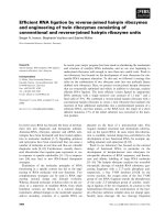
ArgII mRNA was strongly induced in IRF-3 5D-
expressing Jurkat cells, compared with control cells
(Fig. 1A). Furthermore, a dramatic induction of ArgII
was detected by immunoblot in IRF-3 5D-expressing
Jurkat cells at 24 h, and was sustained throughout
doxycycline treatment (Fig. 1B). Arginase activity was
likewise greatly increased after IRF-3 5D expression
by doxycycline, with a profile that mirrored protein
expression (Fig. 1C).
ArgII expression and enzymatic activity are
induced in Jurkat and Raw 264.7 cells infected
with paramyxovirus
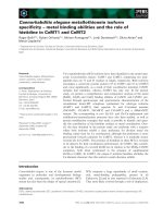
The up-regulation of ArgII was next studied in the con-
text of SeV infection, a negative single-strand RNA
paramyxovirus known to be a strong activator of IRF-
3 phosphorylation [23]. ArgII protein expression and
arginase activity were detected at 24 h and increased
5–10-fold between 48 and 60 h (Fig. 2A). At the
mRNA concentration, ArgII was induced 7 h after SeV
infection (Fig. 4A), suggesting a delay between mRNA
induction and protein detection. Inducible ArgII
expression has been previously described in macro-
phages [17–20], therefore we examined it in RAW 264.7
macrophages after SeV infection. As shown in Fig. 2B,
ArgII protein concentration and enzymatic activity
were also increased 5–10-fold 24–48 h after infection.
This shows for the first time that the ArgII gene is
inducible after SeV infection.
N. Grandvaux et al. IRF-3-mediated antiviral response involves spermine
FEBS Journal 272 (2005) 3120–3131 ª 2005 FEBS 3121
ArgII induction in response to virus infection
is IFN-independent
IRF-3-regulated genes may be activated as part of the
early or delayed phase of the antiviral response [8].
Indeed, these genes are modulated through ISRE
consensus sites, which can be targeted by ISGF3, in
response to IFN stimulation or by IRFs. As IRF-3 5D
alone is not sufficient to induce IFN production [24],
the result described above suggested that IFN was not
involved in ArgII expression. To directly assess whe-
ther ArgII up-regulation could be amplified by IFN
production, Jurkat cells were treated with type 1 IFN
(1000 UÆmL
)1
) for 0–48 h. ArgII protein concentra-
tions were increased by virus infection but not by IFN
treatment, whereas the IFN-responsive ISG56 gene
was induced by both virus and IFN, indicating that
virus-induced ArgII expression was IFN-independent
(Fig. 3).
ArgI and ornithine decarboxylase (ODC) are not
induced in response to virus infection
As the two isoforms of arginase, I (hepatic isoform)
and II (extrahepatic isoform), may contribute to the
arginase activity measured in the previous experiment,
A
B
C
Fig. 1. IRF-3 5D-inducible expression of ArgII. RtTA-Neo-IRF-3 5D
and rt-TA-IRF-3 5D Jurkat cells were induced with doxycycline for
the indicated time in the presence of IFN-neutralizing antibodies.
(A) Total RNA was extracted and subjected to RT-PCR analysis for
ArgII and GAPDH expression. (B) Whole-cell extracts (50 lg) were
subjected to SDS ⁄ PAGE and analyzed by immunoblotting with anti-
bodies against ArgII. Membranes were stripped and reprobed with
antibodies against IRF-3 and actin. (C) Cells were lyzed and ana-
lyzed for arginase activity by colorimetric assay, as described in
Experimental procedures, through measurement of the production
of urea. A
540
was measured and arginase activity was determined
as mUÆ(mg protein)
)1
. This experiment is representative of three
experiments and is expressed as mean ± SEM from triplicate de-
terminations.
AB
Fig. 2. Virus-inducible expression of ArgII in T lymphocytes and
macrophages. Jurkat cells (A) and Raw 264.7 cells (B) were infec-
ted with SeV (40 HAU per 10
6
cells) for the indicated times. Cell
lysates were analyzed for arginase activity. A
540
was measured,
and arginase activity was determined as mUÆ(mg protein)
)1
. This
experiment is representative of three experiments and is expressed
as mean ± SEM from triplicate determinations. In the lower panels,
whole-cell extracts (50 lg) were subjected to SDS ⁄ PAGE and ana-
lyzed by immunoblotting with antibodies against ArgII. Membranes
were stripped and reprobed with antibodies against actin.
Fig. 3. IFN-independent expression of ArgII. Jurkat cells were trea-
ted with either SeV for 48 h or with type I IFN (1000 UÆmL
)1
) for
0–48 h. Whole-cell extracts (50 lg) were resolved by SDS ⁄ PAGE
and transferred to nitrocellulose membrane. The membrane was
probed with antibodies against ArgII. After being stripped, mem-
branes was reprobed with antibodies against ISG56 and actin.
IRF-3-mediated antiviral response involves spermine N. Grandvaux et al.
3122 FEBS Journal 272 (2005) 3120–3131 ª 2005 FEBS
regulation of ArgI in the context of virus infection was
also analyzed. No increase in ArgI mRNA (Fig. 4A)
or protein levels (Fig. 4B) was observed in Jurkat cells
in response to SeV infection.
ArgII is involved in the biosynthesis of natural poly-
amines (putrescine, spermidine and spermine) through
conversion of l-arginine into l-ornithine [16]. The lat-
ter is in turn used by ODC to produce putrescine, the
precursor of spermidine and spermine. To further ana-
lyze the regulation of the polyamine-synthetic pathway
in virus infection, ODC expression in SeV-infected
Jurkat cells was studied. Kinetic analysis of ODC
mRNA by RT-PCR (Fig. 4A) and ODC protein con-
centration by immunoblot (Fig. 4C) revealed that
ODC expression was not regulated at the mRNA or
protein level after virus infection. Similarly, in IRF-
3 5D-expressing Jurkat cells, ODC was not up-regula-
ted at the protein level (data not shown).
Spermine inhibits vesicular stomatitis virus
(VSV) replication in Jurkat T cells
To assess whether natural polyamines have a direct
effect on viral replication, VSV, a negative single-
strand RNA rhabdovirus which strongly stimulates the
IFN pathway and also induces ArgII expression (data
not shown), was used in the next experiment. Jurkat
cells were infected with VSV for 14 h in the presence
or absence of increasing concentrations of putrescine,
spermidine and spermine and assayed for virus repli-
cation using a sensitive, quantitative plaque assay
(Fig. 5A,B). In the absence of polyamine, the VSV titer
reached 2.3 · 10
6
plaque-forming units (pfu)ÆmL
)1
,
whereas in the presence of physiological concentrations
of spermine [20,25,26], the virus titer decreased in a
dose-dependent manner. At a concentration of 25 lm,
the VSV titer was reduced to 5.4 · 10
4
pfuÆmL
)1
, and
at concentration of 100 lm, the virus titer was reduced
more than 3 logs, to 6.3 · 10
2
pfuÆmL
)1
. In the pres-
ence of spermidine, the titer of VSV was slightly
decreased to 5 · 10
5
pfuÆmL
)1
at a concentration of
100 lm, whereas putrescine did not affect virus yield.
Immunoblot analysis of cells treated in the presence of
25 lm and 100 lm polyamine confirmed that spermine
treatment dramatically inhibited the expression of
VSV glycoprotein, nucleocapsid, polymerase and mat-
rix proteins (G, N, P and M) during the lytic cycle
(Fig. 5C).
Spermine antiviral effect is dependent
on apoptosis
IRF-3 5D has been shown to mediate apoptosis
[24,27], and several reports have also described a role
for ArgII and ⁄ or polyamine in the regulation of apop-
tosis [21,22]. Thus, the possibility that the antiviral
effect of spermine is mediated by induction of apop-
tosis was analyzed. For this purpose, the effect of
spermine (50 lm) on viral replication was analyzed in
the presence of Z-VAD-FMK, a general inhibitor of
caspase activity, or Me
2
SO (control). In the presence
of Me
2
SO, virus titer was significantly decreased by
spermine compared with untreated cells (Fig. 6, lanes 2
and 3). However, when cells were pretreated with
A
B
C
Fig. 4. Induction of ArgII by SeV. (A) Total RNA was extracted from
Jurkat cells infected with SeV (40 HAUÆmL
)1
) for the indicated
times or from mouse liver tissue. Time-course expression of mRNA
from ArgI, ArgII and ODC was analyzed by RT-PCR. (B, C) Whole-
cell extracts from Jurkat cells infected with SeV for the indicated
times and from mouse liver and kidney tissues were resolved by
SDS ⁄ PAGE and transferred to nitrocellulose membrane. Mem-
branes were probed with antibodies against ArgI (B) or human
ODC (C). After being stripped, membranes were reprobed with
antibodies against actin. Mouse liver and kidney tissues, respect-
ively, were used as positive and negative control for ArgI expres-
sion [22].
N. Grandvaux et al. IRF-3-mediated antiviral response involves spermine
FEBS Journal 272 (2005) 3120–3131 ª 2005 FEBS 3123
Z-VAD-FMK, virus titer was comparable in the
absence and presence of spermine (Fig. 6, lanes 4 and
5). This shows that activation of caspases is an essen-
tial component of the antiviral effect triggered by sper-
mine. To directly demonstrate that spermine enhanced
virus-induced apoptosis, annexin V ⁄ propidium iodide
(PI) staining of apoptotic cells was quantified in VSV-
infected Jurkat T cells in the absence or presence of
spermine. As shown in Fig. 7, the presence of spermine
during VSV infection strongly potentiated virus-
induced apoptosis. At 8 h postinfection, VSV-induced
apoptosis was low (2.6% annexin V
+
⁄ PI
–
and 3.1%
annexin V
+
⁄ PI
+
), whereas in the presence of spermine
significant levels of apoptotis were detected (7.9%
annexin V
+
⁄ PI
–
and 30.4% annexin V
+
⁄ PI
+
). Intere-
stingly, spermine alone induced significant apoptosis
(3.5% annexin V
+
⁄ PI
–
and 15.9% annexin V
+
⁄ PI
+
).
No effect of spermidine or putrescine was observed
(data not shown). Thus, spermine was the only natural
polyamine with the capacity to induce apoptosis and
to augment apoptosis during virus infection.
A
B
C
Fig. 5. Spermine treatment inhibits VSV replication. Jurkat cells
were infected with VSV (m.o.i. 0.001) for 14 h in serum-free med-
ium in the absence or presence of the indicated concentration of
putrescine (triangles), spermidine (squares) or spermine (circles).
Supernatants were analyzed for VSV titer using a standard plaque
assay. Plaques were counted and titers calculated as pfuÆmL
)1
(A).
(B) Representative plaque assays from cells treated with 100 l
M
putrescine, spermidine or spermine. (C) Whole-cell extracts (20 lg)
from cells treated with 25 l
M and 100 lM polyamine in (A) were
analyzed by immunoblotting using antibodies against VSV.
Fig. 6. The spermine antiviral effect requires caspase activation.
Jurkat cells were pretreated with Z-VAD-FMK (100 l
M) or an equal
volume of Me
2
SO for 1 h before infection with VSV (m.o.i. 0.001)
for 14 h in serum-free medium in the absence or presence of sper-
mine (50 l
M). Supernatants were analyzed for VSV titer using a
standard plaque assay. Plaques were counted and titers calculated
as pfuÆmL
)1
. Values are representative of two experiments and are
expressed as mean ± SEM from triplicate determinations. Note
that the difference in the quantitative effect of spermine (compare
with Fig. 5) on virus titer is due to the presence of Me
2
SO (data
not shown).
IRF-3-mediated antiviral response involves spermine N. Grandvaux et al.
3124 FEBS Journal 272 (2005) 3120–3131 ª 2005 FEBS
Spermine and spermidine are induced in
IRF-3 5D-expressing, but not virus-infected,
Jurkat cells
Finally, to evaluate whether polyamines, and partic-
ularly spermine, were produced in response to IRF-3
activation, rtTA-IRF-3 5D-Jurkat cells were treated
with doxycycline for 30 h, and the pool of intracellular
polyamines was measured by dansylation and LC ⁄ MS
analysis as described in Experimental Procedures. As
shown in Fig. 8A, production of spermine and spermi-
dine was significantly induced in IRF-3 5D-expressing
Jurkat cells compared with control cells. Intracellular
polyamine content was also measured after virus infec-
tion, and polyamine production was not induced after
SeV infection (Fig. 8B) or VSV infection (data not
shown). Thus, the final products of the polyamine-
biosynthetic pathways, spermine and spermidine, are
produced in response to IRF-3 activation, but not
during SeV or VSV infection.
Discussion
In previous studies, we showed that IRF-3 mediates an
antiviral response in an IFN-independent manner, in
part due to the IRF-3-dependent expression of ISGs,
such as ISG-54, 56 and 60. We now report that activa-
tion of IRF-3 stimulates the ArgII gene in an IFN-
independent manner. ArgII is a mitochondrial enzyme
involved in the polyamine synthesis pathway through
the catalysis of l-ornithine production from l-arginine.
Of the natural polyamines, spermine and to a lesser
extent spermidine, possess antiviral activities resulting
from their potential to induce apoptosis, and both
10
0
10
1
10
2
10
3
AnnexinY-FITC
10
4
10
0
10
1
10
2
Pl-FL2
10
3
10
4
10
0
10
1
10
2
Pl-FL2
10
3
10
4
10
0
10
1
10
2
Pl-FL2
10
3
10
4
10
0
10
1
10
2
Pl-FL2
10
3
10
4
10
0
10
1
10
2
10
3
AnnexinY-FITC
10
4
10
0
10
1
10
2
10
3
AnnexinY-FITC
NG050206.017
NG050206.021
NG050206.022
NG050206.018
10
4
10
0
10
1
10
2
10
3
AnnexinY-FITC
10
4
Fig. 7. Spermine potentiates VSV-induced apoptosis. Jurkat T cells
were infected with VSV (m.o.i. 0.01) in the absence or presence of
100 l
M spermine. At the indicated times, cells were harvested and
double-stained with FITC–annexin V ⁄ PI as indicated in Experimental
procedures. The upper panel represents the percentage of cells
that were annexin V positive (annexin V
+
⁄ PI
–
and annexin V
+
⁄ PI
+
)
by flow cytometry. Plots in the lower panel illustrate the 8 h time
point. Data are representative of two independent experiments.
A
B
Fig. 8. IRF-3 5D expression, but not SeV infection, triggers polyam-
ine production in Jurkat cells. (A) rt-TA-IRF-3 5D Jurkat cells were
left uninduced (light-shaded bars) or induced with doxycycline
(1 lgÆmL
)1
) for 30 h (dark-shaded bars). (B) Jurkat cells were left
untreated (light-shaded bars) or infected with SeV (80 HAU per 10
6
cells) for 52 h (dark-shaded bars). Cells were harvested, and per-
chloric acid extracts were used to quantify the intracellular concen-
tration of spermine, spermidine and putrescine as described in
Experimental procedures. These results are representative of two
independent experiments, each with duplicate measurements. The
SE was estimated by the percentage of variation observed over the
two independent experiments.
N. Grandvaux et al. IRF-3-mediated antiviral response involves spermine
FEBS Journal 272 (2005) 3120–3131 ª 2005 FEBS 3125
polyamines were induced in response to the expression
of a constitutively active form of IRF-3.
This study shows for the first time that ArgII expres-
sion is up-regulated in the context of virus infection.
Previous studies reported the induction of ArgII in
response to LPS, cAMP, or H. pylori [20,28–30], with
ArgII expression up-regulated at mRNA, protein and
activity levels after H. pylori infection. Furthermore,
ArgI and ODC expression were not up-regulated at
the transcriptional level after H. pylori infection [20], a
result that correlates with the present experiments in
virus-infected cells. In Jurkat T cells, basal level ODC
mRNA and protein expression was observed, and this
was not modulated after virus infection.
The pathways involved in ArgII gene regulation are
not well characterized, but a role for NF-jB has been
suggested based on the use of chemical inhibitors; pyr-
rolidine dithiocarbamate was shown to inhibit ArgII
induction in rat alveolar macrophages stimulated with
LPS, whereas ArgII expression in LPS-stimulated
Raw264.7 cells was not inhibited by pyrrolidine dithio-
carbamate [28]. In Raw 264.7 cells cocultured with
H. pylori, ArgII expression was inhibited by MG-132
[20], suggesting indirectly an involvement of NF-jBin
ArgII regulation. Our study is thus the first direct
demonstration of the involvement of IRF-3 in ArgII
regulation in response to virus infection. IRF-3 is also
activated in response to LPS in a TLR-4-dependent
mechanism [31,32]; thus IRF-3 may also participate in
the LPS-mediated or H. pylori-mediated induction of
ArgII via a TLR-4-dependent pathway.
The role of polyamines in apoptosis is controversial;
both induction of and protection against apoptosis by
polyamines have been demonstrated [21,22]. In agree-
ment with the present study, an apoptosis process
dependent on ArgII and ODC was reported in
response to H. pylori infection of macrophages [20].
The present study describes a role for ArgII up-
regulation and the polyamine-synthesis pathway in
IRF-3 5D-induced apoptosis. Although IRF-3 can sti-
mulate apoptosis in Jurkat cells [24], the molecular
mechanisms responsible for triggering it in response to
IRF-3 have not been defined. ISG56 was induced in
response to IRF-3, and because ISG56 is involved in
the inhibition of protein translation and cell prolifer-
ation [33,34], it may participate in IRF-3-mediated
apoptosis. Another potential mechanism involves sper-
mine, which induced apoptosis in Jurkat cells and
enhanced virus-induced apoptosis at physiological con-
centrations [20,25,26]. Polyamines are known to modu-
late DNA–protein interactions; specifically, spermine
has been shown to induce NF-jB activation in breast
cancer cells [35,36], whereas Oct-1 binding was inhib-
ited by polyamine [37]. Polyamine depletion inhibited
TNF-a-induced JNK activation and subsequently pre-
vented caspase-3 activation in intestinal epithelial IEC-
6 cells, thereby delaying TNF-a-induced apoptosis [38].
As both NF-jB and JNK pathways are activated by
virus infection, these pathways may be targets of the
pro-apoptotic activity of spermine.
Spermine and to a lesser extent spermidine inhibited
VSV multiplication, but inhibition was abolished when
cells were treated with the caspase inhibitor, Z-VAD-
FMK, suggesting that spermine-mediated apoptosis
may be part of the host antiviral response. Further-
more, enhanced virus-induced apoptosis occurred in
the presence of spermine (Fig. 7). However, we cannot
rule out the possibility that spermine production
in vivo in response to virus infection induces sufficient
apoptosis to limit the levels of virus multiplication,
thus mimicking an antiviral effect. An alternative
mechanism, that spermine acts by inhibition of virus
entry, was examined using recombinant VSV-GFP
virus, and virus entry was not inhibited by spermine
(data not shown).
A limited number of studies have examined the rela-
tionship between polyamine production and herpes
virus replication. Polyamine depletion was shown to
block human cytomegalovirus replication [39,40],
whereas inhibition of polyamine biosynthesis produced
different effects on herpes simplex virus (HSV)-1,
HSV-2 or pseudorabies virus replication [41–43]. HSV
inhibited polyamine biosynthesis by inhibiting protein
synthesis, whereas human cytomegalovirus infection
induced spermine and spermidine expression in fibro-
blasts [41,44]. Another study reported induction of
ArgI and ArgII mRNA in the cornea during HSV
infection, but protein concentrations and arginase
activity were not analyzed [45]. Conversely, proteose–
peptone-activated and IFNc-activated macrophages
exhibited increased arginase activity and were resistant
to HSV infection by a mechanism that was prevented
by the addition of arginine, suggesting an essential role
for arginase in antiviral activity [46,47]. In retrospect,
however, these results may simply reflect the consump-
tion of arginine by inducible nitric oxide synthase,
which competes with arginase for the arginine sub-
strate, to produce nitric oxide, an antiviral compound
produced by macrophages [48,49].
Spermine, spermidine and putrescine are induced in
response to IRF-3 5D expression, but not in response to
SeV or VSV infection, although these two viruses trigger
IRF-3 phosphorylation ⁄ activation. Based on this surpri-
sing result, it is possible that SeV and VSV may have
evolved strategies to antagonize polyamine synthesis
and to evade the polyamine-mediated apoptotic
IRF-3-mediated antiviral response involves spermine N. Grandvaux et al.
3126 FEBS Journal 272 (2005) 3120–3131 ª 2005 FEBS
response. The molecular mechanisms used by viruses to
block polyamine synthesis are under investigation.
In conclusion, this study shows for the first time the
induction of ArgII mRNA, protein and enzymatic
activity in the context of virus infection in an IRF-3-
dependent and IFN-independent manner. Moreover,
expression of a constitutively active form of IRF-3
leads to induction of spermine, which possesses pro-
apoptotic and antiviral activities. These results thus
illustrate a potential new mechanism by which IRF-3
contributes to the development of the antiviral state.
Experimental procedures
Reagents
Spermine, spermidine, putrescine, 1-phenylpropane-1,2-dione-
2-oxime (ISPF) and doxycycline were from Sigma. Human
recombinant IFN type 1 was from Sigma (Oakville, Ontario,
Canada). Z-VAD-FMK was from BioMol.
Cell culture and infection
Jurkat cells (ATCC, Manassas, VA, USA) were grown in
RPMI-1640 medium (wisent, St jean batiste de Roaville,
Quebec, Canada) containing 10% heat-inactivated fetal
bovine serum and antibiotics. Vero cells (ATCC) and RAW
264.7 (ATCC) cells were grown in DMEM medium (wisent)
supplemented with 10% heat-inactivated fetal bovine serum
and antibiotics. rtTA-Neo-IRF-3 and rtTA-IRF-3 5D
Jurkat cells [24] were grown in RPMI-1640 medium con-
taining 10% heat-inactivated fetal bovine serum, glutamine,
antibiotics, 2.5 lgÆmL
)1
puromycin and 400 lgÆmL
)1
G418
(Gibco, Burlington, Ontario, Canada). Twenty hours before
stimulation, cells were seeded in fresh medium at 0.5 · 10
6
cellsÆmL
)1
. Induction with doxycycline was performed at
1 lgÆmL
)1
for the indicated time in the presence of neutral-
izing antibodies against type I IFNs as described [14].
Treatment with IFN-a was performed at 1000 UÆmL
)1
for
16 h in complete medium. SeV infection (Cantell strain, 40
HAU per 10
6
cells) was carried out for 2 h in serum-free
medium and further cultured for the indicated time in com-
plete medium.
RT-PCR analysis
Total RNA from exponentially growing cells stimulated as
described above and from mouse liver tissues was isolated
using homogenization in TRIzol reagent (Gibco). Total
RNA (1 lg) was reverse-transcribed in a final volume of
100 lL (Advantage RT-PCR kit; Clontech, Mountain View,
CA, USA), and 20 lL was used for PCR amplification using
the following primers: human and murine ArgII, 5¢-GAT
CTGCTGATTGGCAAGAGACAA-3¢ and 5¢-CTAAATTC
TCACACGTGCTTGATT-3¢ [50], 362 bp; human and
murine ArgI, 5¢-ATTGGCTTGAGAGACGTGGACCCT-3¢
and 5¢-TTGCAACTGCTGTGTTCACTGTTC-3¢, 369 bp;
human ODC, 5¢ -TGTTGCTGCTGCCTCTACGTT-3¢ and
5¢-GCTGGCATCCTGTTCCTCTACTT-3¢, 138 bp [51];
human b-actin, 5¢-ACAATGAGCTGCTGGTGGCT-3¢ and
5¢-GATGGGCACAGTGTGGGTGA-3¢; murine b-actin,
5¢-TGGAATCCTGTGGCATCCATGAAAC-3¢ and 5¢-TA
AAACGCAGCTCAGTAACCGTCCG-3¢. Human GAPDH
primers were included in the Advantage RT-PCR kit.
Immunoblot analysis
Cells were washed twice in NaCl ⁄ P
i
and lyzed in 50 mm
Tris ⁄ HCl, pH 7.4, containing 1% Nonidet P40, 0.25%
sodium deoxycholate, 150 mm NaCl, 1 mm EDTA supple-
mented with 1 mm phenylmethanesulfonate fluoride,
5 lgÆmL
)1
aprotinin and 5 lgÆmL
)1
leupeptin (lysis buffer)
for 15 min on ice. Mouse liver and kidney total protein
extracts were prepared by Dounce homogenization of tis-
sues in lysis buffer and centrifugation at 10 000 g for
30 min at 4 °C. Supernatants were used as total protein
extracts. Whole cell extracts (50 lg) or mouse tissue
extracts (50 lg) were separated by SDS ⁄ PAGE and trans-
ferred to nitrocellulose membrane (Bio-Rad, Mississauga,
Ontario, Canada). The membrane was blocked in NaCl ⁄ P
i
containing 0.05% Tween 20 and 5% nonfat dry milk for
1 h and incubated with primary antibody, anti-(IRF-3 FL-
425) Ig (1 l g ÆmL
)1
; Santa Cruz), anti-ArgII (1 : 1000) Ig
[52], anti-ArgI Ig (1 : 1000) [53], anti-(ODC sc-21515) Ig
(1 lgÆmL
)1
; Santa Cruz), anti-ISG56 Ig (1 : 1000; a gift
from Dr G. Sen, Lemer Research Institute, Cleveland
Clinic Foundation, Cleveland, OH, USA) or anti-(a-actin)
Ig (Chemicon) in blocking solution. After five 5-min washes
in NaCl ⁄ P
i
containing 0.05% Tween 20, the membranes
were incubated for 1 h with horseradish peroxidase-conju-
gated goat anti-rabbit, goat anti-mouse or rabbit anti-goat
IgG (1 : 2000–1 : 10000) in blocking solution. Immunoreac-
tive proteins were visualized by enhanced chemilumines-
cence (Perkin-Elmer, Woodbridge, Ontario, Canada).
Measurement of arginase enzymatic activity
Arginase activity was measured by colorimetric assay [54].
Cells (10
5
) were lyzed in 50 lL 0.1% Triton containing
5 lg antipain, 5 lg pepstatin, and 5 lg aprotinin. After
30 min at room temperature, 50 lL10mm MnCl
2
⁄ 50 mm
Tris ⁄ HCl, pH 7.5 was added, and the lysate was activated
at 55 °C for 10 min. Arginine hydrolysis was performed
at 37 °C for 60 min by mixing 25 lL previously activated
lysate with 25 lL 0.5 m arginine, pH 9.7. The reaction was
stopped by the addition of 400 lL acidic mixture
H
2
SO
4
⁄ H
3
PO
4
⁄ H
2
O (1 : 3 : 7, v ⁄ v ⁄ v). For quantification of
urea produced, 25 lL 9% ISPF was added and incubated
N. Grandvaux et al. IRF-3-mediated antiviral response involves spermine
FEBS Journal 272 (2005) 3120–3131 ª 2005 FEBS 3127
for 45 min at 100 °C. After 10 min in the dark, A
540
was
measured. A standard curve was obtained by adding
100 lL urea (1.8–30 lg) to 400 lL acidic mixture and
25 lL ISPF. Proteins in the lysate were quantified using the
Bradford assay (Bio-Rad). Arginase activity was determined
as mUÆ(mg protein)
)1
[equivalent to lmol ureaÆmin
)1
Æ(mg
protein)
)1
].
VSV plaque assay
Jurkat T cells were infected with VSV at a multiplicity of
infection (m.o.i.) of 0.001 for 1 h in serum-free medium.
After two washes in NaCl ⁄ P
i
, infection was pursued in
serum-free medium in the absence or presence of putrescine,
spermidine or spermine, and supernatant was harvested
at 14 h postinfection. In experiments where Z-VAD-FMK
was used, the reagent was used at 100 lm for 1 h before
infection, and maintained at this concentration during the
infection. Serial dilutions of the supernatant were used to
infect confluent plates of Vero cells in serum-free medium.
After 1 h infection, the medium was removed and replaced
by 3% methylcellulose. After plaques had formed, the meth-
ylcellulose was removed and the cells were fixed with 4%
formaldehyde for 1 h and stained with 0.2% crystal violet
in 20% ethanol. Plaques were counted, averaged and multi-
plied by the dilution factor to determine viral titer as
pfuÆmL
)1
. Virus protein was detected in cells by immuno-
blot as described above using antibodies against VSV (a gift
from John Bell, Ottawa, CA, USA).
Detection of early and late apoptosis
(annexin V/PI staining)
Jurkat T cells stimulated as described above were harvested
at different time points and resuspended in 50 lL cold
NaCl ⁄ P
i
. Apoptosis was detected by reaction with fluorescein
isothiocyanate (FITC)-conjugated annexin V and PI. Stain-
ing was performed by the addition of cold staining mixture
containing 500 lL binding buffer (10 m m Hepes, pH 7.4,
150 mm NaCl, 5 mm KCl, 1 mm MgCl
2
, 1.8 mm CaCl
2
),
1 lL FITC–annexin V and 1 lLPI(1mgÆmL
)1
) for 5 min.
Acquisition was performed on a FACScan flow cytometer
(BD Biosciences, Mountain View, CA, USA) using FL-1 and
FL-2 detectors. Analysis was performed using the cellquest
software (BD Biosciences). Cells exhibiting annexin V
–
⁄ PI
+
staining were considered necrotic, those showing annex-
in V
+
⁄ PI
–
staining were recognized as early apoptotic cells,
and annexin V
+
⁄ PI
+
cells were taken as late apoptotic.
Measurement of intracellular polyamine
concentration
After treatment, cells were harvested, washed three times
with NaCl ⁄ P
i
, and disrupted by sonication in 0.2 m perchlo-
ric acid. After centrifugation at 3000 g for 10 min, perchlo-
ric supernatants and protein precipitates were stored at
)80 °C until analyzed within 1 month. The dansylation pro-
cedure was performed by a previously described method [55]
using 1,10-diaminododecane as internal standard. Aliquots
(200 l L) of the perchloric supernatants were allowed to
react with 4 vol. dansyl chloride in acetone (5 mgÆmL
)1
)in
the presence of solid sodium carbonate. After the dansyla-
tion reaction (12 h at room temperature), excess dansyl
chloride was removed by reaction with proline. The cyclo-
hexane extract containing the dansyl derivatives was evapor-
ated to dryness, and the residue resuspended in 200 lL
acetonitrile.
The LC ⁄ MS was supplied with chem station 1100 soft-
ware (Agilent Technologie; Massy-Palaiseau, Wilmington,
DE, USA). Nitrogen gas was generated using a Jun-air
model 2000–25M air compressor (Buffalo Grove, IL, USA)
connected to a UHPLCMS Model nitrogen generator
(Domnick Hunter France, S.A., Villefranche-sur-Saoˆ ne,
France). Dansylated polyamine was analyzed by flow injec-
tion analysis without performing a separation with a LC
column [56]. For flow injection analysis ⁄ MS measurements,
30-lL samples were directly injected from the HP1100 ser-
ies autosampler without LC separation into a stream of
water ⁄ acetonitrile (9 : 1, v ⁄ v) at a flow rate of 0.5 mLÆ
min
)1
. The following parameters were used for detec-
tion: sec ⁄ scan cycle, 1.46; threshold, 150; step size, 0.35; ion
mode positive; gain, 9.9; capillary voltage, +3000 V; cor-
ona current, 6 lA; drying gas flow rate, 6 LÆmin
)1
; drying
gas temperature, 300 °C; nebulizer pressure, 30 psig; vapor-
izer temperature, 400 °C. Selected ion monitoring mode
data masses were obtained with an atmospheric pressure
chemical ionization source to monitor the protonated par-
ent ions [M + H]
+
;atm ⁄ z 555.2 for bidansyl-putrescine,
m ⁄ z 845.3 for tridansyl-spermidine, m ⁄ z 1135.4 for tetra-
dansyl-spermine and m ⁄ z 639.3 for the bidansylated inter-
nal standard 1–10, diaminododecane. Ionic intensities,
deduced from the area under each selective peak, were cor-
rected with respect to that of the internal standard. Poly-
amine concentrations were determined by using calibration
curves obtained from known amounts of a mixture contain-
ing the four polyamines dansylated and extracted under the
same conditions. Two independent polyamine-dansylation
experiments were performed, and each polyamine measure-
ment was performed in duplicate.
Acknowledgements
We thank Dr M. Mori and Dr J. Bell for reagents
used in this study. We also thank Laurence Lejeune
and Ste
´
phanie Olie
`
re for excellent technical help with
FACS analyses, and members of the Molecular Oncol-
ogy Group of the Lady Davis Institute for helpful dis-
cussions. This work was supported by grants to J.Hi.
IRF-3-mediated antiviral response involves spermine N. Grandvaux et al.
3128 FEBS Journal 272 (2005) 3120–3131 ª 2005 FEBS
from the Canadian Institutes of Health Research and
CANVAC, the Canadian Network for Vaccines and
Immunotherapeutics. N.G. was supported by a post-
doctoral FRSQ fellowship, J.Ha. and B.R.T. by an
NSERC studentship, R.L. by a FRSQ Chercheur
Boursier, and J.Hi. by a CIHR Senior Scientist award.
References
1 Samuel CE (2001) Antiviral actions of interferons. Clin
Microbiol Rev 14, 778–809.
2 Sen GC (2001) Viruses and interferons. Annu Rev
Microbiol 55, 255–281.
3 Servant MJ, Grandvaux N & Hiscott J (2002) Multiple
signaling pathways leading to the activation of inter-
feron regulatory factor 3. Biochem Pharmacol 64, 985–
992.
4 Chu WM, Ostertag D, Li ZW, Chang L, Chen Y, Hu
Y, Williams B, Perrault J & Karin M (1999) JNK2 and
IKKbeta are required for activating the innate response
to viral infection. Immunity 11, 721–731.
5 Iordanov MS, Paranjape JM, Zhou A, Wong J,
Williams BR, Meurs EF, Silverman RH & Magun BE
(2000) Activation of p38 mitogen-activated protein
kinase and c-Jun NH(2)-terminal kinase by double-
stranded RNA and encephalomyocarditis virus: involve-
ment of RNase L, protein kinase R, and alternative
pathways. Mol Cell Biol 20, 617–627.
6 Sharma S, tenOever BR, Grandvaux N, Zhou GP, Lin
R & Hiscott J (2003) Triggering the interferon antiviral
response through an IKK-related pathway. Science 300,
1148–1151.
7 Fitzgerald KA, McWhirter SM, Faia KL, Rowe DC,
Latz E, Golenbock DT, Coyle AJ, Liao SM & Maniatis
T (2003) IKKepsilon and TBK1 are essential compo-
nents of the IRF3 signaling pathway. Nat Immunol 4,
491–496.
8 Grandvaux N, tenOever BR, Servant MJ & Hiscott J
(2002) The interferon antiviral response: from viral inva-
sion to evasion. Curr Opin Infect Dis 15, 259–267.
9 Marie I, Durbin JE & Levy DE (1998) Differential viral
induction of distinct interferon-alpha genes by positive
feedback through interferon regulatory factor-7. EMBO
J 17, 6660–6669.
10 Sato M, Suemori H, Hata N, Asagiri M, Ogasawara K,
Nakao K, Nakaya T, Katsuki M, Noguchi S, Tanaka
N & Taniguchi T (2000) Distinct and essential roles of
transcription factors IRF-3 and IRF-7 in response to
viruses for IFN-alpha ⁄ beta gene induction. Immunity
13, 539–548.
11 Lin R, Genin P, Mamane Y & Hiscott J (2000) Selec-
tive DNA binding and association with the CREB
binding protein coactivator contribute to differential
activation of alpha ⁄ beta interferon genes by interferon
regulatory factors 3 and 7. Mol Cell Biol 20, 6342–
6353.
12 Nicholl MJ, Robinson LH & Preston CM (2000) Acti-
vation of cellular interferon-responsive genes after infec-
tion of human cells with herpes simplex virus type 1.
J Gen Virol 81, 2215–2218.
13 Nakaya T, Sato M, Hata N, Asagiri M, Suemori H,
Noguchi S, Tanaka N & Taniguchi T (2001) Gene
induction pathways mediated by distinct irfs during
viral infection. Biochem Biophys Res Commun 283,
1150–1156.
14 Grandvaux N, Servant MJ, tenOever B, Sen GC,
Balachandran S, Barber GN, Lin R & Hiscott J
(2002) Transcriptional profiling of interferon regula-
tory factor 3 target genes: direct involvement in the
regulation of interferon-stimulated genes. J Virol 76,
5532–5539.
15 Lin R, Mamane Y & Hiscott J (1999) Structural and
functional analysis of interferon regulatory factor 3:
localization of the transactivation and autoinhibitory
domains. Mol Cell Biol 19, 2465–2474.
16 Jenkinson CP, Grody WW & Cederbaum SD (1996)
Comparative properties of arginases. Comp Biochem
Physiol B Biochem Mol Biol 114, 107–132.
17 Corraliza IM, Soler G, Eichmann K & Modolell M
(1995) Arginase induction by suppressors of nitric oxide
synthesis (IL-4, IL-10 and PGE2) in murine bone-
marrow-derived macrophages. Biochem Biophys Res
Commun 206, 667–673.
18 Salimuddin Nagasaki A, Gotoh T, Isobe H & Mori M
(1999) Regulation of the genes for arginase isoforms
and related enzymes in mouse macrophages by lipopoly-
saccharide. Am J Physiol 277, E110–E117.
19 Gotoh T, Sonoki T, Nagasaki A, Terada K, Takiguchi
M & Mori M (1996) Molecular cloning of cDNA for
nonhepatic mitochondrial arginase (arginase II) and
comparison of its induction with nitric oxide synthase in
a murine macrophage-like cell line. FEBS Lett 395,
119–122.
20 Gobert AP, Cheng Y, Wang JY, Boucher JL, Iyer RK,
Cederbaum SD, Casero RA Jr, Newton JC & Wilson
KT (2002) Helicobacter pylori induces macrophage
apoptosis by activation of arginase II. J Immunol 168,
4692–4700.
21 Thomas T & Thomas TJ (2001) Polyamine in cell
growth and cell death: molecular mechanisms and thera-
peutic applications. Cell Mol Life Sci 58, 244–258.
22 Schipper RG, Penning LC & Verhofstad AA (2000)
Involvement of polyamine in apoptosis. Facts and con-
troversies: effectors or protectors? Semin Cancer Biol 10,
55–68.
23 Lin R, Heylbroeck C, Pitha PM & Hiscott J (1998)
Virus dependent phosphorylation of the IRF-3 tran-
scription factor regulates nuclear translocation, trans-
N. Grandvaux et al. IRF-3-mediated antiviral response involves spermine
FEBS Journal 272 (2005) 3120–3131 ª 2005 FEBS 3129
activation potential and proteasome mediated degrada-
tion. Mol Cell Biol 18, 2986–2996.
24 Heylbroeck C, Balachandran S, Servant MJ, DeLuca C,
Barber GN, Lin R & Hiscott J (2000) The IRF-3 tran-
scription factor mediates Sendai virus-induced apopto-
sis. J Virol 74, 3781–3792.
25 Igarashi K & Kashiwagi K (2000) Polyamine: myster-
ious modulators of cellular functions. Biochem Biophys
Res Commun 271, 559–564.
26 Coburn RF, Jones DH, Morgan CP, Baron CB &
Cockcroft S (2002) Spermine increases phosphatidylino-
sitol 4,5-bisphosphate content in permeabilized and non-
permeabilized HL60 cells. Biochim Biophys Acta 1584,
20–30.
27 Weaver BK, Ando O, Kumar KP & Reich NC (2001)
Apoptosis is promoted by the dsRNA-activated factor
(DRAF1) during viral infection independent of the
action of interferon or p53. FASEB J 15, 501–515.
28 Wang WW, Jenkinson CP, Griscavage JM, Kern RM,
Arabolos NS, Byrns RE, Cederbaum SD & Ignarro LJ
(1995) Co-induction of arginase and nitric oxide
synthase in murine macrophages activated by lipopoly-
saccharide. Biochem Biophys Res Commun 210, 1009–
1016.
29 Gotoh T & Mori M (1999) Arginase II downregulates
nitric oxide (NO) production and prevents NO-mediated
apoptosis in murine macrophage-derived RAW 264.7
cells. J Cell Biol 144 , 427–434.
30 Wei LH, Morris SM Jr, Cederbaum SD, Mori M &
Ignarro LJ (2000) Induction of arginase II in human
Caco-2 tumor cells by cyclic AMP. Arch Biochem
Biophys 374, 255–260.
31 Kawai T, Takeuchi O, Fujita T, Inoue J, Muhlradt PF,
Sato S, Hoshino K & Akira S (2001) Lipopolysacchar-
ide stimulates the MyD88-independent pathway and
results in activation of IFN-regulatory factor 3 and the
expression of a subset of lipopolysaccharide-inducible
genes. J Immunol 167, 5887–5894.
32 Doyle SE, O’Connell R, Vaidya SA, Chow EK, Yee K
& Cheng G (2003) Toll-like receptor 3 mediates a more
potent antiviral response than Toll-like receptor 4.
J Immunol 170, 3565–3571.
33 Guo J, Hui DJ, Merrick WC & Sen GC (2000) A new
pathway of translational regulation mediated by eukar-
yotic initiation factor 3. EMBO J 19, 6891–6899.
34 Geiss G, Jin G, Guo J, Bumgarner R, Katze MG & Sen
GC (2001) A comprehensive view of regulation of gene
expression by double-stranded RNA-mediated cell sig-
naling. J Biol Chem 30, 30.
35 Shah N, Thomas T, Shirahata A, Sigal LH & Thomas
TJ (1999) Activation of nuclear factor kappaB by poly-
amine in breast cancer cells. Biochemistry 38, 14763–
14774.
36 Shah N, Thomas TJ, Lewis JS, Klinge CM, Shirahata
A, Gelinas C & Thomas T (2001) Regulation of estro-
genic and nuclear factor kappa B functions by poly-
amine and their role in polyamine analog-induced
apoptosis of breast cancer cells. Oncogene 20 , 1715–
1729.
37 Panagiotidis CA, Artandi S, Calame K & Silverstein SJ
(1995) Polyamine alter sequence-specific DNA–protein
interactions. Nucleic Acids Res 23, 1800–1809.
38 Bhattacharya S, Ray RM, Viar MJ & Johnson LR
(2003) Polyamines are required for activation of c-Jun
NH2-terminal kinase and apoptosis in response to
TNF-alpha in IEC-6 cells. Am J Physiol Gastrointest
Liver Physiol 285, G980–G991.
39 Tyms AS & Williamson JD (1982) Inhibitors of poly-
amine biosynthesis block human cytomegalovirus
replication. Nature 297, 690–691.
40 Gibson W, van Breemen R, Fields A, LaFemina R &
Irmiere A (1984) d,l-alpha-Difluoromethylornithine
inhibits human cytomegalovirus replication. J Virol 50,
145–154.
41 McCormick FP & Newton AA (1975) Polyamine meta-
bolism in cells infected with herpes simplex virus. J Gen
Virol 27, 25–33.
42 Wang HC & Wong ML (2003) Lytic infection of pseu-
dorabies virus in the presence of spermine, spermidine,
or DFMO. Virus Res 94, 121–127.
43 Pohjanpelto P, Sekki A, Hukkanen V & von Bonsdorff
CH (1988) Polyamine depletion of cells reduces the
infectivity of herpes simplex virus but not the infectivity
of Sindbis virus. Life Sci 42, 2011–2018.
44 Clarke JR & Tyms AS (1991) Polyamine biosynthesis in
cells infected with different clinical isolates of human
cytomegalovirus. J Med Virol 34, 212–216.
45 Mistry SK, Zheng M, Rouse BT & Morris SM Jr
(2001) Induction of arginases I and II in cornea during
herpes simplex virus infection. Virus Res 73, 177–182.
46 Wildy P, Gell PG, Rhodes J & Newton A (1982) Inhibi-
tion of herpes simplex virus multiplication by activated
macrophages: a role for arginase? Infect Immun 37,
40–45.
47 Sethi KK (1983) Contribution of macrophage argi-
nase in the intrinsic restriction of herpes simplex
virus replication in permissive macrophage cul-
tures induced by gamma-interferon containing
products of activated spleen cells. Immunobiology 165,
459–474.
48 MacLean A, Wei XQ, Huang FP, Al-Alem UA, Chan
WL & Liew FY (1998) Mice lacking inducible nitric-
oxide synthase are more susceptible to herpes simplex
virus infection despite enhanced Th1 cell responses.
J Gen Virol 79, 825–830.
49 Benencia F & Courreges MC (1999) Nitric oxide and
macrophage antiviral extrinsic activity. Immunology 98,
363–370.
50 Zhang C, Hein TW, Wang W, Chang CI & Kuo L
(2001) Constitutive expression of arginase in microvas-
IRF-3-mediated antiviral response involves spermine N. Grandvaux et al.
3130 FEBS Journal 272 (2005) 3120–3131 ª 2005 FEBS
cular endothelial cells counteracts nitric oxide-mediated
vasodilatory function. FASEB J 15 , 1264–1266.
51 Brabender J, Lord RV, Danenberg KD, Metzger R,
Schneider PM, Uetake H, Kawakami K, Park JM,
Salonga D, Peters JH, et al. (2001) Upregulation of
ornithine decarboxylase mRNA expression in Barrett’s
esophagus and Barrett’s-associated adenocarcinoma.
J Gastrointest Surg 5, 174–181, discussion 182.
52 Ozaki M, Gotoh T, Nagasaki A, Miyanaka K, Takeya
M, Fujiyama S, Tomita K & Mori M (1999) Expression
of arginase II and related enzymes in the rat small intes-
tine and kidney. J Biochem (Tokyo) 125, 586–593.
53 Sonoki T, Nagasaki A, Gotoh T, Takiguchi M, Takeya
M, Matsuzaki H & Mori M (1997) Coinduction of
nitric-oxide synthase and arginase I in cultured rat
peritoneal macrophages and rat tissues in vivo by
lipopolysaccharide. J Biol Chem 272, 3689–3693.
54 Corraliza IM, Campo ML, Soler G & Modolell M
(1994) Determination of arginase activity in macro-
phages: a micromethod. J Immunol Methods 174,
231–235.
55 Seiler N (1970) Use of the dansyl reaction in biochemi-
cal analysis. Methods Biochem Anal 18, 259–337.
56 Gaboriau F, Havouis R, Moulinoux JP & Delcros JG
(2003) Atmospheric pressure chemical ionization-mass
spectrometry method to improve the determination of
dansylated polyamine. Anal Biochem 318, 212–220.
N. Grandvaux et al. IRF-3-mediated antiviral response involves spermine
FEBS Journal 272 (2005) 3120–3131 ª 2005 FEBS 3131