Báo cáo khoa học: Death inducer obliterator protein 1 in the context of DNA regulation Sequence analyses of distant homologues point to a novel functional role docx
Bạn đang xem bản rút gọn của tài liệu. Xem và tải ngay bản đầy đủ của tài liệu tại đây (347.03 KB, 7 trang )
HYPOTHESIS
Death inducer obliterator protein 1 in the context of DNA
regulation
Sequence analyses of distant homologues point to a novel functional
role
Ana M. Rojas
1,2
, Luis Sanchez-Pulido
1
, Agnes Fu
¨
tterer
2
, Karel H. M. van Wely
2
, Carlos Martinez-A
2
and Alfonso Valencia
1
1 Protein Design Group, CNB ⁄ CSIC, Madrid, Spain
2 Department of Immunology and Oncology, CNB ⁄ CSIC, Madrid, Spain
Apoptosis has an important role in development, tissue
homeostasis, and host defense, among other functions
[1]. Death inducer obliterator 1 [DIDO1; also termed
DIO-1, death-associated transcription factor 1 (DATF-
1)] is a protein described in humans and mice. DIDO1
was initially identified by differential display in WOL-1
pre-B cells undergoing apoptosis following interleukin-
7 starvation. Developmental studies in chicken models
show that its misexpression disrupts limb development
[2]. When overexpressed, DIDO1 translocates to the
nucleus and subsequently triggers apoptosis [3].
DIDO1 is present in all tissues and its levels are up-
regulated during apoptosis. The DIDO1 gene compri-
ses splice variants of various lengths; each variant
encodes distinct protein domain architectures that
share a canonical bipartite nuclear localization signal
and a PHD domain (Zn finger) at the N-terminal
region. The long isoform also contains transcription fac-
tor S domain II (TFS2M) (regulatory) and Spen para-
log and ortholog C-terminal domain (SPOC) (protein
interaction) [4] domains at the C-terminal region.
Besides the functions attributed to these structural ele-
ments, very little is known about their role in DIDO
activity.
The combination of domains (domain shuffling)
is a driving force in Eukarya in the acquisition of
Keywords
CGBP; DATF1; DIDO1; DIO; SPP1
Correspondence
A. Rojas, Centro Nacional de
Biotecnologı
´
a ⁄ CSIC, Darwin 3, Cantoblanco,
E-28049, Madrid, Spain
Fax: +34 91 ⁄ 585 4506
Tel: +34 91 ⁄ 585 4669
E-mail:
(Received 5 April 2005, revised 6 May 2005,
accepted 10 May 2005)
doi:10.1111/j.1742-4658.2005.04759.x
Death inducer obliterator protein 1 [DIDO1; also termed DIO-1 and
death-associated transcription factor 1 (DATF-1)] is encoded by a gene
thus far described only in higher vertebrates. Current gene ontology
descriptions for this gene assign its function to an apoptosis-related pro-
cess. The protein presents distinct splice variants and is distributed ubiqui-
tously. Exhaustive sequence analyses of all DIDO variants identify distant
homologues in yeast and other organisms. These homologues have a role
in DNA regulation and chromatin stability, and form part of higher com-
plexes linked to active chromatin. Further domain composition analyses
performed in the context of related homologues suggest that DIDO-
induced apoptosis is a secondary effect. Gene-targeted mice show altera-
tions that include lagging chromosomes, and overexpression of the gene
generates asymmetric nuclear divisions. Here we describe the analysis of
these eukaryote-restricted proteins and propose a novel, DNA regulatory
function for the DIDO protein in mammals.
Abbreviations
CGBP, CpG binding protein; COMPASS, Complex proteins associated with Set1; DATF-1, death-associated transcription factor 1; DIDO,
death inducer obliterator; HMMER, hidden Markov model profile; ING3, Inhibitor of growth protein 3; MLL, mixed lineage leukemia; PHD,
plant homeodomain; SPEN, split ends domain; SPOC, Spen p aralog and ortholog C-terminal domain; SPP1, suppressor of PRP protein 1;
s-Zf, small zinc finger; TFS2M, transcription factor S domain II.
FEBS Journal 272 (2005) 3505–3511 ª 2005 FEBS 3505
biological ‘complexity’ in terms of regulatory path-
ways. Indeed, breaking down a whole protein into its
component domains to analyse its composition is a
more appealing approach to infer functions [5,6] when
data extracted from the whole protein are limited or
inadequately informative. We therefore conducted
domain analyses to obtain new insights from domain
distribution, with the support of sequence and phylo-
genetic analyses, as well as experimental data.
By extensive sequence analysis, we found a set of
DIDO1 homologues in the context of DNA binding
and chromatin regulation. These homologues include
members of the CpG binding protein (CGBP) family
of proteins that bind DNA at unmethylated CpG
islands [7], which are always located to active chroma-
tin. Other proteins identified are fungal homologues,
of which suppressor of PRP protein (SPP1) (alias
Cps40) is a well-characterized component of the Set1
complex (alias COMPASS) in Saccharomyces cerevisiae
[8]. This complex is implicated in leukemia [9] by
covalent modifications of chromatin.
Results and Discussion
Sequence analyses of all splice variants link DIDO1 to
other distantly related protein families involved in
DNA binding and chromatin stability. Moreover,
additional experimental data place the DIDO protein
in the context of cell division. Our analyses strongly
suggest that DIDO-related apoptosis occurs as a result
of alterations in DNA regulation caused by chromatin
instability.
Computational analyses
Although all splice variants share common domains,
long regions of the protein are not identified using
automatic searches (PFAM and SMART tools). To
obtain further information regarding these regions, we
thus surveyed for the existence of distant DIDO pro-
tein homologues, using these regions as queries for
extensive searches. The murine DIDO PHD domain
[residues 262–423; protein databank (pdb) code:
1WEM] was used as a query to retrieve several homo-
logous sequences and to search databases after profile
building (see Experimental procedures).
The searches found numerous members of different
protein families in the first iteration of a PSI-BLAST
search, with significant e-values of 2e-30, 1e-10 and
1e-08 for DATF-1, PHF3 and CGBP, respectively.
Given the broad distribution and functional repertoire
for the PHD domain in the databases, we studied its
evolution (Fig. 1) in several representative PHD
domain-containing sequences, including members of
DIDO, CGBP, and fungal sequences. We conducted
major phylogenetic analyses with more than 200 PHD
domains retrieved in the initial PSI-BLAST search to
locate the overall position of the DIDO PHD in the
tree (data not shown). From these results, we extracted
representatives of each family, obtained at significant
PSI-BLAST e-values, to conduct more restricted phy-
logenetic analyses. We then selected three DIDO
sequences, two representatives of the PHF family, six
representatives of the CGBP family, and seven fungal
representatives (Fig. 1). To root the tree, we used inhi-
bitor of growth protein 3 (ING3) PHD, which is
another domain involved in chromatin binding and
clearly more divergent (e-value 0.019) from DIDO and
the other sequences. As the alignment length is very
short and divergent, we conducted probabilistic analy-
ses, based on Bayesian inference, to perform phylo-
genies. As seen in the tree (Fig. 1), the branch of the
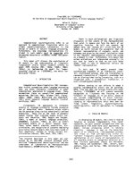
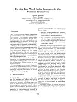
Fig. 1. Phylogenetic analyses of PHD domains. Representative
PHD domains were aligned. Numbers indicate the frequency of
clade probability values. Only values over 0.75 are shown. Black
geometrical shapes are additional domains, as indicated. ANOGA,
Anopheles gambiae; ASHGOS, Ashbya gossypii; BRARE, Brachyda-
nio rerio; CANDGLA, Candida glabrata; CIONA, Ciona intestinalis;
DEBHANSE, Debaryomyces hansenii; DROME, Drosophila melano-
gaster; KLULAC, Kluyveromyces lactis; SACHER, Saccharomyces
cerevisiae; SCHPO, Schizosaccharomyces pombe; YARLIPO, Yar-
rowia lipolytica. ING3_MOUSE is the outgroup. DATF1_MOUSE is
the SwissProt entry (DIDO1).
Sequence analyses of DIDO A. M. Rojas et al.
3506 FEBS Journal 272 (2005) 3505–3511 ª 2005 FEBS
fungal sequences is clearly distant from the other bran-
ches, which was indeed predicted. The other main
group, obtained 97% of the time in 20 740 explored
trees, contained the DIDO PHD sequences clustered
with the PHF and the CGBP proteins. As seen in the
tree, two fungal PHD domains corresponded to an
SPP1 cluster at the basal branch of the tree. Identical
topologies were obtained by using distance methods
and neighbor-joining trees (data not shown).
The sequences of DIDO1, CGBP and SPP1 were
realigned (supplementary Fig. S1), and a cysteine-rich
short motif spanning approximately 25 residues was
detected downstream of the PHD domain. We termed
this new region dPHD; it is well conserved among
the sequences and always follows the PHD domain.
Using PSI-BLAST, the dPHD of DIDO1 hits CGBP_
MOUSE at the second iteration, with an e-value of
2e-06 (inclusion threshold of 0.03). In SPP1, the cor-
responding dPHD segment was used to obtain fungal
sequences, and its profile hit the Q6PGZ4 protein
(zebrafish CGBP) at an hidden Markov model profile
(HMMER) e-value of 0.086 (Fig. 2). When using the
combined profile of CGBP and fungal sequences, the
murine DIO was hit at an HMMER e-value of 0.083.
The statistical robustness is in agreement with the
PHD phylogenetic distribution (Fig. 1), in which the
SPP1 representatives are at the basal branch of CGBP
and DIO.
The GCBP proteins contain a PHD domain, fol-
lowed by a DNA-binding domain (the zf-CXXC) and
the newly described dPHD region (supplementary
Fig. S1). This family is involved in DNA binding at
unmethylated CpG islands in active chromatin, and
these proteins are essential for mammalian develop-
ment [7]. In addition, CGBP subcellular distribution is
identical to that of the human trithorax protein, sug-
gesting that they may be components of a multimeric
complex analogous to the Saccharomyces histone-
methylating Set1 complex, which contains CGBP and
trithorax homologues [10]. The members of the tritho-
rax group encompass various subclasses of gene regu-
latory factors [11]; one subclass involves chromatin
remodeling activity. Another subclass, the trxG, is
poorly understood and includes trithorax itself, Ash1,
and Ash2 [12]; these latter are homologues of compo-
nents of the yeast COMPASS ⁄ SET1 complex. Some
functional features have been reported [13,14] for the
trithorax complex proteins in the context of domain
composition. Recent analyses of trithorax ⁄ mixed
lineage leukemia (MLL) and its relationship with
COMPASS suggest a linkage between leukemogenesis
and covalent modifications of chromatin [9]. Little is
known, however, of the functional role of the remain-
ing subclass members.
In this study, the yeast protein SPP1 (alias cps40), a
member of the COMPASS complex [10], was statisti-
cally related for the first time to DIDO1 and CGBP
proteins (Fig. 2). SPP1 also contains a PHD domain
followed immediately by dPHD (Figs 1 and 3). The
domain architectures concur with the phylogenetic dis-
tribution of PHD (Fig. 1) and the HMMER values of
dPHD (Fig. 2). In both cases, the fungal sequences
appear to be more closely related to CGBP than to
DIDO1, although CGBP contains the CXXC signature
between the two domains, probably as a result of
recombination processes; this signature is missing in
DIDO1 and SPP1. The absence of the CXXC signa-
ture distinguishes SPP1 from the CGBP family (Figs 1,
3 and S1); otherwise, the fungal sequences could well
constitute CGBP homologues.
The presence of a TFS2M domain is also indicative
of a possible function for the DIDO1 long isoform
(Figs 3 and S2). It is the second domain of the elonga-
tion factor, TFSII [15], that stimulates RNA poly-
merase II to transcribe through regions of DNA. The
structure of domain III of this elongation factor is also
solved structurally [16], as a Zn-binding domain. Both
domains II and III constitute the minimal transcrip-
tionally active fragment and are required simulta-
neously to maintain transcription.
The PHF3 protein was recovered in initial analyses
and also contains a TFS2M domain showing the same
architecture as the DIDO1 long isoform, although lack-
ing the dPHD. The PHF3 protein is expressed ubi-
quitously in normal tissues, and its expression is
dramatically reduced or lost in human glioblastoma
(a malignant astrocytic brain tumor) [17,18]. Down-
stream of TFS2M, a very small motif was detected
containing two histidine and two cysteine residues
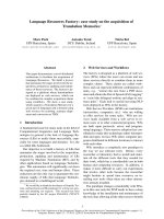
Fig. 2. HMMER e-values between the dPHD domain-containing
families. Numbers correspond to HMMER e-values from global pro-
file search results that connect the families. Arrows indicate profile
search direction.
A. M. Rojas et al. Sequence analyses of DIDO
FEBS Journal 272 (2005) 3505–3511 ª 2005 FEBS 3507
(Fig. S2), for which we propose the name small Zinc
finger (s-Zf). This region is too small to assess with any
confidence, based on statistical terms. Nonetheless, fur-
ther searches using other methods (such as pattern
matching) were conducted in databases, from which no
conclusive results were obtained (data not shown). This
region is present in PHF3, in another protein
(Q8NBC6), and in the DIDO1 long isoform, however,
and appears to be restricted to mammals. This architec-
ture in some way resembles the distribution of TFSII
domains II and III.
Our surveys provided statistically significant e-values
connecting the PHD domain-containing families
DIDO1, CGBP, and the fungal family that we term
SPP1. All these proteins contain a small, previously
undetected domain downstream of the PHD, which
we called dPHD, that allows connection of these famil-
ies (despite the small length of alignment) at reliable
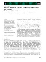
Fig. 3. Domain dissection of death inducer obliterator protein (DIDO) proteins. Sequence names are SwissProt ⁄ TrEMBL identifiers. PHD
(dark blue), dPHD (green ⁄ blue), CXXC (orange), TFS2M (purple), sZf (pink), SPOC (black ⁄ red), RRM (mauve). The central boxed protein is
DIDO (DATF_HUMAN). The dPHD region connects DIDO with CGBP and SPP1 families (upper panel), and with a fly homolog, Q9VG78
(below the DIDO box). TFS2M–sZf links DIDO with PHF3_HUMAN (center panel). The SPOC domain connects to the SPEN family (lower
panel) [4]. CXXC, CGBP-specific; BRK, fly specific; and RRM, SPEN-specific. DATF_HUMAN is the SwissProt identifier for DIDO.
Sequence analyses of DIDO A. M. Rojas et al.
3508 FEBS Journal 272 (2005) 3505–3511 ª 2005 FEBS
e-values (Fig. 2). It is noteworthy that the retrieved
proteins all appear to have a role in DNA regulation,
in the context of chromatin stability, and to form part
of higher complexes linked to active chromatin.
The DIDO1 gene has been linked to the split ends
domain (SPEN) family of proteins [4], involved in
transcriptional repression via their C-terminal domain,
SPOC. Here we show that additional protein families,
CGBP and SPP1, are linked to this gene by two
domains (Fig. 3).
Experimental analyses
The localization of the DIDO1 gene in the context
of chromatin stability was addressed experimentally.
Although the experiments conducted were preliminary,
they indicated the involvement of this gene in cell divi-
sion. This is consistent with our observations, that the
DIDO1 gene shares two domains with CGBP and
SPP1, both proteins being bound to active chromatin
and involved in DNA regulation; in addition, the yeast
protein, SPP1, is well characterized by tandem affinity-
purification experiments.
Ectopically expressed DIDO1 associated with chro-
matin throughout the cell cycle (Fig. 4A,B), causing a
high incidence of asymmetric divisions. Cells from
DIDO1-targeted mice show a notable incidence of
lagging chromosomes (10 of 237; 4.2%) during ana-
phase (Fig. 4C,D), which was not observed in cul-
tures of wild-type cells (0 of 140; 0.0%). Although
merotelic kinetochore attachment to centromeres is
generally considered to be a major cause of lagging
chromosomes, they can also be caused by changes in
chromatin composition [19,20]. As DIDO1 associates
with chromatin in general, and not only in centro-
meric regions, chromatin instability is the most prob-
able explanation for the lagging chromosomes in
DIDO1-targeted cells.
Targeting of the DIDO1 locus leads to genomic
instability, as shown by the occurrence of lagging chro-
mosomes in mitosis. The domains targeted in mice are
PHD and dPHD, which are domains shared with
CGBP and SPP1. Ectopic DIDO1 expression leads to
a high incidence of asymmetric divisions. The reported
DIDO-induced apoptosis could thus be a consequence
of alterations in DNA regulation or chromatin stabil-
ity. A similar case is the Suv39h histone methyltrans-
ferase, in which both deletion and overexpression lead
to alterations in pericentric chromatin, chromosome
missegregation in mitosis and meiosis, and apoptosis
[19,21].
Concluding remarks
Computational analyses, combined with some basic
and preliminary experimental assays (some of which
we show here), enable us to hypothesize an additional
functional role for DIDO1. In this case, apoptosis
induction should be explained within the context of
DNA regulation, especially considering that the apop-
tosis induced by DIDO1 requires protein translocation
to the nucleus. Although functional analyses of indi-
vidual domains have not been addressed, the global
context of the domain analyses allows us to draw a
more general picture of the involvement of this gene
in nuclear processes. Nonetheless, the data presented
here build a hypothesis that should be experimentally
addressed in detail.
Experimental procedures
Computational analyses
The complete protein sequences of human DIDO isoforms 1
and 2 were searched against PFAM [22,23] and SMART
[24] databases to automatically detect domains; they were
further used as queries against NCBI databases using PSI-
BLAST [25,26]. Complete gene sequences were found only
in mouse or human; only partial fragments were found in
other organisms. We first performed BLAST searches
against unfinished genomes from NBCI [27], then against
Fig. 4. Chromosomal instability in death inducer obliterator protein
(DIDO)-overexpressing and DIDO-targeted cells. (A, B) Ectopic
expression of DIDO1. (C) Normal anaphase of a wild-type mouse
cell. (D) Mitosis in DIDO-targeted cells, showing lagging chromo-
somes during anaphase (arrowhead).
A. M. Rojas et al. Sequence analyses of DIDO
FEBS Journal 272 (2005) 3505–3511 ª 2005 FEBS 3509
EST databases, with further EST assembly of reliable hits.
Any new sequence was incorporated into profiles to improve
profile quality. Profile-based sequence searches were per-
formed against the nonredundant and Uniref90 protein
databases with the corresponding global hidden Markov
models [28] (HMMer version 2.3.2 PVM). Alignments were
generated by using T-COFFEE and checked manually [29].
Phylogenies of the PHD domain were obtained by using
probabilistic approaches [30] (Mr Bayes 3 version), which
run for 1 000 000 generations in four independent Markov
chains. When convergence was reached, a total of 20 740
trees were explored to further construct a consensus tree.
Numbers indicate the frequency of clade probability values.
Green fluorescent protein (GFP)–DIDO-expressing
cell lines
To construct the GFP–DIDO1 fusion, human DIDO1
cDNA was transferred from pGEMT (Promega, Madison,
WI, USA) to pEGFP-C1 (Clontech, Mountain View, CA,
USA) by using unique SpeI and ApaI sites. This yields a plas-
mid expressing DIDO1 in-frame with GFP under control of
the cytomegalovirus (CMV) promoter. NIH 3T3 cells were
cultured in Dulbecco’s modified Eagle’s medium (DMEM)
supplemented with 10% (v ⁄ v) fetal bovine serum and antibi-
otics. To generate stable cell lines, 10
5
cells were seeded in
each well of a six-well plate (BD Falcon, San Jose, CA, USA)
and transfected with plasmid DNA (1 lg) and FuGene 6
(3 ll; Roche, Indianapolis, IN, USA), as recommended by
the manufacturer. Cells were selected by incubation with
500 lgÆmL
)1
G418 for 2 weeks, and clones with efficient
expression, as judged by fluorescence microscopy, were used
for further experiments. To visualize GFP-DIDO1, 5 · 10
4
cells were seeded on glass coverslips. After 48 h, cells were
formaldehyde fixed, mounted in Vectashield containing
4,6-diamino-2-phenylindole (DAPI) (Vector Laboratories,
Burlingame, CA, USA), and studied by standard fluores-
cence microscopy.
Analysis of anaphases in embryonic fibroblasts
To determine the occurrence of lagging chromosomes
during anaphase, low-passage mouse embryonic fibroblast
cultures from targeted (PHD and dPHD regions) and
wild-type mice were fixed with methanol and acetic acid,
mounted in Vectashield containing DAPI, and studied by
standard fluorescence microscopy. Lagging chromosomes
were scored only when no attachment whatsoever to either
of the chromosome pools could be detected in anaphase.
Acknowledgements
We thank Catherine Mark for editorial assistance. This
work was financed, in part, by the 6th EU Framework
Program Project IMPAD QLGI-CT-2001-01536, MEC
and GenFun LSHG-CT-2004-503567. The Department
of Immunology and Oncology was founded and is sup-
ported by the Spanish Council for Scientific Research
(CSIC) and by Pfizer.
References
1 Aravind L, Dixit VM & Koonin EV (1999) The
domains of death: evolution of the apoptosis machinery.
Trends Biochem Sci 24, 47–53.
2 Garcia-Domingo D, Leonardo E, Grandien A, Martinez
P, Albar JP, Izpisua-Belmonte JC & Martinez AC
(1999) DIO-1 is a gene involved in onset of apoptosis in
vitro, whose misexpression disrupts limb development.
Proc Natl Acad Sci USA 96, 7992–7997.
3 Garcia-Domingo D, Ramirez D, Gonzalez de Buitrago
G & Martinez AC (2003) Death inducer-obliterator 1
triggers apoptosis after nuclear translocation and cas-
pase upregulation. Mol Cell Biol 23 , 3216–3225.
4 Sanchez-Pulido L, Rojas AM, van Wely KH, Martinez
AC & Valencia A (2004) SPOC: a widely distributed
domain associated with cancer, apoptosis and transcrip-
tion. BMC Bioinformatics 5, 91.
5 Copley RR, Doerks T, Letunic I & Bork P (2002) Pro-
tein domain analysis in the era of complete genomes.
FEBS Lett 513, 129–134.
6 Ponting CP & Dickens NJ (2001) Genome cartography
through domain annotation. Genome Biol 2, Comment
2006.
7 Voo KS, Carlone DL, Jacobsen BM, Flodin A &
Skalnik DG (2000) Cloning of a mammalian transcrip-
tional activator that binds unmethylated CpG motifs
and shares a CXXC domain with DNA methyltransfer-
ase, human trithorax, and methyl-CpG binding domain
protein 1. Mol Cell Biol 20, 2108–2121.
8 Miller T, Krogan NJ, Dover J, Erdjument-Bromage H,
Tempst P, Johnston M, Greenblatt JF & Shilatifard A
(2001) COMPASS: a complex of proteins associated
with a trithorax-related SET domain protein. Proc Natl
Acad Sci USA 98, 12902–12907.
9 Tenney K & Shilatifard A (2005) A COMPASS in
the voyage of defining the role of trithorax ⁄ MLL-con-
taining complexes: Linking leukemogensis to covalent
modifications of chromatin. J Cell Biochem 95,
429–436.
10 Roguev A, Schaft D, Shevchenko A, Pijnappel WW,
Wilm M, Aasland R & Stewart AF (2001) The
Saccharomyces cerevisiae Set1 complex includes an Ash2
homologue and methylates histone 3 lysine 4. EMBO
J 20, 7137–7148.
11 Kennison JA (1995) The Polycomb and trithorax group
proteins of Drosophila: trans-regulators of homeotic
gene function. Annu Rev Genet 29, 289–303.
Sequence analyses of DIDO A. M. Rojas et al.
3510 FEBS Journal 272 (2005) 3505–3511 ª 2005 FEBS
12 Shearn A (1989) The ash-1, ash-2 and trithorax genes
of Drosophila melanogaster are functionally related.
Genetics 121, 517–525.
13 Mazo AM, Huang DH, Mozer BA & Dawid IB (1990)
The trithorax gene, a trans-acting regulator of the
bithorax complex in Drosophila, encodes a protein with
zinc-binding domains. Proc Natl Acad Sci USA 87,
2112–2116.
14 Tripoulas N, LaJeunesse D, Gildea J & Shearn A (1996)
The Drosophila ash1 gene product, which is localized at
specific sites on polytene chromosomes, contains a SET
domain and a PHD finger. Genetics 143, 913–928.
15 Morin PE, Awrey DE, Edwards AM & Arrowsmith CH
(1996) Elongation factor TFIIS contains three structural
domains: solution structure of domain II. Proc Natl
Acad Sci USA 93, 10604–10608.
16 Qian X, Jeon C, Yoon H, Agarwal K & Weiss MA
(1993) Structure of a new nucleic-acid-binding motif in
eukaryotic transcriptional elongation factor TFIIS.
Nature 365, 277–279.
17 Fischer U, Struss AK, Hemmer D, Michel A, Henn W,
Steudel WI & Meese E (2001) PHF3 expression is fre-
quently reduced in glioma. Cytogenet Cell Genet 94,
131–136.
18 Struss AK, Romeike BF, Munnia A, Nastainczyk W,
Steudel WI, Konig J, Ohgaki H, Feiden W, Fischer U
& Meese E (2001) PHF3-specific antibody responses in
over 60% of patients with glioblastoma multiforme.
Oncogene 20, 4107–4114.
19 Peters AH, O’Carroll D, Scherthan H, Mechtler K, Sauer
S, Schofer C, Weipoltshammer K, Pagani M, Lachner
M, Kohlmaier A et al. (2001) Loss of the Suv39h histone
methyltransferases impairs mammalian heterochromatin
and genome stability. Cell 107, 323–337.
20 Cimini D, Mattiuzzo M, Torosantucci L & Degrassi
F (2003) Histone hyperacetylation in mitosis prevents
sister chromatid separation and produces
chromosome segregation defects. Mol Biol Cell 14,
3821–3833.
21 Shen WH & Meyer D (2004) Ectopic expression of the
NtSET1 histone methyltransferase inhibits cell expan-
sion, and affects cell division and differentiation in
tobacco plants. Plant Cell Physiol 45, 1715–1719.
22 Sonnhammer EL, Eddy SR, Birney E, Bateman A &
Durbin R (1998) Pfam: multiple sequence alignments
and HMM-profiles of protein domains. Nucleic Acids
Res 26, 320–322.
23 Bateman A, Birney E, Cerruti L, Durbin R, Etwiller L,
Eddy SR, Griffiths-Jones S, Howe KL, Marshall M &
Sonnhammer EL (2002) The Pfam protein families data-
base. Nucleic Acids Res 30, 276–280.
24 Letunic I, Copley RR, Schmidt S, Ciccarelli FD, Doerks
T, Schultz J, Ponting CP & Bork P (2004) SMART 4.0:
towards genomic data integration. Nucleic Acids Res 32,
Database issue, D142–144.
25 Altschul SF, Madden TL, Schaffer AA, Zhang J,
Zhang Z, Miller W & Lipman DJ (1997) Gapped
BLAST and PSI-BLAST: a new generation of protein
database search programs. Nucleic Acids Res 25,
3389–3402.
26 Altschul SF & Koonin EV (1998) Iterated profile
searches with PSI-BLAST – a tool for discovery in pro-
tein databases. Trends Biochem Sci 23, 444–447.
27 Cummings L, Riley L, Black L, Souvorov A, Resen-
chuk S, Dondoshansky I & Tatusova T (2002) Genomic
BLAST: custom-defined virtual databases for complete
and unfinished genomes. FEMS Microbiol Lett 216,
133–138.
28 Eddy SR (1998) Profile hidden Markov models. Bio-
informatics 14, 755–763.
29 Notredame C, Higgins DG & Heringa J (2000) T-Cof-
fee: a novel method for fast and accurate multiple
sequence alignment. J Mol Biol 302, 205–217.
30 Ronquist F & Huelsenbeck JP (2003) MrBayes 3: Baye-
sian phylogenetic inference under mixed models. Bioin-
formatics 19, 1572–1574.
Supplementary material
The following material is available online.
Fig. S1. Multiple alignment of DIDO, CGBP and SPP1
proteins. Additional proteins were included. Names are
SwissProt or sptrembl identifiers, with added common
species name: Chick, Gallus gallus; Fugu, Fugu rubripes;
Brare, Danio rerio; Anoga, Anopheles gambiae; Drome,
Drosophila melanogaster; Sacce, Saccharomyces cerevisi-
ae; Schizo, Schizosaccharomyces pombe; Ciona, Ciona
intestinalis; Caeel, Caenorhabditis elegans; Caebri,
Caenorhabditis briggsae. The DIDO1 EST consensus
sequence was reconstructed manually by assembling
ESTs. Boxed, vertebrate-restricted. Red-boxed sequence
names are CGBP, showing a specific CXXC motif
absent in other sequences (a solid red box above the
alignment). A solid dark blue box indicates the PHD
domain, where rectangles and boxes indicate secondary
structural elements from the DIDO1 mouse structure
(pdb code: 1WEM); a solid green ⁄ blue box identifies the
newly identified dPHD domain. DATF1_MOUSE and
DATF_HUMAN are the SwissProt identifiers for
DIDO.
Fig. S2. Multiple alignment of death inducer oblitera-
tor protein (DIDO), PHF3 and yeast proteins. Addi-
tional proteins were included. Naming conventions are
as in Fig. S1. The solid purple box indicates the
TFS2M domain. Pink-boxed sequence names are pro-
teins containing the newly identified s-Znf signature
(solid pink box above the alignment). The solid
black ⁄ red box identifies the SPOC domain.
A. M. Rojas et al. Sequence analyses of DIDO
FEBS Journal 272 (2005) 3505–3511 ª 2005 FEBS 3511