Báo cáo khoa học: BCR kinase phosphorylates 14-3-3 Tau on residue 233 pdf
Bạn đang xem bản rút gọn của tài liệu. Xem và tải ngay bản đầy đủ của tài liệu tại đây (283.37 KB, 10 trang )
BCR kinase phosphorylates 14-3-3 Tau on residue 233
Samuel J. Clokie
1
, Kin Y. Cheung
1
, Shaun Mackie
1,
*, Rodolfo Marquez
2
, Alex H. Peden
1,
†
and Alastair Aitken
1
1 School of Biomedical and Clinical Laboratory Sciences, University of Edinburgh, UK
2 School of Life Sciences, University of Dundee, UK
The term breakpoint cluster region (BCR) refers to an
area of 5.8 kb on chromosome 22 that by a reciprocal
translocation event with the oncogene Abl, from chro-
mosome 9, produces the chimera BCR–Abl [1]. It is
this reciprocal translocation event that creates an aber-
rant chromosome called the Philadelphia chromosome
(ph
1
) that is the hallmark of chronic myeloid leukaemia
(CML) and which is found in over 90% of patients
with CML [2]. BCR–Abl proteins can vary in size,
depending on the breakpoint within the BCR. The
resultant fusion protein, containing different amounts
of the BCR gene fused to ABL, gives rise to different
clinical outcomes with ranging clinical severity [3,4].
The constitutively active tyrosine kinase activity of
Abl, essential for the progression of CML [5], has been
the focus of many studies to find an effective inhibitor
[6], of which the compound Gleevec or Imatinib has
proved to be highly successful. However, the many
varied domains of BCR are also essential for the trans-
forming potential of BCR–Abl [1].
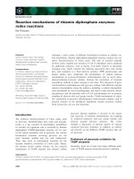
The normal BCR product is 160 kDa and contains
a number of domains (Fig. 1) (reviewed in [1]). These
include an oligomerization domain [7], an atypical S ⁄ T
kinase domain [8–10], a Src homology 2 (SH2)-binding
domain [11], guanine nucleotide exchange factor
(GEF) domain [12,13] and a GTPase activity (GAP)
Keywords
14-3-3 isoforms; phosphorylation; BCR
kinase; protein interactions
Correspondence
A. Aitken, School of Biomedical and Clinical
Laboratory Sciences, Darwin Building,
University of Edinburgh, King’s Buildings,
Mayfield Road, Edinburgh EH8 9XD, UK
Fax/Tel: +44 131 650 5357
E-mail:
Present addresses
*Psychiatric Genetics Section, Molecular
Medicine Centre, University of Edinburgh, UK
†The National Creutzfeldt–Jakob Disease
Surveillance Unit, Western General Hospital,
Edinburgh, UK
(Received 21 February 2005, revised 4 May
2005, accepted 13 May 2005)
doi:10.1111/j.1742-4658.2005.04765.x
The breakpoint cluster region protein, BCR, has protein kinase activity
that can auto- and trans-phosphorylate serine, threonine and tyrosine resi-
dues. BCR has been implicated in chronic myelogenous leukaemia as well
as important signalling pathways, and as such its interaction with 14-3-3 is
of major interest. 14-3-3s and f isoforms have been shown previously to be
phosphorylated in vitro and in vivo by BCR kinase on serine and threonine
residue(s) but site(s) were not determined. Phosphorylation of 14-3-3 iso-
forms at distinct sites is an important mode of regulation that negatively
affects interaction with Raf kinase and Bax, and potentially influences the
dimerization of 14-3-3. In this study we have further characterized the
BCR)14-3-3 interaction and have identified the site phosphorylated by
BCR. We show here that BCR interacts with at least five isoforms of
14-3-3 in vivo and phosphorylates 14-3-3s on Ser233 and to a lesser extent
14-3-3f on Thr233. We have previously shown that these two isoforms are
also phosphorylated at this site by casein kinase 1, which, in contrast to
BCR, preferentially phosphorylates 14-3-3f.
Abbreviations
BCR, breakpoint cluster region; CK1, casein kinase 1; CML, chronic myeloid leukaemia; D4476, 4-{4-[2,3-dihydro-benzo (1,4)dioxin-6-yl]-5-
pyridin-2-yl-1H-imidazol-2-yl}benzamide; ERBIN, ERB2 interacting protein; GAP, GTPase activity; GEF, guanine nucleotide exchange factor;
JNK, c-Jun N-terminal kinase; ph
1
, Philadelphia chromosome; PI3K, phosphatidylinositol 3-kinase; PKC, protein kinase C; SH2, Src homology
2; XPB, xeroderma pigmentosum group B protein.
FEBS Journal 272 (2005) 3767–3776 ª 2005 FEBS 3767
domain [14]. BCR binds 14-3-3 [9], Xeroderma pig-
mentosum group B protein (XPB) [15] and chromatin
[16]. BCR binds to Grb2 when phosphorylated on
Tyr177 in the SH2 binding domain, thus linking it to
a role in the Ras pathway [11]. Recently a functional
PDZ binding domain was identified in BCR, associ-
ating through a motif consisting of S-T-E-V, with the
ERB2 interacting protein (ERBIN) [17].
The 14-3-3 family forms protein complexes involved
in neurodegeneration, apoptosis, signal transduction,
trafficking and secretion [18–20]. In many cases, these
complexes show a distinct preference for a particular
isoform(s) of 14-3-3. 14-3-3 proteins are established
adaptors of signalling proteins that bind primarily,
but not solely, to proteins containing phosphorylated
serine ⁄ threonine residues. Using a phosphopeptide
library, an optimal motif for 14-3-3 binding was identi-
fied as R(S)XpS ⁄ TXP [21] which was later refined to
RXXXpS ⁄ TXP where pS is phosphoserine and X is
any amino acid [22]. The crystal structures of 14-3-3
dimers [23,24] led to identification of the binding site
of the novel phosphopeptide motif RSX
1,2
SpXP and
unphosphorylated motifs [22,25].
Recent findings also show that the mechanism of
interaction is more complex than simply acting
through the phosphoserine ⁄ threonine motif. Nonphos-
phorylated binding motifs can also be of high affinity
and may show more isoform-dependence in their inter-
action [25]. Some well-characterized interacting pro-
teins such as Raf kinase have been shown to have
additional binding site(s) for 14-3-3 on their cysteine-
rich regions. BCR also binds via a serine-rich region.
Binding of a protein through two distinct binding
motifs to a dimeric 14-3-3 may also be essential for
full interaction [26]. Dimerization with specific iso-
forms in vivo has important implications for the role
of 14-3-3 in the formation of signalling complexes [19],
and phosphorylation of specific 14-3-3 isoforms can
also regulate interactions [18,20].
The BCR protein has four potential R(S)XXpSXP
motifs [27] and the association with 14-3-3 is of major
biological significance due to their respective involve-
ment in signalling pathways including the association
with Raf kinase [28]. 14-3-3 has been shown to bind
the p110 subunit of phosphatidylinositol 3-kinase
(PI3K) [29] and the authors suggested that 14-3-3 neg-
atively regulates the activity of PI3K in activated
T cells by ‡ 50%. Interestingly the authors noted
enhanced binding of 14-3-3s to PI3K with inclusion of
the tyrosine phosphatase inhibitor pervanadate to the
lysis buffer, suggesting that 14-3-3 may bind through
phosphotyrosine residues as well as phosphoserine ⁄
threonine residues.
We showed that a and d were phosphorylated
forms of b and f, respectively, and are more than
50% phosphorylated on Ser185 in brain 14-3-3 [30],
but we find no evidence for phospho-forms in a wide
range of other tissue types and cell lines. Casein
kinase 1 (CK1, reviewed in [31,32]) colocalizes in neu-
rons with synaptic vesicle markers and can phos-
phorylate some synaptic vesicle associated proteins.
We identified CK1a as the brain kinase that phos-
phorylated 14-3-3f on Thr233 [33]. 14-3-3s and yeast
14-3-3s (BMH1 and 2) were also phosphorylated
on the equivalent sites. In vivo phosphorylation of
14-3-3f at this site negatively regulates its binding to
c-Raf and may be important in Raf mediated signal
transduction [28,33].
The b, g and f isoforms of 14-3-3 (but not e and c
although they also contain serine at the equivalent site)
are phosphorylated by a sphingosine-dependant kinase,
SDK1, now identified as the kinase domain of protein
kinase C (PKC) d produced after caspase-3 cleavage
[34].
Phosphorylation of 14-3-3 by BCR could affect the
ability of 14-3-3 to bind other signalling proteins; for
example we have shown that phosphorylation of 14-3-3
by CK1 negatively regulates binding to Raf in vivo [33].
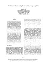
Fig. 1. Domains of BCR. Possible 14-3-3 binding sites are indicated as filled circles on top. These and the kinase domain are located within
exon 1. The positions of the RacGAP, GEF, oligomerization and SH
2
binding domains are indicated; the tyrosine residues in SH
2
domains by
short lines.
BCR kinase phosphorylates 14-3-3s on residue 233 S. J. Clokie et al.
3768 FEBS Journal 272 (2005) 3767–3776 ª 2005 FEBS
Ser185 is located in the tertiary structure adjacent to
residue 233 [19], and Gotoh’s group [35] have recently
shown that activated c-Jun N-terminal kinase (JNK)
promotes Bax translocation to the mitochondria
through phosphorylation of 14-3-3r and f at sites equiv-
alent to Ser185, which led to the dissociation of Bax.
Expression of phosphorylation defective mutants of
14-3-3 blocked JNK-induced Bax translocation to mito-
chondria, cytochrome c release and apoptosis.
14-3-3s isoform has been shown to interact with full
length BCR and with BCR-Abl [9]. The authors indi-
cated that 14-3-3s was a substrate for the BCR serine-
threonine kinase activity and in this study we have
determined the site to be residue 233. This is of major
potential physiological relevance since this C-terminal
region has recently been proposed as a general inhib-
itor of 14-3-3–ligand interactions [36]. The observation
here that BCR phosphorylates 14-3-3 on the same
residue, 233, as CK1 indicates a conserved mode of
regulation, whereby phosphorylation could affect the
ability of 14-3-3 to bind target proteins.
Results
BCR associates with 14-3-3 isoforms in vitro and
in vivo
14-3-3 isoforms s and f and b have previously been
shown to interact with BCR [9,10]. To investigate the
possibility that additional isoforms may also interact
with BCR, two approaches were taken. Firstly BCR–
FLAG was overexpressed in 293 cells, GST)14-3-3
fusion proteins were incubated with the lysate,
glutathione beads added and the ‘pull downs’ were sub-
jected to SDS ⁄ PAGE and western blotted using anti-
FLAG (Fig. 2A). Recombinant GST fusion constructs
of all 14-3-3 isoforms pulled down BCR–FLAG from
transfected cells which verified that all 14-3-3 isoforms
have the ability to interact with BCR. However, relat-
ively more BCR associated with the 14-3-3g and c iso-
form constructs (Fig. 2A). Pull down experiments were
repeated, with consistent results. The example shown
was carried out at a time when the phosphorylation site
had been identified, which is the reason for the inclu-
sion of the T233Df)14-3-3 construct. Secondly, BCR–
FLAG was overexpressed in cells, immunoprecipitated,
and western blotting used to detect interaction with
endogenous 14-3-3 isoforms. The results show that
B
A
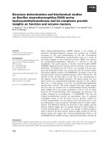
Fig. 2. (A) BCR interacts with all 14-3-3 isoforms in 293 cells.
HEK293 -cells were transfected with BCR–FLAG, lysed and incuba-
ted with the indicated GST)14-3-3 isoform. A loading control for
14-3-3 stained with Ponceau S is shown in the lower panel.
GST)14-3-3f T233D construct was also assayed, right-hand lane.
An equivalent amount of 1% of the lysate used for each incubation
is shown in lane 1, and a GST-only incubation is shown in lane 2.
(B) 293 cells were transfected with BCR–FLAG, the lysates pooled
and divided into seven aliquots for immunoprecipitation with anti-
FLAG Ig. An aliquot containing 1% of the input of each lysate was
western blotted with anti-14-3-3 Igs to verify endogenous levels (top
panel). The input lysate (1%) was western blotted with anti-FLAG
Igs (middle panel) to check expression levels of the BCR construct.
The 14-3-3 isoforms were coimmunoprecipitated with anti-Flag Ig
and each anti-FLAG immunoprecipitation was western blotted with
antibodies specific for a 14-3-3 isoform [54] as indicated (bottom
panel). To demonstrate that 14-3-3 isoforms do not bind nonspecifi-
cally to the resin beads, the left lane is an immunoprecipitation with
control IgG followed by a western blot with antibodies that recog-
nize all 14-3-3 isoforms (PAN).
S. J. Clokie et al. BCR kinase phosphorylates 14-3-3s on residue 233
FEBS Journal 272 (2005) 3767–3776 ª 2005 FEBS 3769
14-3-3b, c, e, f, s and g isoforms associate with BCR–
FLAG (Fig. 2B). Tau14-3-3 is expressed at low levels
in 293 cells; nevertheless interaction with this isoform
can be seen. 14-3-3r is expressed at high levels only in
epithelial cells and is present at such low levels in the
293 cell line that the interaction could not be detected.
Negative controls using nonimmune sera were added to
BCR–FLAG transfections. These showed that none
of the isoforms tested associate with the agarose
bead ⁄ antibody matrix.
As well as verifying the binding of 14-3-3 isoforms
b, f and s shown previously [9,10] we have thus shown
in this study that c, g and e14-3-3 can also associate
with BCR in vivo and in vitro.
BCR phosphorylates 14-3-3s and 14-3-3f in vitro
BCR kinase has previously been shown to phosphory-
late 14-3-3 on serine ⁄ threonine residues [9]. It has also
been shown that BCR when treated with alkaline phos-
phatase reduced ability to associate with 14-3-3 [37].
However it is not known if association of BCR with
14-3-3 facilitates phosphorylation. Two vectors suitable
for mammalian expression containing full length BCR
were produced; an N-terminal GST fusion construct
and a C-terminal FLAG construct. The purpose of cre-
ating a GST N-terminal fusion was to determine whe-
ther the dimerization ability of GST could increase the
kinase activity of BCR, because Maru et al. [38] showed
that GST could replace the oligomerization domain of
BCR. It has also been shown that BCR purifies as an
oligomer [8]. A C-terminal FLAG tag construct was cre-
ated in case the GST itself would create steric hindrance
between BCR and 14-3-3 as substrate. In addition, pro-
duction in mammalian cells would allow any necessary
post-translational modifications such as phosphoryla-
tion and correct processing and folding of BCR. The
tagged BCR transcripts were overexpressed in COS-1
and human embryonic kidney (HEK) 293 cells, and
lysed in NP-40 buffer designed to maintain the phos-
phorylated state of BCR. GST–BCR was affinity puri-
fied using glutathione–Sepharose beads, extensively
washed and incubated with exogenous 14-3-3 under
appropriate assay conditions. In agreement with previ-
ous studies, the 14-3-3s and f isoforms were phosphoryl-
ated (Fig. 3), the latter to a much lower level than s.
None of the other mammalian isoforms b, c, e, g and r
were phosphorylated. BCR–FLAG constructs immuno-
precipitated with M2 a-FLAG antibody gave a slightly
higher level of phosphorylation and so were used for
further studies. There was no difference in substrate spe-
cificity between GST–BCR and BCR–FLAG (data not
shown). Alignment of the mammalian 14-3-3 isoform
sequences indicate that the only Ser ⁄ Thr residues com-
mon to s and f, but not present in the other isoforms,
are S233 in 14-3-3s and T233 in 14-3-3f. Using Ala
mutants of these phosphorylation sites, kinase assays
were carried out as previously. The SerfiAla mutant
(S233A) of 14-3-3s (Fig. 4A) and the ThrfiAla mutant
(T233A) of 14-3-3f were not phosphorylated by BCR
(Fig. 4B). There was no change in phosphorylation
by BCR of the SerfiAla mutant (S185A) of 14-3-3f
(Fig. 4B). The phosphorylation of the 14-3-3f constructs
at residue T233 was very poor in comparison to phos-
phorylation of wild type 14-3-3s and mutation of this
residue to Ala completely abolished phosphorylation.
The lack of phosphorylation of the T233A construct
of 14-3-3f indicates that BCR does not phosphorylate
residue 185 in 14-3-3f, which was shown by Gotoh’s
group to be a substrate for JNK [35].
Ser58, common to all 14-3-3 isoforms except r,is
phosphorylated by a variety of protein kinases (SDK1
[39], PKB [40] and by PKC in a synthetic peptide cor-
responding to residues 49–68 of the other isoforms
[41]). We show that there is complete lack of phos-
phorylation of 14-3-3r (Fig. 3), which acts as a natural
negative control. The S185A mutant of 14-3-3f as well
as the S233A and T233A variants still include Ser58,
which rules out the possibility of Ser58 being a site of
phosphorylation by BCR.
Phosphorylation of 14-3-3 isoforms is not
due to coimmunoprecipitation of CK1
Casein kinase 1 has been shown to phosphorylate
14-3-3s and f specifically on residue 233 both in vitro
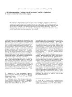
Fig. 3. BCR kinase phosphorylation of isoforms of 14-3-3. GST–
BCR was ‘pulled down’ and a protein kinase assay with each mam-
malian 14-3-3 isoform was carried out, followed by autoradiography
of the SDS ⁄ PAGE. The lower panel shows the loading control of
each 14-3-3 isoform (stained with Coomassie blue).
BCR kinase phosphorylates 14-3-3s on residue 233 S. J. Clokie et al.
3770 FEBS Journal 272 (2005) 3767–3776 ª 2005 FEBS
and in vivo [33]. We have also shown that CK1 associ-
ates with 14-3-3 (S. Clokie and A. Aitken, unpublished
results; [42]). The possibility existed that endogenous
14-3-3 could be acting as a ‘molecular bridge’ between
BCR and endogenous CK1 and that the latter activity
was phosphorylating 14-3-3s. To exclude this possibility,
the CK1 inhibitor CKI-7 was added to kinase assays at
concentrations up to 100 lm. Little effect was seen, even
at the highest concentration, and only slight inhibition
was seen when BCR was preincubated with CKI-7 for
1 h. However the IC
50
of this compound is rather high
and it is possible that it is causing a general inhibition of
kinase activity when used at such high concentrations.
We then used the newly developed inhibitor of CK1
4-{4-[2,3-dihydro-benzo (1,4)dioxin-6-yl]-5-pyridin-2-yl-
1H-imidazol-2-yl}benzamide (D4476) [43]. This has an
IC
50
of approximately 1 lm, and is therefore 10-fold
more inhibitory than CKI-7 towards CK1 and has been
shown to be highly specific [43]. This had no effect on
the phosphorylation of 14-3-3s or f by BCR kinase, but
completely inhibited CK1 assayed in parallel with BCR
(Fig. 5).
Discussion
Reuther et al. [9] showed that 14-3-3 binds to BCR
downstream of residue 297 (Fig. 1), and these authors
alluded to the possibility of 14-3-3 binding elsewhere
on the protein, but to a lesser extent. Indeed BCR has
many potential 14-3-3 binding sites – RASA-S95-RP,
RSG-S301-TS, RL-T310-WPR, RSY-S317-P and RSP-
S371-QN [28,45] – four of them C-terminal to residue
297. Recently the sequence RL-T310-WPR has been
shown to be phosphorylated in vivo [44].
Fig. 5. CK1 specific inhibitors do not affect BCR kinase activity.
The figure shows an autoradiograph of 14-3-3 s wt protein phos-
phorylated by BCR (left 4 lanes) and by CKIa (right 4 lanes) in the
presence and absence of the inhibitors as indicated. The bottom
panels show the 14-3-3 protein levels (Coomassie blue stained) in
the corresponding lanes of the autoradiograph. Dimethylsulfoxide
was included as a vehicle control. CKI-7 and D4476 were both used
at 20 l
M.
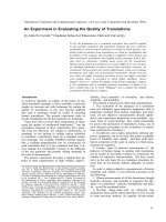
A
B
Fig. 4. Ala mutation at residue 233 abolishes phosphorylation by
BCR in vitro. (A) BCR–FLAG and the empty flag vector were trans-
fected into 293 cells and immunoprecipitated with anti-FLAG Igs as
described in the Experimental procedures. Kinase assays of 14-3-3s
were performed and SDS ⁄ PAGE of the radiolabelled protein was
carried out. The top panel shows an autoradiograph of tau 14-3-3
wild type (left 3 lanes) and tau 14-3-3 S233A (right 3 lanes). The con-
trol (middle lanes) is a transfection with the GST–BCR construct,
which we then attempted to pull down with the anti-FLAG Ig to
verify the specificity of the immunoprecipitation. The bottom panel
shows the 14-3-3 protein levels (Coomassie blue stained) in the cor-
responding lanes of the autoradiograph. (B) A similar experiment
was carried out with the zeta 14-3-3 T233A and zeta S185A con-
structs. Wild type tau and zeta 14-3-3 were phosphorylated in paral-
lel. The bottom panel shows the 14-3-3 protein levels (Coomassie
blue stained) in the corresponding lanes of the autoradiograph.
S. J. Clokie et al. BCR kinase phosphorylates 14-3-3s on residue 233
FEBS Journal 272 (2005) 3767–3776 ª 2005 FEBS 3771
We have now shown that the 14-3-3 isoforms ( b, c,
e, f and g) that are expressed at a detectable level in
293 cells bind BCR in vivo. The fact that 14-3-3s was
detected in the BCR–FLAG immunoprecipitation,
even though not detectable in the 293 lysate shows the
interaction may be of higher affinity than the other
isoforms. We also showed that 14-3-3 isoforms incuba-
ted with a cell lysate containing BCR–FLAG were able
to associate. Therefore, in addition to the s, f and b
isoforms previously shown, 14-3-3 e, g and c can also
interact with BCR. Our results suggest that while there
is the capacity of all 14-3-3 isoforms to bind BCR,
there is a preference for binding certain 14-3-3 iso-
forms. It may be difficult to ascertain true binding spe-
cificities, in vivo, due to the ability of 14-3-3 to form
a limited repertoire of heterodimers [45]. A T233D
mutant of 14-3-3f was incubated with the BCR lysate
(Fig. 2A) to determine whether mimicking a phosphor-
ylated T233 could negatively affect binding, as repor-
ted previously [28,33]. However, the mutant had no
significant effect, possibly due to the fact that some-
times an Asp mutation that introduces a carboxyl
group does not have the same effect as a phosphate
group.
The increased number of 14-3-3 isoforms that are
shown here to bind BCR opens up further potential
roles for BCR in cellular signalling. Even though these
extra 14-3-3 isoforms are not substrates for BCR, they
may well affect BCR activity and ⁄ or subcellular loca-
tion.
Using specific mutants of 14-3-3 we have shown that
BCR phosphorylates the tau isoform on serine 233
only. There is a rational explanation why phosphoryla-
tion at Ser233 in this isoform led to the observation by
Reuther et al. [9] of four phosphopeptide spots on thin
layer electrophoresis. From our own extensive protein
sequence analysis ([46] and A. Aitken, unpublished
results) we have shown that tryptic cleavage of 14-3-3
isoforms produces the following two C-terminal pep-
tides: (R)DNLTLWTSDSAGEECDAAEGAEN(223–
245) and (K)DSTLIMQLL
RDNLTLWTSDSAGEE
CDAAEGAEN(213–245). This is due to partial clea-
vage at Arg223 (underlined). The unique cysteine resi-
due (also underlined) in the tau isoform may undergo
modification, such as partial oxidation to cysteic acid
during thin layer electrophoresis when exposed to air,
which changes its electrophoretic mobility. Phosphory-
lation at residue 233 would yield two radiolabelled
phosphopeptides due to partial cleavage by trypsin,
multiplied by two due to the partially modified cys-
teine residues (which have a more acidic mobility), and
producing a total of four spots on thin layer electro-
phoresis.
BCR has a clear preference for phosphorylation of
14-3-3s rather than 14-3-3f (in agreement with previ-
ous studies [9]), possibly due to increased binding
affinity. In three separate experiments we observed an
approximately 10-fold higher phosphorylation of s
than f 14-3-3 (Fig. 3 and data not shown). This is in
contrast to the preference of CK1a for 14-3-3f over
14-3-3s [34]. It may be worth noting that 14-3-3s, the
major isoform substrate for BCR is expressed in
T-cells to a greater extent than in other tissues [47,48].
Western blots of immunoprecipitated BCR using
phospho-Tyr antibodies showed the presence of phos-
phorylated tyrosine residues (data not shown). One
study has shown that tyrosine phosphorylation on resi-
due 177 (by Fes kinase) actually reduced the associ-
ation with 14-3-3, while at the same time increasing
the SH2 binding to GRB2 [49]. The kinase that phos-
phorylates BCR on Tyr177 in HEK293 cells is cur-
rently not known. A study of the Philadelphia positive
cell line K562 showed that Tyr177 is phosphorylated
in vivo [44], but this residue is a known substrate for
BCR–Abl, also expressed in this cell line [50,51]. The
possibility remains that 14-3-3 association with BCR
may perturb Tyr177 phosphorylation and ⁄ or affect
SH2 binding at this site.
Experimental procedures
Materials
All chemicals and reagents were from Sigma (St Louis,
MO, USA), apart from Redivue [
32
P]ATP[cP] (triethyl-
ammonium salt) from Amersham (Buckinghamshire, UK)
and prestained protein markers from New England Biolabs
(Beverly, MA, USA). Protease inhibitor tablets were from
Roche (Indianapolis, IN, USA); recombinant CK1 was
from Upstate Biotechnology (Lake Placid, NY, USA) and
CKI-7 was from Seikagaku (Tokyo, Japan).
A vector containing the bcr sequence was a kind gift
from O. Witte (Department of Cell Biology, Harvard
Medical School, Boston, MA, USA). The coding sequence
for bcr was amplified by PCR using two oligonucleotides
5¢-GATC
GCGGCCGCGCGCCATGGTGGACCCGGTG
GGCTT-3¢ and 3¢-GATC
GAATTCGACTTCGGTGGAG
AACAGGATGCTCTGTCT-5¢ creating the restriction sites
Not1 and EcoR1, respectively (underlined), and ligated into
the pEBG-2T GST vector for mammalian expression (kind
gift from D. Alessi, University of Dundee, UK) creating an
N-terminally fused bcr construct. Two oligonucleotides
(5¢-GATC
GAATTCATGGTGGACCCGGTGGGCTTCG-3¢
and 3¢-GATC
GCGGCCGCTTAGACTTCGGTGGAGAA
CAGGATGCTCTGTCT-5¢)wereusedtoproducebcrcDNA,
containing the restriction sites EcoR1 and Not1 for ligation
BCR kinase phosphorylates 14-3-3s on residue 233 S. J. Clokie et al.
3772 FEBS Journal 272 (2005) 3767–3776 ª 2005 FEBS
into the pCMV-4A vector (Stratagene, La Jolla, CA, USA),
producing a C-terminal f usion w ith the FLAG tag.
The cDNAs for 14-3-3 isoforms were from various
sources. 14-3-3b is an IMAGE clone (4843961 ⁄ gi14060448),
and was subcloned from the supplied vector (pOTB7) PCR
with two oligonucleotides: 5¢-GATC
GAATTCATGACAA
TGGATAAAAGTGAGCTGGTA-3¢ and 3¢-GATC
GTC
GACTTAGTTCTCTCCCTCCCCAG-5¢, creating an EcoR1
and a Sal1 restriction site, respectively (underlined). The
PCR product was inserted into pGEX-4T1 (Amersham),
creating an N-terminal GST fusion. 14-3-3g, 14-3-3c and
14-3-3r were a gift from H. Leffers (University of Copenha-
gen, Denmark), the g and c clones were present as an N-ter-
minal GST fusion in the vector pGEX-2TK (Amersham).
The 14-3-3r was subcloned from the vector pGPT-delta 6
using the oligonucleotides 5¢-GATCGAATTCATGGAGA
GAGCCAGTCTGATC-3¢ and 3¢-GATCGTCGACTCAG
CTCTGGGGCTCCT-5¢ creating an EcoR1 site and Sal1
site, respectively (underlined). The PCR product was inserted
into pGEX-4T1. 14-3-3f was from a human T-cell cDNA lib-
rary and has been produced as an N-terminal GST fusion in
the pGEX-2T vector [33,47,52]. 14-3-3e was produced as an
N-terminal maltose binding protein (MBP) fusion, from a rat
cDNA (accession no. m84416) [53]. 14-3-3s was from a
human source [48,49]. All cDNAs were checked by sequen-
cing both strands (in house sequencing core and Cytomyx,
Cambridge, UK).
Tissue culture and immunoprecipitation
SV40 transformed African green monkey kidney cells (COS-
1) and adenovirus 5E1A ⁄ B transformed human embryonic
kidney (HEK) 293 cells were transiently transfected with
8 lg DNA with 24 lL Lipofectamine 2000 (Invitrogen, Car-
lsbad, CA, USA). Cells were routinely cultured in Dulbecco’s
modified Eagle’s medium (DMEM, Invitrogen) supplemen-
ted with 10% (w ⁄ v) fetal bovine serum (Invitrogen), penicil-
lin, streptomycin and l-glutamine at 1 UÆmL
)1
,1lgÆmL
)1
and 0.292 mgÆmL
)1
, respectively, at 5% (v ⁄ v) CO
2
and 37 °C
until lysis. For transient transfections, 2–4 · 10
6
cells were
added to 100 mm plates, using antibiotic-free media, left
until 80–90% confluent, then incubated for 24 h after addi-
tion of the DNA–Lipofectamine complex at 5% (v ⁄ v) CO
2
,
37 °C. The plates were washed twice with ice cold NaCl ⁄ P
i
and lysed on ice with ice-cold NP-40 buffer using a cell scra-
per. The lysate was clarified by centrifugation at 16 000 g for
30 min at 4 °C, the addition of 50 lL washed Pansorbin A
cells (Calbiochem) for 60 min to remove endogenous IgG,
then a further 30 min at 16 000 g,4°C.
Glutathione ‘pull-down’ immunoprecipitation
kinase assay
Glutathione–Sepharose 4B (Amersham Pharmacia) beads
or a 1 : 1 mix of protein A and G beads (Amersham) were
used to pull down the GST fusion and immunoprecipitate
the FLAG-tagged BCR, respectively. To the clarified cell
lysates, GSH beads were added for 2 h before washing.
For immunoprecipitation with FLAG antibody (M2) the
antibody was incubated in the lysate overnight, then incu-
bated with protein AG beads for 2 h. The beads were then
centrifuged at 8000 g in a benchtop centrifuge for 20 s and
washed three times in lysis buffer [50 mm Tris, pH 7.5,
10% (v ⁄ v) glycerol, 137 mm NaCl, 2 mm b-glycerol
phosphate, 1 mm NaF, 1 mm NaVO
4
,1mm EDTA, 1 mm
dithiothreitol and protease inhibitor cocktail tablet, EDTA-
free (Roche)]. The beads were then washed twice in kinase
assay buffer (see below, without ATP and dithiothreitol).
After the last wash, the beads were resuspended in a final
volume of 25 lL kinase assay buffer, with a final concen-
tration of 50 mm Hepes, pH 7.05, 10 mm MgCl, 20 lm
ATP (containing 10 lCi [
32
P]ATP) and 20 lm dithiothrei-
tol, and 2 lg of 14-3-3 isoform was used for each assay.
The reaction was carried out for 30 min at 30 °C and
stopped in Laemmli buffer prior to SDS ⁄ PAGE, followed
by autoradiography.
Casein kinase 1 inhibitors
CKI-7 was dissolved in dimethylsulfoxide as a 10 mm
stock. For preincubation experiments with CKI-7, during
the last wash of the IP, BCR–FLAG immunoprecipitates
were turned end over end while suspended in kinase assay
buffer including 100 lm CKI-7 (minus ATP). Where stated,
CKI-7 was added just prior to addition of the substrate
(20 lm). D4476 inhibitor was dissolved in dimethylsulfoxide
to a stock of 1 mm and was used at 20 lm in the final
assay. This was added immediately prior to addition of
the substrate. No preincubation with D4476 was required
to observe an inhibitory effect. Dimethylsulfoxide (2 lL)
was used as a vehicle control.
Recombinant protein purification
All GST)14-3-3 fusion cDNAs were transformed into
E. coli BL21(DE3)pLysS competent cells (Novagen, Madi-
son, WI, USA), using the appropriate antibiotic. The cells
were grown at 37 °C until an attenuation of 0.9, then
induced using isopropyl thio-b-d-galactoside (ICN, Costa
Mesa, CA, USA) for 3.5 h at 30 °C, in a shaking incuba-
tor. The same procedure was used for the MBP)14-3-3e,
but with the addition of glucose at 2 gÆL
)1
at all stages.
Cell pellets, resuspended in lysis buffer [NaCl ⁄ P
i
,1mm
phenylmethanesulfonyl fluoride, 1 mm EDTA, 1 mm dithio-
threitol, protease inhibitor tablet and 0.1% (v ⁄ v) Triton],
were sonicated six times for 30 s with amplitude of
5 microns. The Triton X-100 concentration was increased
to 1%; the cell suspensions were rotated for 30 min at 4 °C
and clarified by centrifugation at 16 000 g for 30 min. The
supernatant was then passed through a 0.22 lm filter and
S. J. Clokie et al. BCR kinase phosphorylates 14-3-3s on residue 233
FEBS Journal 272 (2005) 3767–3776 ª 2005 FEBS 3773
the GST fusion protein was recovered from the lysate using
glutathione–Sepharose 4B beads (Amersham). The beads
were washed extensively and the 14-3-3 cleaved from the
GST tag using 50 U thrombin (Sigma) or 50 U Factor Xa
(New England Biolabs) for MBP)14-3-3e, for each litre of
original culture. The 14-3-3 was then concentrated and buf-
fer-exchanged into NaCl ⁄ P
i
containing protease inhibitors
(Roche) using a Vivaspin 10K MWCO concentrator and
stored in small aliquots at )70 °C until required.
Acknowledgements
A vector containing the bcr sequence was a kind gift
from Owen Witte (Department of Cell Biology, Har-
vard Medical School, Boston, MA, USA). We thank
Sir Philip Cohen and Carol MacKintosh (MRC pro-
tein phosphorylation unit, University of Dundee) for
the suggestion to use D4476 and for the use of lab
facilities. The pEBG-2T GST vector for mammalian
expression was a kind gift from Dario Alessi.
References
1 Laurent E, Talpaz M, Kantarjian H & Kurzrock R
(2001) The BCR gene and philadelphia chromosome-
positive leukemogenesis. Cancer Res 61, 2343–2355.
2 Nowell PC & Hungerford DA (1960) Chromosome stu-
dies on normal and leukemic human leukocytes. J Natl
Cancer Inst 25, 85–109.
3 Li WJ, Dreazen O, Kloetzer W, Gale RP & Arlinghaus
RB (1989) Characterization of bcr gene products in
hematopoietic cells. Oncogene 4, 127–138.
4 McLaughlin J, Chianese E & Witte ON (1989) Alterna-
tive forms of the BCR-ABL oncogene have quantita-
tively different potencies for stimulation of immature
lymphoid cells. Mol Cell Biol 9, 1866–1874.
5 Lugo TG, Pendergast AM, Muller AJ & Witte ON (1990)
Tyrosine kinase activity and transformation potency of
bcr-abl oncogene products. Science 247, 1079–1082.
6 Salesse S & Verfaillie CM (2002) BCR ⁄ ABL: from
molecular mechanisms of leukemia induction to treat-
ment of chronic myelogenous leukemia. Oncogene 21,
8547–8559.
7 McWhirter JR, Galasso DL & Wang JY (1993) A
coiled-coil oligomerization domain of BCR is essential
for the transforming function of BCR-Abl oncoproteins.
Mol Cell Biol 13, 7587–7595.
8 Maru Y & Witte ON (1991) The BCR gene encodes a
novel serine ⁄ threonine kinase activity within a single
exon. Cell 67, 459–468.
9 Reuther GW, Fu H, Cripe LD, Collier RJ & Pendergast
AM (1994) Association of the protein kinases c-BCR
and BCR-Abl with proteins of the 14-3-3 family.
Science 266, 129–133.
10 Braselmann S & McCormick F (1995) BCR and Raf
form a complex in vivo via 14-3-3 proteins. EMBO J
14, 4839–4848.
11 Pendergast AM, Quilliam LA, Cripe LD, Bassing CH,
Dai Z, Li N, Batzer A, Der Rabun KMCJ, Schlessinger J
et al. (1993) BCR-ABL-induced oncogenesis is mediated
by direct interaction with the SH2 domain of the GRB-2
adaptor protein. Cell 75, 175–185.
12 Adams JM, Houston H, Allen J, Lints T & Harvey R
(1992) The hematopoietically expressed vav proto-onco-
gene shares homology with the dbl GDP-GTP exchange
factor, the bcr gene and a yeast gene (CDC24) involved
in cytoskeletal organization. Oncogene 7, 611–618.
13 Ron D, Zannini M, Lewis M, Wickner RB, Hunt LT,
Graziani G, Tronick SR, Aaronson SA & Eva A (1991)
A region of proto-dbl essential for its transforming
activity shows sequence similarity to a yeast cell cycle
gene, CDC24, and the human breakpoint cluster gene,
bcr. New Biol 3, 372–379.
14 Diekmann D, Brill S, Garrett MD, Totty N, Hsuan J,
Monfries C, Hall C, Lim L & Hall A (1991) BCR
encodes a GTPase-activating protein for p21rac. Nature
351, 400–402.
15 Takeda N, Shibuya M & Maru Y (1999) The BCR-
ABL oncoprotein potentially interacts with the xero-
derma pigmentosum group B protein. Proc Natl Acad
Sci USA 96, 203–207.
16 Wetzler M, Talpaz M, Yee G, Stass SA, Van Etten RA,
Andreeff M, Goodacre AM, Kleine HD, Mahadevia
RK & Kurzrock R (1995) Cell cycle-related shifts in
subcellular localization of BCR: association with mitotic
chromosomes and with heterochromatin. Proc Natl
Acad Sci USA 92, 3488–3492.
17 Boisguerin P, Leben R, Ay B, Radziwill G, Moelling K,
Dong L & Volkmer-Engert R (2004) An improved
method for the synthesis of cellulose membrane-bound
peptides with free C termini is useful for PDZ domain
binding studies. Chem Biol 11, 449–459.
18 Fu H, Subramanian RR & Masters SC (2000) 14-3-3
proteins: structure, function, and regulation. Annu Rev
Pharmacol Toxicol 40, 617–647.
19 Aitken A (2002) Functional specificity in 14-3-3 isoform
interactions through dimer formation and phosphoryla-
tion. Chromosome location of mammalian isoforms and
variants. Plant Mol Biol 50, 993–1010.
20 MacKintosh C (2004) Dynamic interactions between 14
and 3–3s and phosphoproteins regulate diverse cellular
processes. Biochem J 381, 329–342.
21 Muslin AJ, Tanner JW, Allen PM & Shaw AS (1996)
Interaction of 14-3-3 with signaling proteins is
mediated by the recognition of phosphoserine. Cell 84,
889–897.
22 Yaffe MB, Rittinger K, Volinia S, Caron PR, Aitken A,
Leffers H, Gamblin SJ, Smerdon SJ & Cantley LC
BCR kinase phosphorylates 14-3-3s on residue 233 S. J. Clokie et al.
3774 FEBS Journal 272 (2005) 3767–3776 ª 2005 FEBS
(1997) The structural basis for 14-3-3: phosphopeptide
binding specificity. Cell 91, 961–971.
23 Xiao B, Smerdon SJ, Jones DH, Dodson GG, Soneji Y,
Aitken A & Gamblin SJ (1995) Structure of a 14-3-3
protein and implications for coordination of multiple
signalling pathways. Nature 376, 188–191.
24 Liu D, Bienkowska J, Petosa C, Collier RJ, Fu H &
Liddington R (1995) Crystal structure of the zeta iso-
form of the 14-3-3 protein. Nature 376, 191–194.
25 Henriksson ML, Francis MS, Peden A, Aili M, Stefans-
son K, Palmer R, Aitken A & Hallberg B (2002) A non-
phosphorylated 14-3-3 binding motif on exoenzyme S
that is functional in vivo. Eur J Biochem 269, 4921–4929.
26 Tzivion G & Avruch J (2002) 14-3-3 proteins: active
cofactors in cellular regulation by serine ⁄ threonine
phosphorylation. J Biol Chem 277, 3061–3064.
27 Aitken A (1996) 14-3-3 and its possible role in coordina-
ting multiple signalling pathways. Trends Cell Biol 6,
341–347.
28 Rommel C, Radziwill G, Lovric J, Noeldeke J,
Heinicke T, Jones D, Aitken A & Moelling K (1996)
Activated Ras displaces 14-3-3 protein from the amino
terminus of c-Raf-1. Oncogene 12, 609–619.
29 Bonnefoy-Berard N, Liu YC, von Willebrand M,
Sung A, Elly C, Mustelin T, Yoshida H, Ishizaka K &
Altman A (1995) Inhibition of phosphatidylinositol
3–kinase activity by association with 14-3-3 proteins in
T cells. Proc Natl Acad Sci USA 92, 10142–10146.
30 Aitken A, Howell S, Jones D, Madrazo J & Patel Y
(1995) 14-3-3 alpha and delta are the phosphorylated
forms of raf-activating 14-3-3 beta and zeta. In vivo
stoichiometric phosphorylation in brain at a
Ser-Pro-Glu-Lys MOTIF. J Biol Chem 270, 5706–
5709.
31 Gross SD & Anderson RA (1998) Casein kinase I: spa-
tial organization and positioning of a multifunctional
protein kinase family. Cell Signal 10, 699–711.
32 Vielhaber E & Virshup DM (2001) Casein kinase I:
from obscurity to center stage. IUBMB Life 51, 73–78.
33 Dubois T, Rommel C, Howell S, Steinhussen U, Soneji
Y, Morrice N, Moelling K & Aitken A (1997) 14-3-3 is
phosphorylated by casein kinase I on residue 233.
Phosphorylation at this site in vivo regulates Raf ⁄ 14-3-3
interaction. J Biol Chem 272, 28882–28888.
34 Hamaguchi A, Suzuki E, Murayama K, Fujimura T,
Hikita T, Iwabuchi K, Handa K, Withers DA, Masters
SC, Fu H & Hakomori S (2003) Sphingosine-dependent
protein kinase-1, directed to 14-3-3, is identified as the
kinase domain of protein kinase C delta. J Biol Chem
278, 41557–41565.
35 Tsuruta F, Sunayama J, Mori Y, Hattori S, Shimizu S,
Tsujimoto Y, Yoshioka K, Masuyama N & Gotoh Y
(2004) JNK promotes Bax translocation to mitochon-
dria through phosphorylation of 14-3-3 proteins. EMBO
J 23, 1889–1899.
36 Truong AB, Masters SC, Yang H & Fu H (2002) Role
of the 14-3-3 C-terminal loop in ligand interaction.
Proteins 49, 321–325.
37 Michaud NR, Fabian JR, Mathes KD & Morrison DK
(1995) 14-3-3 is not essential for Raf-1 function: identifi-
cation of Raf-1 proteins that are biologically activated
in a 14-3-3- and Ras-independent manner. Mol Cell Biol
15, 3390–3397.
38 Maru Y, Afar DE, Witte ON & Shibuya M (1996) The
dimerization property of glutathione S-transferase par-
tially reactivates BCR-Abl lacking the oligomerization
domain. J Biol Chem 271, 15353–15357.
39 Megidish T, Cooper J, Zhang L, Fu H & Hakomori S
(1998) A novel sphingosine-dependent protein kinase
(SDK1) specifically phosphorylates certain isoforms of
14-3-3 protein. J Biol Chem 273, 21834–21845.
40 Powell DW, Rane MJ, Chen Q, Singh S & McLeish
KR (2002) Identification of 14-3-3zeta as a protein
kinase B ⁄ Akt substrate. J Biol Chem 277, 21639–21642.
41 Dubois T, Howell S, Amess B, Kerai P, Learmonth M,
Madrazo J, Chaudhri M, Rittinger K, Scarabel M,
Soneji Y & Aitken A (1997) Structure and sites of phos-
phorylation of 14-3-3 protein: role in coordinating sig-
nal transduction pathways. J Protein Chem 16, 513–522.
42 Pozuelo Rubio M, Geraghty KM, Wong BH, Wood
NT, Campbell DG, Morrice N & MacKintosh C (2004)
14-3-3-affinity purification of over 200 human phospho-
proteins reveals new links to regulation of cellular meta-
bolism, proliferation, and trafficking. Biochem J
43 Rena G, Bain J, Elliott M & Cohen P (2004) D4476, a
cell-permeant inhibitor of CK1, suppresses the site-spe-
cific phosphorylation and nuclear exclusion of FOXO1a.
EMBO Report 5, 60–65.
44 Salomon AR, Ficarro SB, Brill LM, Brinker A, Phung
QT, Ericson C, Sauer K, Brock A, Horn DM, Schultz
PG & Peters EC (2003) Profiling of tyrosine phosphory-
lation pathways in human cells using mass spectro-
metry. Proc Natl Acad Sci USA 100, 443–448.
45 Chaudhri M, Scarabel M & Aitken A (2003) Mammalian
and yeast 14-3-3 isoforms form distinct patterns of dimers
in vivo. Biochem Biophys Res Commun 300, 679–685.
46 Toker A, Sellers LA, Amess B, Patel Y, Harris A &
Aitken A (1992) Multiple isoforms of a protein kinase
C inhibitor (KCIP-1 ⁄ 14-3-3) from sheep brain. Amino
acid sequence of phosphorylated forms. Eur J Biochem
206, 453–461.
47 Nielsen PJ (1991) Primary structure of a human protein
kinase regulator protein. Biochim Biophys Acta 1088,
425–428.
48 Jones DH, Martin H, Madrazo J, Robinson KA,
Nielsen P, Roseboom PH, Patel Y, Howell SA & Aitken
A (1995) Expression and structural analysis of 14-3-3
proteins. J Mol Biol 245, 375–384.
49 Peters KL & Smithgall TE (1999) Tyrosine phosphory-
lation enhances the SH2 domain-binding activity of
S. J. Clokie et al. BCR kinase phosphorylates 14-3-3s on residue 233
FEBS Journal 272 (2005) 3767–3776 ª 2005 FEBS 3775
BCR and inhibits BCR interaction with 14-3-3 proteins.
Cell Signal 11, 507–514.
50 Puil L, Liu J, Gish G, Mbamalu G, Bowtell D, Pelicci
PG, Arlinghaus R & Pawson T (1994) BCR-Abl onco-
proteins bind directly to activators of the Ras signalling
pathway. EMBO J 13, 764–773.
51 Lozzio CB & Lozzio BB (1975) Human chronic myelo-
genous leukemia cell-line with positive Philadelphia
chromosome. Blood 45, 321–334.
52 Jones DH, Ley S & Aitken A (1995) Isoforms of 14-3-3
protein can form homo- and heterodimers in vivo and
in vitro: implications for function as adapter proteins.
FEBS Lett 368, 55–58.
53 Roseboom PH, Weller JL, Babila T, Aitken A, Sellers
LA, Moffett JR, Namboodiri MA & Klein DC (1994)
Cloning and characterization of the epsilon and zeta
isoforms of the 14-3-3 proteins. DNA Cell Biol 13, 629–
640.
54 Martin H, Rostas J, Patel Y & Aitken A (1994) Subcel-
lular localisation of 14-3-3 isoforms in rat brain using
specific antibodies. J Neurochem 63, 2259–2265.
BCR kinase phosphorylates 14-3-3s on residue 233 S. J. Clokie et al.
3776 FEBS Journal 272 (2005) 3767–3776 ª 2005 FEBS