Báo cáo khoa học: The propeptide in the precursor form of carboxypeptidase Y ensures cooperative unfolding and the carbohydrate moiety exerts a protective effect against heat and pressure pot
Bạn đang xem bản rút gọn của tài liệu. Xem và tải ngay bản đầy đủ của tài liệu tại đây (295.04 KB, 7 trang )
The propeptide in the precursor form of carboxypeptidase Y ensures
cooperative unfolding and the carbohydrate moiety exerts
a protective effect against heat and pressure
Michiko Kato
1
, Yasuhiro Sato
1
, Kumiko Shirai
1
, Rikimaru Hayashi
1,
*, Claude Balny
2
and Reinhard Lange
2
1
Laboratory of Biomacromolecular Chemistry, Division of Applied Life Sciences, Graduate School of Agriculture, Kyoto University,
Japan;
2
INSERM U128, IFR 122, Montpellier, France
The heat- and pressure-induced unfolding of the glycosyl-
ated and unglycosylated forms of mature carboxypeptidase
Y and the precursor procarboxypeptidase Y were analysed
by differential scanning calorimetry and/or by their intrinsic
fluorescence in the temperature range of 20–75 °Corthe
pressure range of 0.1–700 MPa. Under all conditions, the
precursor form showed a clear two-state transition from a
folded to an unfolded state, regardless of the presence of the
carbohydrate moiety. In contrast, the mature form, which
lacks the propeptide composed of 91 amino acid residues,
showed more complex behaviour: differential scanning
calorimetry and pressure-induced changes in fluorescence
were consistent with a three-step transition. These results
show that carboxypeptidase Y is composed of two structural
domains, which unfold independently but that procarb-
oxypeptidase Y behaves as a single domain, thus ensuring
cooperative unfolding. The carbohydrate moiety has a
slightly protective role in heat-induced unfolding and a
highly protective role in pressure-induced unfolding.
Keywords: carboxypeptidase Y; fluorescence spectrometry;
pressure unfolding; procarboxypeptidase Y; thermal
unfolding.
Carboxypeptidase Y (CPY), a member of the serine
carboxypeptidase family, is a 61-kDa vacuolar enzyme
obtained from Saccharomyces cerevisiae [1]. This enzyme is
synthesized in the form of procarboxypeptidase Y (pro-
CPY) and sorted to the vacuole via the Golgi apparatus
where it undergoes carbohydrate modification. ProCPY has
an N-terminal extension (propeptide) of 91 residues [2,3],
compared to the mature CPY. This propeptide structure is
essential for folding both in vivo and in vitro,aswellasfor
maintaining CPY in an inactive form [4–6]. The mature and
precursor forms are glycoproteins [7], which contain % 16%
carbohydrates [8]: the four carbohydrate chains are of
similar sizes and are bound to asparagine residues at
Asn-Xaa-Thr glycosylation sites [9–12]. The genetic replace-
ment of these asparagine residues by alanine residues
produces unglycosylated (Dgly) CPY [13] and proCPY with
no change in their activities.
The reason why the presence of the propeptide is
important for the correct folding of CPY and role that the
large amount of carbohydrate moiety plays on the stability
and function of CPY has not been fully clarified. To answer
these questions, we examined the folding/unfolding of
mature and precursor CPY as well as their unglycosylated
forms using temperature and pressure as the structural
perturbant. Compared to heat, pressure studies have been
used to obtain complementary information concerning
protein–solvent interactions [14,15], the unfolded states of
proteins [16,17], and protein folding pathways [18].
Our analytical techniques involved the use of differential
scanning calorimetry (DSC) and protein fluorescence as a
function of temperature and pressure. The intrinsic fluor-
escence of CPY is due mainly to tryptophan and, to a lesser
extent, tyrosine residues [19]. The shape and the wavelength
of the emission maximum reflects the polarity of the
environment of these residues, which can be conveniently
assessed by the centre of spectral mass, <m>, which
corresponds to the wave-number of the emission maximum,
normalized by the fluorescence intensity [17]. CPY contains
10 tryptophan and 24 tyrosine residues, which are distri-
buted evenly throughout the entire protein molecule, and
the propeptide of proCPY contains two tryptophan and two
tyrosine residues. Upon protein unfolding, these residues
come into contact with solvent water and the increase in
polarity is evidenced by the observed decrease in <m>.
The present study leads to the conclusion that the
propeptide plays a role in the unfolding mechanism,
ensuring cooperative structural transitions, and that the
carbohydrate moiety serves to stabilize the protein structure,
especially against pressure. These results imply the biologi-
cal significance of the CPY maturation process.
Correspondence to M. Kato, Division of Applied Life Sciences,
Graduate School of Agriculture, Kyoto University, Kyoto 606-8502,
Japan. Fax: +81 75 7536128, Tel.: + 81 75 7536495,
E-mail:
Abbreviations: CPY, carboxypeptidase Y; Dgly, unglycosylated
carboxypeptidase Y; DSC, differential scanning calorimetry;
proCPY, procarboxypeptidase Y.
Enzymes: carboxypeptidase Y (EC 3.4.12.1).
*Present address: Department of Food Science and Technology,
College of Bioresource Science, Nippon University, Fujisawa,
Kanagawa 252-8510, Japan.
(Received 22 May 2003, revised 12 August 2003,
accepted 1 October 2003)
Eur. J. Biochem. 270, 4587–4593 (2003) Ó FEBS 2003 doi:10.1046/j.1432-1033.2003.03860.x
Experimental procedures
Proteins
CPY was prepared from baker’s yeast as described previ-
ously [1] or was obtained from Oriental Yeast Co. (Lot
21003805) (Osaka, Japan) and proCPY was prepared as the
same manner as CPY, with minor modifications. Dgly CPY
and Dgly proCPY, in which the asparagine residues at
positions 13, 87, 168 and 368 (sequence number of CPY)
had been replaced by alanine residues, were expressed in
the proteinase A, B, and CPY-deficient strain, BJ2168, of
S. cerevisiae transformed by plasmid pTSY3 for CPY and
mutated pTSY3 for proCPY, and purified as described
previously [13].
Measurement of fluorescence
Fluorescence under pressure or temperature was recorded
with an Aminco-Bowmann Series 2 luminescence spectro-
meter (SLM Co.), equipped with a thermostated high-
pressure resistant cell accommodating a round quartz
cuvette (5 mm inner diameter) [20,21] or with a Shimadzu
RF-5300PC spectrofluorimeter accommodating a thermo-
stated square quartz cuvette (5-mm light path). The
excitation wavelength was 280 nm (4-nm bandpass). Emis-
sion spectra were recorded between 310 and 410 nm (4-nm
bandpass, in steps of 1 nmÆs
)1
). The fluorescence intensities
were corrected for volume contraction of the sample due to
solvent compressibility [22]. The protein concentration was
0.1 mgÆmL
)1
in 50 m
M
Mops buffer (pH 7.0) for all
experiments, as the pK of the Good’s buffer, that includes
Mops, is relatively independent of pressure [23]. Spectral
changes were quantified by determining the centre of
spectral mass, <m>, as defined by Weber and coworkers
in Eqn (1) [24].
<m> ¼ Rm
i
F
i
=RF
i
ð1Þ
where m
i
is the wave-number and F
i
is the fluorescence
intensity at m
i
.
Temperature or pressure change
Temperature or pressure was increased in steps of 5 °C
or 50 MPa, respectively. The sample was allowed to
equilibrate for 5 min prior to each spectral recording.
Reversibility was measured 1 h after cooling the sample
from the highest temperature to 25 °C, or after releasing
the pressure from the highest pressure to ambient
pressure.
DSC
DSC was performed by using a VP-DSC microcalorimeter
(MicroCal Inc.) with a scan rate of 1.0 °CÆmin
)1
.Protein
(1.0 mgÆmL
)1
), dissolved in 0.1
M
phosphate buffer pH 7.0,
was dialysed against the same buffer overnight. The
solutions inside and outside the dialysis tube were used as
the protein and the reference solutions, respectively. The
solutions were degassed under vacuum prior to applying to
the DSC cell. Heating curves were corrected for the baseline.
DH
cal
and DH
v
(van’t Hoff enthalpy) were determined from
the scanned data using the
ORIGIN
software program
(version 4.0) (MicroCal Inc.).
Qualitative thermodynamic parameters
for temperature- and pressure-induced unfoldings
The <m> values of the unfolding reaction were fitted
against temperature in the frame of simple two-state
transitions between the native and denatured states,
according to Eqn (2):
<m> ¼ð<m
n
> À <m
d
>Þ=½1 þ e
À½ðDHÀTDSÞ=RT
g
þ <m
d
> ð2Þ
where <m>, <m
n
>, and <m
d
> are the observed
<m>, <m> for the native state, and <m> of the
denatured state, respectively. The correlation coefficient
of the fitting was 0.999 or higher in all cases. DH and DS
were determined from Eqn (2) and DG
T
and T
m
were
derived from Eqns (3 and 4), respectively:
DG
T
¼ DH À TDS ð3Þ
T
m
¼ DH=DS ð4Þ
Plots of <m> against pressure were similarly fitted
according to Eqn (5):
<m> ¼ð<m
n
> À <m
d
>Þ=½1 þ e
À½ðDGpþPDVÞ=RT
þ <m
d
>
ð5Þ
where DG
p
and DV are the Gibbs free energy change at T
(298 K) and 0.1 MPa and the volume change at T,
respectively. The correlation coefficient of the fitting was
0.999 or higher in all cases.
DG
p
and DV were determined from Eqn (5) and P
m
was
derived from Eqn (6):
P
m
¼ÀDG
p
=DV ð6Þ
Results
Temperature-induced unfolding of CPY and proCPY
DSC analysis of Dgly proCPY revealed a perfectly sym-
metrical single peak (Fig. 1A), indicating that the thermal
unfolding process of the precursor form follows a two-state
transition. The ratio of the unfolding enthalpy (DH
cal
)tothe
van’t Hoff enthalpy (DH
v
) was 1.05 (DH
cal
and DH
v
values
were 585 and 557 kJÆmol
)1
, respectively). In contrast, a
DSC analysis of the mature form (CPY) revealed an
apparently symmetrical single peak but the ratio of DH
cal
/
DH
v
wasdeterminedtobe1.74(DH
cal
and DH
v
values were
765 and 440 kJÆmol
)1
, respectively) and the peak was
deconvoluted into two peaks with T
m1
of 57.0 and T
m2
of
62.1 °C, as shown by the dashed lines of Fig. 1B. This
strongly suggests that the thermal unfolding of CPY
involves a multistate transition.
The temperature dependent fluorescence data for pro-
CPY and CPY, as well as their unglycosylated forms
showed two-state transitions. The carbohydrate moiety
appeared to increase the heat stability of proCPY slightly
but had no effect on mature CPY: the temperature of half
transition, T
m
,ofproCPYandDglyproCPYwere54.5and
4588 M. Kato et al. (Eur. J. Biochem. 270) Ó FEBS 2003
51.0 °C, respectively (Fig. 2A). Moreover, even in the native
state, Dgly proCPY exhibited a <m> value lower by
150 cm
)1
than its glycosylated form (Fig. 2A, double-
headed arrow a). Interestingly, the T
m
values of CPY and
Dgly CPY, which were almost identical, were higher by 4
and 7 °C, respectively, than the corresponding values of
proCPY and Dgly proCPY (Fig. 2B), indicating that the
precursor form was less thermally stable than the mature
form, regardless of the carbohydrate moiety.
After the temperature was lowered from the highest
temperature tested to 25 °C, the <m> values for CPY, Dgly
CPY, proCPY, and Dgly proCPY were partially reversible
(open and closed triangles, Fig. 2).
Pressure-induced unfolding of CPY and proCPY
The pressure-induced changes in <m>ofproCPYand
Dgly proCPY up to 700 MPa at 25 °C were perfectly
cooperative (Fig. 3A). These precursor forms showed
simple two-state transitions characterized by a P
m
of
253 MPa for proCPY and 164 MPa for Dgly proCPY with
a parallel change in the <m> values of approximately
600 cm
)1
. This large difference in P
m
between proCPY and
Dgly proCPY (% 90 MPa) indicates that the carbohydrate
moiety contributes to the effective stabilization of proCPY
against pressure.
In contrast to the two-state transition of the precursor
form, mature CPY showed a multistate transition: a first
transition in the 0.1–150 MPa range, a second from 150
to 450 MPa, and a third at pressures above 500 MPa
(Fig. 3B). The first transition was small with a half
transition, P
m1
, of 50 MPa or lower. The second transition
could be fitted to a theoretical curve of a two-state
transition with a half transition, P
m2
, of 345 MPa (Fig. 3B,
inset showing a magnified change in <m>). The third
transition was incomplete, even at 700 MPa, with an
estimated half transition, P
m3
,of500MPaorhigher.The
<m> values of CPY and Dgly CPY decreased from
29 160 cm
)1
to 29 010 cm
)1
as the pressure increased to
700 MPa at 25 °C (Fig. 3B). This pressure-induced
decrease in <m>of150cm
)1
was significantly smaller
than that observed for the thermal-induced unfolding
reaction (400 cm
)1
). This suggests that pressure does not
induce the complete unfolding of the structures of mature
CPY even at 700 MPa and 25 °C. However, the pressure-
induced unfolding of the mature CPY clearly showed a
multistep transition at 60 °CwithP
m1
, P
m2
,andP
m3
of
50 MPa or lower, 194 MPa, and 492 MPa, respectively
(Fig. 3C).
The pressure-induced transition of Dgly CPY also
showed at least a three-step transition for pressures up to
Fig. 1. DSC profiles of (A) Dgly proCPY and
(B) CPY. Solid and dashed lines indicate
observed and deconvoluted curves, respect-
ively.
Fig. 2. Temperature-induced changes in the centre of the spectral mass
<m>of (A) proCPY (d) and Dgly proCPY (s)and(B)CPY(d)and
Dgly CPY (s). Fluorescence of the enzymes (concentration of
enzymes, 0.1 mgÆmL
)1
) was measured at 310–410 nm and excited at
280 nm. Solid lines show the best-fit curves for the two-state transition
model (Eqn 2). Triangles indicate <m>1haftercoolingfromthe
highest temperature to 25 °C. m and n indicate glycosylated and
unglycosylated forms, respectively. See Experimental procedures.
Ó FEBS 2003 Propeptide of proCPY ensures cooperative unfolding (Eur. J. Biochem. 270) 4589
700 MPa (Fig. 3B, open circles). However, the P
m2
value
of Dgly CPY (P
m
, 302 MPa) was lower by 43 MPa than
the corresponding value for the glycosylated CPY (P
m
,
345 MPa), indicating that the carbohydrate moiety has a
slight protective effect on the pressure-induced unfolding of
CPY. This finding is consistent with results reported by
Dumoulin et al. [25].
After the pressure was released from the highest pressure
tested, the <m> values for CPY, Dgly CPY, proCPY and
Dgly proCPY were partially reversible (open and closed
triangles in Fig. 3B).
Discussion
Structural properties of mature form (CPY)
As far as can be seen in the experiments involving the
stepwise increase in temperature and pressure, the heat-
induced unfolding of CPY and DglyCPYshowedatwo-
state transition which is typical of a cooperative unfolding
(Fig. 2B), but their pressure-induced unfoldings showed a
multistate transition (Fig. 3B). The pressure-induced
change in <m> induced at relatively low pressures of up
to 150 MPa is small with no increase in ANS-binding
fluorescence, with approximately 80% of the catalytic
activity being retained [25]; a large conformational change
induced by higher pressures at 150–500 MPa (Fig. 3B, solid
line) shows a two-state transition, accompanied by an
increase in ANS-binding fluorescence and a loss of
enzymatic activity [25], indicating exposure of the hydro-
phobic core to the solvent; further conformational change
induced by higher pressures of 500–700 MPa is not
complete, even at 700 MPa. Such a complex pressure-
induced transition has been observed and interpreted as a
reflection of Ômultiple molten globule-like state transitionsÕ
[26–29].
The difference between the heat- and pressure-unfold-
ing of CPY described above may be due to its two
domains (the b-sheet-rich and the helix-rich domains [9])
(Fig.4).ThefactthataDSCpeakofCPYwas
deconvoluted into two peaks (Fig. 1B) suggests that
CPY contains two domains, which are differently heat
sensitive. Thus, it can be concluded that the mature form
of CPY essentially unfolds in a multistate transition by
temperature and pressure, regardless of the presence of
the carbohydrate moiety. Probably the two domains
unfold with similar activation energies but with different
activation volumes.
Structural properties of the precursor form (proCPY)
The temperature-induced unfolding of proCPY and Dgly
proCPY followed a two-state transition even in the DSC
experiments (Fig. 1A), showing a cooperative unfolding
(Fig. 2A). Their pressure-induced unfoldings also clearly
followed a two-state transition (Fig. 3A).
Although the X-ray crystal structure of proCPY has not
yet been solved, it is naturally anticipated that the cleft of
the active site will be located in the interface between the
two structural domains of CPY and would be filled by the
propeptide, thus uniting the two domains in a body, as if
the entire structure of proCPY were composed of a single
domain. Although the change in fluorescence for the
unfolded CPY and proCPY were partially reversible in the
present experiments (Figs 2 and 3), it has been reported
that neither changes in the secondary structure nor the
activity of CPY are irreversible but those of proCPY are
reversible [5]. These results support the view that the
mature form, CPY, is composed of two independent
domains the sensitivities of which to temperature and
pressure are different from those of each other. In contrast,
the precursor form, proCPY, consists of a single domain,
which exhibits a two-state transition for heating and high
pressure.
Fig. 3. Pressure-induced changes of the centre of the spectral mass of
(A) proCPY (d)andDgly proCPY (s)at25°C(B)CPY(d)and
Dgly CPY (s)at25°C, and (C) CPY at 60 °C(d). Insert shows an
enlargement of the ordinate. Triangles indicate <m> 1 h-later after
pressure-release from highest pressure to 0.1 MPa. m and n indicate
glycosylated and unglycosylated forms, respectively. Solid lines show
the best-fit curves for the two-state transition model (Eqn 5). Three
fit curves are applied in C. See Experimental procedures for other
details.
4590 M. Kato et al. (Eur. J. Biochem. 270) Ó FEBS 2003
Thermodynamic properties of CPY and proCPY
Thermodynamic parameters were calculated based on Eqns
(2–6) to compare qualitatively the temperature and pressure
effects of the four proteins, and summarized in Table 1.
In both thermal and pressure unfolding, the T
m
, P
m
, DG
T
and DG
P
values for CPY are higher than those of proCPY,
regardless of the extent of glycosylation (Table 1). This
indicates that the mature CPY is more stable to heating and
high pressure than the precursor proCPY. This is supported
by the higher DH value of CPY, compared to that of
proCPY.
It is interesting to note that protein stability is not
necessarily dependent on the number of structural domains,
but this issue may be extended to the biological meaning of
the structure of proCPY. proCPY must be rather unstable
in vivo because it is a precursor to an active enzyme, and the
in vivo structure is either the native or denatured form
(without the presence of intermediate structures) because
only the native form leads to an active enzyme, while others
are effectively digested by intracellular proteases.
Contributions of the carbohydrate moiety
The specific activities of glycosylated and unglycosylated
CPY are the same as previously reported [13] with the same
<m>valuesat20°C and 0.1 MPa (Fig. 2B). However, the
<m> values of the precursor were higher by 100 cm
)1
than
that of Dgly proCPY (double-headed arrow a, Fig. 2A),
indicating that the carbohydrates in proCPY shield some of
the tryptophan and tyrosine residues, which are exposed to
the solvent. This is evidence that high concentrations of
glucose increase the <m> value of N-acetyl tryptophan-
amide (see below).
Although the T
m
values for CPY and Dgly CPY were
nearly the same, DG
T
and DH for CPY were higher than the
corresponding values for Dgly CPY (Table 1), indicating
that glycosylation causes the unfolding to be energetically
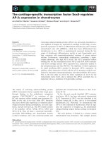
Fig. 4. Ribbon diagram of CPY showing two
structural domains [9]. When the catalytic triad
shown by the CPK model is placed in the
centre of the model, the CPY structure is
divided into a b-sheet rich domain on the
left side and an a-helix rich domain on the
right side.
Ó FEBS 2003 Propeptide of proCPY ensures cooperative unfolding (Eur. J. Biochem. 270) 4591
unfavourable. The T
m
of proCPY was higher by 4 °Cthan
that of Dgly proCPY, indicating that the carbohydrate
moiety exerts a slightly protective effect on the thermal
unfolding of proCPY.
The P
m
value for CPY was higher than that of its
unglycosylated form, though the DV and DG
P
values were
almost the same. The P
m
value of proCPY was higher than
that of its unglycosylated form. This is due to the higher DV
value of the unglycosylated form, according to Eqn (6) (see
Experimental procedures). This is consistent with a more
pronounced conformational change and/or a more pro-
nounced hydration upon unfolding of the unglycosylated
form.
At high pressure, in the glycosylated forms the carbohy-
drate moiety of CPY and proCPY would be hydrated to
compensate the volume contraction and the protein portion
is minimally hydrated. However, in the unglycosylated
forms the protein portion would be directly hydrated to
ensure the corresponding volume contraction. Hence, the
protein portion of the unglycosylated forms would be
more heavily hydrated under high pressure, resulting in
instability.
Thedifferencein<m> values for the glycosylated and
unglycosylated forms of CPY and proCPY at 75–80 °C
(double-headed arrows b, Fig. 2A and a, Fig. 2B, respect-
ively) is caused by the presence of the carbohydrate moiety,
because the <m>forN-acetyl tryptophanamide is increased
by 100 cm
)1
in a 16% glucose solution (T. Maki, M. Kato,
and R. Hayashi, unpublished data). Tryptophan and tyro-
sine residues (Y17, Y20, Y82, W84, and W369) in CPY
would be perturbed by the carbohydrate moiety, thus
increasing their fluorescence, since they are in close proximity
to the carbohydrate-attachment sites, N13, 87, and 368.
In conclusion, the mature enzyme, CPY, unfolds in a
multistate transition, but the precursor, proCPY, unfolds in
a two-state transition, indicating that CPY is composed of
two structural domains, while proCPY would be composed
of a single fragile domain. The propeptide of the proenzyme
would be located at the interface of the two domains thus
combining them into one body, to ensure structural
cooperativity.
Acknowledgements
We are grateful for the technical assistance of C. Valentin in the high-
pressure experiments. The authors are grateful to G. Jung for his initial
work on the construction of the expression plasmid.
References
1. Hayashi, R. (1976) Carboxypeptidase Y. Methods Enzymol. 45,
568–587.
2. Valls, L.A., Hunter, C.P., Rothman, J.H. & Stevens, T.H. (1987)
Protein sorting in yeast: The localization determinant of yeast
vacuolar carboxypeptidase Y residues in the propeptide. Cell. 48,
887–897.
3. Jung, G., Ueno, H. & Hayashi, R. (1999) Carboxypeptidase Y:
Structural basis for protein sorting and catalytic triad. J. Biochem.
126, 1–6.
4. Ramos, C., Winther, J.R. & Kielland-Brandt, M.C. (1994)
Requirement of the propeptide for in vivo formation of active
yeast carboxypeptidase Y. J. Biol. Chem. 269, 7006–7012.
5. Winther, J.R. & Sørensen, P. (1991) Propeptide of carboxy-
peptidase Y provides a chaperonin-like function as well as
inhibition of enzymatic activity. Proc. Natl Acad. Sci. USA 88,
9330–9334.
6. Winther, J.R., Sørensen, P. & Kielland-Brandt, M.C. (1994)
Refolding of a carboxypeptidase Y folding intermediate in vitro
by low-affinity binding of the proregion. J. Biol. Chem. 269,
22007–22013.
7. Hata, T., Hayashi, R. & Doi, E. (1967) Purification of yeast
proteinases. Part III. Isolation and physicochemical properties of
yeast proteinase A and C. Agric. Biol. Chem. 31, 357–367.
8. Aibara, S., Hayashi, R. & Hata, T. (1971) Physical and chemical
properties of yeast proteinase C. Agric. Biol. Chem. 35, 658–666.
9. Endrizzi, J.A., Breddam & K., Remington, S.J. (1994) 2.8-A
˚
structure of yeast serine carboxypeptidase Y. Biochemistry 33,
11106–11120.
10. Hasilik, A. & Tanner, W. (1978) Biosynthesis of the vacuolar
yeast glycoprotein carboxypeptidase Y. Eur. J. Biochem. 85,
599–608.
11. Hashimoto, C., Cohen, R.E., Zhang, W J. & Ballou, C.E. (1981)
Carbohydrate chains on yeast carboxypeptidase Y are phos-
phorylated. Proc.NatlAcad.Sci.USA78, 2244–2248.
12. Trimble, R.B., Maley, F. & Chu, F.K. (1983) Glycoprotein bio-
synthesis in yeast. J. Biol. Chem. 258, 2562–2567.
Table 1. Summary of qualitative thermodynamic parameters for the temperature- and pressure-induced transitions of CPY, Dgly CPY, proCPY and
Dgly proCPY calculated by fluorescence analysis. Errors are within 6%. n.d., not determined.
Peptidase
Temperature-induced transition at 0.1 MPa Pressure-induced transition at 25 °C
T
m
(°C)
DH
(kJÆmol
)1
)
DS
(kJÆmol
)1
ÆK
)1
)
DG
T
(kJÆmol
)1
)
P
m
(MPa)
DV
(mlÆmol
)1
)
DG
P
(kJÆmol
)1
)
CPY 58.6 405 1.22 41.4
First transition < 50, < 50
a
n.d. n.d.
Second transition 345, 334
b
, 194
a
)75.8, )61
b
, )117
a
26.2, 20.6
b
, 22.8
a
Third transition > 500, 492
a
)24.5
a
12.1
a
Dgly CPY 58.4 323 0.97 32.4
First transition < 50 n.d. n.d.
Second transition 302, 282
b
)72.3, ) 80
b
21.9, 22.9
b
Third transition > 500 n.d. n.d.
proCPY 54.5 185 0.57 16.9 253 )42.8 10.8
Dgly proCPY 51.0 176 0.54 12.8 164 )49.3 8.1
a
Obtained at 60 °C.
b
Obtained at 25 °C (Dumoulin et al. [25]).
4592 M. Kato et al. (Eur. J. Biochem. 270) Ó FEBS 2003
13. Shimizu, H., Ueno, H. & Hayashi, R. (1999) Role of carbohydrate
moiety in carboxypeptidase Y: Structural study of mutant enzyme
lacking carbohydrate moiety. Biosci. Biotechnol. Biochem. 63,
1045–1050.
14. Zhou, J.M., Zhu, L. & Balny, C. (2000) Inactivation of creatine
kinase by high pressure may precede dimer dissociation. Eur. J.
Biochem. 267, 1247–1253.
15. Ruan,K.,Xu,C.,Yu,Y.,Li,J.,Lange,R.,Bec,N.&Balny,C.
(2003) The thermodynamic analysis of protein stabilization by
sucrose and glycerol against pressure-induced unfolding. Eur. J.
Biochem. 270, 1654–1661.
16. Ruan,K.,Xu,C.,Yu,Y.,Li,J.,Lange,R.,Bec,N.&Balny,C.
(2001) Pressure-exploration of the 33-kDa protein from spinach
photosystem II particle. Eur. J. Biochem. 268, 2742–2750.
17. Ruan, K., Lange, R., Bec, N. & Balny, C. (1997) A stable partly
denatured state of trypsin induced by high hydrostatic pressure.
Biochem. Biophys. Res. Commun. 239, 150–154.
18. Torrent, J. (2003) Alternative prion structural changes revealed by
high pressure. Biochemistry 42, 1318–1325.
19. Hamaguchi, K. (1992) Physico-Chemical Properties of Amino Acid
Chains. Japan Scientific Society Press, Tokyo.
20. Bec, N., Villa, A., Tortora, P., Mozhaev, V.V., Balny, C. & Lange,
R. (1996) Enhanced stability of carboxypeptidase from Sulfolobus
solfataricus at high pressure. Biotechnol. Lett. 18, 482–488.
21. Lange, R., Frank, J., Saldana, J L. & Balny, C. (1996) Fourth
derivative UV-spectroscopy of proteins under high pressure. I.
Factors affecting the fourth derivative spectrum of the aromatic
amino acids. Eur. Biophys. J. 24, 277–283.
22. Gibson, R.E. & Loeffler, O.H. (1941) Pressure-volume-tempera-
ture relations in solutions. V. The energy-volume coefficients of the
carbon tetrachloride, water and ethylene glycol. J. Am. Chem. Soc.
63, 898–906.
23. Neuman, R.C. Jr,, Kauzmann, W. & Zipp, A. (1973) Pressure
dependence of weak acid ionization in aqueous buffers. J. Phys.
Chem. 77, 2687–2691.
24. Silva, J., Miles, E. & Weber, G. (1986) Pressure dissociation and
conformational drift of the b dimer of tryptophane synthase.
Biochemistry 25, 5780–5786.
25. Dumoulin, M., Ueno, H., Hayashi, R. & Balny, C. (1999) Con-
tribution of the carbohydrate moiety to conformational stability
of the carboxypeptidase Y: High pressure study. Eur. J. Biochem.
262, 475–483.
26. Kunugi, S., Yanagi, Y., Kitayaki, M., Tanaka, N. & Uehara-
Kunugi, Y. (1997) Effects of high-pressure on the activity and
spectroscopic properties of carboxypeptidase Y. Bull. Chem. Soc.
Jpn. 70, 1459–1463.
27. Masson, P. & Cle
´
ry, C. (1996) Pressure-induced molten globule
states of proteins. In High Pressure Bioscience and Biotechnology
(Hayashi, R. & Balny, C., eds), pp. 117–126. Elsevier Science B.V.,
the Netherlands.
28. Ptitsyn, O.B. (1995) Molten globule and protein folding. Adv.
Prot. Chem. 47, 83–229.
29. Trovaslet, M., Dallet-Choisy, S., Meersman, F., Heremans, K.,
Balny, C. & Legoy, M D. (2003) Fluorescence and FTIR study of
pressure-induced structural modifications of horse liver alcohol
dehydrogenase (HLADH). Eur. J. Biochem. 270, 119–128.
Ó FEBS 2003 Propeptide of proCPY ensures cooperative unfolding (Eur. J. Biochem. 270) 4593