Báo cáo khoa học: Phosphorylation of NF-jB proteins by cyclic GMP-dependent kinase A noncanonical pathway to NF-jB activation potx
Bạn đang xem bản rút gọn của tài liệu. Xem và tải ngay bản đầy đủ của tài liệu tại đây (435.52 KB, 12 trang )
Phosphorylation of NF-jB proteins by cyclic GMP-dependent kinase
A noncanonical pathway to NF-jB activation
Bin He
1
and Georg F. Weber
1,2
1
Department of Radiation Oncology, New England Medical Center, Boston, MA, USA;
2
Immunology Program,
Sackler School of Graduate Biomedical Research, Tufts University Medical School, Boston, MA, USA
The transcription factor NF-jB is activated in cellular stress
responses. This requires rapid regulation of its function,
which is accomplished, in part, by various modes of phos-
phorylation. Even though diverse DNA binding subunits of
NF-jB proteins may transactivate from distinct recognition
sequences, the differential regulation of transcription from
the large number of NF-jB responsive sites in various gene
promoters and enhancers has been incompletely understood.
The cyclic GMP-dependent kinase (PKG) is an important
mediator of signal transduction that may induce gene
expression through cAMP response element binding protein
(CREB) and through other, yet undefined, mechanisms. We
have previously characterized a signal transduction pathway
that leads to activation-induced cell death in T-lymphocytes
and involves the activation of PKG. Here we demonstrate
that the NF-jB proteins p65, p49 (also called p52), and p50
are specific substrates for this kinase. PKG dose-dependently
increases the transactivating activity of p65 from the NF-jB
consensus sequence. It also mediates dose-dependently an
increase in transcriptional activity by p49 or p50 from a
unique CCAAT/enhance binding protein (C/EBP)-associ-
ated NF-jB site, but not from the consensus site. Phos-
phorylation of p65, p50, or p49 does not alter their
subcellular distribution. Because the release of cytosolic
p65/p50 heterodimers into the nucleus is by itself insufficient
to differentiate all the numerous NF-jBpromoter
sequences, phosphorylation of the DNA-binding subunits
reveals a form of differential regulation of NF-jB activity
and it implies a novel pathway for PKG-induced gene
transcription. These observations may bear on mechanisms
of programmed cell death in T-lymphocytes. They may also
be relevant to ongoing efforts to induce cancer cell apoptosis
through activation of PKG.
Keywords: protein kinases; signal transduction; superanti-
gens; transcription factors.
The transcription factor NF-jB [1,2] mediates a wide range
of cellular stress responses. It induces the gene expression
of cytokines, acute phase proteins, and adhesion molecules.
In T-lymphocytes, NF-jB regulates activation and activa-
tion-induced cell death [3] and it contributes to T-cell
selection at the double positive stage [4,5]. A large number
of gene promoters and enhancers contain NF-jB binding
sites. This poses the question how selectivity of NF-jB-
dependent transactivation is accomplished during specific
modes of lymphocyte activation. While the release of
p65/p50 into the nucleus is insufficient to differentiate
among diverse binding sites it is clear that there are
mechanisms, which control the interactions of NF-jBwith
its various recognition sequences.
The biologic roles of NF-jB necessitate a rapid activation
of the preformed cytosolic complex, which is typically
accomplished by phosphorylation. NF-jB phosphorylation
is functionally relevant on three levels. Firstly, it targets the
inhibitor I-jB for degradation. Even though phosphoryla-
tion of I-jB is not sufficient to dissociate the complexes of
I-jBandNF-jB in vivo, phosphorylation on serines 32 or
36 is a prerequisite for I-jB degradation in the ubiquitin-
proteasome pathway [6]. Secondly, phosphate residues
contribute to the processing of the p100 and p105 NF-jB
precursor proteins for p50 and p49/p52. A prerequisite for
the proteolytic cleavage of p105 is the phosphorylation of
serines 894 and 908, which are potential recognition sites for
proline-directed serine/threonine kinases, including cyclin-
dependent kinases and Erk2 kinase [7]. The phosphoryla-
tion of the C-terminal region of p105 may be mediated by
cyclic AMP-dependent protein kinase or protein kinase C
[8]. Finally, direct phosphorylation of the DNA-binding
NF-jB subunits can regulate their functions. Phosphoryla-
tion of the DNA-binding NF-jB subunits may modulate
DNA binding affinity, transactivation, or the interaction
with other regulatory proteins. A PKA recognition
sequence within the Rel homology domain, which contains
DNA binding sites and nuclear localization signals, has
been linked to the transformation of avian spleen cells by
v-rel and to the cytoplasmic retention of c-Rel in chicken
embryo fibroblasts [9]. PKA activates NF-jB in a manner
that is independent of I-jB phosphorylation, does not
impair NF-jB interaction with unmodified p105, and
induces transactivation [8,10]. This is accomplished through
the recruitment of CREB-binding protein (CBP)/p300 by
Correspondence to G. F. Weber, Department of Radiation and
Cancer Biology, New England Medical Center, NEMC #824,
750 Washington Street, Boston, MA 02111, USA.
Fax: 617 636 1766, Tel.: 617 636 9013,
E-mail:
Abbreviations: PKA, cyclic AMP-dependent kinase; PKG, cyclic
GMP-dependent kinase.
(Received 20 November 2002, revised 19 February 2003,
accepted 14 March 2003)
Eur. J. Biochem. 270, 2174–2185 (2003) Ó FEBS 2003 doi:10.1046/j.1432-1033.2003.03574.x
phosphorylated p65 [11–13]. Some investigators have not
found p65, p50, or p52 to be phosphorylated by PKA or
PKC [8,14]. While p65 and p50 have candidate recognition
sequences for PKA or PKG, p49/p52 does not have such a
consensus site [9,15], however, related sequences are found
in all three molecules.
The enzyme cyclic GMP-dependent kinase (cGK, PKG)
is an important mediator of intracellular signal transduc-
tion, involved in such diverse processes as the regulation of
blood vessel tone, platelet aggregation, and long-term
potentiation in memory formation [16–18]. Furthermore,
PKG may regulate apoptosis positively [3,19–22] or negat-
ively [23], possibly depending on other modulating bio-
chemical events [24]. Efforts have been made to induce
cancer cell apoptosis with sulindac sulfone (exisulind,
Aptosyn), which activates PKG [25–27]. The down-regula-
tion of PKG levels in neoplastic ovarian epithelial cells [28]
may contribute to enhanced tumor cell survival. Although
PKG, like cyclic AMP-dependent kinase (PKA), can
phosphorylate and activate the transcription factor CREB,
the connection of PKG to gene expression is incompletely
understood. Recently, the localization of the PKG form I in
the cytosol and the nucleus was reported [29,30], suggesting
a broader role for PKG in the regulation of gene transcrip-
tion. Our previous studies [3] suggested that NF-jBmaybe
activated by PKG. Here we demonstrate that p49, p50,
and p65 are substrates for the kinase and we analyze the
mechanisms by which PKG induces NF-jB activation.
Materials and methods
Reagents
Recombinant human NF-jB p49 and p50 were obtained
from Promega. The a-isozyme of cGMP-dependent protein
kinase, purified from bovine lung or recombinant bovine,
was purchased from Promega or Calbiochem.
The following oligonucleotides were utilized in gel shift
reactions after radiolabeling with T4 polynucleotide kinase:
NF-jB consensus (Promega, sense 5¢-AGTTGAGGGGA
CTTTCCCAGGC-3¢), OCT1 (Promega, sense 5¢-TGTCG
AATGCAAATCACTAGAA-3¢), H2K (sense 5¢-GGATC
CCGGTCGGGGGATTCCCCATCTCGG-3¢), j enhan-
cer (sense 5¢-AGCAGAGGGGACTTTCCGAGGC-3¢).
The custom made oligonucleotides were obtained as single
stranded and were annealed to double stranded probes after
phosphorylation with T4 polynucleotide kinase and
[c-
32
P]ATP. Double-stranded poly(dI-dC).poly(dI-dC) was
purchased from Pharmacia. The cGMP-dependent kinase
inhibitor Rp-8-pCPT-cGMPS was purchased from Biolog.
The reporter constructs used in this study included a
commercial NF-jB luciferase reporter (pNF-jB-luc,
Clontech) that contains four consensus NF-jBsites.A
luciferase reporter containing the C-reactive protein
promoter (pC/EBP-luc) and a relevant control with a
mutated NF-jB p50 binding site (pC/EBP-mP50-luc)
were generously provided by D. Samols (Dept Biochem-
istry, Case Western Reserve University, Cleveland, OH,
USA). The plasmids containing human p49, p50, and
p65 were obtained from the NIH AIDS Reagent
Repository. J. Stavnezer generously provided the murine
p50 DNA.
Kinase reaction
The enzymatic activity of cGMP-dependent kinase was
analyzed in kinase reaction buffer (250 m
M
Mes, pH 6.9,
2m
M
EGTA, 5 m
M
magnesium acetate, 50 m
M
NaCl,
10 mgÆmL
)1
BSA, 100 m
M
dithiothreitol, 2 m
M
protein
kinase A inhibitor peptide) with 1 mgÆmL
)1
Kemptide
substrate (LRRASLG) and 1 m
M
[c-
32
P]ATP (30–40
c.p.m.Æpmol
)1
). The reaction was performed in the presence
or absence of 200 l
M
cyclic GMP at room temperature
for 3 min. The reaction was terminated by spotting 50 lL
onto Whatman P-81 filter paper and immediate immers-
ionin10mL75m
M
H
3
PO
4
for 2 min. This was followed
by five washes in 10 mL 75 m
M
H
3
PO
4
, air drying, and
scintillation counting. For the analysis of PKG activity in
cell lyzates, the PKA inhibitor peptide (Sigma) was present.
Phosphorylation of PKG substrates (232 ngÆlL
)1
for
recombinant NF-jB p49 and p50; 500 ngÆlL
)1
for p65)
was performed in 100 m
M
Tris/HCl,pH7.5,20m
M
sodium chloride, 10 m
M
dithiothreitol, 2 m
M
magnesium
acetate, 200 l
M
ATP, at room temperature for 15 min.
The kinase concentration was 3.5 UÆlL
)1
, while cyclic
GMP was present at 200 l
M
. Mops buffer was not used
because it is incompatible with the electrophoretic mobi-
lity shift assay after transfer. There was no loss of
PKG enzymatic activity in Tris buffer at the indicated
concentration.
293T cells (1 · 10
6
per 100 mm diameter Petri dish) were
transiently transfected with 0.3 lg pFLAG-p49 or pRSV-
p65 with CaCl
2
. Twenty-four hours after transfection, the
cells were lyzed in 0.5 mL RIPA buffer (50 m
M
Tris/HCl
pH 7.5, 150 m
M
NaCl, 1% NP-40, 0.5% Na-deoxycholate,
0.1% sodium dodecyl sulfate) and precleared with 30 lL
20% protein A agarose beads overnight. One microgram of
anti-FLAG (mouse IgG) or anti-p65 Ig (rabbit polyclonal
IgG) were added for 2 h at 4 °C followed by pulling-down
with 30 lL 20% protein A agarose beads for an additional
1 h. The agarose beads were pelleted at 14 000 g for 1 min
and washed four times in cold RIPA buffer, then twice in
detergent-free buffer (50 m
M
Tris/HCl pH 7.5, 150 m
M
NaCl). Kinase reaction buffer was directly added to the
pelleted beads with or without PKG and cGMP as indicated
plus 1 lL[c-
32
P]ATP for 15 min at room temperature. The
reaction solutions were then resolved on 8% reducing
denaturing SDS-polyacrylamide gel and transferred to
poly(vinylidene difluoride) (PVDF) membranes for auto-
radiographic exposure and Western blotting.
Electrophoretic mobility shift assay
DNA binding was assessed by electrophoretic mobility
shift according to standard protocols. The reaction mixture
contained 10 m
M
Tris/HCl, pH 7.5, 1 m
M
EDTA, pH 8.0,
35 m
M
NaCl, 50 lgÆmL
)1
poly(dI-dC)Æpoly(dI-dC), and
5% glycerol plus labeled probe. The DNA binding
proteins were transferred from the kinase reaction mixture.
The reaction was incubated at room temperature for
20 min before separation on a native 4% polyacrylamide
gel. For supershift, the appropriate antibodies (0.05–
0.1 lg) were incubated with the nuclear extracts for
10 min at room temperature before adding DNA binding
buffer.
Ó FEBS 2003 Phosphorylation of NF-jB by PKG (Eur. J. Biochem. 270) 2175
Cloning of relevant gene products
To clone mouse PKG Ia cDNA, frozen mouse kidney tissue
was homogenized and total RNA was isolated by using
RNeasy mini kit from Qiagen (Valencia, CA, USA)
following the manufacturer’s protocol. One microgram
of total RNA was used for cDNA synthesis with Super-
script II RNase H
–
reverse transcriptase (Gibco BRL,
USA). The coding sequence of PKG Ia was amplified
with the primers 5¢-AGCATGGGCACCCTGCGGGAT
TTA-3¢ and 5¢-ATTAGAAGTCTATGTCCCAGCCTGA
GTTG-3¢. The amplified product was cloned into the vector
pCR3.1 (Invitrogen Carlsbad, CA) followed by subcloning
into the vector pEF6/His B (Invitrogen, Carlsbad, CA).
Sequence fidelity and accurate reading frame were verified
by DNA sequencing analysis.
Targeted mutations in p65 were generated in positions
276 and 305 by PCR cloning with the Quickchange site
directed mutagenesis kit (Stratagene) according to the
protocol by the manufacturer. The sense mutagenic oligo-
nucleotides used were 5¢-GCGGCGGCCTGCCGACCGG
GAGCTCAGT-3¢ for S276A and 5¢-AAACGTAAAAG
GGCATATGAGACCTTCAAGAGCATC-3¢ for T305A
(mutations in bold). The accuracy of the mutations was
confirmed by DNA sequencing.
p49 (obtained from the NIH AIDS Reagent Repository)
was Flag-tagged at the 5¢-end by PCR using the pri-
mers 5¢-CTGCAGCATGGACTACAAGGACGACGA
TGACAAGGAGAGTTGCTACAACCCAGGTCTG-3¢
and 5¢-GAGAGTTGCTACAACCCAGGTCTG-3¢
with pRSV-p49 as a template. The amplified fragment
was cloned into the vector pCR3.1 and sequence fidelity was
confirmed by DNA sequencing.
Reporter gene assays
293T cells were plated at 1 · 10
6
cells per 100 mm
diameter Petri dish and were grown for 24 h before
transfection with CaCl
2
. The commercial pNF-jB-luc
reporter (Clontech, Palo Alto, CA, USA) contains four
NF-jB response elements and was used at 0.5 lgper
transfection (to amplify the signal for the analysis of
transactivation by endogenous p65, we used 2 lgof
reporter DNA). The common internal transfection
standard Renilla in pRL-SV40 (10 ng per transfection)
served as a control for transfection efficiency. This
reporter construct was not sensitive to cotransfection of
PKG or NF-jB and provided stable reference values.
Renilla was not used in transfection experiments with the
noncommercial reporter constructs because the lumines-
cence intensity is too high compared to the specific
readout. Twenty-four hours after transfection, the cells
were harvested in 1 mL reporter lysis buffer (Promega)
and dual luciferase reporter assays were performed
following the protocol provided by the manufacturer.
Lyzates were diluted 1 : 40 and 10 lLwereusedfor
measurement in a luminometer (Turner Designs TD-20/20).
In reporter gene experiments without Renilla, lumine-
scene was measured in 40 lg (total protein) of lyzate. The
protein concentrations were determined by the BCA
protein assay reagent kit (Pierce). As confirmation of
protein expression, 20 lg of the same lyzates were also used
for separation on 8% SDS-polyacrylamide gels followed by
Western blotting on PVDF membranes. PKG kinase
activity in the lyzates was confirmed by phosphorylation
of the standard substrate LRRASLG (ÔKemptideÕ)where
indicated.
Western blotting
Cells were lyzed in RIPA (50 m
M
Tris/HCl pH 7.5,
150 m
M
NaCl, 1% NP-40, 0.5% Na-deoxycholate, 0.1%
sodium dodecyl sulfate) or NTEN buffer (20 m
M
Tris/
HCl, pH 8.0, 120 m
M
NaCl, 0.5% NP40) containing
1m
M
phenylmethanesulfonyl fluoride, 10 lgÆmL
)1
pep-
statin, and 1 m
M
dithiothreitol. The lyzates were centri-
fuged at 15 000 g for 5 min and the protein
concentration was determined in the supernatants.
Twenty micrograms of total protein were resolved on
reducing denaturing SDS/polyacrylamide gels and trans-
ferred to PVDF membranes. The membranes were
probed with appropriate antibodies followed by horse-
radish peroxidase-conjugated secondary antibodies and
development using enhanced chemiluminescence. Anti-
p65 Ig (C-terminus, rabbit polyclonal), anti-p50 Ig
(rabbit antiserum), and anti-p52 Ig (rabbit antiserum,
used to detect p49) were obtained from Upstate
Biotechnology (Lake Placid, NY, USA). Anti-PKG Ig
(C-terminal, amino acids 657–671, rabbit polyclonal) was
purchased from Calbiochem (La Jolla, CA, USA).
Reprobing of the membranes with anti-tubulin Ig
(mouse IgG isotype, Sigma) served as additional loading
control.
For pull-down assays, transiently transfected 293T cells
were lyzed in 0.5 mL NTEN buffer. The lyzates were
precleared at 4 °Cwith25lL 20% agarose beads, before
addition of 0.5–1 lg of the indicated antibody for 2 h and
precipitation with 25 lL 20% agarose beads for 1 h. The
beads were washed, resuspended in SDS/PAGE sample
buffer, and the bound proteins were resolved on 8% SDS/
polyacrylamide gels. The proteins of interest were detected
by Western blotting.
Cells
Reporter assays were performed by transient transfections
of 293T cells. O3 is a CD4
+
T-helper cell clone derived
from BALB/c mice after in vitro selection for proliferation
to ovalbumin in association with BALB/c antigen-present-
ing cells [31]. O3 cells express V
b
6 and respond to
conventional antigen (ovalbumin) and to the retroviral
superantigen MTV-7 (Mls-1
a
) [32]. The AF3.G7 hybri-
doma was generated by fusing beef insulin immune
C57BL/6 lymph node cells with the BW5147 thymoma
line. It bears V
b
6andV
a
3.2 and responds to MTV-7
according to interleukin-2 production [33]. After stimula-
tion by conventional antigen or superantigen, the T-cells
were obtained by passage through Cell-ect
TM
columns
(Biotex Laboratories Inc., Edmonton, Alberta, Canada)
for the preparation of nuclear extracts. As control for
activation, measurement of
3
H-thymidine incorporation by
O3 cells after stimulation with mitomycin C-treated LBB
cells as antigen-presenting cells was performed as described
previously [32].
2176 B. He and G. F. Weber (Eur. J. Biochem. 270) Ó FEBS 2003
Results
NF-jB is activated by PKG in T-lymphocytes
T-lymphocytes proliferate in response to engagement of
their antigen receptor by conventional antigen peptide. We
have previously described an alternative signal transduc-
tion pathway, associated with the T-cell antigen receptor
that is induced by superantigen and leads to activation of
PKG and activation-induced cell death [3]. Because
T-lymphocyte stimulation is often characterized by NF-jB
translocation to the nucleus and binding to cognate DNA
sequences, we compared the induction of NF-jB following
stimulation by conventional antigen or retroviral super-
antigen in the T-cell clone O3. Consistent with previous
observations [34], the induction of NF-jB by physiologic
T-lymphocyte stimulation is moderate. In electrophoretic
mobility shift assays measuring the binding to a NF-jB
consensus probe, the larger and smaller NF-jB complexes
were induced by the conventional antigen. In contrast,
predominantly the smaller complex, represented by the
lower band on the gel, was induced by stimulation with
superantigen. This was not due to quantitative differences
in stimulation because both modes of T-cell activation
induced comparable levels of tritiated thymidine incorpor-
ation in the same experiment (Fig. 1A). To confirm the
dependence of the superantigen mediated induction of
NF-jB, we treated the O3 clones with the cell permeable
PKG inhibitor Rp-8-pCPT-cGMPS before stimulation.
Because superantigen induces predominantly the faster
migrating band the gel shift analysis was performed with
the H2K probe, which has a higher affinity to p50/p52
than the consensus probe. Expectedly, the presence of the
PKG inhibitor suppressed the superantigen-dependent
induction of NF-jB but had no effect on T-cell stimulation
by conventional antigen. To test whether PKG could
induce the lower NF-jB band, we treated nuclear extracts
from resting O3 T-cell clones or AF3.G7 T-cell hybridoma
cells with PKG and cGMP and found it to induce DNA
binding of the lower band to a NF-jB consensus
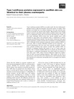
Fig. 1. Correlation between PKG activity and NF-jB induction in
T-lymphocyte activation. (A,leftpanel)TimecourseofNF-jB
induction after activation of O3 T-cell clones. O3 cells were stimulated
by the conventional antigen ovalbumin (OVA) or retroviral super-
antigen (MTV-7) for 0, 2, or 4 h. The T-cells were selected for pre-
paration of nuclear extracts, which were then subjected to incubation
with a
32
P-labelled NF-jB consensus probe followed by electropho-
retic mobility shift assay. The relative intensity of the slower and faster
migrating bands after stimulation with MTV or OVA was quantitated
by densitometric measurement. The relative density units for the lower
band are O3 7.0, OVA 2 h 9.8, OVA 4 h 12.8, MTV 2 h 7.9, MTV 4 h
9.7.TherelativedensityunitsfortheupperbandareO36.9,OVA2h
10.5, OVA 4 h 13.3, MTV 2 h 5.5, MTV 4 h 6.7. Comparable levels of
T-cell stimulation were confirmed by tritiated thymidine incorporation
(c.p.m.). (A, right panel) Before stimulation for 4 h, the O3 cells were
pretreated with the inhibitor Rp-8-pCPT-cGMPS. After T-cell selec-
tion, nuclear extracts were prepared and analyzed by gel shift assay for
binding to the H2K probe. (B) Nuclear extracts from the AF3.G7
hybridoma or the O3 clone were phosphorylated in vitro by PKG plus
cGMP followed by analysis of DNA binding to an oligonucleotide
containing the H2K sequence in gel shift assays. The nuclear extract
from O3 cells that had been treated with plate-bound anti-CD3e
antibody served as a positive control for induction of the faster
migrating NF-jB band. Note that the resting levels of NF-jBbinding
are relatively high in O3 cells, because the clone depends on the pre-
sence of interleukin-2 in the growth medium, whereas the hybridoma
AF3.G7 does not. (C) The induction by PKG plus cGMP of DNA
binding by AF3.G7 cytosol is inhibitable by addition of high con-
centrations of a competing PKG substrate peptide, GRTGRRNSI
(ÔPKI substrateÕ,amountsarel
M
). In addition, cGMP and PKG did
not affect Octamer-1 binding in the same experiment (not shown).
Similar results were obtained in at least three additional experiments.
Ó FEBS 2003 Phosphorylation of NF-jB by PKG (Eur. J. Biochem. 270) 2177
oligonucleotide (Fig. 1B). A similar induction was seen in
AF3.G7 cytosol and this was inhibitable by increasing
amounts of the competing PKG substrate peptide
GRTGRRNSI (ÔPKI substrateÕ) (Fig. 1C). We therefore
set out to investigate the role of PKG in the induction of
NF-jB.
PKG increases the transactivating activity of NF-jB
proteins from distinct recognition sites
We asked whether PKG can alter the transactivating activity
of p65 as judged by luciferase assays with a commercial NF-
jB luciferase reporter that contains four consensus NF-jB
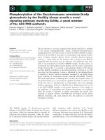
Fig. 2. PKG increases the transactivating activity of p65 from consensus sites but not from a nonconsensus NF-jBreporter.(A) 293T cells were
transiently transfected with 300 ng pRSV-p65 and increasing amounts of pEF6/HisB-PKG as indicated. Transactivation from the cotransfected
pNF-jB-luc reporter (500 ng) was measured by luciferase activity. The values are normalized to luminescence induced by 10 ng cotransfected
Renilla construct pRL-SV40 and the results (fold induction) are presented as mean ± standard deviation of three samples (top panel). The protein
expression of the transfected molecules correlated with the amounts of DNA introduced into the cells, while tubulin (used as a loading control)
remained constant (bottom panel). The PKG activity in 20 lg of cell lyzates, as judged by phosphorylation of LRRASLG peptide (ÔkemptideÕ)
in vitro, reflected the amounts of PKG transfected. The kinase activity is indicated as mean ± standard deviation (middle panel). (B) 293T cells
were transiently cotransfected with pC/EBP-wt-luc reporter (1 lg) and pRSV-p65 (0.3 lg) with or without 3 lg pEF6/HisB-PKG. Transfection of
0.5 lg pRSV-p50 with pC/EBP-wt-luc reporter (1 lg) served as a positive control. Twenty-four hours after transfection, the cells were harvested in
reporter lysis buffer. Forty micrograms of lyzate samples were used for luciferase assays by luminometer and the values obtained for the vector
control group were normalized to 1. The results represent mean ± standard deviation of triplicate samples (top panel). Similar results were
obtained in three independent experiments. Twenty micrograms of lyzates were used for Western blotting to confirm the expression levels of the
transfected proteins (bottom panel). (C) 293T cells were transiently transfected with increasing amounts of PKG in conjunction with either 2 lg
NF-jB consensus reporter or 2 lg pC/EBP-wt-luc reporter. Twenty-four hours after transfection, the cells were harvested in reporter lysis buffer.
Luciferase activity was measured in 40 lg of lyzate samples and the values obtained for the vector control group were normalized to 1. The results
represent mean ± standard deviation of triplicate samples.
2178 B. He and G. F. Weber (Eur. J. Biochem. 270) Ó FEBS 2003
sites. Cotransfection of increasing amounts of PKG dose-
dependently enhanced the transactivation by transfected
p65. The results were consistent with the protein expres-
sion levels and kinase activities in the cell lyzates
(Fig. 2A). Therefore, PKG increases the transactivating
ability of p65.
We also performed cotransfection experiments with a
reporter that contains a nonconsensus NF-jB binding site.
Consistent with earlier reports [35–37], p65 does not
transactivate the nonconsensus motif associated with
C/EBP, which is found in the C-reactive protein promoter.
In this case, the lack of transactivation is not overcome
by cotransfection of PKG (Fig. 2B).
We noted that the transfection of PKG alone was
sufficient to increase the consensus reporter activity by
approximately fourfold (compare Fig. 2A). We therefore
tested whether PKG could stimulate transactivation by
the endogenous NF-jB. 293T cells express substantial
amounts of endogenous p65, but very little p49 and p50.
Consistently, transfected PKG increased the activity of
the consensus reporter, but not of the C/EBP-associated
promoter, in a dose-dependent manner, reaching
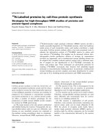
Fig. 3. Transactivation by p50/p49 from a nonconsensus sequence is
enhanced by PKG. (A) Transactivation by transfected p50 of a luci-
ferase reporter gene containing a nonconsensus NF-jBsitethat
overlaps with a C/EBP site (sequence in top panel). 293T cells were
transiently cotransfected with 1 lg pC/EBP-wt-luc reporter (or the
control construct pC/EBP-mp50-luc, in which the p50 binding site is
mutated, represented as pC/EBP (mP50)) with 0.5 lgpRSV-p50and
the indicated amounts of pEF6/HisB-PKG. Twenty-four hours after
transfection, the cells were harvested in reporter lysis buffer and 40 lg
lyzate samples were used for luciferase assays. The results, measured as
fold induction, are presented as mean ± standard deviation of three
samples and the values obtained for the vector control group have been
normalized to 1. The transcriptional activity is induced dose-depend-
ently by cotransfection of increasing doses of PKG (second panel from
top). Twenty micrograms of the lyzates were analyzed for PKG kinase
activity (third panel from top), while another 20 lg of lyzates were
used for Western blotting to confirm the protein expression levels
(fourth panel). PKG activity in cell lyzate and Western blotting for the
transfected molecules served as transfection controls. (B) PKG
enhances p49-mediated transactivation from the C/EBP-associated
nonconsensus site. 293T cells were transiently cotransfected with 1 lg
pC/EBP-wt-luc reporter and 0.5 lgpRSV-p49plusincreasing
amounts of pEF6/HisB-PKG for analysis of luciferase reporter gene
activity. The results are represented as fold induction and the values
obtained for the vector control group are normalized to 1. Western
blotting confirmed the expression levels of the transfected molecules
(bottom panel). (C) Neither p49 (top panel) nor p50 (bottom panel)
transactivate from the commercial NF-jB reporter gene containing
four consensus sites. 293T cells were transiently cotransfected with
0.5 lgpNF-jB-luc plus 0.3 lgp49or0.3lgpRSV-p50plusincreas-
ing amounts of pEF6/HisB-PKG. 10 ng of the Renilla construct pRL-
SV40 was also cotransfected to normalize the data for transfection
efficiency. Transactivation by p65 (0.3 lgpRSV-p65)inthesame
experiment is shown as a positive control (at the chosen concentrations
of reporter DNA, PKG induces a less than twofold induction of
transactivation by endogenous p65). Expression of the transfected
proteins was confirmed by Western blotting. All panels show the
results of one representative experiment from at least three replicates.
Ó FEBS 2003 Phosphorylation of NF-jB by PKG (Eur. J. Biochem. 270) 2179
approximately fivefold increase in luminescence readout
(Fig. 2C).
The NF-jB protein p50 contains a DNA binding
domain, but no transactivation domain. Nevertheless,
transactivation may be observed after transfection of p50
into cells, presumably due to its binding to endogenous
interaction partners. Those include most prominently p65,
but also Bcl-3 [38]. In addition, p50-dependent transactiva-
tion can occur from a nonconsensus site in conjunction with
C/EBP [35,36]. We used the nonconsensus reporter con-
struct in transient cotransfection assays. No reporter activity
was induced by PKG alone, whereas p50 dose-dependently
increased the luciferase activity (data not shown).
Co-transfection of PKG with low amounts of p50 (0.3 lg
DNA) dose-dependently enhanced its transactivating acti-
vity (Fig. 3A), consistent with an increased affinity of p50 to
this DNA sequence after phosphorylation by PKG. Com-
parable results were obtained with murine p50 (data not
shown). P49 and p50 are related NF-jB subunits [39]. We
found p49 to also transactivate from the nonconsensus
NF-jB site in a manner that could be increased dose-
dependently by cotransfected PKG (Fig. 3B).
We then tested whether p50 or p49 transactivate the
luciferase reporter that contains four NF-jB consensus sites
and whether transactivation under these conditions might
be modulated by PKG. Luciferase activity was not induced
by transfection of p49 or p50 alone (under the conditions
used here, PKG enhances the transactivation by endo-
genous p65 less than twofold). Furthermore, cotransfection
of p50 or p49 with increasing amounts of PKG did not lead
to measurable transactivation from the NF-jB consensus
luciferase reporter (Fig. 3C).
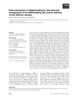
Fig. 4. The NF-jB proteins p49, p50, and p65
are substrates for cGMP-dependent kinase.
(A) Substrate phosphorylation of p49 or p50
depends on the presence of PKG and is
enhanced by the addition of cGMP, whereas
cGMP in the absence of the kinase does not
mediate measurable incorporation of phos-
phate. Autophosphorylation of PKG is rep-
resented as the upper band on all gels and
reflects a specificity control for effects on the
enzyme. (B) Phosphorylation of p49 or p50 by
PKG (as well as PKG autophosphorylation) is
reversible by titration of a competing substrate
peptide for the kinase (GRTGRRNSI), but
not a control peptide with a mutated serine
(GRTGRRNAI). (C) An analog of cGMP,
Rp-8-pCPT-cGMPS, which can act as an
inhibitor of PKG, reverses the enzymatic
phosphorylation of p49 or p50. Consistent
with the competition for binding to the kinase
between cGMP and Rp-8-pCPT-cGMPS, the
inhibition is more complete in the absence of
cGMP than in its presence. Autophosphory-
lation of PKG serves as a positive control for
kinase activity. (D) Recombinant His-tagged
p65 was phosphorylated by PKG in vitro in
the absence or in the presence of cGMP (top
panel). 293T cells were transiently transfected
with p65 or FLAG-tagged p49. The cells were
lyzedinRIPAbufferandthetransfected
molecules were pulled down by antibodies to
the p65 or Flag. Kinase reaction buffer plus
[c-
32
P]-ATP was directly added to the pelleted
beads with or without PKG and cGMP for
15 min at room temperature. The reaction
mixtures were analyzed by autoradiography
and Western blotting (bottom panel).
2180 B. He and G. F. Weber (Eur. J. Biochem. 270) Ó FEBS 2003
In summary, the transactivation experiments using
reporter assays indicated that PKG can enhance the
transcriptional activity of the DNA binding NF-jBproteins
p65, p50, and p49 from their cognate recognition sites. In
contrast, PKG does not confer transactivating potential
from the, respectively, noncognate NF-jB sequences.
Because the proteins, p49, p50, and p65 direct transcription
from distinct DNA sequences, their activation by PKG
enhances their differential effects.
P49, p50, and p65 are substrates for cyclic GMP-
dependent kinase
A possible mechanism to account for PKG-enhanced
transactivation by NF-jB is the phosphorylation of the
DNA binding proteins by the enzyme. We therefore tested
whether p49, p50, and p65 are substrates of the kinase. NF-
jB p49 and p50 were phosphorylated by cyclic GMP-
dependent kinase and the phosphorylation levels were
enhanced by addition of cyclic GMP (Fig. 4A). The
substrate peptide GRTGRRNSI (ÔPKI substrateÕ), but not
the control peptide GRTGRRNAI, inhibited phosphory-
lation of p49 or p50 as well as autophosphorylation of
cGMP-dependent kinase in a dose-dependent manner
(Fig. 4B). The kinase reactions on p49 and p50 were also
inhibited by the cGMP-dependent kinase inhibitor Rp-8-
pCPT-cGMPS at a high concentration. Consistent with the
competitive function of the compound, the inhibition was
complete in the absence of cGMP but partial in the presence
of cGMP (Fig. 4C). There is substantial substrate overlap
between PKA and PKG. Cyclic AMP-dependent kinase
also phosphorylated p50 with comparable efficiency, but
p49 was phosphorylated strongly by cGMP-dependent
kinase and only very weakly by PKA (data not shown).
Fig. 5. PKG binds NF-kB proteins. (A) 293T cells were transfected
with vector or p65, or were cotransfected with NF-jBp65plusPKG.
Alternatively, p49 was transfected with or without PKG. The cells
were lyzed in NTEN buffer. After preclearing, immunoprecipitation
was performed with anti-p65 antibody or anti-Flag antibody (for pull-
down of p49). The immunoprecipitates and 10% of the input were
resolved on SDS/PAGE and the resulting Western blot was probed
with antibodies to p65, to p52 (recognizes p49), and to PKG.
Detectable levels of endogenous p65 are expressed in 293T cells and are
recognized by the specific antibody, accounting for the band on the p65
blot from untransfected cells. Similar results were obtained in a repeat
experiment. No bands were detected with control immunoglobulin or
in a sample without cell lyzate. (B) 293T cells were transiently trans-
fected by calcium phosphate precipitation with vector control or 3 lg
pEF6-PKG.After24 h,thecellswerelyzedinNTENbuffer.PKGwas
pulled down, the bound proteins were resolved on SDS/PAGE, and
the Western blot was probed with antibodies to PKG and to p65. The
input (2%) is shown in the two left lanes. The two right lanes show the
no cell control and the pull-down with an irrelevant antibody,
respectively. (C) 293T cells were transiently transfected with 3 lg
vector, pRSV-p50, pRSV-p49, or pRSV-p65. After 24 h, the cells were
lyzed for immunoprecipitation. The left panel (Input) shows the
Western blots of whole cell lyzates as a control for transfection effi-
ciency. The right panel (IP) coimmunoprecipitated PKG (top row) and
precipitated or coprecipitated p65 (second row). The immunoglobulin
heavy chain interferes with the detection of pulled-down p50 or p49.
Therefore, the successful immunoprecipitation in these cases was
confirmed by Western blotting for the endogenous precursor proteins
p100, which is recognized by anti-p52 antiserum, and p105, which is
recognized by antibody to p50 (bottom rows). Immunoprecipitation
with an irrelevant antibody and immunoprecipitation without cells
served as negative controls (two right lanes).
Ó FEBS 2003 Phosphorylation of NF-jB by PKG (Eur. J. Biochem. 270) 2181
Similar to p49 and p50, bacterial recombinant His-tagged
p65 was phosphorylated by PKG in vitro and phosphate
incorporation was enhanced by the presence of cGMP
(Fig. 4D, top panel). We expressed p49 and p65 by transient
transfection in 293T cells. P65 was immunoprecipitated with
an anti-p65 Ig and p49 was immunoprecipitated with
an antibody to Flag-tag. The pulled-down proteins were
phosphorylated by PKG in vitro.BothNF-jB subunits
incorporated radioactive phosphate, although the phos-
phorylation of p65 was substantially weaker than the
phosphorylation of p49 (Fig. 4D, bottom panel).
We further confirmed the interaction between p65 or p49
and PKG in vivo by coimmunoprecipitation of the kinase
with an antibody to either p65 or Flag-tag (for p49) after
cotransfection of the kinase with either of the NF-jB
proteins (Fig. 5A). Because 293T cells express substantial
levels of NF-jB p65, we tested whether transfected PKG
could pull down endogenous p65. The immunoprecipitation
with anti-PKG antibody efficiently yielded endogenous as
well as transfected PKG. In both cases, endogenous NF-jB
p65 was bound, and the band intensity on Western blot
correlated to the amount of PKG present (Fig. 5B). To
extend this analysis, we transfected 293T cells with p65, p50,
or p49, immunoprecipitated with antibodies specific to the
transfected gene products, and probed for pulled-down
endogenous PKG. In all cases, coimmunoprecipitation of
substantial amounts of PKG was detected. Due to the high
expression levels of p65 in 293T cells, the antibody to p65
also pulled-down PKG from untransfected cells. Not
unexpectedly, the immunoprecipitations with anti-p50 and
anti-p52 Ig pulled down endogenous p65, suggesting the
possibility of a trimeric complex containing PKG, p65, and
p50/p49 (Fig. 5C).
The phosphorylation of p65 occurs on nonconsensus
sites
P65 is a substrate for PKA [11], an enzyme, whose substrate
specificity is similar to PKG. Likely recognition sites for
both enzymes are in positions serine 276 and threonine 305
on p65, and serine 276 has been demonstrated to be
phosphorylated by cyclic AMP-dependent kinase [11]. We
mutated both candidate phosphorylation sites. Phosphory-
lation of the serine in position 276 is known to be essential
for p65 dependent transactivation [11]. In accord with these
previous observations, the transactivating activity was
diminished moderately by the mutation T305A and sub-
stantially by S276A, however, cotransfection of PKG led
to comparable dose-dependent increases in reporter activity
in all cases (Fig. 6A). Consistent with this observation,
synthetic peptides covering the threonine 305 (EKRKRT
YETF)ortheserine276(MQLRRPSDRE) did not
incorporate radioactive phosphate during incubation with
the kinase, whereas the standard substrate peptide
LRRASLG (ÔKemptideÕ) did (Fig. 6B). We also phospho-
rylated bacterial recombinant p65-His [40] with PKG
in vitro and were able to compete the kinase reaction with
the substrate peptide GRTGRRNSI, but not with the
peptides covering threonine 305 or serine 276 (Fig. 6C).
These results suggest the hypothesis that PKG phos-
phorylates p65 in positions distinct from the amino acids
276 and 305.
Fig. 6. The phosphorylation of p65 by PKG does not occur on the
consensus recognition sites. (A) 293T cells were transiently cotrans-
fected with 0.5 lgpNF-jB-luc and 0.3 lgpRSV-p65oritsmutants
T305A or S276A plus increasing amount of pEF6/HisB-PKG.
Co-transfected 10 ng of the Renilla construct pRL-SV40 served as a
control for transfection efficiency. Twenty-four hours after transfec-
tion, the cells were harvested in reporter lysis buffer and 10 lLof1 : 40
diluted lyzates were assayed for luciferase activity. The data are pre-
sented as fold induction with the values obtained from the vector
(pEF6/HisB) transfected cells normalized to 1. The inset shows the
PKG-dose dependent increase in transactivation by p65S276 A on an
adjusted scale. (B) Ten micrograms of the synthetic peptides EK-
RKRTYETF (T305p65), MQLRRPSDRE (S276p65), or Kemptide
were incubated with or without 1 U PKG plus 200 l
M
cGMP plus
1m
M
[c-
32
P]ATP in total volume of 100 lL for 3 min at room tem-
perature. One half of the reaction volume was spotted onto filter paper
and the reaction was stopped by washes in phosphoric acid. The levels
of peptide phosphorylation were determined by scintillation counting
and the results are presented as mean values ± standard deviation of
three samples. (C) Recombinant p65 (10 lgÆmL
)1
) was phosphoryl-
ated by 1 U PKG in kinase reaction buffer at room temperature for
15 min, in the presence or absence of increasing concentrations of the
synthetic peptides GRTGRRNSI (ÔPKI substrateÕ), or EKRKRT
YETF (T305p65), or MQLRRPSDRE (S276p65). The extent of p65
phosphorylation and PKG autophosphorylation were determined by
resolution on reducing denaturing 8% SDS/polyacrylamide gel fol-
lowed by autoradiography.
2182 B. He and G. F. Weber (Eur. J. Biochem. 270) Ó FEBS 2003
Phosphorylation of p50 by PKG directly impacts
its DNA binding characteristics
Changes in transactivating activity may reflect alterations
in DNA binding affinity. We studied the effects of PKG
on oligonucleotide binding by NF-jB proteins in elec-
trophoretic mobility shift assays. Cyclic GMP-dependent
kinase is inactive under standard gel shift assay condi-
tions. Conversely, gel shift assays cannot be performed in
the PKG reaction buffer. We therefore adjusted the kinase
reaction buffer so that we could phosphorylate NF-jB
proteins and then transfer an aliquot to the standard
DNA binding buffer for analysis of phosphorylation-
dependent changes in the DNA binding characteristics.
Phosphorylation by cGMP-dependent kinase did not
affect the binding of p49 or p50 to the H2K probe, to
which these proteins already have high affinity without
being phosphorylated [41]. In contrast, in vitro binding of
recombinant p50 to the NF-jB consensus sequence or to
the nonconsensus NF-jB-C/EBP sequence was increased
by PKG (Fig. 7A,B). The binding affinity of nuclear
extracts from 293T cells transiently transfected with p50
and PKG to the same probes was similarly increased
(Fig. 7C,D). The specificity of the main DNA binding
band from transfectants of p50 or p50 plus PKG was
confirmed by supershift (Fig. 7D).
The subcellular localization of NF-jB subunits
is not affected by phosphorylation with PKG
NF-jB is an inducible transcription factor, which is retained
in the cytosol in resting cells. It was therefore possible that
the phosphorylation of DNA binding subunits might affect
their nuclear import as would be reflected in their subcel-
lular distribution. Transfection of increasing amounts of
PKG did not alter the relative fractions of cotransfected
p49, p50, or p65 in the cytosols and nuclei. Although not
definitive, these observations made phosphorylation-
induced changes in the transport and half-lives of these
NF-jB subunits unlikely (Fig. 8).
Discussion
The incorporation of phosphate into p49 or p50 depends on
PKG, is enhanced by cGMP, can be competed by a PKG
substrate peptide but not by a control peptide, and is
inhibitable by a cGMP analog with inhibition being more
efficient in the absence of cGMP than in its presence. The
sum of these observations indicates that NF-jB p49 and p50
are specific substrates for PKG. Similarly, phosphorylation
of p65 is dependent on the kinase, is increased in the presence
of cGMP, and is competed out by a standard substrate
peptide. Cyclic GMP-dependent kinase, like cyclic AMP-
dependent kinase, has a preference for the phosphorylation
of serines or threonines found close to at least two
consecutive N-terminal basic residues. The standard PKG
recognition site is (R,K)(R,K)X(S,T). It is important to note,
however, that there are a number of exceptions to this rule.
The p50 precursor p105 has PKG recognition sites in
positions 335 and 940 (GenBank accession numbers M57999
or NM_003998). Although p49 (GenBank accession number
A57034) does not contain any PKG consensus sites, there are
five similar sites with the sequence X(R,K)X(S,T) in p49
(amino acids 76, 195, 201, 231, 430) that may conceivably
serve as candidate recognition motifs. Interestingly, we have
found p49 to be a poor substrate for PKA. The consensus
sites for PKG in p65 are at positions 276 and 305 (GenBank
accession number M62399). The phosphorylation of amino
Fig. 7. PKG dependent phosphorylation of p50 increases its binding affinity to consensus and nonconsensus (jB-C/EBP) sequences. (A and B)
Recombinant p50, 25 ng per sample, were phosphorylated at room temperature for 15 min by 1 U purified PKG with or without 200 l
M
cGMP in
total volume of 10 lL. The reaction mixtures were transferred to DNA binding buffer and incubated for additional 20 min with the indicated
32
P-labeled probes. The reactants were resolved on native 4% polyacrylamide gels and exposed to autoradiography film. (C and D) 293T cells were
transiently transfected with 0.3 lg pRSV-p50 with or without 3 lg pEF6/HisB-PKG. Twenty-four hours after transfection, the cells were har-
vested, washed in NaCl/P
i
, and nuclear extracts were prepared. Ten micrograms of nuclear protein was used for electrophoretic mobility shift assay
with radiolabeled NF-jB consensus or OCT1 oligonucleotides (C), or with radiolabeled jB-C/EBP oligonucleotides (D). The identity of the major
DNA-binding band was confirmed by supershift with 50 ng anti-p50 antiserum, added to the nuclear extracts at room temperature for 10 min
before DNA binding (D). The upper arrow indicates the supershifted band.
Ó FEBS 2003 Phosphorylation of NF-jB by PKG (Eur. J. Biochem. 270) 2183
acid 276 by PKA is of major importance for p65 transcrip-
tional activity. Remarkably, our experiments indicate that
PKG phosphorylates p65 in positions other than the two
consensus sites. The differential phosphorylation of p49 by
PKG but much less by PKA, and the phosphorylation of p65
by PKA and PKG on distinct residues implies multiple
modes of regulation of NF-jB function. It also indicates that
the position, in which phosphorylation occurs, is an import-
ant determinant for transactivation.
Recognition sequences for NF-jB have been identified in
the promoters of a large number of genes. Beside the
consensus sequence, additional NF-jB binding sites are
known and may differ in their affinities to various subunits
of the transcription factor. The release of cytosolic p65/p50
heterodimers into the nucleus is by itself insufficient to
differentiate among the numerous NF-jBpromoter
sequences. Our results suggest a mechanism of differential
transactivation of various NF-jB dependent genes by
phosphorylation of the DNA-binding subunits. Phosphory-
lation of p65 by PKG induces transcription from the
consensus sequence, whereas phosphorylation of p50 or p49
induces transcription from a C/EBP-associated nonconsen-
sus site, but not from the consensus sequence. PKG 1 has
been shown to be located in the cytosol as well as in the
nucleus [29]. It may therefore exert its regulatory role before
or after translocation of p65 and p50 to the nucleus.
Beside the well-studied induction of CREB, there has
been little evidence for the activation of transcription factors
by cGMP-dependent kinase. Transfection of BHK cells
with PKG causes transactivation of the c-fos promoter,
mediated most notably through SRE, FAP, and CRE [42].
TheconnectionofPKGandNF-jB may have broad
implications, as a pathway that involves phospholipase A
2
,
hydroxyl radical, guanylate cyclase, and PKG has been
mapped to T-cell signal transduction leading to apoptosis [3]
as well as to long-term potentiation in the central nervous
system [18]. Cellular NF-jB activation by H
2
O
2
has been
shown to involve protein phosphorylation and therefore to
be mediated indirectly via induction of a kinase [8]. The
cGMP-dependent kinase may thus provide a link for
the NF-jB activation by reactive oxygen species because the
activity of guanylate cyclase, which synthesizes the cofactor
cGMP, can be regulated by redox reactions.
Acknowledgements
This study was supported by National Institutes of Health research
grant CA76176 and Department of Defense breast cancer grant
DAMD17-98-1-8060 to G. F. W. The authors are indebted to Ranjan
Sen for substantial helpful discussions throughout the project. Janet
Stavnezer generously provided the murine p50 DNA. The plasmids
containing human p49, p50, and p65 were obtained from the NIH
AIDS Reagent Repository. The reporter constructs for the noncon-
sensus p50 site were a generous gift from David Samols. Thomas
Maniatis generously provided the purified recombinant p65. We thank
Peter Brodeur for critically reading the manuscript.
References
1. Sen, R. & Baltimore, D. (1986) Multiple nuclear factors inter-
act with the immunoglobulin enhancer sequences. Cell 46, 705–
716.
2. Sen, R. & Baltimore, D. (1986) Inducibility of j immunoglobulin
enhancer-binding protein NF-jB by a posttranslational mechan-
ism. Cell 47, 921–928.
3. Weber, G.F., Abromson-Leeman, S. & Cantor, H. (1995) A sig-
nalling pathway coupled to T cell receptor ligation by MMTV
superantigen leading to transient activation and programmed cell
death. Immunity 2, 363–372.
4. Kim, D., Xu, M., Nie, L., Peng, X.C., Jimi, E., Voll, R.E.,
Nguyen, T., Ghosh, S. & Sun, X.H. (2002) Helix-loop-helix pro-
teins regulate pre-TCR and TCR signaling through modulation of
Rel/NF-jB activities. Immunity 16, 9–21.
5. Mora, A.L., Stanley, S., Armistead, W., Chan, A.C. & Boothby,
M. (2001) Inefficient ZAP-70 phosphorylation and decreased
thymic selection in vivo result from inhibition of NF-jB/Rel.
J. Immunol. 167, 5628–5635.
6. Brown, K., Gerstberger, S., Carlson, L., Franzoso, G. & Sieben-
list, U. (1995) Control of IjB-alpha proteolysis by site-specific,
signal-induced phosphorylation. Science 267, 1485–1488.
7. Fujimoto,K.,Yasuda,H.,Sato,Y.&Yamamoto,K.(1995)A
role for phosphorylation in the proteolytic processing of the
human NF-jB1 precursor. Gene 165, 183–189.
8. Naumann, M. & Scheidereit, C. (1994) Activation of NF-jB
in vivo is regulated by multiple phosphorylations. EMBO J. 13,
4597–4607.
9. Mosialos, G., Hamer, P., Capobianco, A.J., Laursen, R.A. &
Gilmore, T.D. (1991) A protein kinase-A recognition sequence is
Fig. 8. The subcellular distribution of NF-jBproteinsisnotaffectedby
PKG. 293T cells were transiently cotransfected with 0.3 lgpRSV-50
(top panel), pRSV-p49 (middle panel), or pRSV-65 (bottom panel) plus
increasing amounts of pEF6/HisB-PKG. Twenty-four hours after
transfection, the cells were harvested and washed once with NaCl/P
i
for
preparation of nuclear extracts and cytosols. The fractions, at 10 lg
protein per lane, were resolved on reducing denaturing 8% polyacryl-
amide gels, transferred to PVDF membranes, and probed with specific
antibodies to the indicated proteins. In the top panel, NF-jBp105is
shown as a marker protein for cytosol to indicate proper fractionation.
2184 B. He and G. F. Weber (Eur. J. Biochem. 270) Ó FEBS 2003
structurally linked to transformation by p59v-rel and cytoplasmic
retention of p68c-rel. Mol. Cell Biol. 11, 5867–5877.
10. Ghosh, S. & Baltimore, D. (1990) Activation in vitro of NF-jBby
phosphorylation of its inhibitor IjB. Nature 344, 678–682.
11. Zhong,H.,SuYang,H.,Erdjument-Bromage,H.,Tempst,P.&
Ghosh, S. (1997) The transcriptional activity of NF-jBisregu-
lated by the IjB-associated PKAc subunit through a cyclic AMP-
independent mechanism. Cell 89, 413–424.
12. Zhong, H., Voll, R.E. & Ghosh, S. (1998) Phosphorylation of
NF-jB p65 by PKA stimulates transcriptional activity by pro-
moting a novel bivalent interaction with the coactivator CBP/
p300. Mol. Cell 1, 661–671.
13. Zhong, H., May, M.J., Jimi, E. & Ghosh, S. (2002) The Phos-
phorylation status of nuclear NF-jB determines its association
with CBP/p300 or HDAC-1. Mol. Cell 9, 625–636.
14. Hayashi, T., Sekine, T. & Okamoto, T. (1993) Identification of a
new serine kinase that activates NFjB by direct phosphorylation.
J. Biol. Chem. 268, 26790–26795.
15. Schmid,R.M.,Perkins,N.D.,Duckett,C.S.,Andrews,P.C.&
Nabel, G.J. (1991) Cloning of an NF-jB subunit which sti-
mulates HIV transcription in synergy with p65. Nature 352,
733–736.
16. Hofmann, F., Ammendola, A. & Schlossmann, J. (2000) Rising
behind NO: cGMP-dependent protein kinases. J. Cell Sci. 113,
1671–1676.
17. Ruth, P. (1999) Cyclic GMP-dependent protein kinases: under-
standing in vivo functions by gene targeting. Pharmacol. Ther. 82,
355–372.
18. Weber, G.F. (1999) Final common pathways in neurodegenerative
diseases: regulatory role of the glutathione cycle. Neurosci.
Biobehav. Rev. 23, 1079–1086.
19. Chiche, J.D., Schlutsmeyer, S.M., Bloch, D.B., de la Monte, S.M.,
Roberts, J.D. Jr, Filippov, G., Janssens, S.P., Rosenzweig, A. &
Bloch, K.D. (1998) Adenovirus-mediated gene transfer of cGMP-
dependent protein kinase increases the sensitivity of cultured
vascular smooth muscle cells to the antiproliferative and pro-
apoptotic effects of nitric oxide/cGMP. J. Biol. Chem. 273, 34263–
34271.
20. Loweth, A.C., Williams, G.T., Scarpello, J.H. & Morgan, N.G.
(1997) Evidence for the involvement of cGMP and protein kinase
G in nitric oxide-induced apoptosis in the pancreatic B-cell line,
HIT-T15. FEBS Lett. 400, 285–288.
21. Taimor, G., Hofstaetter, B. & Piper, H.M. (2000) Apoptosis
induction by nitric oxide in adult cardiomyocytes via cGMP-
signaling and its impairment after simulated ischemia. Cardiovasc.
Res. 45, 588–594.
22. Weber, G.F. (2002) Altered lymphocyte calcium signaling and
age-related diseases. In Calcium Homeostasis and Signaling in
Aging. Advances in Cell Aging and Gerontology,Vol.10(Mattson,
M.P., ed), pp. 127–145. Elsevier, Amsterdam.
23.Takuma,K.,Phuagphong,P.,Lee,E.,Mori,K.,Baba,A.&
Matsuda, T. (2001) Anti-apoptotic effect of cGMP in cultured
astrocytes: inhibition by cGMP-dependent protein kinase of
mitochondrial permeable transition pore. J. Biol. Chem. 276,
48093–48099.
24. Canals, S., Casarejos, M.J., de Bernardo, S., Rodriguez-Martin, E.
& Mena, M.A. (2001) Glutathione depletion switches nitric
oxide neurotrophic effects to cell death in midbrain cultures:
implications for Parkinson’s disease. J. Neurochem. 79, 1183–1195.
25. Liu, L., Li, H., Underwood, T., Lloyd, M., David, M., Sperl, G.,
Pamukcu, R. & Thompson, W.J. (2001) Cyclic GMP-dependent
protein kinase activation and induction by exisulind and CP461 in
colon tumor cells. J. Pharmacol. Exp. Ther. 299, 583–592.
26. Piazza, G.A., Thompson, W.J., Pamukcu, R., Alila, H.W.,
Whitehead,C.M.,Liu,L.,Fetter,J.R.,Gresh,W.E.Jr,Klein-
Szanto,A.J.,Farnell,D.R.,Eto,I.&Grubbs,C.J.(2001)
Exisulind, a novel proapoptotic drug, inhibits rat urinary bladder
tumorigenesis. Cancer Res. 61, 3961–3968.
27. Thompson,W.J.,Piazza,G.A.,Li,H.,Liu,L.,Fetter,J.,Zhu,B.,
Sperl, G., Ahnen, D. & Pamukcu, R. (2000) Exisulind induction of
apoptosis involves guanosine 3¢,5¢-cyclic monophosphate phos-
phodiesterase inhibition, protein kinase G activation, and
attenuated beta-catenin. Cancer Res. 60, 3338–3342.
28. Wong, A.S., Kim, S.O., Leung, P.C., Auersperg, N. & Pelech, S.L.
(2001) Profiling of protein kinases in the neoplastic transformation
of human ovarian surface epithelium. Gynecol. Oncol. 82, 305–311.
29. Gudi, T., Lohmann, S.M. & Pilz, R.B. (1997) Regulation of gene
expression by cyclic GMP-dependent protein kinase requires
nuclear translocation of the kinase: identification of a nuclear
localization signal. Mol. Cell Biol. 17, 5244–5254.
30. Gudi, T., Casteel, D.E., Vinson, C., Boss, G.R. & Pilz, R.B. (2000)
NO activation of fos promoter elements requires nuclear
translocation of G-kinase I and CREB phosphorylation but is
independent of MAP kinase activation. Oncogene 19, 6324–6333.
31. Friedman, S., Sillcocks, D. & Cantor, H. (1987) Alloreactivity
ofanOVA-specificT-cellclone.I.StimulationbyclassIIMHC
and novel non-MHC B-cell determinants. Immunogenetics 26,
193–203.
32. Weber, G.F. & Cantor, H. (1994) Phosphatidylinositol synthesis
is a proximal event in intracellular signalling coupled to T-cell
receptor ligation: Differential induction by conventional antigen
and superantigen. J. Immunol. 152, 4433–4443.
33. Spinella, D.G., Hansen, T.H., Walsh, W.D., Behlke, M.A.,
Tillinghast, J.P., Chou, H.S., Whiteley, P.J., Kapp, J.A., Pierce,
C.W., Shevach, E.M. & Loh, D.Y. (1987) Receptor diversity of
insulin-specific T cell lines from C57BL (H-2
b
)mice.J. Immunol.
138, 3991–3995.
34. Kang, S.M., Tran, A.C., Grilli, M. & Lenardo, M.J. (1992) NF-jB
subunit regulation in nontransformed CD4
+
lymphocytes.
Science 256, 1452–1456.
35. Cha-Molstad, H., Agrawal, A., Zhang, D., Samols, D. & Kush-
ner, I. (2000) The Rel family member P50 mediates cytokine-
induced C-reactive protein expression by a novel mechanism.
J. Immunol. 165, 4592–4597.
36. Agrawal, A., Cha-Molstad, H., Samols, D. & Kushner, I. (2001)
Transactivation of C-reactive protein by IL-6 requires synergistic
interaction of CCAAT/enhancer binding protein beta (C/EBP
beta) and Rel p50. J. Immunol. 166, 2378–2384.
37. Proesch, S., Heine, A.K., Volk, H D. & Krueger, D.H. (2001)
CCAAT/enhancer binding proteins a and b negatively influence
the capacity of tumor necrosis factor a to up-regulate the human
cytomegalovirus IE1/2 enhancer/promoter by nuclear factor jB
during monocyte differentiation. J. Biol. Chem. 276, 40712–40720.
38. Bours, V., Franzoso, G., Azarenko, V., Park, S., Kanno, T.,
Brown, K. & Siebenlist, U. (1993) The oncoprotein Bcl-3 directly
transactivates through jB motifs via association with DNA-
binding p50B homodimers. Cell 72, 729–739.
39. Liptay, S., Schmid, R.M., Perkins, N.D., Meltzer, P., Altherr,
M.R.,McPherson,J.D.,Wasmuth,J.J.&Nabel,G.J.(1992)
Related subunits of NF-jB map to two distinct loci associated
with translocations in leukemia, NFKB1 and NFKB2. Genomics
13, 287–292.
40. Kim, T.K. & Maniatis, T. (1997) The mechanism of transcrip-
tional synergy of an in vitro assembled interferon-b enhanceosome.
Mol. Cell 1, 119–129.
41. Perkins, N.D., Schmid, R.M., Duckett, C.S., Leung, K., Rice,
N.R. & Nabel, G.J. (1992) Distinct combinations of NF-jB
subunits determine the specificity of transcriptional activation.
Proc.NatlAcad.Sci.USA89, 1529–1533.
42. Gudi, T., Huvar, I., Meinecke, M., Lohmann, S.M., Boss, G.R. &
Pilz, R.B. (1996) Regulation of gene expression by cGMP-
dependent protein kinase. J. Biol. Chem. 271, 4597–4600.
Ó FEBS 2003 Phosphorylation of NF-jB by PKG (Eur. J. Biochem. 270) 2185