Báo cáo khoa học: Covalent and three-dimensional structure of the cyclodextrinase from Flavobacterium sp. no. 92 pdf
Bạn đang xem bản rút gọn của tài liệu. Xem và tải ngay bản đầy đủ của tài liệu tại đây (555.02 KB, 10 trang )
Covalent and three-dimensional structure of the cyclodextrinase
from
Flavobacterium
sp. no. 92
Hanna B. Fritzsche, Torsten Schwede and Georg E. Schulz
Institut fu
¨
r Organische Chemie und Biochemie, Albert-Ludwigs-Universita
¨
t, Freiburg im Breisgau, Germany
Starting with oligopeptide sequences and using PCR, the
gene of the cyclodextrinase from Flavobacterium sp. no. 92
wasderivedfromthegenomicDNA.Thegenewas
sequenced and expressed in Escherichia coli; the gene pro-
duct was purified and crystallized. An X-ray diffraction
analysis using seleno-methionines with multiwavelength
anomalous diffraction techniques yielded the refined 3D
structure at 2.1 A
˚
resolution. The enzyme hydrolyzes a(1,4)-
glycosidic bonds of cyclodextrins and linear malto-oligo-
saccharides. It belongs to the glycosylhydrolase family no. 13
and has a chain fold similar to that of a-amylases, cyclo-
dextrin glycosyltransferases, and other cyclodextrinases. In
contrast with most family members but in agreement with
other cyclodextrinases, the enzyme contains an additional
characteristic N-terminal domain of about 100 residues. This
domain participates in the formation of a putative D
2
-sym-
metric tetramer but not in cyclodextrin binding at the active
center as observed with the other cyclodextrinases. More-
over, the domain is located at a position quite different from
that of the other cyclodextrinases. Whether oligomerization
facilitates the cyclodextrin deformation required for hydro-
lysisisdiscussed.
Keywords: calcium-binding site; cyclodextrin degradation;
glycosylhydrolase family no. 13; oligomerization; X-ray
analysis.
Cyclodextrins (CDs) are cyclic malto-oligosaccharides of at
least six to generally eight glucosyl units linked via a(1,4)-
glycosidic bonds. Their ability to form inclusion complexes
with numerous small hydrophobic molecules is used in
various applications such as microencapsulation of drugs [1]
and chromatographic separation of chiral compounds [2].
The increasing application of CDs has stimulated an interest
in the mechanisms of their degradation, particularly as they
are resistant to hydrolysis by most a-amylases.
Several CD-degrading enzymes have been isolated [3–8].
They are from various bacterial sources and generally prefer
CDs but also accept other maltodextrins, converting them
to a broad spectrum of products. It has been suggested that
these cyclodextrinases (EC 3.2.1.54) should be combined
with maltogenic amylases (EC 3.2.1.133) and neopullula-
nases(EC3.2.1.135) intoasingle enzymeclass [9]because their
catalytic properties differ only partially and not distinctly.
One characteristic of the CD-degrading enzymes is their
additional N-terminal domain, which in neopullulanase
from Thermoactinomyces vulgaris (TVA-II) [10], maltogenic
amylase from Thermus sp. (ThMA) [11] and cyclodextrinase
from Bacillus sp. I-5 (BaCD) [9] participates in dimer
formation. In these dimers, the N-terminal domain of one
subunit contacts the active center of the other subunit and
participates in CD binding. Moreover, it constricts the
active-center pocket, affecting substrate specificity, for
instance, by excluding large molecules such as starch
[11–13]. Beyond the common dimerization, BaCD forms a
hexamer of these dimers, i.e. a dodecamer in solution [9].
Here we investigate the cyclodextrinase from Flavobac-
terium sp. no. 92 (CDase) which hydrolyzes CDs and short
linear malto-oligosaccharides at comparable rates. The
enzyme exhibits only minor hydrolytic activity on the
a(1,4)-linkages of starch [14] and pullulan [15] but shows
considerable transglycosylation activity [16]. The sequence
and 3D structure of the enzyme is presented and compared
with related proteins.
Experimental procedures
Isolation and sequencing of the gene
The enzyme was purified from Flavobacterium sp. no. 92 as
described [4]. The N-terminal amino-acid sequence was
determined to be AAPTAIEHMEPPFW using Edman
degradation in a gas phase sequencer (Applied Biosystems).
Furthermore the enzyme was cleaved with CNBr, and the
sequences of the six resulting peptides were analyzed. One of
the fragments, with the sequence MPDRFANGDPSND,
was selected because it showed 60% amino-acid sequence
identity with several a-amylases and cyclodextrin glycosyl-
transferases (CGTases) in the SWISSPROT Data Bank. On
the basis of these two peptides the following two primers
were constructed (S denotes a C/G mixture and R stands for
Correspondence to G. E. Schulz, Institut fu
¨
r Organische Chemie und
Biochemie, Albertstr. 21, Freiburg im Breisgau, D-79104, Germany.
Fax: + 49 761 203 6161, Tel.: + 49 761 203 6058,
E-mail:
Abbreviations: BaCD, cyclodextrinase from Bacillus sp. I-5; CD,
cyclodextrin, i.e. cyclic malto-oligosaccharide of six or more glucosyl
groups; CDase, cyclodextrinase from Flavobacterium sp. no. 92;
CGTase, cyclodextrin glycosyltransferase; TAKA, a-amylase from
Aspergillus oryzae; ThMA, cyclodextrin-degrading maltogenic
amylase from Thermus sp.; TVA-I, a-amylase 1 from Thermoactino-
myces vulgaris; TVA-II, neopullulanase from Thermoactinomyces
vulgaris.
(Received 28 January 2003, revised 21 March 2003,
accepted 2 April 2003)
Eur. J. Biochem. 270, 2332–2341 (2003) Ó FEBS 2003 doi:10.1046/j.1432-1033.2003.03603.x
A/G): 5¢-GCSCCSACSGCSATCGAGCACATGGA-3¢
(residues
2
APTAIEHME) and 3¢-TACGGSCTRGCSAA
GCGSTTG-5¢ (reverse, residues
(113)
MPDRFAN). Using
these two primers in a PCR amplification with genomic
DNA from the Flavobacterium as the template, a 350 bp
DNA fragment was produced. Using the random primer
method [17], this fragment was taken as a template to
produce [
32
P]dCTP-labeled probes.
Genomic DNA from Flavobacterium sp. no. 92 was
prepared using a slightly modified protocol of Sambrook
et al. [18], partially digested with Sau3A and fractionated by
sucrose gradient centrifugation. Fragments ranging from 7
to 12 kb were used to prepare a genomic library in kZAP
Express DNA (Stratagene). The recombinant phages were
packaged in vitro with Gigapack II Packaging Extract
(Stratagene) and plated on Escherichia coli XL1-Blue MRF¢
(Stratagene) to a final concentration of 5000 pfu per plate
(diameter 15 cm). As determined by blue/white selection,
the library contained 55 000 independent plaques including
10% wild-type phages without inserts. Positive plaques were
identified by in situ hybridization with the radiolabeled
probe. They were subcloned in vivo into pBK-CMV
phagemides (Stratagene) by coinfection with the helper
phage M13 Exassist (Stratagene), and then analyzed with
restriction enzymes. A clone with the complete gene was
isolated. With the use of the dideoxy method [19], the gene
sequence was determined by PAGE and by more advanced
methods (SeqLab, Go
¨
ttingen, Sweden). The complete DNA
sequence has been deposited in the EMBL Nucleotide
Sequence Database under accession code AJ489171.
Expression, purification and crystallization
The CDase gene without the signal sequence was subcloned
into the expression vector pET22b+ (Novagene) using
restriction enzymes EcoRI and NdeI. Thereby, the first
alanine of the mature enzyme was replaced by a methionine.
The CDase gene was then expressed in E. coli strain
BL21(DE3). Cells were grown at 25 °C in Luria–Bertani
broth supplemented with 100 lgÆmL
)1
ampicillin and
induced at an A
600
of 0.4–0.5 by adding 0.1 m
M
isopropyl
thio-b-
D
-galactoside. They were harvested 4.5 h after
induction, resuspended in buffer A (50 m
M
Hepes,
pH 6.5, 2 m
M
CaCl
2
), and disrupted using a French press.
After centrifugation, the supernatant was diluted 1 : 1 with
buffer A and loaded on to a cation-exchange column
(Source 30S; Pharmacia). The enzyme was eluted from the
column at 120 m
M
within a 0–200 m
M
NaCl gradient in
buffer A and was identified using SDS/PAGE. The main
fractions were pooled and concentrated to 6 mgÆmL
)1
protein. The yield of the purified protein was 8 mg per litre
of culture medium. For the crystallization experiments, the
CDase was dialyzed against deionized water.
For phasing the X-ray reflections with the multiwave-
length anomalous diffraction method, all methionines were
replaced with seleno-methionines. For this purpose, the
CDase was expressed at 25 °C in the methionine-auxo-
trophic E. coli strain B834(DE3) using a culture medium
containing seleno-
D
,
L
-methionine at a concentration of
50 mgÆL
)1
[20,21]. The purification procedure was similar to
that of the wild-type enzyme, but 3 m
M
dithiothreitol was
added to all buffers and solutions to avoid oxidation of the
incorporated seleno-methionines. The yield of Se-labeled
CDase was 6 mg per litre of culture medium and thus only
slightly lower than that of the wild-type enzyme.
Crystallization was carried out by the hanging drop
vapor diffusion method using a sparse matrix screen
(Hampton Research, La Jolla, CA, USA)
1
. After optimiza-
tion, the best crystal conditions for the wild-type enzyme
were a 1 : 1 mixture of a 6 mgÆmL
)1
protein solution and
the reservoir buffer containing 50 m
M
Hepes, pH 7.5, and
3.8
M
NaCl. The Se-labeled CDase crystallized under the
same conditions, except for the addition of 3 m
M
dithio-
threitol. Crystals appeared within 2 days and grew to final
dimensions of 500 · 200 · 100 lm
3
. For cryoprotection,
15% glycerol was added just before the crystals were
mounted in a cryo-loop and shock-frozen. The crystals of
wild-type and Se-labeled CDase were isomorphous.
Structure determination, phasing and refinement
Preliminary data for wild-type CDase crystals and heavy
atom derivatives were collected on a wire-frame detector
(X-1000; Bruker-Nicolet, Karlsruhe, Germany)
2
using a
rotating anode (RU200B; Rigaku, Tokyo, Japan)
3
. The final
data, however, were collected with an Se-labeled CDase
crystal at synchrotron beamline BW7A (EMBL-outstation,
DESY Hamburg) at three different wavelengths, which
were selected on the basis of an X-ray fluorescence spectrum
taken from the same crystal. Data were processed and
scaled with the program suite
HKL
[22] bringing Friedel pairs
to the same scale. The positions of 48 selenium atoms were
determined with program
SHELX
-
D
[23]. Phases were cal-
culated with
SHELX
-
E
[24]andinasecondrunalsowith
program
SHARP
/
AUTOSHARP
[25]. The two resulting density
maps were of equal quality.
The model was built by a combination of
ARP
/
WARP
[26]
and manual operations using program
O
[27]. The complete
model was refined by simulated annealing with noncrystal-
lographic symmetry (NCS) restraints using program
CNS
[28]. Several refinement cycles with individual isotropic
B-factors followed. Water molecules were either automati-
cally identified by program
CNS
or manually introduced
using program
O
. The final refinement was performed using
the
TLS
approach in
REFMAC
[29] without NCS restraints.
Program
LSQMAN
[30] was used for structural alignments.
Figures were produced with
MOLSCRIPT
[31] and
RASTER3D
[32]. The co-ordinates and structure factors are deposited in
the Protein Data Bank under accession code 1H3G.
Results and discussion
DNA and polypeptide sequence
The CDase gene consists of 1857 bases of which the first 54
bases code for a signal sequence for protein translocation
into the periplasm. The DNA sequence agreed with the
independently established amino-acid sequences of seven
peptides. The derived amino-acid sequence is given in Fig. 1
except for the 18-residue signal peptide. The native mature
protein consists of 601 residues with an M
r
of 67 946. On
the basis of sequence similarity, it belongs to the glyco-
sylhydrolase family no. 13 [33]. The four conserved
segments of family no. 13 (Fig. 1) represent the calcium
Ó FEBS 2003 Cyclodextrinase structure (Eur. J. Biochem. 270) 2333
site, Ca-I, and the three invariant catalytic acids, Asp311,
Glu340 and Asp418.
The 102 N-terminal residues of CDase form a domain
that is missing in most other members of family no. 13.
However, it is also present in the other structurally
established CD-degrading enzymes neopullulanase TVA-II
[10], maltogenic amylase ThMA [11], cyclodextrinase BaCD
[9], and a second neopullulanase that resembles TVA-II, but
is not yet available from the Protein Databank [34].
Furthermore, it is present in the a-amylase TVA-I [13],
a trehalohydrolase [35], and an isoamylase [36]. This
N-terminal domain is not present in the CD-producing
CGTases, which, however, contain about 150 additional
C-terminal residues that probably mediate starch binding
[37–39].
3D structure
The crystals of Se-labeled CDase belong to space group
R32 with unit cell dimensions a ¼ b ¼ 181.3 A
˚
and
c ¼ 231.5 A
˚
at 100 K and two CDase molecules in
the asymmetric unit. They have a packing parameter of
2.6 A
˚
3
/Da, which is 4% smaller than that of the isomor-
phous wild-type crystals at 100 K. The wild-type crystals
failed to show a comparable diffraction quality and were
therefore not further analyzed. Data collection statistics are
given in Table 1. The structural refinement yielded a
crystallographic R factor of 18.8% and an R
free
of 22.3%
with over 90% of the residues in the most favored region of
a Ramachandran plot (Table 2). The resulting model is
depicted in Fig. 2. The Ca backbones of the two molecules
canbesuperimposed,withanrmsdof0.31A
˚
, indicating
conformational homogeneity. Met1 is removed during
expression in E. coli; Ala2, Glu600 and Ala601 have no
density. The overall real space map correlation coefficient
was 0.95 [30]. The model includes about 0.6 water molecules
per residue, which is appropriate at 2.1 A
˚
resolution. The
B factor plot for both molecules is shown in Fig. 3. The
peaks are almost exclusively in loop regions.
Following the assignments in related enzymes, CDase
was divided into four domains (Fig. 2). As a member of the
glycosylhydrolase family no. 13, its chain fold is similar to
that of known a-amylases consisting of a central TIM barrel
[40] (domain A, residues 103–516), with a 60-residue insert
after the third strand of the b-barrel (domain B, 223–282)
and a C-terminal domain (domain C, 517–601). In addition,
CDase contains an N-terminal domain (1–102), which
assumes a characteristic b-sandwich structure composed of
the antiparallel strands b1tob8. The N-terminal domain is
connected by an extended 10-residue linker to the TIM
barrel. It contacts the bulk of the molecule at helices a6, a7
and at domain B.
Domain A harbors the active center at the C-termini of the
TIM barrel b-strands. The loops at the C-terminal barrel end
connecting b-strands with the following a-helices are longer
and more complex than the loops at the opposite barrel end.
The lengths of the b-strands in the barrel vary from two (b14)
to seven (b11) residues. As in other enzymes of family no. 13,
the regularity of the CDase TIM barrel is broken by the
a-helices after the sixth b-barrel strand where helix a9
extends in the direction of the preceding strand b14 and only
the next helix a10 runs in the opposite direction (Fig. 2).
For historical reasons the large loop between the third
strand of the TIM barrel (b11) and helix a6 is called domain
B (indicated in Fig. 2). This inserted domain participates in
substrate binding and is rather variable. It is considered to
play a role in determining the enzyme specificity [41]. The
end of the TIM barrel domain A is connected to domain C
forming two antiparallel sandwiched b-sheets. Between
them, the sheet contacting the TIM barrel at helices a9, a10,
a12 and a13 contains strands b17, b18, b19 and b24,
whereas the solvent-exposed second b-sheet harbors strands
b20, b21, b22 and b23. The b-sheet at the interface to
domain A has lower B factors (Fig. 3) and is structurally
much better conserved within the family than the other
Fig. 1. Amino-acid sequence of native mature CDase derived from the
DNA sequence. Independently established peptide sequences are
underlined. The four conserved segments at the calcium-binding site
Ca-I and at the three invariant catalytic residues (inverted) Asp311,
Glu340 and Asp418 are given in bold letters. Residues shown in lower
case are not included in the model. In the analyzed enzyme, Ala1 was
replaced by Met1 which, however, was removed during protein
expression in E. coli.
Table 1. Data collection for phasing with multiwavelength anomalous diffraction.
Data set Peak Inflection point High energy remote
Wavelength (A
˚
) 0.9795 0.9797 0.9393
Resolution
a
(A
˚
) 24–2.4 (2.5–2.4) 26.7–2.4 (2.5–2.4) 25–2.1 (2.18–2.08)
Number of observations 862078 829496 629678
Unique reflections
a
58931 (5892) 58981 (5898) 86572 (8625)
Completeness
a
(%) 99.8 (99.8) 99.9 (99.9) 99.9 (99.9)
R
sym-I
a
(%) 7.7 (21) 5.1 (22) 6.0 (40)
Multiplicity
a
14.5 (14.4) 14.1 (14.1) 7.3 (7.3)
Average I/r
I
a
10.5 (3.4) 11.9 (3.1) 10.4 (1.9)
a
Values in parentheses refer to the outermost shell.
2334 H. B. Fritzsche et al.(Eur. J. Biochem. 270) Ó FEBS 2003
sheet, which supports the proposal that domain C stabilizes
the TIM barrel.
Like most other members of family no. 13, CDase
contains Ca
2+
ions. One of the two Ca
2+
ions in CDase is
at site Ca-I, which is widely conserved forming the first of
the four sequence fingerprints shown in Fig. 1 [42]. The
removal of Ca-I was shown to promote proteolysis [43,44].
Ca-I is co-ordinated by the side chains of Asp280 and
Ser222 at the beginning and end of domain B as well as by
the main-chain oxygens of Tyr315 (domain A) and Thr270
(domain B) and by two water molecules. Ser222 of the Ca-I
sequence fingerprint is specific for CDase, where it replaces
a highly conserved asparagine. At its position between
domains A and B, Ca-I stabilizes the conformation of
domain B together with residues Tyr315, Phe274 and others
that are directly or indirectly involved in substrate binding.
Ca-I is missing in the three CD-degrading enzymes TVA-II,
ThMA and BaCD.
The second and third calcium-binding sites of family no.
13 enzymes show greater variation [42,43,45]. The second
calcium site of CDase is called Ca-II. It is also present in the
CD-producing CGTase [38] and in the CD-degrading TVA-
II [13], but not in the CD-degrading enzymes ThMA and
BaCD nor in the majority of a-amylases. Ca-II is located in
the loop between a1anda2 of domain A (Fig. 2) and
Table 2. Refinement statistics. Values in parentheses refer to the
outermost shell.
Resolution range (A
˚
) 21–2.1 (2.13–2.08)
Number of reflections 78164 (4817)
Protein atoms 10239
Calcium ions 4
Water molecules 695
Average B factor (A
˚
2
)41
R
cryst
(%) 18.8 (21.9)
R
free
(%) (test set of 1991 reflections) 22.3 (27.8)
Rmsd bond lengths (A
˚
)/angles (°) 0.016/1.34
Ramachandran angles in:
Most favored region (%) 90.2
Allowed [generally allowed] region (%) 9.5 [0.3]
Fig. 2. Stereoview of a ribbon plot of CDase showing the N-terminal domain (red), the TIM-barrel domain A (blue), the inserted domain B (green) and
the C-terminal domain C (orange). The two Ca
2+
ions are represented by black spheres. The active center is indicated by the three invariant catalytic
residues (Fig. 1). All a-helices and b-strands are labeled.
Fig. 3. B-factor plots of the main chains of the two molecules of CDase in the asymmetric unit. The averages of molecules A (solid line) and B (broken
line) are 39 A
˚
2
and 44 A
˚
2
, respectively. The a-helices and b-strands are given for reference. Helices a1 through a14 and strands b9throughb16
comprise the TIM barrel. Domain B is inserted between b11 and a6. The seven 3
10
-helices are not indicated.
Ó FEBS 2003 Cyclodextrinase structure (Eur. J. Biochem. 270) 2335
co-ordinated by the side chains of Asp125, Asp146, Asn119
and Asn124, the main-chain oxygens of Gly144 and
Asp121, and by a water molecule. Ca-II stabilizes a surface
region far away from the active center. The B factors of both
Ca
2+
ions in both CDase molecules are similar to those of
the surrounding atoms. This indicates that the sites are fully
occupied in the crystal, even though the protein was
dialyzed against deionized water before crystallization and
the crystallization buffer lacked calcium.
Oligomerization
In the crystal, CDase forms putative D
2
-symmetric
tetramers containing two noncrystallographic and one
crystallographic twofold axes. The oligomerization was
derived from the large contact areas formed within each
tetramer and the small contacts between neighboring tetra-
mers, making the crystal look like an assembly of
tetramers. As shown in Fig. 4, the tetramer contact across
the crystallographic twofold axis is 1590 A
˚
2
in size, which
is in the normal range of oligomer interfaces. The other
internal contact is formed by the N-terminal domains and
measures 520 A
˚
2
, which exceeds an average crystal
packing contact. The large difference between these two
interface areas classifies the tetramer as a weak dimer of
strong dimers. A preliminary size-exclusion chromato-
graphy run (Sephacryl 300S, 100 m
M
Hepes, pH 7.5) at
0.15
M
NaClcomparedwith3.8
M
NaCl in the crystals
showed a dominating dimer mixed with other oligomers.
Similar runs under other conditions have yet to be
performed to determine the detailed oligomerization
pattern in solution.
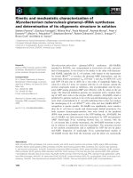
Fig. 4. D
2
-symmetric tetramer structure of CDase in the crystal together with the symmetry axes. (A) Front view placing the crystallographic twofold
axis horizontally in the paper plane. The crystallographic axis runs through the large interface and the vertical noncrystallographic axis runs
through the small interface between the N-terminal domains. One subunit is given in the colors and in an orientation similar to Fig. 2. A b-CD
(orange) derived from a superposition with the complex between b-CD and the homologous enzyme TVA-II [47] marks the active center. (B) View
from the left side of (A), which is along the crystallographic twofold axis, showing a smooth silhouette.
2336 H. B. Fritzsche et al.(Eur. J. Biochem. 270) Ó FEBS 2003
The chain fold of the N-terminal domain of CDase is
similar to that of the related CD-degrading enzymes TVA-II
[10], ThMA [11] and BaCD [9]. However, the positions of
these domains relative to the respective TIM barrel are
completely at variance as shown in Fig. 5. The N-terminal
domains of TVA-II, ThMA and BaCD attach to the active
center of the other subunit and participate in substrate
selection [11,12]. This dimer interface is not related to either
of the two interfaces in the CDase tetramer. It seems very
unlikely that the deviating domain position in CDase is
a packing artefact caused by domain swapping during
crystallization because the interface between the N-terminal
domain and the protein remainder (domains A and B)
amounts to 1390 A
˚
2
, which is much larger than a common
packing contact.
Comparison with related enzymes
The relationships within the group of CD-degrading
enzymes were evaluated by a comprehensive chain-fold
comparison. The comparison was extended to the structur-
ally related TVA-I [13], the CD-producing CGTases [38,39]
and a-amylase from Aspergillus oryzae (TAKA) [46], which
was taken as a well-known representative of family no. 13.
In principle, all comparisons could have been performed
with the amino-acid sequences alone, as the glycosylhydro-
lase families are defined by the sequences. However, this
method suffers from the rather arbitrary placing of the gaps.
Therefore, we took account of the geometry and first
derived the group of structurally equivalent residues in a
chain-fold superposition and then counted the number of
identical residues within this group. The results are given in
Table 3.
The most obvious result of this comparison is the close
relationship between TVA-II, ThMA and BaCD, which
can be almost completely structure-aligned, giving rise to
about 50% identical residues. As mentioned above, these
three enzymes also form similar dimers (with a further
hexameric association in BaCD) and have similar catalytic
activities. Therefore, we classify them as the TVA-II group.
When comparing CDase with this group, only 380 of the
600 residues can be structure-aligned, and only 28% of
the aligned residues are identical. This renders CDase an
outlier among the structurally established CD-degrading
enzymes. As for the other enzymes, a-amylase TVA-I
shows considerable structural similarity to the TVA-II
group, although its function differs greatly (Table 3).
Moreover, the data reveal that the CD-degrading enzymes
are more similar to the CD-producing CGTase than to the
a-amylase TAKA.
A more obvious difference between CDase and the others
is the deviating spatial position of its N-terminal domain
Fig. 5. Stereoview of the superposition of
CDase(coloredasinFig.2)withTVA-II
(black, Ca
2+
at Ca-II grey) given as
Ca-backbone plots. The completely different
positions of the N-terminal domains and the
differences in domain B near Ca-I (right) are
clearly visible.
Table 3. Chain-fold comparisons within glycosylhydrolase family no. 13. The upper right triangle shows the number of Ca atoms aligned within the
3A
˚
distance criterion in superpositions of the complete polypeptide chains using program
LSQMAN
(30). The numbers in parentheses are the
percentages of identical residues in the aligned segments. For CDase, CGTase and TAKA, only domains A, B and C could be superimposed with
any of the other enzymes. The lower left triangle shows the respective numbers for a separate superposition series involving only the N-terminal
domains.
CDase TVA-II ThMA BaCD TVA-I CGTase TAKA
CDase 387 (29) 387 (28) 376 (26) 371 (25) 380 (26) 361 (25)
TVA-II 47 (11) 547 (47) 552 (47) 428 (37) 378 (25) 369 (21)
ThMA 47 (13) 121 (35) 570 (54) 472 (32) 366 (24) 368 (26)
BaCD 48 (6) 119 (32) 123 (49) 421 (37) 376 (22) 354 (26)
TVA-I 54 (6) 98 (18) 98 (26) 102 (25) 367 (25) 377 (24)
CGTase – – – – – 401 (25)
Ó FEBS 2003 Cyclodextrinase structure (Eur. J. Biochem. 270) 2337
(Fig. 5). A superposition restricted to the N-terminal
domains showed that those of the TVA-II group can be
almost fully structure-aligned, resulting in 40% amino-
acid residue identities (Table 3). TVA-I is somewhat outside
the TVA-II group, but can still be well aligned. However,
the N-terminal domain of CDase aligns only with about
half of its residues, shows almost no sequence identity
(Fig. 6 and Table 3), and clearly differs from the TVA-II
group with respect to sequence, chain fold, and position.
The N-terminal domains of the trehalohydrolase [35] and
the isoamylase [36] have a similar chain fold to that of
CDase and the TVA-II group, but they are barely related to
any of them (data not shown). Interestingly, the general
location of the N-terminal domains of these two outliers
[35,36] corresponds to that of CDase.
A superposition of the highly variable B domains, which
participate in the active center, is given in Fig. 7. CDase has
a very long extension, whereas CGTase and TAKA have
intermediate ones. In contrast, the TVA-II group and
TVA-I have a much smaller B domain. The large B domains
of CDase, CGTase and TAKA are fixed by Ca-I, which is
absent in the TVA-II group with their small B domains. The
surprisingly large difference between CDase on one hand
and the TVA-II group on the other corresponds to the
different oligomeric structures. CDase uses the long exten-
sion of its B domain to make an intimate contact across the
strong dimer interface with domains A and C of the other
subunit. In contrast, the TVA-II group dimer attaches the B
domain to an N-terminal domain of the other subunit,
which restricts the size of the B domain (Fig. 7).
Active center
The active center of CDase is depicted in Fig. 8, which
includes the superimposed structure of a TVA-II dimer with
bound b-CD [47]. Interestingly, the superposition causes a
Fig. 6. Structural alignment of the N-terminal domain of CDase with those of TVA-II [10], ThMA [11], BaCD [9], TVA-I [13], a trehalohydrolase [35]
and an isoamylase [36], which are the only structurally similar domains within glycosylhydrolase family no. 13. CD-degrading activity has been
reported for the top four enzymes. The secondary structure of CDase is given, and every 10th amino acid residue is marked by a dot. Residues
87–175 of the isoamylase have been omitted (marked #). All residues that superimpose within the 3 A
˚
distance criterion of program
LSQMAN
[30]
are underlined. For reference, strand b9 of the TIM barrel has been included, and the alignments with the TIM barrels are given in bold.
Fig. 7. Superposition of the inserted B domains
of CDase (green, His251 marked by a ball),
TVA-II (red), CGTase (blue) and TAKA (grey)
as aligned on the TIM barrels. TVA-II, ThMa
and BaCD are so similar that only one of them
was drawn out for clarity. As TVA-I varies
only slightly from TVA-II, it was omitted. The
chain direction is indicated by the N* and C*
ends.
2338 H. B. Fritzsche et al.(Eur. J. Biochem. 270) Ó FEBS 2003
clash between the long B-domain extension of CDase
(His251, Fig. 7) and the N-terminal domain of the other
subunit of the TVA-II dimer (Tyr45¢). It has been suggested
that the N-terminal domain of the TVA-II group [9–11]
confers CD specificity because it covers one side of the bound
CD [47]. In CDase, this role is fulfilled by the B domain of the
same subunit. Therefore, it is sterically impossible for CDase
to form the same dimer as the TVA-II group.
As the polypeptide superposition of Fig. 8 places the
three catalytic residues of CDase (Fig. 1) within less than
1A
˚
of the positions of those of TVA-II, and as the active
centers closely resemble each other, the CD molecule bound
to TVA-II can be expected to bind at a similar position in
the CDase structure. The hydrolysis of CD should start by
Glu340 protonating a bridge oxygen of the cyclic substrate.
However, the distance between Glu340 and the next bridge
oxygen is 6 A
˚
, which is much too long. A similar distance to
a bound CD has been observed in CGTase [48], where,
however, it has been demonstrated that a linear malto-
oligosaccharide binds much deeper in the pocket at the
required 3 A
˚
distance to the Glu340 equivalent [49].
Moreover, the conformation at the scissile bond in a CD
complex with CGTase showed a substantial deviation from
the circular symmetry [48]. These observations indicate that
the observed CD-binding position in TVA-II is most likely
displaced by about 3 A
˚
. For catalysis, the CD molecule has
to be pushed 3 A
˚
deeper into the active-center pocket and
deformed at its scissile bond [48]. Such a CD position has
not yet been observed in any crystal structure. It would
enable Phe274 of CDase (or Phe286 of TVA-II) to rotate
around its Ca–Cb bond and enter the CD hollow, as has
been implied for TVA-II [10] and for Tyr195 of CGTase
[49]. Crystal experiments to clarify the CD position in
CDase are under way.
The required induced fit and deformation of the bound
CD need energy, part of which may be derived from
co-operative effects in the CDase tetramer (or TVA-II
dimer) association. This proposal is consistent with the
observation of a higher rate of CD hydrolysis for dimeric
ThMA than for the monomeric ThMA [12]. Although such
an energy source is conceivable for the TVA-II group in
which the bound CD contacts the N-terminal domain of the
other subunit, it is also possible for CDase in which the
bound CD is very close to the long B-domain extension
(Fig. 7) as well as to A-domain and C-domain residues of
the other subunit (Fig. 4A). In fact, the interface mediating
the strong dimer association would appear to explain the
particularly long B-domain extension of CDase. In con-
clusion, the dimer association may help to overcome
the conformational activation energy barrier during CD
hydrolysis.
Acknowledgements
We thank H. Bender for drawing our attention to the enzyme, E. Schiltz
for amino-acid sequence analyses, M. Ru
¨
ckels for preparing the
initializing 350-bp fragment and C. Vonrhein for help with
SHARP
/
AUTOSHARP
. Moreover, we are grateful for the contributions of
S. Thorspecken, A. Dorowski, B. Phillips, S. Jelakovic, C. Schleberger
and M. Mrosek at early stages of the analysis, and we thank the
beamline staff of the EMBL-outstation (DESY Hamburg) for help with
the data collection. The project was supported by the European
Commision under BIO4-98-0022 (AGADE) and by the Deutsche
Forschungsgemeinschaft under GRK-434.
References
1. Loftsson, T. & Brewster, M.E. (1996) Pharmaceutical applications
of cyclodextrins. 1. Drug solubilisation and stabilization. J. Pharm.
Sci. 85, 1017–1025.
2. Vetter, W. & Schurig, V. (1997) Enantioselective determination
of chiral organochlorine compounds in biota by gas chromato-
graphy on modified cyclodextrins. J. Chromatogr. Sect. A 774,
143–175.
3. Saha, B.C. & Zeikus, J.G. (1992) Cyclodextrin degrading enzymes.
Starch/Sta
¨
rke 44, 321–315.
4. Bender, H. (1993) Purification and characterization of a cyclo-
dextrin-degrading enzyme from Flavobacterium sp. Appl. Micro-
biol. Biotechnol. 39, 714–719.
5. Oguma, T., Matsuyama, A., Kikuchi, M. & Nakano, E. (1993)
Cloning and sequence analysis of the cyclodextrinase gene from
Bacillus sphaericus and expression in Escherichia coli cells. Appl.
Microbiol. Biotechnol. 39, 197–203.
Fig. 8. Active-center region in a superposition of CDase (blue with light green domain B) with the TVA-II dimer (grey with dark green domain B and
pink N-terminal domain). The N-terminal domain of the other subunit of the TVA-II dimer is shown in red including Tyr45¢. The bound b-CD
molecule is from a complex with TVA-II [47]. Active-center residues of CDase are given as ball-and-stick models.
Ó FEBS 2003 Cyclodextrinase structure (Eur. J. Biochem. 270) 2339
6. Abe, J., Onitsuka, N., Nakano, T., Shibata, Y., Hizukuri, S. &
Entani, E. (1994) Purification and characterization of periplasmic
alpha-amylase from Xanthomonas campestris K-11151. J. Bacter-
iol. 176, 3584–3588.
7. Bender, H. (1995) Purification and characterisation of a soluble,
cytoplasmic decycling maltodextrinase from Lactobacillus sp.
strain 26X, isolated from kitchen waste water. Appl. Microbiol.
Biotechnol. 43, 838–843.
8. Feederle, R., Pajatsch, M., Kremmer, E. & Bo
¨
ck, A. (1996)
Metabolism of cyclodextrins by Klebsielle oxytoca m5a1: puri-
fication and characterisation of a cytoplasmatically located
cyclodextrinase. Arch. Microbiol. 165, 206–212.
9. Lee, H S., Kim, M S., Cho, H S., Kim, J I., Kim, T J., Choi,
J H., Park, C., Lee, H S., Oh, B H. & Park, K H. (2002)
Cyclomaltodextrinase, neopullulanase and maltogenic amylase
are nearly indistinguishable from each other. J. Biol. Chem. 277,
21891–21897.
10. Kamitori,S.,Kondo,S.,Okuyama,K.,Yokota,T.,Shimura,Y.,
Tonozuka, T. & Sakano, Y. (1999) Crystal structure of Thermo-
actinomyces vulgaris R 47 a-amylase II (TVA II) hydrolyzing
cyclodextrins and pullulan at 2.6 A
˚
resolution. J. Mol. Biol. 287,
907–921.
11. Kim, J S., Cha, S S., Kim, H J., Kim, T J., Ha, N C., Oh,
S T., Cho, H S., Cho, M J., Kim, M J., Lee, H S., Kim, J W.,
Choi, K.Y., Park, K H. & Oh, B H. (1999) Crystal structure of a
maltogenic amylase provides insights into a catalytic versatility.
J. Biol. Chem. 274, 26279–26286.
12. Kim,T J.,Nguyen,V.D.,Lee,H S.,Kim,M J.,Cho,H Y.,
Kim, Y W., Moon, T W., Park, C.S., Kim, J W., Oh, B H.,
Lee, S B., Svensson, B. & Park, K H. (2001) Modulation of the
multisubstrate specificity of Thermus maltogenic amylase
by truncation of the N-terminal domain and by a salt-induced shift
of the monomer/dimer equilibrium. Biochemistry 40, 14182–
14190.
13.Kamitori,S.,Abe,A.,Othaki,A.,Kaji,A.,Tonozuka,T.&
Sakano, Y. (2002) Crystal structures and structural comparison of
Thermoactinomyces vulgaris R 47 a-amylase1(TVAI)at1.6A
˚
resolution and a-amylase 2 (TVA II) at 2.3 A
˚
resolution. J. Mol.
Biol. 318, 443–453.
14. Bender, H. (1994) Studies of the action pattern on potato starch
of the decycling maltodextrinase from Flavobacterium sp, 92.
Carbohydr. Res. 263, 137–147.
15. Bender, H. (1994) Studies of the degradation of pullulan by the
decycling maltodextrinase of Flavobacterium sp, 92. Carbohydr.
Res. 260, 119–130.
16. Bender, H. (1994) Studies of the transglycosylation reaction
catalysed by the decycling maltodextrinase of Flavobacterium sp,
92 with malto-oligosaccharides and cyclodextrins. Carbohydr. Res.
263, 123–135.
17. Feinberg, A.P. & Vogelstein, B. (1983) A technique for radio-
labeling DNA restriction endonuclease fragments to high specific
activity. Anal. Biochem. 132, 6–13.
18. Sambrook, J., Fritsch, E.F. & Maniatis, T. (1989) Molecular
Cloning: A Laboratory Manual, 2nd edn. Cold Spring Harbor
Laboratory Press, Cold Spring Harbor, NY.
19. Sanger, F. (1981) Determination of nucleotide sequences in DNA.
Science 214, 1205–1210.
20. LeMaster, D.M. & Richards, F.M. (1985) 1H)15N heteronuclear
NMR studies of Escherichia coli thioredoxin in samples isotopi-
cally labeled by residue type. Biochemistry 24, 7263–7268.
21. Hendrickson, W.A., Horton, J.R. & Le Master, D.M. (1990)
Selenomethionyl proteins produced for analysis by multi-
wavelength anomalous diffraction (MAD): a vehicle for direct
determination of three-dimensional structure. EMBO J. 9, 1665–
1672.
22. Otwinowski, Z. & Minor, W. (1997) Processing of X-ray diffrac-
tion data collected in oscillation mode. Methods Enzymol. 276,
307–326.
23. Uson, I. & Sheldrick, G.M. (1999) Advances in direct methods for
protein crystallography. Curr. Opin. Struct. Biol. 9, 642–648.
24. Sheldrick, G.M., Hauptman, H.A., Weeks, C.M., Miller, R.
& Uson, I. (2001) Ab Initio phasing. International Tables for
Crystallography (Rossmann, M.G. & Arnold, E., eds), Vol. F,
pp. 333–345. Kluwer Academic Publishers, Dordrecht.
25. de la Fortelle, E. & Bricogne, G. (1997) Maximum-likelihood
heavy-atom parameter refinement for multiple isomorphous
replacement and multiwavelength anomalous diffraction methods.
Methods Enzymol. 276, 472–494.
26. Perrakis, A., Sixma, T.K., Wilson, K.S. & Lamzin, V.S. (1997)
wARP: improvement and extension of crystallographic phases by
weighted averaging of multiple refined dummy atomic models.
Acta Crystallogr. Sect. D 53, 448–455.
27. Jones, T.A., Zou, J.Y., Cowan, S.W. & Kjeldgaard, M. (1991)
Improved methods for building protein models in electron density
maps and the location of errors in these models. Acta Crystallogr.
Sect A 47, 110–119.
28. Bru
¨
nger, A.T., Adams, P.D., Clore, G.M., Delano, W.L., Gros,
P., Grosse-Kunstleve, R.W., Jiang, J S., Kuszewski, J., Nilges, N.,
Pannu, N.S., Read, R.J., Rice, L.M., Simonson, T. & Warren,
G.L. (1998) Crystallography and NMR system: a new software
suite for macromolecular structure determination. Acta Crystal-
logr. Sect. D 54, 901–921.
29. Winn, M., Isupov, M. & Murshudov, G.N. (2000) Use of TLS
parameters to model anisotropic displacements in macromolecular
refinement. Acta Crystallogr. Sect. D 57, 122–133.
30. CCP4 Collaborative Computational Project, 4
4
(1994) The CCP4
suite: programs for protein crystallography. Acta Crystallogr.
Sect. D 50, 760–763.
31. Kraulis, P.J. (1991)
MOLSCRIPT
: a program to produce both
detailed and schematic plots of protein structures. J. Appl. Crys-
tallogr. 24, 946–950.
32. Merritt, E.A. & Bacon, D.J. (1997) Raster3D photorealistic
molecular graphics. Methods Enzymol. 277, 505–524.
33. Henrissat, B. & Bairoch, A. (1996) Updating the sequence-based
classification of glycosyl hydrolases. Biochem. J. 316, 695–696.
34. Hondoh, H., Kuriki, T. & Matsuura, Y. (2002) Three-dimensional
structure of Bacillus stearothermophilus neopullulanase. Biologia
(Bratisl.), 57, 77–82.
35. Feese, M.D., Kato, Y., Tamada, T., Kato, M., Komeda, T.,
Miura, Y., Hirose, M., Hondo, K., Kobayashi, K. & Kuroki, R.
(2000) Crystal structure of glycosyltrehalose trehalohydrolase
from the hyperthermophilic archaeum Sulfolobus solfataricus.
J. Mol. Biol. 301, 451–464.
36. Katsuya, Y., Mezaki, Y., Kubota, M. & Matsuura, Y. (1998)
Three-dimensional structure of Pseudomonas isoamylase at 2.2 A
˚
resolution. J. Mol. Biol. 281, 885–897.
37. Hofmann, B.E., Bender, H. & Schulz, G.E. (1989) Three-dimen-
sional structure of cyclodextrin glycosyltransferase from Bacillus
circulans at 3.4 A
˚
resolution. J. Mol. Biol. 209, 793–800.
38. Klein, C. & Schulz, G.E. (1991) Structure of cyclodextrin glyco-
syltransferase refined at 2.0 A
˚
resolution. J. Mol. Biol. 217, 737–
750.
39. Leemhuis, H., Dijkstra, B.W. & Dijkhuizen, L. (2003) Thermo-
anaerobacterium thermosulfurigenes cyclodextrin glycosyltransfer-
ase. Mechanism and kinetics of inhibition by acarbose and
cyclodextrins. Eur. J. Biochem. 270, 155–162.
40. Banner, D.W., Bloomer, A.C., Petsko, G.A., Phillips, D.C.,
Pogson, C.I., Wilson, I.A., Corron, P.H., Furth, A.J., Milman,
J.D., Offord, R.E., Priddle, J.D. & Waley, S.G. (1975) Structure of
chicken muscle triose phosphate isomerase determined crystal-
2340 H. B. Fritzsche et al.(Eur. J. Biochem. 270) Ó FEBS 2003
lographically at 2.5 A
˚
resolution using amino acid sequence data.
Nature (London) 255, 609–614.
41. MacGregor, E.A., Janecek, S. & Svensson, B. (2001) Relationship
of sequence and structure to specificity in the a-amylase family of
enzymes. Biochim. Biophys. Acta 1546, 1–20.
42. Boel, E., Brady, L., Brzozowski, A.M., Derewenda, Z., Dodson,
G.G.,Jensen,V.J.,Petersen,S.B.,Swift,H.,Thim,L.&Wolike,
H.F. (1990) Calcium binding in a-amylases: an X-ray diffraction
study at 2.1 A
˚
resolution of two enzymes from Aspergillus.
Biochemistry 29, 6244–6249.
43. Fujimoto,Z.,Takase,K.,Doui,N.,Momma,M.,Matsumoto,T.
& Mizuno, H. (1998) Crystal structure of a catalytic-site mutant
a-amylase from Bacillus subtilis complexed with maltopentaose.
J. Mol. Biol. 277, 393–407.
44. Machius, M., Wiegand, G. & Huber, R. (1995) Crystal structure
of calcium-depleted Bacillus licheniformis a-amylase at 2.2 A
˚
resolution. J. Mol. Biol. 246, 545–559.
45. Kadziola, A., Abe, J., Svensson, B. & Haser, R. (1994) Crystal
and molecular structure of barley a-amylase. J. Mol. Biol. 239,
104–121.
46. Matsuura, Y., Kusunoki, M., Harada, W. & Kakudo, M. (1984)
Structure and possible catalytic residues of Taka-amylase A.
J. Biochem. (Tokyo) 95, 697–702.
47. Kondo, S., Ohtaki, A., Tonozuka, T., Sakano, Y. & Kamitori, S.
(2001) Studies on the hydrolyzing mechanism for cyclodextrins of
Thermoactinomyces vulgaris R 47 a-amylase 2 (TVA II). X-ray
structure of the mutant E354A complexed with b-cyclodextrin,
and kinetic analyses on cyclodextrins. J. Biochem. (Tokyo) 129,
423–428.
48. Schmidt,A.K.,Cottaz,S.,Driguez,H.&Schulz,G.E.(1998)
Structure of cyclodextrin glycosyltransferase complexed with a
derivative of its main product b-cyclodextrin. Biochemistry 37,
5909–5915.
49. Parsiegla, G., Schmidt, A.K. & Schulz, G.E. (1998) Substrate
binding to a cyclodextrin glycosyltransferase and mutations
increasing the cyclodextrin production. Eur. J. Biochem. 255,
710–717.
Ó FEBS 2003 Cyclodextrinase structure (Eur. J. Biochem. 270) 2341