Báo cáo khoa học: A simple protocol to study blue copper proteins by NMR pot
Bạn đang xem bản rút gọn của tài liệu. Xem và tải ngay bản đầy đủ của tài liệu tại đây (390.14 KB, 10 trang )
A simple protocol to study blue copper proteins by NMR
Ioannis Gelis
1
, Nikolaos Katsaros
1
, Claudio Luchinat
2,3
, Mario Piccioli
2,4
and Luisa Poggi
2,4
1
NCSR Demokritos, Institute of Physical Chemistry, Agia Paraskevi Attikis, Greece;
2
Magnetic Resonance Center,
3
Department of Agricultural Biotechnology and
4
Department of Chemistry, University of Florence, Italy
In the case of oxidized plastocyanin from Synechocystis sp.
PCC6803, an NMR approach based on classical two and
three dimensional experiments for sequential assignment
leaves unobserved 14 out of 98 amino acids. A protocol
which simply makes use of tailored versions of 2D HSQC
and 3D CBCA(CO)NH and CBCANH leads to the identi-
fication of nine of the above 14 residues. The proposed
protocol differs from previous aproaches in that it does not
involve the use of unconventional experiments designed
specifically for paramagnetic systems, and does not exploit
the occurrence of a corresponding diamagnetic species in
chemical exchange with the blue copper form. This protocol
is expected to extend the popularity of NMR in the struc-
tural studies of copper (II) proteins, allowing researchers to
increase the amount of information available via NMR on
the neighborhood of a paramagnetic center without requi-
ring a specific expertise in the field. The resulting 3D spectra
are standard spectra that can be handled by any standard
software for protein NMR data analysis.
Keywords: blue copper proteins; NMR spectroscopy; struc-
tural biology; paramagnetic proteins; plastocyanin.
There is a strong interest in the structural biology commu-
nity for the study of copper trafficking and copper
homeostasis [1–11]. This involves the understanding of the
role of metal ions in protein folding and misfolding related
diseases [12–20], as well as the understanding at the atomic
level of protein–protein interactions in electron-transfer
processes [21–29]. Within this framework the search for
methodological advancements in NMR spectroscopy tail-
ored to the structural characterization of copper(II) proteins
may play a significant role.
In paramagnetic metalloproteins, NMR signals of pro-
tons close to the metal ions are broadened, sometimes
beyond detection, by the presence of the paramagnetic
center [30,31]. The extent of paramagnetic induced line
broadening depends on the electronic relaxation times of the
metal center [32–35]. Tetragonal Cu(II), found in Type II
centers, has long electronic relaxation times [36] which make
the NMR lines of residues belonging to the coordination
sphere broad beyond detection [37,38]. When Cu(II) adopts
a trigonal geometry, such as that provided by two histidines
and one cysteine residue in Type I centers, or blue copper
centers, the electronic relaxation times are about one order
of magnitude shorter. Hence, signals belonging to Cu(II)
first coordination sphere, although severely broadened,
become observable [39]. Because of the axial symmetry of
the g-tensor in Type I copper centers [40], the pseudocontact
contribution to the observed shifts is negligible and para-
magnetic shifts arise only from through-bond spin density
delocalization from the metal to the ligands. Therefore, in
Type I copper centers, pseudocontact shifts can not be used
for structural purposes, unlike many other classes of
metalloproteins [41–43]. As a partial compensation of such
a drawback, the shifts can be safely interpreted on the basis
of the chemical shift index [44].
It was recently shown that solution structures of
copper(II) proteins can be obtained [45]. To this end, the
standard protocol for solution structure of biomacromole-
cules has been substantially augmented by a number of non
conventional strategies for resonances assignments and by
the use of paramagnetism-based constraints for structure
calculations [46]. This approach often requires specific
expertise in the field of electron relaxation and hyperfine
interaction [42,46–53] and, in some case, specific hardware
[54]. As a consequence, NMR structural characterization of
paramagnetic metalloproteins is routinely performed only
in a limited number of laboratories [31,55–63].
We would like to present here a different perspective of
the NMR study of paramagnetic proteins and to emphasize
the fact that paramagnetic proteins should not necessarily
be considered as a different field with respect to mainstream
biomolecular NMR. We will discuss the information
content of basic 2D and 3D experiments when they are
collected using a different choice of experimental parameters
with respect to the standard ones. The additional experi-
ments that we propose are deliberately restricted to simple
modifications of the pulse sequences that are routinely used
for resonance assignment, like CBCA(CO)NH [64,65] and
CBCANH [66,67], in such a way that their implementation
does not require any special expertise. This approach should
extend significantly the detectability of resonances that sense
the hyperfine interaction and therefore should substantially
increase the number of assignments in the proximity of
the paramagnetic center that can be obtained within a
standard protocol [68,69]. The modifications discussed here
to CBCA(CO)NH and CBCANH experiments do not
substantially alter the coherence transfer pathway with
Correspondence to M. Piccioli, Via L. Sacconi 6,
50019 Sesto Fiorentino, Florence, Italy.
Fax: + 39 055457 4253, Tel.: + 39 055457 4265,
E-mail: fi.it
Abbreviations: INEPT, insensitive nuclei enhanced by polarization
transfer; PFG, pulsed field gradients.
(Received 12 June 2002, revised 25 October 2002,
accepted 27 November 2002)
Eur. J. Biochem. 270, 600–609 (2003) Ó FEBS 2003 doi:10.1046/j.1432-1033.2003.03400.x
respect to the scheme originally proposed by Bax and
coworkers [64], but they make the two sequences much
more effective in the presence of contributions to relaxation
arising from the hyperfine interaction.
The test system chosen is the blue copper protein
plastocyanin from the cyanobacterium Synechocystis sp.
PCC6803. It contains a typical Type I center extensively
spectroscopically characterized [70–72]. Previous NMR
studies showed that this is an excellent system to address
the efficiency of nonconventional NMR approaches to
obtain structural information [45,73].
In the present work we will demonstrate that an approach
which does not require any hardware or software dedicated
to paramagnetic systems can substantially improve the
available assignments close to the copper (II) ion without
recourse to metal substitution.
Materials and methods
Protein expression and purification
The expression and purification of Synechocystis sp.
PCC6803 plastocyanin in Escherichia coli was performed as
previously described [74]. Uniformly
13
C,
15
N-labeled over-
expressed plastocyanin was obtained from M9 minimal
medium containing (
15
NH
4
)
2
SO
4
as the sole nitrogen source
and [
13
C
6
]
D
-glucose as the sole carbon source. Samples for
NMR spectroscopy (2 m
M
)werepreparedin50 m
M
sodium
phosphate buffer (either in 90% H
2
O, 10% D
2
O or in 100%
D
2
O) at pH 5.2. Complete oxidation of the protein was
achieved using a slight excess of ferricyanide, subsequently
removed by gel filtration. The samples were kept at 4 °Cin
between measurements.
NMR Spectroscopy
Experiments were performed at 295 K on Bruker Avance
spectrometers operating at 700 and 800 MHz. Diamagnetic
1
H-
13
CHSQCand
1
H-
15
N HSQC [75] experiments were
performed. The number of real data points acquired were 512
in the t
1
dimension (
13
Cand
15
N), and 2048 in acquisition (t
2
dimension). Spectral widths of 11 p.p.m. for
1
H dimension,
80 p.p.m. for
13
C dimension and 50 p.p.m. for
15
Ndimen-
sion were used. For both experiments, 4 scans and a recycle
delay of 800 ms were used. Echo-antiecho acquisition [76]
was used to perform quadrature detection in t
1
dimension.
Sensitivity improvement [77,78] and crush gradients during
the insensitive nuclei enhanced by polarization and transfer
(INEPT) and inverse INEPT mixing were also used.
Two dimensional tailored
1
H-
13
CHSQCand
1
H-
15
N
HSQC experiments were performed to detect fast relaxing
signals [79]. All delays (INEPT transfer and recycle) were
shortened to 1.6 ms and 100 ms, respectively, in order to
detect resonances near the paramagnetic center. Two
dimensional nonselective inversion-recovery
1
H-
15
NHSQC
experiments (
15
N IR-HSQC) were performed to measure
nonselective longitudinal relaxation rates of protons [79]. In
order to measure T
1
values of very fast relaxing protons,
INEPT transfer and relaxation delays were shortened to
1.6 ms and 200 ms, respectively. Eight points were collected
to fit T
1
values, with the following inversion recovery delays:
2, 4, 8, 16, 32, 64, 128 and 256 ms.
A three dimensional HNCO experiment [80] was per-
formed to assign backbone resonances. For the above
experiment spectral windows of 11 p.p.m. for
1
H, 50 p.p.m.
for
15
N, and 30 p.p.m. for
13
C dimensions were typically
used. The number of real data points acquired were 128 in
the t
1
dimension (
13
C), 64 in the t
2
dimension (
15
N), and
1024 in acquisition (t
3
dimension). Three dimensional
CBCA(CO)NH [64,65] and CBCANH [66,67] experiments
were carried out to sequentially assign
13
C resonances.
Spectral widths of 11 p.p.m. for
1
H dimension, 76 p.p.m.
for
13
C dimension and 41 p.p.m. for
15
N dimension were
used. The number of real data points acquired were 64
points in the
15
N dimension, 256 in the
13
C dimension, and
1024 in acquisition (t
3
dimension) for both experiments. A
recycle delay of 800 ms was used and 8 scans per increment
were collected.
All the data were zero-filled in the indirect dimensions
and apodized using cosine squared functions. Linear
prediction was always applied in the indirect dimension.
All NMR data were processed with the Bruker
XWINNMR
software packages. The program
SPARKY
3 (T. D. Goddard
and D. G. Kneller, University of California, San Francisco,
USA) was used for the analysis of all NMR spectra.
Theory
A classical approach toward structure determination in a
paramagnetic metalloprotein does not provide information
in the proximity of the metal center [81,82], even when
careful and extensive studies are performed using double
and triple labeled samples [61,83].
CBCA(CO)NH and CBCANH are among the most
popular experiments for sequential assignment of macro-
molecules in solution [65,66]. CBCA(CO)NH spectra con-
nect HN(i) with C
b
(i-1) and C
a
(i-1) resonances, while
CBCANH spectra connect HN(i) with C
b
(i), C
a
(i), C
b
(i-1)
and C
a
(i-1) resonances, the inter residue peaks being lower
in intensity than the intra residue peaks. The standard
versions of both experiments make use of several INEPT
transfer delays, crush gradients, flip back pulses, sensitivity
improvement schemes and echo-antiecho gradient selection.
Each of the above building blocks requires coherence
transfer delays during which the magnetization of interest is
relaxed. In the case of paramagnetic molecules, the presence
of the unpaired electron makes large contributions to
nuclear relaxation for nuclei nearby. As a consequence,
CBCA(CO)NH and CBCANH are expected to be unsuit-
able for the study of such systems. However, a series of
modifications can be planned that make the two sequences
exploitable.
The optimization of polarization transfer and recycle
delays in heteronuclear experiments has been extensively
discussed elsewhere, as well as the choice of the number of
scans and data points in t
1
, t
2
and t
3
dimensions [46,84]. On
such bases, the NH reverse INEPT and the CH INEPT
transfer delays were shortened to 1.6 ms and 1.8 ms,
respectively, in the CBCA(CO)NH experiment, while only
the NH transfer delay was shortened to 1.6 ms in the
CBCANH. The building blocks of the pulse sequences
related to the coherence transfers pathway C
b
-C
a
-CO-N or
C
b
-C
a
-N were not modified with respect to the standard
version of the sequence. A recycle delay of 300 ms was used
Ó FEBS 2003 Blue copper proteins studied by NMR (Eur. J. Biochem. 270) 601
and 64 scans were collected for both experiments. With
respect to the diamagnetic version of the experiments, the
number of data points in the
15
Nand
13
C dimensions were
reduced from 64 to 48 and from 256 to 128, respectively.
Besides the choice of INEPT transfer delays, other
modifications can be introduced with respect to the
diamagnetic version of the sequences.
The sensitivity improvement scheme (SI) [78] makes use,
during the reverse INEPT, of a double spin-echo which
allows the detection of both antiphase components N
x
H
z
and N
y
H
z
created during
15
N evolution, thus giving a 2
1/2
improvement of the signal to noise ratio [78]. This scheme
has twice the duration of a normal reverse INEPT, and
different relaxation mechanisms are operative on the
various coherence transfer pathways that transform the
two above components to observable magnetizations. Even
if the transfer delays are shortened, as already extensively
discussed [84], the occurrence of a strong contribution to
relaxation may be such that, for fast relaxing signals, the
elimination of sensitivity improvement scheme gives a better
S/N.
The use of pulsed field gradients (PFGs) within a pulse
sequence to detect paramagnetic signals may be critical. In
general, their use to clean observable magnetization from
spurious peaks has no drawbacks, provided that PFG do
not entail additional delays [85]. However, in the case of
echo-antiecho detection schemes [76], the gradient selection
requires that two additional gradients are placed in the
sequences, together with two additional 180° pulses and
refocusing delays. Because hyperfine relaxation depends on
c
2
X
, where X is the involved nucleus, the loss of signal
intensity is critical in those coherence transfer steps in which
1
H R
2
relaxation is involved [86]. This is of course the case of
the period immediately preceding t
3
acquisition. Similar
considerations hold for the use of crush gradients during the
INEPT and reverse INEPT steps. In this case, the loss due
to relaxation depends on R
1
. Therefore the use of crush
gradients for fast relaxing signals is less destructive that the
gradients needed in the echo-antiecho scheme. Of course, a
major drawback expected from the elimination of gradient
selection and crush gradients is that there is no efficient
water suppression scheme left in the sequence. To overcome
this problem, a Watergate scheme, with short gradients in
order to be compatible with the short delays of the reverse
INEPT step [87] can be reintroduced in the final reverse
INEPT step.
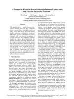
The calculated effects of the stepwise removal of the crush
gradients, echo-antiecho and sensitivity improvement
schemes are shown in Fig. 1. The calculations are per-
formed for the transfer function from a
15
N nucleus to a
bound proton in either the CBCANH or CBCA(CO)NH
pulse schemes. The proton is considered to be at 6 A
˚
from
the copper(II) center, assuming a s
s
¼ 0.5 ns [73] and a
s
r
¼ 5.9 ns [74]. Under these conditions R
2
c. ¼ 600 s
)1
,
while R
1
is about 5 times smaller. If we use the standard
values for duration and recovery of gradients of 1 ms and
0.5 ms, respectively, the transfer function has a maximum at
about 1 ms (Fig. 1A). Its intensity is about 2% of the
intensity expected for the corresponding peak in a normal
reverse INEPT when relaxation is neglected. Elimination of
the crush gradients, during which
1
H R
1
relaxation occurs,
leads to a gain in intensity of about 15% (Fig. 1B).
The most important effect arises from the elimination of
the echo-antiecho scheme. The effect of removing the echo-
antiecho building block is observed in the calculated transfer
functions shown in Fig. 1C. Considering as a test case the
signal discussed above, the replacement of the echo-antiecho
block with any other quadrature detection scheme that does
not rely on gradient selection of coherences, increases signal
intensity by about a factor of five. Of course the relative gain
in intensity is reduced when, in the diamagnetic version of
the sequence, shorter gradients and recovery delays are
used. When gradient and recovery delays in the diamagnetic
experiment are shortened down to 150 lsand100ls,
respectively, the gain of signal intensity under the above
conditions is still of about a factor of two. This shows that
even if very short values of gradient and recovery delays are
used within the diamagnetic version of CBCA(CO)NH and
CBCANH (and this would not be the ÔdefaultÕ choice in the
absence of fast relaxation), the use of echo-antiecho
quadrature detection is not recommended with respect to
States-TPPI [88] or any other quadrature detection scheme
methods that does not rely on gradient selection of
coherences.
Finally the effects of the replacement of the sensitivity
improvement step with the usual reverse INEPT step is
illustrated in the transfer function shown in Fig. 1D. It can
be seen that the single reverse INEPT step, not only gives
about a 10% increase in the maximum of the transfer
function with respect to the sensitivity improvement scheme
but also it gives a transfer function which is much less
sensitive to optimization of the transfer delay, as observed in
Fig. 1 when transfer delays longer than 1.8–2 ms are
considered.
Fig. 1. Calculated transfer functions for the NH reverse INEPT transfer
step of CBCA(CO)NH or CBCANH experiments with: (A) diamagnetic
pulse sequence, using sensitivity improvement detection scheme and echo-
antiecho quadrature detection (all applied gradients were 1 ms with a
recovery delay of 0.5 ms); (B) same as (A) without the use of crush
gradient occurring in between the 90° pulses; (C) same as (B) without the
echo-antiecho detection, i.e. with the elimination of the additional delays
needed for the gradients of the echo-antiecho; (D) same as (C) with the
removal of the SI scheme. All transfer functions are normalized with
respect to a normal reverse INEPT under optimized condition for the
transfer delay and neglecting losses due to
1
H-
15
N relaxation. Transfer
functions have been calculated for a
1
H signal of a proton at about 6 A
˚
from the metal center (R
2
¼ 600 s
)1
, R
1
¼ 120 s
)1
assuming a
s
s
¼ 0.5 ns and a s
r
¼ 5.9 ns).
602 I. Gelis et al. (Eur. J. Biochem. 270) Ó FEBS 2003
Results and discussion
Spectral assignment of oxidized plastocyanin:
the standard approach
Synechocystis sp. PCC6803 plastocyanin was overexpressed
in E. coli to obtain large amounts of
13
C,
15
N-enriched
protein. The already available assignment of
1
Hand
15
N
resonances [45] was extended to
13
C resonances of backbone
and side chains by a combination of classic triple resonance
experiments. 3D HNCO [89], CBCA(CO)NH [64,65] and
CBCANH [66,67] were collected at 700 and 800 MHz
spectrometers. The analysis of these spectra has lead to the
assignment of 73% of C¢, 81% of C
a
and 79% of C
b
.
Because of broadening effects induced by the paramagnetic
center, no sequential backbone assignment is available [45]
in the loop regions encompassing residues 7–8, 38–42,
61–62, and 82–88.
Detection of fast relaxing signals:
15
N- and
13
C-HSQC
experiments
Tailored versions of
1
H-
15
N HSQC [90],
1
H-
13
CHSQC[79],
CBCA(CO)NH and CBCANH were used to detect reso-
nances in the proximity of Cu(II).
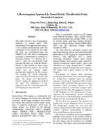
The comparison of two
1
H-
15
N HSQC spectra recorded
with different recycle and polarization transfer delays allows
to identify 14 resonances that clearly experience a substan-
tial gain in signal intensity when comparing a diamagnetic
HSQC experiment with a tailored experiment. The overlay
of the two spectra is shown in Fig. 2, and the 14 resonances
are highlighted. Of these, 7 are observed with much lower
intensity in the diamagnetic experiment while 7 were
completely missing in the diamagnetic experiment. The
former 7 signals were already assigned in a previous study
[45], and correspond to residues Leu14, Phe16, Asn34,
Lys35, Ser37, Ile41 and Ala89. The seven new signals are
listed in Table 1.
In order to measure the proton T
1
values of the
previously unobserved fast relaxing signals detected in the
tailored
1
H-
15
N HSQC, a series of two dimensional
nonselective inversion-recovery
1
H-
15
N HSQC experiments
was performed [79]. As our present interest is focused on
relatively fast relaxing signals, we used for the inversion
recovery experiment a recycle delay of 200 ms. Therefore
the inversion recovery experiment gave fully reliable results
only for those resonances having a T
1
values < 60 ms. The
T
1
values obtained for the above signals, together with the
1
Hand
15
Nshifts,arealsoreportedinTable1.
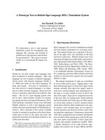
Similarly to the
1
H-
15
N HSQC experiment, the compari-
son of two
1
H-
13
C HSQC spectra recorded with different
recycle and polarization transfer delays allows to identify 11
resonances that clearly experience a substantial gain in
signal intensity when comparing a diamagnetic HSQC
experiment with a tailored experiment. Of these, four belong
to C
a
s peaks 1–4 in Table 2 and 7 to C
b
s peaks 5–11 in
Table 2. They are also highlighted in Fig. 3A and 3B.
Detection of fast relaxing signals: tailored CBCA(CO)NH
and CBCANH
A 3D version of these experiments tailored as discussed
above to optimize the detection of fast relaxing signals has
been performed. The new peaks identified through
1
H-
15
N
HSQC were monitored in CBCA(CO)NH. While in the
diamagnetic version of CBCA(CO)NH experiment only
Fig. 2. Overlay of diamagnetic and tailored
1
H-
15
NHSQCspectra.Fourteen resonances
are highlighted. The seven signals completely
missing in the diamagnetic experiment are
labelled A-G, while the seven observed with
much lower intensity in the diamagnetic
experiment are labelled with their corres-
ponding assignment.
Table 1. Previously unobserved signals found in the tailored
1
H-
15
N
HSQC.
dN (p.p.m) dHN (p.p.m) T
1
(ms)
A 108.29 8.80 34.0
B 118.52 7.24 17.7
C 121.62 8.1 > 60
D 126.57 8.6 25.3
E 128.23 8.28 40.9
F 107.45 8.1 27.8
G 125.02 9.23 55.1
Ó FEBS 2003 Blue copper proteins studied by NMR (Eur. J. Biochem. 270) 603
one of the peaks (signal C) listed in Table 1 was observed,
the tailored CBCA(CO)NH has allowed us to observe
connectivities with previous amino acid for 5 out of 7
residues, as reported in Table 3. As far as signal C is
concerned, the very weak connectivities observed in the
diamagnetic version of CBCA(CO)NH are observed with
much larger intensity (a factor of 2) in the tailored
experiment.
Similar considerations hold for CBCANH. The sensiti-
vity of CBCANH is expected to be smaller than CBCA
(CO)NH, as already proven extensively in diamagnetic
systems. None of the peaks listed in Table 1 was observed in
the diamagnetic experiment while four out of seven gave
connectivities in the tailored CBCANH, as reported in
Table 3, which summarizes the information obtained using
modified CBCA(CO)NH and CBCANH.
Assignment of fast relaxing signals
The assignment of the new signals found in the tailored
1
H-
15
N HSQC can be performed considering the following:
(a) limited number of missing assignments in the
1
H-
15
N
HSQC spectrum (14, listed in Table 4); (b) C
b
and C
a
chemical shifts provide substantial information on the
nature of the amino acid under investigation [44,91,92]. Of
course, such assignment is feasible only under the assump-
tion that contributions arising form pseudocontact shifts are
negligible with respect to the chemical shift index tolerance
[93]. As outlined above, this is a very reasonable assumption
as shown by the available literature on Cu(II) proteins
[39,94].
Let us consider signal A [Table 3]: the intra residue C
a
peak at 43.8 p.p.m. shows unambiguously that signal A
belongs to a Gly residue, while inter residue C
a
and C
b
peaks at 58.3 and 27.9 p.p.m. are primarily consistent with
Met, Arg or His residues. Therefore the only possible
assignment is Gly8, preceded by Met7. In previous works
only some sparse
1
H assignments were available for residues
7, 8, 61 and 62 [45].
No assignment can be performed for signal B, for which
no connectivities are found in both CBCA(CO)NH and
CBCANH.
Signal C shows no connectivities in the CBCANH
spectrum, but the inter residue C
a
peak found in the
CBCA(CO)NH at 43.1 p.p.m. is only consistent with a Gly
as preceding residue. Given the limited number of missing
assignments, this is in agreement only with the assignment
of signal C as the HN of Leu61, preceded by Gly60.
Signal D shows in the CBCA(CO)NH spectrum inter
residue peaks at 56.7 and 36.3 p.p.m., while among the intra
residue peaks only the C
a
is found in the CBCANH at
59.2 p.p.m. These connectivities perfectly fit the assignment
of signal D as NH of Val42, preceded by Ile41. The
identification of Val42 is also confirmed by the pattern
observed in the CBCA(CO)NH for Phe43, which presents
inter residue connectivities at 59.2 and 30.8 p.p.m.
As far as signal E is concerned, the four peaks corres-
ponding to intra and inter residue C
a
and C
b
do not permit a
fully consistent assignment. Inter and intra residue C
a
’s are
observed at 52.2 and 55 p.p.m., respectively, and they match
with His86-Arg87 residues. This assignment is supported by
Table 2. Signals that experience a substantial gain in signal intensity in
the tailored
1
H-
13
C HSQC compared with the diamagnetic experiment.
d
1
H (p.p.m) d
13
C (p.p.m) Assignment
1 5.44 56.2 Val15 (C
a
-H
a
)
2 5.14 58.0 Met7 (C
a
-H
a
)
3 4.92 42.3 Gly88 (C
a
-H
a
)
4 4.67 43.0 Gly8 (C
a
-H
a
)
5 1.27 37.3 Tyr 81 (C
b
-H
b
)
6 0.93 37.9 Arg87 (C
b
-H
b
)
a
7 0.72 37.3 Tyr 81 (C
b
-H
b
)
8 2.66 33.8 Val15 (C
b
-H
b
)
9 1.00 31.2 Val42 (C
b
-H
b
)
10 2.97 30.1 –
11 ) 0.30 11.6 Ile41 (C
d
-H
d
)
a
Tentative assignment.
Fig. 3. Overlay of diamagnetic and tailored
1
H-
13
CHSQCspectra.(A) C
a
region. (B) C
b
region. The 11 resonances that substantially
increase their intensity in the tailored experi-
ment are highlighted and labelled 1–11.
604 I. Gelis et al. (Eur. J. Biochem. 270) Ó FEBS 2003
the inter residue C
b
, which is observed at 35.6 p.p.m. (a
typical His region), but does not fit with the intra residue C
b
that is observed at 38 p.p.m., i.e. out of the region where C
b
of Arg residues are expected to fall. Therefore, we assign
signal E as the HN of Arg 87 only tentatively.
The
15
N shift of signal F is only consistent with a Gly
residue. As Gly8 has been already identified as signal A,
signal F can be safely assigned as the only other glycine
residue missing, i.e. Gly88, even if no connectivities are
found in both CBCA(CO)NH and CBCANH.
For signal G, in the CBCANH spectrum only the intra
residue connectivities are observed while those with the
previous residue are observed only in CBCA(CO)NH. The
intra residue peaks at 48.9 p.p.m. for the C
a
and 17.3 p.p.m.
for C
b
are only consistent with an Ala residue, while side
chain carbons observed from signal G in CBCA(CO)NH
(53.2 and 40.3 p.p.m) are only consistent with an Asn or a
Leu residues. The only possible assignment for signal G is
thus Ala62 NH, preceded by Leu61.
In summary, the tailored experiments described above
allowed us to detect and assign six new HN signals that were
previously completely unobserved. Another seven signals
showed a sizable increase in their S/N ratio. With the only
exception of signal B, all these newly identified signals in the
tailored
1
H-
15
N HSQC could be assigned.
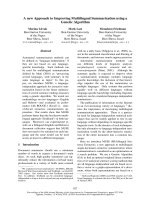
Figure 4 shows, as an example, comparison of diamag-
netic and tailored CBCA(CO)NH as far as signal D is
concerned. As observed, the two spectra are processed and
displayed with the same resolution. While the two peaks
arising from signal D are unambiguously detected in the
paramagnetic spectrum, there is no evidence of them in the
diamagnetic experiment.
Some of the
13
C resonances that were identified as arising
from the proximity of the paramagnetic center can be also
identified in the tailored
1
H-
13
C HSQC. This is the case of
the H
a
-C
a
peaks 1–4 shown in Fig. 3A, whose shifts match
with the C
a
resonances of Val15, Met7, Gly88 and Gly8.
Analogous considerations hold for the 7 H
b
-C
b
resonances
identified (Fig. 3B), which are assigned on the basis of the
already available
1
H assignment [45]. The only exception to
this criterion is peak 6 which has a C
b
shift that corresponds
to the identified Arg87 C
b
and for which no.
1
H assignment
is available. Therefore, we tentatively assign peak 6 as
Table 3. Connectivities found for signals A-G in tailored
CBCA(CO)NH and CBCANH spectra.
HN(i)
dC
a
(i-1)
(p.p.m)
dC
b
(i-1)
(p.p.m)
dC
a
(i)
(p.p.m)
dC
b
(i)
(p.p.m)
A 58.3 27.9 43.8
B
C 43.1
D 56.7 36.3 59.2
E 52.2 35.6 55 38
F
G 53.2 40.3 48.9 17.3
Table 4. New assignments obtained for oxidized plastocyanin from
Synechocystis sp. PCC6803. Copper(II) ligands are highlighted in bold.
In the right column N-Cu and H
N
-Cu distances are reported for each
amino acid.
dN
(p.p.m)
dNH
(p.p.m)
dH
a
(p.p.m)
dC
a
(p.p.m)
dH
b
(p.p.m)
dC
b
(p.p.m)
N, HN
distances
(A
˚
)
Met7 5.14 58.3 27.9 7.9–8.4
Gly8 108.29 8.80 4.67 43.8 6.7–7.0
Val15 5.44 56.3 2.68 33.1 7.9–8.5
His39 4.9–5.8
Asn40 4.6–3.7
Val42 126.57 8.6 59.2 30.8 8.1–7.3
Leu61 121.62 8.1 53.2 40.3 10.3–11.3
Ala62 125.02 9.23 48.9 17.3 8.6–7.7
Tyr82 8.2–8.4
Cys83 5.6–5.8
Glu84 6.6–7.0
His86 52.2 35.6 5.7–5.4
Arg87 128.23 8.28 55 38 7.2–7.5
Gly88 107.45 8.1 4.92 42.3 8.9–8.9
Fig. 4. Strip plot of tailored (left) and diamagnetic (right) CBCA
(CO)NH spectra in the region corresponding to signal D. While inter-
residue C
a
and C
b
peaks are present in the tailored spectrum, no
correlation is found in the diamagnetic one.
Ó FEBS 2003 Blue copper proteins studied by NMR (Eur. J. Biochem. 270) 605
Arg87 C
b
-H
b
and we identify an H
b 1
H signal of Arg87 at
0.93 p.p.m. No assignment is proposed for peak 10. All the
new assignments are summarized in Table 4.
A simple NMR protocol is an important tool to study
paramagnetic proteins
Plastocyanin is a good model system to address features of
paramagnetic copper proteins in terms of assignment
strategy. Indeed, the previously available assignment on
oxidized plastocyanin from Synechocystis sp. PCC6803 [45]
was obtained through a combination of methods which
basically rely on saturation transfer techniques [73]. Within
such a frame, dedicated experiments overcome the difficul-
ties arising from the presence of the paramagnetic center
and, eventually, permit the assignment for most of the
amino acids, including those directly bound to the copper
ion. Such nonconventional experiments include saturation
transfer [95,96] from signals broadened beyond detection
[97], mono dimensional NOEs over
1
H signals very broad
and shifted in the region 100/)50 p.p.m. [98,99], NOESY
and TOCSY experiments that allowed several
1
H assign-
ment only on the basis of relative line broadening (i.e. based
on a metal-to proton distance predictable by means of
relaxation rates) [79], NOESY cross peaks between protons
that were not identified in a classical sequential assignment
work [100], the occurrence of signals with unusual chemical
shift behaviour [39].
The above approach, which had lead to extensive
assignment of paramagnetic copper proteins even in the
first coordination sphere, required the occurrence of
favourable exchange rates between the oxidized form and
the reduced diamagnetic form. Of course, such requirements
limit the application of the approach. Therefore we have
designed experiments to extend the assignment of plasto-
cyanin without relying on its reduced state and without any
specific aprioriknowledge.
A standard approach to resonance assignment, i.e.
CBCA(CO)NH, and CBCANH, applied on plastocyanin
permitted the identification of 80 out of 94 non proline
residues [85%] with 14 amino acid for which no information
were available. All missing residues belong to the northern
loops of the protein surrounding the copper ion and fall
within a 11 A
˚
sphere from the metal center. The protocol
proposed in the present work allowed assignment of 9 out of
the above 14 residues. Indeed, no information was obtained
only for two of the three strong ligands of copper(II) (His39
and Cys83), for Asn40, whose HN group is directly involved
in a hydrogen bond with the copper-bound Cys83 S
c
atom
[45,101–103], and for Tyr82 and Glu84. It is noteworthy
that both C
a
and C
b
resonances of the binding residue His86
can be assigned. This permits the identification of reso-
nances as close as 3.6 A
˚
from the copper center without
relying on any knowledge on the electron-nucleus coupling.
Missing residues also provide a picture of the electron
spin density delocalization on the ligands. Experimental
evidence and theoretical calculations show that a larger
amount of spin density is expected on Cys83 [97,104–106].
Consistently, not only Cys83 but also the surrounding
residues (Tyr82, Glu84) are missing in the present assign-
ment. Electron spin density is delocalized also through the
H-bond between Cys83 Sc and Asn40. This makes Asn40
unobservable. The missing assignment of Asn40 prevents, in
turn, the identification of the preceding residue His39.
Indeed both
14
N ENDOR [107] and
1
H NMR data [45] on
plastocyanin indicate that metal bound imidazoles from
His86 and His39 experience a similar spin density delocali-
zation, thus supporting the hypothesis that the H-bond
between Cys83–Asn 40 is indeed responsible for the non
identification of His39 with this approach.
In summary, such an approach allows identification, in a
sequence specific fashion, 89 out of 94 non proline residues
(95%) providing 89%, 87% and 92% of the assignment of
C
a
,C
b
and N–H, respectively. With the above approach we
can reach metal-to-nucleus distances of 7.2, 3.6, and 7.5 A
˚
,
for H, C
a
and N, respectively.
Conclusions
In the case of the oxidized plastocyanin from Synechocystis
sp. PCC6803, an NMR approach based on classical two
and three dimensional experiments for sequential assign-
ment leaves unobserved 14 residues out of 98 amino acids. A
protocol that simply makes use of tailored version of 2D
HSQC and 3D CBCA(CO)NH and CBCANH leads to the
identification of 9 of the above 14 residues. Although it is
clear that such improvement does not circumvent all the
limitations arising from the presence of an oxidized copper
center and actually still prevents the complete characteriza-
tion of the first coordination sphere, we should stress that
the approach proposed allows those structural biologists
that are not experts nor familiar with paramagnetic proteins
to substantially increase their knowledge.
Acknowledgements
We are grateful to Prof. Ivano Bertini for his advice and support. The
expression system of Synechocystis sp. PCC6803 plastocyanin was a
generous gift of Prof S. Ciurli. This work was supported by the
European Union Research and Training Network (RTN) Project
ÔCross correlation between the fluctuations of different interactions: a
new avenue for biomolecular NMRÕ (Contract no. HPRN-CT-2000–
00092). I.G. is a Fellow of the Marie Curie Training Site ÔNMR in
Inorganic Structural BiologyÕ, contract no. HPMT-2000–000137.
Support from PARABIO (HPRT-CT-00009) is acknowledged.
References
1. Brown,D.R.,Qin,K.,Herms,J.W.,Madlung,A.,Manson,J.,
Strome, R., Fraser, P.E., Kruck, T.A., Von Bohlen, A., Schulz-
Schaeffer, W., Giese, A., Westaway, D.&Kretzschmar, H.A. (1997)
The cellular prion protein binds copper in vivo. Nature 684–687.
2. Poulos, T.L. (1999) Helping copper find a home. Nat Struct. Biol.
6, 709–711.
3. Viles, J.H., Cohen, F.E., Prusiner, S.B., Goodin, D.B., Wright,
P.E. & Dyson, H.J. (1999) Copper binding to the prion protein:
structural implications of four identical cooperative binding sites.
Proc.NatlAcad.Sci.USA96, 2042–2047.
4. O’Halloran, T.V. & Culotta, V.C. (2000) Metallochaperones: An
Intracellular Shuttle Service for Metal Ions. J. Biol. Chem. 275,
25057–25060.
5. Goto, J.J., Zhu, H., Sanchez, R.J., Gralla, E.B. & Valentine, J.S.
(2000) Loss of in vitro metal ion binding specificity in mutant
copper-zinc superoxide dismutase associated with familial amyo-
trophic lateral sclerosis. J. Biol. Chem. 14, 1007–1014.
606 I. Gelis et al. (Eur. J. Biochem. 270) Ó FEBS 2003
6. Harrison, M.D., Jones, C.E., Solioz, M. & Dameron, C.T. (2000)
Intracellular copper routing: the role of copper chaperones. Trends
Biochem. Sci. 25, 29–32.
7. Heaton, D., Nittis, T., Srinivasan, C. & Winge, D.R. (2000)
Mutational analysis of the mitochondrial copper metallochaper-
one Cox17. J. Biol. Chem. 275, 37582–37587.
8. McMahon, H.E., Mange, A., Nishida, N., Creminon, C., Casa-
nova, D. & Lehmann, S. (2001) Cleavage of the amino terminus of
the prion protein by reactive oxygen species. J. Biol. Chem. 276,
2286–2291.
9. Quaglio, E., Chiesa, R. & Harris, D.A. (2001) Copper converts the
cellular prion protein into a protease-resistant species that is dis-
tinct from the scrapie isoform. J. Biol. Chem. 276, 11432–11438.
10. Rosenzweig, A.C. (2001) Copper delivery by metallochaperone
proteins. Acc Chem Res. 34, 119–128.
11. Hayward, L.J., Rodriguez, J.A., Kim, J.W., Tiwari, A., Goto, J.J.,
Cabelli, D.E., Valentine, J.S. & Brown, R.H.J. (2002) Decreased
metallation and activity in subsets of mutant superoxide dis-
mutases associated with familial ALS. J. Biol. Chem. 277, 15923–
15931.
12. Hughson, F.M., Wright, P.E. & Baldwin, R.L. (1987) Structural
characterization of a partly folded apomyoglobin intermediate.
Science 249, 1544–1548.
13. Arcus, V.L., Vuilleumier, S., Freund, S.M.V., Bycroft, M. &
Fersht, A.R. (1995) A comparison of the pH, urea, and tem-
perature-denaturated states of barnase by heteronuclear NMR:
implications for the initiation of protein folding. J. Mol. Biol. 254,
305–321.
14. Frank, M.K., Clore, G.M. & Gronenborn, A.M. (1995) Structural
and dynamic characterization of the urea denatured state of the
immunoglobin binding domain of streptococcal protein G by
multidimensional heteronuclear NMR spectroscopy. Protein Sci.
4, 2605–2615.
15. Serrano, L. (1995) Comparison between the phi distribution of
amino acids in the protein data base and NMR data indicates that
amino acids have various phi propensities in the random coil
conformation. J. Mol. Biol. 254, 322–333.
16. Bertini, I., Cowan, J.A., Luchinat, C., Natarajan, K. & Piccioli,
M. (1997) Characterization of a partially unfolded high potential
iron protein relevant to the folding pathway and cluster stability.
Biochemistry 36, 9332–9339.
17. Zhang, O., Kay, L.E., Shortle, D. & Forman-Kay, J.D. (1997)
Comprehensive NOE characterization of a partially folded large
fragment of staphylococcal nuclease Delta131Delta, using NMR
methods with improved resolution. J. Mol. Biol. 272, 9–20.
18. Bertini, I., Luchinat, C., Piccioli, M. & Soriano, A. (1998)
Folding properties of iron sulfur proteins. (Dedicated to Prof.
O. Yamauchi). Inorg. Chim. Acta 283, 12–16.
19. Dyson, H.J. & Wright, P.E. (1998) Equilibrium NMR studies
of unfolded and partially folded proteins. Nat. Struct. Biol. 5,
499–503.
20. Kuhn, T. & Schwalbe, H. (2000) Monitoring the Kinetics of Ion-
Dependent Protein Folding by Time-Resolved NMR Spectro-
scopy at Atomic Resolution. J. Am. Chem. Soc. 122, 6169–6174.
21. Farrar, J.A., Neese, F., Lappalainen, P., Kroneck, P.M.H.,
Saraste, M., Zumft, W.G. & Thomson, A.J. (1996) The Electronic
Structure of Cu
A
: a novel mixed-valence dinuclear copper elec-
tron-transfer center. J. Am. Chem. Soc. 118, 11501–11514.
22. Klomp, L.W., Lin, S.J., Yuan, D., Klausner, R.D., Culotta, V.C.
& Gitlin, J.D. (1997) Identification and functional expression of
HAH1, a novel human gene involved in copper homeostasis.
J. Biol. Chem. 272, 9221–9226.
23. Van Pouderoyen, G., Cigna, G., Rolli, G., Cutruzzola, F.,
Malatesta, F., Silvestrini, M.C., Brunori, M. & Canters, G.W.
(1997) Electron-transfer properties of Pseudomonas aeruginosa
[Lys44,Glu64]azurin. Eur. J. Biochem. 247, 322–331.
24. Casareno, R.L., Waggoner, D.J. & Gitlin, J.D. (1998) The copper
chaperone CCS directly interacts with copper/zinc superoxide
dismutase. J. Biol. Chem. 273, 23625–23628.
25. Wittung-Stafshede, P., Malmstro
¨
m, B.G., Sanders, D., Fee, J.A.,
Winkler, J.R. & Gray, H.B. (1998) Effect of redox state on the
folding free energy of a thermostable electron-transfer metallo-
protein: the CuA domain of cytochrome oxidase from Thermus
thermophilus. Biochemistry 37, 3172–3177.
26. Hamza, I., Schafer, M., Klomp, L.W. & Gitlin, J.D. (1999)
Interaction of the copper chaperone HAH1 with the Wilson dis-
ease protein is essential for copper homeostasis. Proc. Natl Acad.
Sci. USA 96, 13363–13368.
27. Huffman, D.L. & O’Halloran, T.V. (2000) Energetics of Copper
Trafficking Between the Atx1 Metallochaperone and the Intra-
cellularCopper-transporter, Ccc2. J. Biol. Chem.275, 18611–18614.
28. Crowley, P.B., Otting, G., Schlarb-Ridley, B., Canters, G.W. &
Ubbink, M. (2001) Hydrophobic interactions in a cyanobacterial
plastocyanin-cytochrome f complex. J. Am. Chem. Soc. 123,
10444–10453.
29. Banci, L., Bertini, I., Del Conte, R., Markey, J. & Ruiz-Duen
˜
as,
F.J. (2001) Copper trafficking: the solution structure of Bacillus
subtilis CopZ. Biochemistry 40, 15660–15668.
30. Bertini, I. & Luchinat, C. (1996) NMR of paramagnetic sub-
stances. Coord. Chem. Rev. 150, 1–225.
31. Bertini, I., Luchinat, C. & Parigi, G. (2001) Solution NMR of
Paramagnetic Molecules. Elsevier, Amsterdam.
32. Banci,L.,Bertini,I.&Luchinat,C.(1991)Nuclear and Electron
Relaxation. The Magnetic Nucleus-Unpaired Electron Coupling in
Solution. VCH, Weinheim.
33. Xia, B., Westler, W.M., Cheng, H., Meyer, J., Moulis, J M. &
Markley, J.L. (1995) Detection and Classification of Hyperfine-
Shifted
1
H,
2
H, and
15
N Resonances from the Four Cysteines That
Ligate Iron in Oxidized and Reduced Clostridium pasteurianum
Rubredoxin. J. Am. Chem. Soc. 117, 5347–5350.
34. Wilkens, S.J., Xia, B., Weinhold, F., Markley, J.L. & Westler,
W.M. (1998) NMR investigations of Clostridium pasteurianum
rubredoxin. Origin of hyperfine
1
H,
2
H,
13
Cand
15
NNMRche-
mical shifts in iron-sulfur proteins as determined by comparison of
experimental data with hybrid density functional calculations.
J. Am. Chem. Soc. 120, 4806–4814.
35. Hurley, J.K., Weber-Main, A.M., Hodges, A.E., Stankovic, M.T.,
Benning, M.M., Holden, H.M., Cheng, H., Xia, B., Markley, J.L.,
Genzor, C., Gomez-Moreno, C., Hafezi, R. & Tollin, G. (1997)
Iron-sulfur cluster cysteine-to-serine mutants of Anabaena 2Fe-2S-
ferredoxin exhibit unexpected redox properties and are competent
in electron transfer to ferredoxin: NADP+ reductase. Biochem-
istry 36, 15109–15117.
36. Fee, J.A. & Briggs, R.G. (1975) Studies on the reconstitution of
bovine erythrocyte superoxide dismutase. V. Preparation and
properties of derivatives in which both zinc and copper sites
contain copper. Biochim. Biophys. Acta 400, 439–439.
37. Lippard, S.J., Burger, A.R., Ugurbil, K., Pantoliano, M.W. &
Valentine, J.S. (1977) Nuclear magnetic resonance and chemical
modification studies of bovine erythrocyte superoxide dismutase:
evidence for zinc-promoted organization of the active site struc-
ture. Biochemistry 16, 1136–1141.
38. Bertini, I., Lanini, G., Luchinat, C., Messori, L., Monnanni, R. &
Scozzafava, A. (1985) Investigation of Cu
2
Co
2
SOD and its anion
derivatives.
1
H NMR and electronic spectra. J. Am. Chem. Soc.
107, 4391–4396.
39. Kalverda, A.P., Salgado, J., Dennison, C. & Canters, G.W. (1996)
Analysis of the paramagnetic copper (II) site of amicyanin by
1
H
NMR spectroscopy. Biochemistry 35, 3085–3092.
40. Penfield, K.W., Gewirth, A.A. & Solomon, E.I. (1985) Electronic
structure and bonding of the blue copper site in plastocyanin.
J. Am. Chem. Soc. 107, 4519–4529.
Ó FEBS 2003 Blue copper proteins studied by NMR (Eur. J. Biochem. 270) 607
41. Banci, L., Bertini, I., Cremonini, M.A., Gori Savellini, G., Luch-
inat, C., Wu
¨
thrich,K.&Gu
¨
ntert, P. (1998) PSEUDODYANA for
NMR structure calculation of paramagnetic metalloproteins using
torsion angle molecular dynamics. J. Biomol. NMR 12, 553–557.
42. Turner, D.L., Brennan, L., Chamberlin, S.G., Louro, R.O. &
Xavier, A.V. (1998) Determination of solution structures of
paramagnetic proteins by NMR. Eur. Biophys. J. 27, 367–375.
43. Gochin, M. & Roder, H. (1995) Protein Structure Refinement
based on Paramagnetic NMR shifts. Applications to Wild-Type
and mutants forms of cytochrome c. Protein Sci. 4, 296–305.
44. Wishart, D.S., Sykes, B.D. & Richards, F.M. (1992) The chemical
shift index: a fast and simple method for the assignment of protein
secondary structure through NMR spectroscopy. Biochemistry 31,
1647–1651.
45. Bertini, I., Ciurli, S., Dikiy, A., Ferna
´
ndez, C.O., Luchinat, C.,
Safarov, N., Shumilin, S. & Vila, A.J. (2001) The first solution
structure of an oxidized paramagnetic copper (II) protein: the case
of plastocyanin from the cyanobacterium Synechocystis PCC6803.
J. Am. Chem. Soc. 123, 2405–2413.
46. Bertini, I., Luchinat, C. & Piccioli, M. (2001) Paramagnetic probes
in metalloproteins. turning limitations into advantages. Meth
Enzymol. 339, 314–340.
47. Boisbouvier, J., Gans, P., Blackledge, M., Brutscher, B. & Marion,
D. (1999) Long-range structral information in NMR studies of
paramagnetic molecules from electron spin-nuclear spin cross-
correlated relaxation. J. Am. Chem. Soc. 121, 7700–7701.
48. Nguyen, B.D., Xia, Z., Yeh, D.C., Vyas, K., Deaguero, H. &
La Mar., G.N. (1999) Solution NMR determination of the ani-
sotropy and orientation of the paramagnetic susceptibility tensor
as a function of temperature for metmyoglobin cyanide: implica-
tions for the population of excited electron states. J. Am. Chem.
Soc. 121, 208–217.
49. Xia, Z., Nguyen, B.D. & La Mar., G.N. (2000) The use of che-
mical shift temperature gradients to establish the paramagnetic
susceptibility tensor orientation: implication for structure
determination/refinement in paramagnetic metalloproteins.
J. Biomol. NMR 17, 167–174.
50. Hus, J.C., Marion, D. & Blackledge, M. (2000) De novo
determination of protein structure by nmr using orientational and
long-range order restraints. J. Mol. Biol. 298, 927–936.
51. Tsan, P., Caffrey, M., Daku, M.L., Cusanovich, M., Marion, D. &
Gans, P. (2001) Magnetic susceptibility tensor and heme contact
shifts determinations in the Rhodobacter capsulatus ferricyto-
chrome c’: NMR and magnetic susceptibility studies. J. Am. Chem
Soc. 123, 2231–2242.
52. Epperson, J., Ming, L J., Baker, G. & Newkome, G. (2001)
Paramagnetic cobalt (II) as an NMR probe of dendrimer struc-
ture: mobility and cooperativity of dendritic arms. J. Am. Chem
Soc. 123, 8583–8592.
53. Piccioli, M. (1996) The application of selective-excitation pulse
sequences in NMR spectroscopy of paramagnetic proteins.
J. Magn. Reson. B110, 202–204.
54. Luchinat, C., Piccioli, M., Pierattelli, R., Engelke, F.,
Marquardsen, T. & Ruin, R. (2001) Development of NMR
intrumentation to achieve excitation of large bandwidths in high
resolution spectra at high-fields. J. Magn. Reson. 150, 161–166.
55. Ghose, R. & Prestegard, J.H. (1997) Electron spin-nuclear spin
cross-correlation effects on multiplet splittings in paramagnetic
proteins. J. Magn. Reson. 128, 138–143.
56. Bougault, C.M., Dou, Y., Ikeda-Saito, M., Langry, K.C., Smith,
K.M. & La Mar., G.N. (1998) Solution
1
H NMR study of the
electronic structure and magnetic properties of high-spin ferrous
or deoxy myoglobins. J. Am. Chem. Soc. 120, 2113–2123.
57. Hus, J.C., Marion, D. & Blackledge, M. (2000) De novo
determination of protein structure by NMR using orientational
and long-range order restraints. J. Mol Biol. 298, 927–936.
58. Brennan, L., Turner, D.L., Messias, A.C., Teodoro, M.L., LeGall,
J., Santos, H. & Xavier, A.V. (2000) Structural basis for the net-
work of functional cooperativities in cytochrome c3 from
Desulfovibrio gigas: solution structures of the oxidised and reduced
states. J. Mol. Biol. 298, 61–82.
59. Ubbink, M., Worrall, J.A.R., Canters, G.W., Groenen, E.J.J. &
Huber, M. (2002) Paramagnetic resonance of biological metal
centers. Annu. Rev. Biophys. Biomol. Struct. 31, 393–422.
60. Walker, F.A. (2000) Proton NMR and EPR spectroscopy of
Paramagnetic Metalloporphyrins. In The Porphyrin Handbook
(Kadish, K.M., Smith, K.M. & Guilard, R., eds), pp. 81–183.
Academic Press, San Diego, CA, USA.
61. Vathyam, S., Byrd, R.A. & Miller, A.F. (2000) Mapping the
effects of metal ion reduction and substrate analog binding to
Fe-superoxide dismutase by NMR spectroscopy. Magn. Reson.
Chem. 38, 536–542.
62. Machonkin, T.E., Westler, W.M. & Markley, J.L. (2002)
13
C
[
13
C]2D NMR: a novel strategy for the study of paramagnetic
proteins with slow electronic relaxation times. J. Am. Chem. Soc.
124, 3204–3205.
63. Fernandez, C.O., Cricco, J.A., Slutter, C.E., Richards, J.H., Gray,
H.B. & Vila, A.J. (2002) Axial ligand modulation of the electronic
structures of binuclear cooper sites: analysis of paramagnetic
1
H
NMR spectra of Met160Gln Cu (A). J. Am. Chem. Soc. 123,
11678–11685.
64. Grzesiek, S. & Bax, A. (1992) Correlating backbone amide
and side chain resonances in larger proteins by multiple
relayed triple resonance NMR. J. Am. Chem. Soc. 114, 6291–
6293.
65. Muhandiram, D.R. & Kay, L.E. (1994) Gradient-enhanced triple
resonance three-dimensional NMR experiments with improved
sensitivity. J. Magn. Reson. B103, 203–216.
66. Grzesiek, S. & Bax, A. (1992) An efficient experiment for
sequential backbone assignment of medium-sized isotopically
enriched proteins. J. Magn. Reson. 99, 201–207.
67. Meissner,A.&Sorensen,O.W.(2001)SequentialHNCACBand
CBCANH protein NMR pulse sequences. J. Magn. Reson. 151,
328–331.
68. Wu
¨
thrich, K. (1996) Biological macromolecules: structure deter-
mination in solution. In Encyclopedia of Nuclear Magnetic Res-
onance (Grant, D.M. & Harris, R.K., eds), pp. 932–939. John
Wiley & Sons, Chichester, UK.
69. Riek, R., Wider, G., Billeter, M., Hornemann, S., Glockshuber, R.
&Wu
¨
thrich, K. (1998) Prion protein NMR structure and familial
human spongiform encephalopathies. Proc. Natl Acad. Sci. USA
95, 11667–11672.
70. Adman, E.T. (1985) Structure and functin of blue copper proteins.
In Metalloproteins (Harrison, P., ed.), pp. 1–42. Macmillan, New
York.
71. Adman, E.T. (1991) Copper protein structures. Adv. Prot. Chem.
42, 144–197.
72. Solomon, E.I., Baldwin, M.J. & Lowery, M.D. (1992) Electronic
structures of active sites in copper proteins: contributions to
reactivity. Chem. Rev. 92, 521.
73. Bertini, I., Ciurli, S., Dikiy, A., Gasanov, R., Luchinat, C.,
Martini, G. & Safarov, N. (1999) High-field NMR studies of
oxidized blue copper proteins: the case of spinach plastocyanin.
J. Am. Chem. Soc. 121, 2037–2046.
74. Bertini, I., Bryant, D.A., Ciurli, S., Dikiy, A., Ferna
´
ndez, C.O.,
Luchinat, C., Safarov, N., Vila, A.J. & Zhao, J. (2001) Backbone
dynamics of plastocyanin in both oxidation states. Solution
structure of the reduced form and comparison with the oxidized
state. J. Biol. Chem. 276, 47217–47226.
75. Bodenhausen, G. & Ruben, D.J. (1980) Natural abundance
nitrogen-15 NMR by enhanced heteronuclear spectroscopy.
Chem. Phys. Lett. 69, 185–188.
608 I. Gelis et al. (Eur. J. Biochem. 270) Ó FEBS 2003
76. Kay, L.E., Keifer, P. & Saarinen, T. (1992) Pure absorption
gradient enhanced heteronucler single quantum correlation
spectroscopy with improved sensitivity. J. Am. Chem. Soc. 114,
10663–10665.
77. Cavanagh, J., Palmer, A.G., IIIWright, P.E. & Rance, M.
(1991) Sensitivity improvement in proton-detected two-dimen-
sional heteronuclear relay spectroscopy. J. Magn. Reson. 91,429–
436.
78. Palmer,A.G., IIICavanagh, J., Wright, P.E. & Rance, M. (1991)
Sensitivity improvement in proton-detected two dimension al
heteronuclear correlation NMR spectroscopy. J. Magn. Reson. 93,
151–170.
79. Bertini, I., Couture, M.M.J., Donaire, A., Eltis, L.D., Felli, I.C.,
Luchinat,C.,Piccioli,M.&Rosato,A.(1996)Thesolution
structure refinement of the paramagnetic reduced HiPIP I from
Ectothiorhodospira halophila by using stable isotope labeling and
nuclear relaxation. Eur. J. Biochem. 241, 440–452.
80. Kay, L.E., Ikura, M., Tschudin, R. & Bax, A. (1990) Three-
dimensional triple-resonance NMR spectroscopy of isotopically
enriched proteins. J. Magn. Reson. 89, 496–514.
81. Pochapsky, S.S., Jain, N.U., Kuti, M., Lyons, T.A. & Heymont, J.
(1999) A refined model for the solution structure of oxidized
putidaredoxin. Biochemistry 38, 4681–4690.
82. Baumann, B., Sticht, H., Scha
¨
rpf, M., Sutter, M., Haehnel, W. &
Roesch, P. (1996) Structure of synechococcus-elongatus [Fe
2
S
2
]
ferredoxininsolution.Biochemistry 35, 12831–12841.
83. Vathyam, S., Byrd, R.A. & Miller, A.F. (1999) Assignment of the
backbone resonances of oxidized Fe-superoxide dismutase, a
42 kDa paramagnet-containing enzyme. J. Biomol. NMR 14,293–
294.
84. Piccioli, M. & Poggi, L. (2002) Tailored HCCH-TOCSY experi-
ment for resonance assignment in the proximity of a paramagnetic
center. J. Magn. Reson. 155, 236–243.
85. Skidmore, K. & Simonis, U. (1996) Novel strategy for assigning
hyperfine shifts using pulsed-field gradients heteronuclear multi-
ple-bond correlation spectroscopy. Inorg. Chem. 35, 7470–7471.
86. Bertini, I., Lee, Y M., Luchinat, C., Piccioli, M. & Poggi, L.
(2001) Locating the metal ion in calcium-binding proteins by using
cerium (III) as a probe. Chembiochem. 2, 550–558.
87. Bertini, I., Dalvit, C., Huber, J.G., Luchinat, C. & Piccioli, M.
(1997) ePHOGSY experiment on a paramagnetic protein: location
of the catalytic water molecule in the heme crevice of the oxidized
form of horse heart cytochrome c. FEBS Lett. 415, 45–48.
88. States, D.J., Habenkorn, R.A. & Ruben, D.J. (1982) A two-
dimensional nuclear overhauser experiment with pure absorption
phase in four quadrants. J. Magn. Reson. 48, 286–292.
89. Grzesiek, S. & Bax, A. (1992) Improved 3D triple-resonance
NMR techniques applied to a 31 KDa protein. J. Magn. Reson.
96, 432–440.
90. Bertini, I., Luchinat, C., Macinai, R., Piccioli, M., Scozzafava, A.
& Viezzoli, M.S. (1994) Paramagnetic metal centers in proteins can
be investigated through heterocorrelated NMR spectroscopy.
J. Magn. Reson. B104, 95–98.
91. Wishart, D.S., Sykes, B.D. & Richards, F.M. (1991) Relationship
between nuclear magnetic resonance chemical shift and protein
secondary structure. J. Mol. Biol. 222, 311–333.
92. Wishart, D.S. & Sykes, B.D. (1994) The
13
Cchemicalshift
index: a simple method for the identification of protein secondary
structure using
13
C chemical shift data. J. Biomol. NMR 4,171–
180.
93. Spera, S. & Bax, A. (1991) Empirical correlation between protein
backbone conformation and
13
Ca and C b
13
C nuclear magnetic
resonance chemical shifts. J. Am. Chem. Soc. 113, 5490–5492.
94. Bertini, I., Bren, K.L., Clemente, A., Fee, J.A., Gray, H.B.,
Luchinat, C., Malmstro
¨
m, B.G., Richards, J.H., Sanders, D. &
Slutter, C.E. (1996) The Cu
a
center of a soluble domain from
thermus cytochrome ba
3
:anNMRinvestigationofthepara-
magnetic protein. J. Am. Chem. Soc. 46, 11658–11659.
95. Jensen, M., Hansen, D.F. & Led, J.J. (2002) A general method for
determining the electron self-exchange rates of blue copper pro-
teins by longitudinal NMR relaxation. J. Am. Chem Soc. 124,
4093–4096.
96. Ma, L., Philipp, E. & Led, J.J. (2001) Determination of the elec-
tron self-exchange rates of blue copper proteins by super-WEFT
NMR spectroscopy. J. Biomol. NMR 19, 199–208.
97. Bertini, I., Ferna
´
ndez,C.O.,Karlsson,B.G.,Leckner,J.,Luch-
inat, C., Malmstro
¨
m, B.G., Nersissian, A.M., Pierattelli, R.,
Shipp, E., Valentine, J.S. & Vila, A.J. (2000) Structural informa-
tion through NMR hyperfine shifts in blue copper proteins. J. Am.
Chem. Soc. 122, 3701–3707.
98. Banci, L., Bertini, I., Luchinat, C., Piccioli, M., Scozzafava, A. &
Turano, P. (1989)
1
H NOE studies on dicopper (II) dicobalt (II)
superoxide dismutase. Inorg. Chem. 28, 4650–4656.
99. Goasdoue, N., Riviere, D.G., Correia, I., Convert, O. & Piccioli,
M. (2000) Multiple selective excitation as a tool for NMR studies
of paramagnetic proteins. Magn. Reson. Chem. 38, 827–832.
100. Banci, L., Bertini, I., Eltis, L.D., Felli, I.C., Kastrau, D.H.W.,
Luchinat, C., Piccioli, M., Pierattelli, R. & Smith, M. (1994) The
three dimensional structure in solution of the paramagnetic pro-
tein high-potential iron-sulfur protein I from Ectothiorhodospira
halophila through nuclear magnetic resonance. Eur. J. Biochem.
225, 715–725.
101. Guss, J.M. & Freeman, H.C. (1983) Structure of Oxidized Poplar
Plastocyanin at 1.6 A Resolution. J. Mol. Biol. 169, 521–563.
102. Donaire, A., Salgado, J. & Moratal, J.M. (1998) Determination of
the magnetic axes of cobalt (II) and nickel (II) azurins from 1H
NMR data: influence of the metal and axial ligands on the origin
of magnetic anisotropy in blue copper proteins. Biochemistry 37,
8659–8673.
103. Donaire, A., Jimenez, B., Moratal Mascarell, J.M., Hall, J.F. &
Hasnain, S.S. (2001) Electronic characterization of the oxidized
state of the blue copper protein rusticyanin by 1H NMR: is the
axial methionine the dominant influence for the high redox
potential. Biochemistry 40, 837–846.
104. Randall, D.W., Gamelin, D.R., LaCroix, L.B. & Solomon, E.I.
(2000) Electronic structure contributions to electron transfer in
blue Cu and Cu
A
. JBIC 5, 16–29.
105. LaCroix, L.B., Randall, D.W., Nersissian, A.M., Hoitink,
C.W.G., Canters, G.W., Valentine, J.S. & Solomon, E.I. (1998)
Spectroscopi and geometric variation in perturbed blue copper
centers: electronic structures of stellacyanin and cucumber pasic
protein. J. Am. Chem. Soc. 120, 9621–9631.
106. Solomon, E.I., Penfield, K.W., Gewirth, A.A., Lowery, M.D.,
Shadle, S.E., Guckert, J.A. & LaCroix, L.B. (1996) Electronic
structure of the oxidized and reduced blue copper sites: con-
tributions to the electron transfer pathway, reduction potential,
and geometry. Inorg. Chim. Acta 243, 67–78.
107. Werst, M.M., Davoust, E.E. & Hoffman, B.M. (1991) Ligand spin
densities in blue copper proteins by Q-band
1
Hand
14
N ENDOR
spectroscopy. J. Am. Chem. Soc. 113, 1533–1538.
Supplementary material
The following material is available from http://www.
blackwellpublishing.com/products/journals/suppmat/EJB/
EJB3400/EJB3400sm.htm
Table S1.
13
C assignment obtained for oxidized plastocya-
nin from Synechocystis sp. PCC6803 (BMRB accession
number: 5584).
Ó FEBS 2003 Blue copper proteins studied by NMR (Eur. J. Biochem. 270) 609