Báo cáo khoa học: Relation between domain evolution, specificity, and taxonomy of the a-amylase family members containing a C-terminal starch-binding domain pot
Bạn đang xem bản rút gọn của tài liệu. Xem và tải ngay bản đầy đủ của tài liệu tại đây (491.39 KB, 11 trang )
Relation between domain evolution, specificity, and taxonomy
of the a-amylase family members containing a C-terminal
starch-binding domain
S
ˇ
tefan Janec
ˇ
ek
1
, Birte Svensson
2
and E. Ann MacGregor
3
1
Institute of Molecular Biology, Slovak Academy of Sciences, Bratislava, Slovakia;
2
Department of Chemistry,
Carlsberg Laboratory, Copenhagen Valby, Denmark;
3
Department of Chemistry, University of Manitoba, Winnipeg, Canada
The a-amylase family (glycoside hydrolase family 13;
GH 13) contains enzymes with approximately 30 specifi-
cities. Six types of enzyme from the family can possess
a C-terminal starch-binding domain (SBD): a-amylase,
maltotetraohydrolase, maltopentaohydrolase, maltogenic
a-amylase, acarviose transferase, and cyclodextrin glu-
canotransferase (CGTase). Such enzymes are multidomain
proteins and those that contain an SBD consist of four or
five domains, the former enzymes being mainly hydrolases
and the latter mainly transglycosidases. The individual
domains are labelled A [the catalytic (b/a)
8
-barrel], B, C,
D and E (SBD), but D is lacking from the four-domain
enzymes. Evolutionary trees were constructed for domains
A, B, C and E and compared with the Ôcomplete-sequence
treeÕ. The trees for domains A and B and the complete-
sequence tree were very similar and contain two main
groups of enzymes, an amylase group and a CGTase
group. The tree for domain C changed substantially, the
separation between the amylase and CGTase groups being
shortened, and a new border line being suggested to
include the Klebsiella and Nostoc CGTases (both four-
domain proteins) with the four-domain amylases. In the
ÔSBD treeÕ the border between hydrolases (mainly
a-amylases) and transglycosidases (principally CGTases)
was not readily defined, because maltogenic a-amylase,
acarviose transferase, and the archaeal CGTase clustered
together at a distance from the main CGTase cluster.
Moreover the four-domain CGTases were rooted in the
amylase group, reflecting sequence relationships for the
SBD. It appears that with respect to the SBD, evolu-
tion in GH 13 shows a transition in the segment of
the proteins C-terminal to the catalytic (b/a)
8
-barrel
(domain A).
Keywords: a-amylase family; glycoside hydrolase family 13;
starch-binding domain; evolutionary tree; domain evolution.
The a-amylase family (glycoside hydrolase family 13, with
close relatives in families 70 and 77) consists at present of
enzymes of almost 30 different specificities comprising
hydrolases, transglycosidases and isomerases [1]. All of these
contain a catalytic (b/a)
8
-barrel domain first recognized
in Taka-amylase A, an a-amylase from Aspergillus oryzae
[2]. This fold was confirmed by crystallography for
other specificities, such as cyclodextrin glucanotransferase
(CGTase) [3], oligo-1,6-glucosidase [4], maltotetraohydro-
lase [5], isoamylase [6], neopullulanase [7], maltogenic
a-amylase [8], maltogenic amylase [9], amylomaltase [10],
glycosyltrehalose trehalohydrolase [11], amylosucrase
[12], maltosyltransferase [13], cyclomaltodextrinase [14],
4-a-glucanotransferase [15], and branching enzyme [16].
Structure determinations of family members with yet other
specificities are in progress (e.g. [17,18]). Furthermore,
prediction of the presence of this (b/a)
8
-barrel fold in other
family members has been carried out using unambiguous
sequence similarities, particularly at well-known conserved
sequence motifs [19–22].
In the sequence-based classification of glycoside hydro-
lases [23] family 13 (most typically a-amylases) forms the
GH-H group together with glycoside hydrolase families
(GHs) 70 (glucan sucrase-type glycosyltransferases) and 77
(amylomaltases). These enzymes are multidomain proteins
that contain several characteristic domains in addition to
domain A, the catalytic (b/a)
8
-barrel[1].Mostofthem
possess a domain B that protrudes from the barrel between
the third b-strand and third a-helix and varies greatly in
length, sequence and tertiary structure [20,24]. Domain C,
which immediately succeeds the catalytic barrel, is essen-
tially a b-sandwich structure (e.g. [2–5]), characteristic for
GH 13 members, but missing in GH 77, as shown by the
structure of amylomaltase from Thermus aquaticus [10].
Domain C is, moreover, lacking in its common form in
Correspondence to S
ˇ
.Janec
ˇ
ek, Institute of Molecular Biology,
Slovak Academy of Sciences, Du´ bravska
´
cesta 21,
SK-84551 Bratislava, Slovakia.
Fax: + 421 2 5930 7416, Tel.: + 421 2 5930 7420,
E-mail:
Abbreviations: CBM, carbohydrate-binding module family; CGTase,
cyclodextrin glucanotransferase; GH, glycoside hydrolase family;
SBD, starch-binding domain.
Enzymes: a-amylase (EC 3.2.1.1); maltotetraohydrolase (3.2.1.60);
maltopentaohydrolase (EC 3.2.1 ); maltogenic a-amylase
(EC 3.2.1.133); cyclodextrin glucanotransferase (EC 2.4.1.19);
acarviose transferase (EC 2.4.1 ).
(Received 17 September 2002, revised 18 November 2002,
accepted 28 November 2002)
Eur. J. Biochem. 270, 635–645 (2003) Ó FEBS 2003 doi:10.1046/j.1432-1033.2003.03404.x
GH 70 where the glycosyltransferases have a circularly
permuted catalytic (b/a)
8
-barrel [21]. Several GH 13 mem-
bers contain one or more N-terminal domains preceding the
barrel [19]; such domains have occasionally been named
domain N although they are not all structurally related.
Finally, a group of enzymes in GH 13 contain one or two
additional all-b domains, D and E, at the C-terminal end,
following the above-mentioned domain C. If the enzymes
possess both domains D and E, they do not normally
contain an N-domain, and are thus five-domain proteins
possessing the catalytic (b/a)
8
-barrel (domain A) and the
four domains B, C, D and E. In the case of four-domain
proteins without an N-domain, only domain E (but not
domain D) is present. It should be noted that the function of
domain D is as yet unknown [19,22]. Domain E, however,
was recognized early and has attracted much attention due
to its raw starch-binding function (e.g [25–32]), which
facilitates degradation of starch granules by the enzymes
containing such a domain. Throughout this paper, domain
E is referred to as SBD, the starch-binding domain.
In a classification of carbohydrate-binding modules, this
starch-binding domain is considered to belong to family 20
(CBM 20) [33], and is central to the present study. It is
worth mentioning here that amylolytic enzymes containing
a completely different kind of starch-binding site [34,35] or a
second type of SBD consisting of some sequence repeats of
unknown structure [36,37] are outside the scope of this
work. The SBD of the present study, CBM 20, is well-
known as domain E in CGTases [3,38–41]. It occurs,
however, not only in some enzymes of the GH 13 a-amylase
family but also in certain b-amylases (GH 14), and in the
vast majority of glucoamylases (GH 15), despite the fact
that while GH 13 enzymes bring about retention of
configuration, both b-amylases and glucoamylases are
inverting enzymes and possess catalytic domains that differ
from the (b/a)
8
-barrel characteristic of the a-amylase family
[2,42,43]. This ÔclassicalÕ SBD motif consists of seven
b-strand segments forming an open-sided distorted b-barrel,
as demonstrated by the crystal structures of CGTases from
Bacillus circulans strains 8 and 251 [3,27], Bacillus stearo-
thermophilus [38], Thermoanaerobacterium thermosulfuro-
genes [40], Bacillus sp. strain 1011 [41], and b-amylase from
Bacillus cereus [44], and the NMR solution structure of the
isolated recombinant SBD of glucoamylase from Aspergillus
niger [28].
The SBD is present in approximately 10% of amylo-
lytic enzymes from GHs 13, 14 and 15 [26,30]. In the
a-amylase family, this module has been recognized in
enzymes having six of the almost 30 specificities: a-amy-
lase, maltotetraohydrolase, maltopentaohydrolase, malto-
genic a-amylase, CGTase, and the acarviose transferase
(which has, however, been assigned the same EC number
as CGTase). While the first three enzymes are four-
domain proteins, the latter three have five domains, with
the SBD being the C-terminal domain in all cases.
Furthermore, the presence of the SBD in an amylolytic
enzyme is closely connected with the enzyme origin. Only
microorganisms, in particular filamentous fungi, Gram-
positive bacteria (Firmicutes), Proteobacteria of the
c-subdivision, actinomycetes and Archaea are known to
produce a-amylase family members containing an SBD.
Some species, e.g. among aspergilli or streptomycetes,
produce GH 13 enzymes with an SBD, and others
without this domain. Interestingly, certain mammalian
proteins such as laforin [45,46] and genethonin [47],
having functions completely unrelated to starch hydro-
lysis, were found very recently to exhibit unambiguous
sequence similarity to an SBD, suggesting a more
universal role for this domain.
The present work analyses and compares sequences of
the individual domains of all GH 13 members containing
an SBD. It is documented by their evolutionary trees that
overall the SBD sequences are evolutionarily related
according to the taxonomy of the organisms, while the
accompanying catalytic and other domains when ana-
lysed in the full length sequence, respect the enzyme
specificity. Detailed analysis of evolutionary trees calcu-
lated for individual domains also reveals that a transition
occurs in parts of the proteins which are C-terminal to
domain A, discriminating the various GH 13 hydrolases
from the transglycosidases having four and five domains,
respectively.
Materials and methods
All amino acid sequences of the enzymes studied in this
work are listed in Table 1. Most of the sequences were
retrieved from the SwissProt database and its supplement
TrEMBL [87]. In a few cases, the GenBank [88] was used
(Table 1).
BLAST
[89] was used for performing the searches in the
molecular biology databases (using the default parameters)
to retrieve for comparison all the relevant enzymes from the
a-amylase family having a C-terminal SBD. As query,
the entire sequence of the SBD from B. circulans strain
251 CGTase (610 SGDQVSVRFV VNNATTALGQ
NVYLTGSVSE LGNWDPAKAI GPMYNQVVYQ YP
NWYYDVSV PAGKTIEFKF LKKQGSTVTW EGGS
NHTFTA PSSGTATINV NWQP 713) [39] was used.
Published three-dimensional structures of representatives
of GH 13 were used as templates that served as definition
criteria for individual domains of enzymes listed in Table 1.
These were the a-amylase from A. oryzae [2], CGTases from
B. circulans strain 8 and strain 251 [3,27,39], malto-
tetraohydrolase from Pseudomonas stutzeri [5], and malto-
genic a-amylase from B. stearothermophilus [8]. Some
structural information was extracted also from the Swiss-
Prot database [87] and from sequence-oriented studies
focused on the GH 13 enzymes published previously
[19–22,24–26,30].
All sequence alignments were performed using the
program
CLUSTAL W
[90] and then manually tuned where
required. The method used for building the evolutionary
trees was the neighbour-joining method [91] with the
Phylip format tree output implemented in the
CLUSTAL W
package. The trees were drawn with the program
TREE-
VIEW
[92].
The three-dimensional structure of Bacillus circulans
strain 251 CGTase was retrieved from the Protein Data
Bank [93] under the PDB code 1CDG [39]. The protein
structure was displayed using the program
WEBLABVIEWER-
LITE
(Molecular Simulations, Inc.).
636 S
ˇ
. Janec
ˇ
ek et al.(Eur. J. Biochem. 270) Ó FEBS 2003
Results and discussion
Domain arrangement and linkers
The initial analysis of 40 amino acid sequences of GH 13
members having the ÔclassicalÕ SBD (Table 1) revealed that,
in fact, there are two groups of these enzymes, which are the
five-domain proteins (mostly CGTases, i.e. transglycosi-
dases) and the four-domain proteins (mostly hydrolases).
A few exceptions, however, are observed. The maltogenic
a-amylase from B. stearothermophilus is clearly a hydrolase,
yet contains five domains as shown by sequence studies
[1,20,94] and its three-dimensional structure [8]. In contrast,
the two CGTases from Klebsiella pneumoniae [82] and
Nostoc sp. PCC 9229 [83] lack almost all of a typical domain
D, a fact that differentiates them from the CGTases
produced by bacilli.
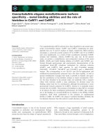
The structural arrangement of domains in a five-domain
member of GH 13 is presented in Fig. 1. No three-
dimensional structure has been determined for a complete
four-domain member, although structures are available
for several a-amylases that consist of domains A, B and C
only [2,95–99]. It should be noted, however, that crystals
have been obtained for the four-domain maltotetrao-
hydrolase from P. stutzeri, but the SBD was found by
X-ray crystallography to be in a disordered state [100].
Figure 1, in addition to illustrating the arrangement of all
domains in the five-domain members of the GH 13, can
be taken as an approximation of the first three domains in
the four-domain members. It also shows the typical
Table 1. The enzymes from the a-amylase family used in the present study.
Enzyme Source Abbr. SwissProt
a
Reference
a-Amylase Aspergillus nidulans Aspnd Q9UV09 Unpublished
Aspergillus kawachii Aspka P13296 [49]
Bacillus sp. TS-23 Bacsp Q59222 [50]
Cryptococcus sp. S2 Crcsp Q92394 [51]
Streptomyces albidoflavus Stral P09794 [52]
Streptomyces griseus Strgr P30270 [53]
Streptomyces lividans TK21 Strli21 O86876 Unpublished
Streptomyces lividans TK24 Strli24 P97179 [55]
Streptomyces venezuelae Strve P22998 [56]
Thermomonospora curvata Thscu P29750 [57]
Maltotetrao-hydrolase Pseudomonas saccharophila Psesa P22963 [58]
Pseudomonas stutzeri Psest P13507 [59]
Maltopentao-hydrolase Pseudomonas sp. KO-8940 Psesp Q52516 [60]
Maltogenic a-amylase Bacillus stearothermophilus Bacst P19531 [61]
Acarviose transferase Actinoplanes sp. SE50 Actsp Q9K5L5 [62]
Cyclodextrin glucanotransferase Bacillus sp. 1–1 Bac11 P31746 [63]
Bacillus sp. 17–1 Bac17 P30921 [64]
Bacillus sp. 38–2 Bac38 P09121 [65]
Bacillus sp. 6.6.3 Bac663 P31747 Unpublished
Bacillus sp. 1011 Bac1011 P05618 [67]
Bacillus sp. A2–5a BacA2 O82984 [68]
Bacillus sp. B1018 Bac1018 P17692 [69]
Bacillus sp. E-1 BacE1 Z34466* [70]
Bacillus sp. KC201 BacKC Q59239 [71]
Bacillus brevis Bacbr O30565 [72]
Bacillus circulans 8 Bacci8 P30920 [73]
Bacillus circulans 251 Bacci251 P43379 [39]
Bacillus circulans A11 BacciA Q9F5W3 Unpublished
Bacillus clarkii Baccl AB082929* [75]
Bacillus licheniformis Bacli P14014 [76]
Bacillus macerans IB7 BacmaIB7 O52766 Unpublished
Bacillus macerans IFO 3490 BacIFO P04830 [78]
Bacillus ohbensis Bacoh P27036 [79]
Bacillus stearothermophilus ET1 Bacst1 Q9ZAQ0 [80]
Bacillus stearothermophilus no. 2 Bacst2 P31797 [81]
Klebsiella pneumoniae Klepn P08704 [82]
Nostoc sp. PCC 9229 Nossp AF497477* [83]
Thermoanaerobacter sp. ATCC53627 Thbsp Z35484* [84]
Thermoanaerobacter thermosulfurogenes Thbth P26827 [85]
Thermococcus sp. B1001 Thcsp Q9UWN2 [86]
a
The accession numbers with * are the numbers from GenBank.
Ó FEBS 2003 a-Amylase family members with starch-binding domain (Eur. J. Biochem. 270) 637
structure of an SBD as its basic features seem well-
conserved [3,8,22,26–30,38–41,44].
While in the five-domain CGTases, the maltogenic
a-amylase, and most probably the acarviose transferase,
the SBD immediately follows the preceding domain D
(Fig. 1), a linker sequence is likely to be necessary in the
four-domain proteins to connect domain C to the SBD.
Possible linker sequences for the a-amylases and malto-
tetrao- and maltopentao-hydrolases are shown in Fig. 2A.
These sequences vary in length from 5–40 amino acid
residues. While the linker in the maltopentaohydrolase is
shown as five residues long, uncertainty exists here because
the preceding sequence segment, which should correspond
to domain C, does not match domain C of any of the other
GH 13 sequences reported to date, being unusually high in
arginine (37 out of 124 residues). The linkers in all cases are
characteristically rich in glycine, serine, threonine and
proline (Fig. 2).
For comparison, in glucoamylases (GH 15) the SBD is
separated from the catalytic domain by a linker (Fig. 2B) of
varying length from a few to more than 50 amino acid
residues [101], the longest linker of 68 residues being found
in A. niger glucoamylase G1 [102]. It should be noted that
there is a strong resemblance between the linkers of
Aspergillus a-amylase and Aspergillus glucoamylases, indi-
cating that taxonomy rather than the specificity may play
a major role in linker design. These longer linkers should be
flexible, while the shorter linkers, particularly those con-
taining proline, may be more rigid.
Evolutionary trees
The differences in the modular organization of the enzymes
studied here (Table 1) are clearly reflected in their evolu-
tionary tree (Fig. 3A) calculated using the complete amino
acid sequences including the SBD. Unambiguously there is
an Ôamylase groupÕ and a ÔCGTase groupÕ in the tree
covering at present the hydrolases (four-domain GH 13
members) and transglycosidases (five-domain members),
respectively. The two CGTases, probably lacking domain D
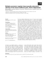
Fig. 2. Linkers connecting the SBD to a preceding domain in amylolytic enzymes. (A) Probable linkers connecting domains C and E in the four-
domain GH 13 members of this study. AAM, a-amylase; M4H, maltotetraohydrolase; M5H, maltopentaohydrolase. Other abbreviations (Aspka,
Aspnd, etc.) are explained in Table 1. (B) For comparison, linkers from GH 15 glucoamylases published in [101] are shown. Aspni GAM,
Aspergillus niger glucoamylase; Horre GAM, Hormoconis resinae glucoamylase; Humgr GAM, Humicola grisea glucoamylase.
Fig. 1. Stereo view of a CGTase as an example of a five-domain member of the a-amylase family having the C-terminal SBD.
638 S
ˇ
. Janec
ˇ
ek et al.(Eur. J. Biochem. 270) Ó FEBS 2003
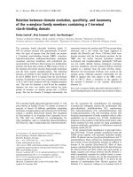
Fig. 3. The evolutionary trees. (A) ÔComplete-sequence treeÕ and (B) trees calculated for individual domains A, B, C and E (SBD). The abbreviations
are explained in Table 1. Colour code: red, CGTases; yellow, acarviose transferase; pink, maltogenic a-amylase; blue, a-amylases from Bacillus and
actinomycetes; light blue, a-amylases from fungi and yeast; green, maltotetraohydrolases and maltopentaohydrolase. A thick dashed line separates
the amylase group from the CGTase group, while the thin dotted line indicates the change of the border between the two parts in the C domain tree
and the SBD tree.
Ó FEBS 2003 a-Amylase family members with starch-binding domain (Eur. J. Biochem. 270) 639
completely (K. pneumoniae; Klepn, and Nostoc sp.
PCC9229; Nossp), are on branches adjacent to each other
and close to the border that separates the two major parts of
the tree. Note that the B. stearothermophilus maltogenic
a-amylase (Bacst) is placed in the ÔCGTase groupÕ of the
tree. This is, however, not surprising as the enzyme has
approximately 60% sequence identity with Bacillus CGT-
ases [1,8,20,61,94] and was recently successfully converted
by protein engineering into a CGTase [103]. Nevertheless,
the unique features discriminating it from the highly similar
Bacillus CGTases are demonstrated by its appearance in a
different cluster (Fig. 3A) together with the only represent-
atives of archaeal CGTases (Thermococcus sp. B1001;
Thcsp) and acarviose transferases (Actinoplanes sp. SE50;
Actsp).
Several groups of closely related sequences can be found
in both parts of the tree, e.g. the a-amylases produced by
streptomycetes or fungi, and the CGTases from the genera
Bacillus and Thermoanaerobacter (Fig. 3A). The a-amylase
from Bacillus sp. TS23 (Bacsp) is on a long branch,
indicating that another bacterial group could emerge in the
future as more sequences become available. In the Ôamylase
groupÕ of the tree the amino acid sequence of the
maltopentaohydrolase from Pseudomonas sp. KO-8940
(Psesp) is more similar to the sequences of a-amylases
originating from the streptomycetes than the two Pseudo-
monas maltotetraohydrolases (Psesa and Psest) (Fig. 3A). It
is worth mentioning that the positions of the malto-
oligosaccharide-producing amylases in the tree shown in
Fig. 3A (the complete-sequence tree) are in agreement with
those found in the evolutionary tree built on the alignment
of short conserved sequence regions extracted only from
domains A and B [20,94]. Both of these trees, i.e. the
complete-sequence tree and the tree based on short
conserved sequences from domains A and B, respect
enzyme specificity.
In order to improve our understanding of evolution-
ary relationships among the GH 13 four- and five-domain
members, partial evolutionary trees were constructed
(Fig. 3B) based on the alignments of the individual domains
A, B, C and E (i.e. the SBD). A tree was not constructed on
the D domain because, as mentioned above, the four-
domain amylases and the two CGTases from Klebsiella and
Nostoc lack this domain.
The tree for domain A, i.e. of the catalytic (b/a)
8
-barrel,
looks very much like the complete-sequence tree shown in
Fig. 3A. In the amylase group of the A domain tree, the
a-amylase from Bacillus sp. TS-23 (Bacsp) is clustered again
together with the two Pseudomonas maltotetraohydrolases,
although it still preserves its own long branch. In the
CGTase group of this tree there are no dramatic changes.
This essentially shared arrangement of the two trees
obviously reflects the fact that domain A constitutes
a substantial part, representing more than 50% of the
consensus sequence length, of the final alignment. More-
over, the domain A contains most of the functionally
important residues which are conserved in the short
sequence motifs [1,19–22,94,104–110].
The tree for domain B is also quite similar to the full-
length tree, albeit with a few small changes. In the amylase
group of the tree, fungal a-amylases have joined the region
of a-amylases from streptomycetes and the Pseudomonas
malto-oligosaccharide-producing amylases to form a more
compact large ÔamylaseÕ cluster. The a-amylase from
Bacillus sp. TS-23 (Bacsp) maintains its own long branch,
but approaches the border between the two groups. In the
CGTase group, the major change concerns the archaeal
CGTase from Thermococcus sp. B1001 (Thcsp) that leaves
the maltogenic a-amylase (Bacst) and joins the Klebsiella
CGTase.
In general it should be pointed out that the overall
arrangement of the trees constructed for domains A and B
(Fig. 3B) are similar to each other and in good agreement
with the complete-sequence tree (Fig. 3A). In the group of
CGTases produced by the genera Bacillus and Thermo-
anaerobacter (the large compact clusters in the CGTases
group of the trees), the longest separated branch is occupied
bytheCGTasefromBacillus clarkii (Baccl) [75], indicating
that this CGTase is at present the most distantly related
CGTase from that group.
The arrangement and clustering of the individual
enzymes and enzyme specificities are substantially changed
in the C domain tree (Fig. 3B) compared to the two partial
trees discussed above and the complete-sequence tree. The
C domain tree suggests that a transition occurs in sequence
segments C-terminal to domain A such that the amylase/
CGTase distinction is altered slightly. Several lines of
evidence support this: (a) the distance separating the
ÔhydrolaseÕ part of the tree from the ÔtransglycosidaseÕ part
has been dramatically shortened in the ÔCdomaintreeÕ;(b)
the two CGTases lacking domain D (Klepn and Nossp)
branch off closer to the four-domain GH 13 members,
suggesting a new border-line between the two parts of the
tree; (c) the Bacillus stearothermophilus maltogenic a-amy-
lase (Bacst) is now rooted deeply in the cluster of Bacillus
and Thermoanaerobacter CGTases; (d) this entire large
CGTase cluster is joined to the rest by a clearly shorter
branch; (e) a-amylases from streptomycetes move closer to
the border.
Some of the findings resulting from the C domain tree are
not surprising and simply reflect the obvious differences
seen in sequences and structures. For example, the ÔisolatedÕ
position of the maltopentaohydrolase from Pseudomonas
sp. KO-8940 is based on its C-domain [60] which is unlike
other GH 13 domain C sequences. Further, the three-
dimensional structure of domain C of maltotetraohydrolase
from P. stutzeri is reported [5] to resemble that of barley
a-amylase [111], a three-domain protein lacking the C-ter-
minal SBD. In all known cases of GH 13 enzymes, domain
Cisab-sheet structure [2–9,11–15,38–41,95–99,111],
although the length of this domain is variable within the
family.
The final partial tree, the ÔSBD treeÕ, lacks the character of
a tree consisting of two groups, i.e. the amylase group and
a CGTase group. It was originally reported [30] for the
evolutionary relationships of the SBDs originating from the
three families GH 13, GH 14, and GH 15 that their
evolutionary tree reflects taxonomy rather than the enzyme
specificity. In this study focused on the GH 13 members the
two four-domain CGTases (Klepn and Nossp) are rooted
obviously in the amylase group of the SBD tree that could
involve also the cluster of acarviose transferase (Actsp),
archaeal CGTase (Thcsp) and the maltogenic a-amylase
(Bacst) due to its longer branch separating it from the
640 S
ˇ
. Janec
ˇ
ek et al.(Eur. J. Biochem. 270) Ó FEBS 2003
compact cluster of Bacillus and Thermoanaerobacter
CGTases (Fig. 3B). Thus for the SBD tree there is not an
obvious border between the hydrolases and transglycosi-
dases, but rather there may be one between the compact
cluster of Bacillus and Thermoanaerobacter CGTases and
the remaining enzymes. The positions of the two CGTases
from K. pneumoniae and Nostoc sp. PCC-9229 are, how-
ever, in agreement with the values of amino acid sequence
identity and similarity of their SBD to SBDs from other
sources (Table 2). This is evident, for example, for the SBD
from Klebsiella CGTase that exhibits more than 42%
identity to the SBD from Pseudomonas maltotetraohydro-
lase (compare Table 2 and the SBD tree in Fig. 3B). This
value is almost 15% higher than that for B. circulans strain
251 CGTase representing the CGTases from bacilli. With
regard to the Nostoc CGTase, it matches best the a-amylase
from Streptomyces griseus, a representative of the a-amy-
lases produced by streptomycetes. The positioning of a
Nostoc CGTase in the assumed amylase group of the SBD
tree (Fig. 3B) very probably reflects rather the values of
sequence similarities (see these values for Bacillus sp. TS-23
and S. griseus a-amylases vs. that for B. circulans strain 251
CGTase in Table 2). Overall the SBD from Nostoc sp. PCC
9229 CGTase exhibits a low degree of both sequence
identity and similarity to the SBDs from all sources studied
here (Table 1), a fact reflected in its long branch in the SBD
tree. For comparison, the values of sequence identity for the
SBD from B. circulans strain 251 CGTase with the SBDs
from the CGTases from Thermococcus sp. B1001, Bacillus
ohbensis,andThermoanaerobacter thermosulfurogenes are
37.3%, 63.8% and 74.0%, respectively. Even for the
acarviose transferase and the maltogenic a-amylase SBDs
compared to the B. circulans strain 251 CGTase SBD these
values are 39.8% and 45.0%, respectively.
Conclusions
When SBD-containing GH 13 members are analysed, a
change in the evolutionary trees from a specificity-deter-
mined relationship at the N-terminal part of the enzymes to
one influenced more by taxonomy at the C-terminal part of
the same enzymes (Figs 3 and 4) can be seen in the present
study. The four- and five-domain members of GH 13 can be
referred to generally as the SBD-containing hydrolases
(mainly a-amylases, but generally classified as EC 3.2.1.x)
and transglycosidases (mainly CGTases, but classified as
EC 2.4.1.x), and with a noticeable small intermediate group
comprising at present the CGTases from K. pneumoniae [82]
and Nostoc sp. PCC 9229 [83] (Fig. 4). The fact that SBD
occurs in GH 13, GH 14, and GH 15 [26] supports the idea
that there has been a separate evolution of this domain [30].
This together with the findings of the present study indicates
a separate evolution of the domains C and E compared to
the domains A and B.
The recent introduction by gene fusion of a Bacillus
CGTase SBD into a Bacillus subtilis a-amylase [112] and of
the fungal SBD including a linker segment of glucoamylase
from A. niger to the barley a-amylase 1 [113,114] promoted
the a-amylase activity towards starch granules by two- to
threefold. The conversion of a CGTase from a transglyco-
sidase into a starch hydrolase was also demonstrated recently
[115]. This work, taken together with theresults of the present
study, as well as with many theoretical and experimental
results on sequence and structure similarities between
amylases and CGTases [19,20,22,26,30,94,116–118], their
phylogenies [20,94,106,119–121], and a novel SBD in an
archaeal CGTase [122] can shed more light on, in general, the
relations between protein evolution and taxonomy of species
[123] and, in particular, the evolution of these industrially
important glycoside hydrolases with possible exploitation for
their development with enhanced performance.
Acknowledgements
This work was financially supported in part by the VEGA grant
no. 2/2057/22 from the Slovak Grant Agency for Science and the
EMBO Short-Term Fellowship to S
ˇ
J.
Table 2. Sequence identity (similarity) in percentage for SBD of the two
CGTases lacking domain D and selected GH 13 members.
Species
Klebsiella
pneumoniae
Nostoc sp.
PCC 9229
Bacillus circulans strain 251 CGT 27.8 (47.2) 24.3 (36.0)
Klebsiella pneumoniae CGT – 15.2 (33.9)
Nostoc sp. PCC 9229 CGT 15.2 (33.9) –
Thermococcus sp. B1001 CGT 16.8 (35.4) 15.5 (28.5)
Actinoplanes sp. SE50 ACT 20.9 (40.0) 18.8 (31.3)
Bacillus stearothermophilus MAA 22.3 (42.0) 23.5 (37.4)
Aspergillus kawachii AAM 27.0 (46.0) 21.6 (33.6)
Bacillus sp. TS-23 AAM 30.8 (53.3) 22.9 (43.1)
Streptomyces griseus AAM 25.2 (46.2) 27.9 (43.2)
Pseudomonas stutzeri M4H 42.6 (62.4) 18.0 (36.0)
Pseudomonas sp. KO-8940 M5H 26.4 (50.9) 18.4 (37.7)
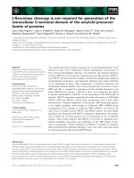
Fig. 4. The proposed relationship between four- and five-domain GH 13
members. It is indicated that there might be a change in domain
evolution from specificity to taxonomy when moving from the
N-terminal to the C-terminal end of a sequence for this particular
group of enzymes.
Ó FEBS 2003 a-Amylase family members with starch-binding domain (Eur. J. Biochem. 270) 641
References
1. MacGregor, E.A., Janec
ˇ
ek, S
ˇ
. & Svensson, B. (2001) Relation-
ship of sequence and structure to specificity in the a-amylase
family of enzymes. Biochim. Biophys. Acta 1546, 1–20.
2. Matsuura, Y., Kusunoki, M., Harada, W. & Kakudo, M. (1984)
Structure and possible catalytic residues of Taka-amylase A.
J. Biochem. 95, 697–702.
3. Klein, C. & Schulz, G.E. (1991) Structure of cyclodextrin glyco-
syltransferase refined at 2.0 A
˚
resolution. J. Mol. Biol. 217,
737–750.
4. Kizaki, H., Hata, Y., Watanabe, K., Katsube, Y. & Suzuki, Y.
(1993) Polypeptide folding of Bacillus cereus ATCC7064 oligo-
1,6-glucosidase revealed by 3.0 A
˚
resolution X-ray analysis.
J. Biochem. 113, 646–649.
5. Morishita, Y., Hasegawa, K., Matsuura, Y., Katsube, Y.,
Kubota, M. & Sakai, S. (1997) Crystal structure of a mal-
totetraose-forming exo-amylase from Pseudomonas stutzeri.
J. Mol. Biol. 267, 661–672.
6. Katsuya, Y., Mezaki, Y., Kubota, M. & Matsuura, Y. (1998)
Three-dimensional structure of Pseudomonas isoamylase at 2.2 A
˚
resolution. J. Mol. Biol. 281, 885–897.
7. Kamitori, S., Kondo, S., Okuyama, K., Yokota, T., Shimura, Y.,
Tonozuka, T. & Sakano, Y. (1999) Crystal structure of Thermo-
actinomyces vulgaris R 47 a-amylase II (TVAII) hydrolyzing
cyclodextrins and pullulan at 2.6 A
˚
resolution. J. Mol. Biol. 287,
907–921.
8. Dauter, Z., Dauter, M., Brzozowski, A.M., Christensen, S.,
Borchert, T.V., Beier, L., Wilson, K.S. & Davies, G.J. (1999)
X-ray structure of Novamyl, the five-domain ÔmaltogenicÕ
a-amylase from Bacillus stearothermophilus: maltose and acar-
bose complexes at 1.7 A
˚
resolution. Biochemistry 38, 8385–8392.
9. Kim, J.S., Cha, S.S., Kim, H.J., Kim, T.J., Ha, N.C., Oh, S.T.,
Cho,H.S.,Cho,M.J.,Kim,M.J.,Lee,H.S.,Kim,J.W.,Choi,
K.Y., Park, K.H. & Oh, B.H. (1999) Crystal structure of a mal-
togenic amylase provides insights into a catalytic versatility.
J. Biol. Chem. 274, 26279–26286.
10. Przylas, I., Tomoo, K., Terada, Y., Takaha, T., Fujii, K.,
Saenger, W. & Strater, N. (2000) Crystal structure of amylo-
maltase from Thermus aquaticus, a glycosyltransferase catalysing
the production of large cyclic glucans. J. Mol. Biol. 296, 873–886.
11. Feese, M.D., Kato, Y., Tamada, T., Kato, M., Komeda, T.,
Miura, Y., Hirose, M., Hondo, K., Kobayashi, K. & Kuroki, R.
(2000) Crystal structure of glycosyltrehalose trehalohydrolase
from the hyperthermophilic archaeum Sulfolobus solfataricus.
J. Mol. Biol. 301, 451–464.
12.Skov,L.K.,Mirza,O.,Henriksen,A.,DeMontalk,G.P.,
Remaud-Simeon,M.,Sarcabal,P.,Willemot,R.M.,Monsan,P.
& Gajhede, M. (2001) Amylosucrase, a glucan-synthesizing
enzyme from the a-amylase family. J. Biol. Chem. 276, 25273–
25278.
13. Roujeinikova, A., Raasch, C., Burke, J., Baker, P.J., Liebl, W. &
Rice, D.W. (2001) The crystal structure of Thermotoga maritima
maltosyltransferase and its implications for the molecular basis of
the novel transfer specificity. J. Mol. Biol. 312, 119–131.
14. Lee, H.S., Kim, M.S., Cho, H.S., Kim, J.I., Kim. T.J., Choi. J.H.,
Park, C., Lee, H.S., Oh, B.H. & Park, K.H. (2002) Cyclomalto-
dextrinase, neopullulanase, and maltogenic amylase are nearly
indistinguishable from each other. J. Biol. Chem. 277, 21891–
21897.
15. Roujeinikova, A., Raasch, C., Sedelnikova, S., Liebl, W. &
Rice, D.W. (2002) Crystal structure of Thermotoga maritima
4-a-glucanotransferase and its acarbose complex: implications for
substrate specificity and catalysis. J. Mol. Biol. 321, 149–162.
16. Abad, M.C., Binderup, K., Rios-Steiner, J., Arni, R.K., Preiss, J.
& Geiger, J.H. (2002) The X-ray crystallographic structure
of Escherichia coli branching enzyme. J. Biol. Chem. 277, 42164–
42170.
17. Kobayashi, M., Kubota, M. & Matsuura, Y. (1999) Crystal-
lization and improvement of crystal quality for X-ray diffraction
of maltooligosyl trehalose synthase by reductive methylation of
lysine residues. Acta Crystallogr. D55, 931–933.
18. Lebbink, J.H.G., Bertoldo, C., Tibbelin, G., Andersen, J.T.,
Duffner, F., Antranikian, G. & Ladenstein, R. (2000) Crystal-
lization and preliminary X-ray crystallographic studies of the
thermoactive pullulanase type I, hydrolyzing a-1,6 glycosidic
linkages, from Fervidobacterium pennivorans Ven5. Acta Crys-
tallogr. D56, 1470–1472.
19. Jespersen, H.M., MacGregor, E.A., Sierks, M.R. & Svensson, B.
(1991) Comparison of the domain-level organization of starch
hydrolases and related enzymes. Biochem. J. 80, 51–55.
20. Jespersen, H.M., MacGregor, E.A., Henrissat, B., Sierks, M.R. &
Svensson, B. (1993) Starch- and glycogen-debranching and
branching enzymes: prediction of structural features of the cata-
lytic (b/a)
8
-barrel domain and evolutionary relationship to other
amylolytic enzymes. J. Protein Chem. 12, 791–805.
21.MacGregor,E.A.,Jespersen,H.M.&Svensson,B.(1996)A
circularly permuted a-amylase-type a/b-barrel structure in glu-
can-synthesizing glucosyltransferases. FEBS Lett. 378, 263–266.
22. Janec
ˇ
ek, S
ˇ
. (2002) How many conserved sequence regions are
there in the a-amylase family? Biologia, Bratislava 57 (Suppl. 11),
29–41.
23. Coutinho, P.M. & Henrissat, B. (1999) Carbohydrate-active
enzymes: an integrated database approach. In Recent Advances in
Carbohydrate Bioengineering (Gilbert, H.J., Davies, G., Henris-
sat, B. & Svensson, B., eds), pp. 3–12. The Royal Society of
Chemistry, Cambridge, UK.
24. Janec
ˇ
ek, S
ˇ
., Svensson, B. & Henrissat, B. (1997) Domain evolu-
tion in the a-amylase family. J. Mol. Evol. 45, 322–331.
25. Tanaka, Y., Ashikari, T., Nakamura, N., Kiuchi, N., Shibano,
Y., Amachi, T. & Yoshizumi, H. (1986) Comparison of amino
acid sequences of three glucoamylases and their structure-func-
tion relationships. Agric. Biol. Chem. 50, 965–969.
26. Svensson, B., Jespersen, H., Sierks, M.R. & MacGregor, E.A.
(1989) Sequence homology between putative raw-starch binding
domains from different starch-degrading enzymes. Biochem. J.
264, 309–311.
27. Penninga, D., van der Veen, B.A., Knegtel, R.M.A., van Hijum,
S.A.F.T.,Rozeboom,H.J.,Kalk,K.H.,Dijkstra,B.W.&
Dijkhuizen, L. (1996) The raw starch binding domain of cyclo-
dextrin glycosyltransferase from Bacillus circulans strain 251.
J. Biol. Chem. 271, 32777–32784.
28. Sorimachi, K., Le Gal-Coeffet, M.F., Williamson, G., Archer,
D.B. & Williamson, M.P. (1997) Solution structure of the gran-
ular starch binding domain of Aspergillus niger glucoamylase
bound to b-cyclodextrin. Structure 5, 647–661.
29. Southall, S.M., Simpson, P.J., Gilbert, H.J., Williamson, G. &
Williamson, M.P. (1999) The starch-binding domain from glu-
coamylase disrupts the structure of starch. FEBS Lett. 447, 58–60.
30. Janec
ˇ
ek, S
ˇ
.&S
ˇ
evc
ˇ
ı
´
k, J. (1999) The evolution of starch-binding
domain. FEBS Lett. 456, 119–125.
31. Sauer, J., Sigurskjold, B.W., Christensen, U., Frandsen, T.P.,
Mirgorodskaya, E., Harrison, M., Roepstorff, P. & Svensson, B.
(2000) Glucoamylase: structure/function relationships, and pro-
tein engineering. Biochim. Biophys. Acta 1543, 275–293.
32. Giardina, T., Gunning, A.P., Juge, N., Faulds, C.B., Furniss,
C.S., Svensson, B., Morris, V.J. & Williamson, G. (2001) Both
binding sites of the starch-binding domain of Aspergillus niger
glucoamylase are essential for inducing a conformational change
in amylose. J. Mol. Biol. 313, 1149–1159.
33. Coutinho, P.M. & Henrissat, B. (1999) The modular structure of
cellulases and other carbohydrate-active enzymes: an integrated
642 S
ˇ
. Janec
ˇ
ek et al.(Eur. J. Biochem. 270) Ó FEBS 2003
database approach. In Genetics, Biochemistry and Ecology of
Cellulose Degradation (Ohmiya, K., Hayashi, K., Sakka, K.,
Kobayashi, Y., Karita, S. & Kimura, T., eds), pp. 15–23. Uni
Publishers Co, Tokyo, Japan.
34. Søgaard, M., Kadziola, A., Haser, R. & Svensson, B. (1993) Site-
directed mutagenesis of histidine 93, aspartic acid 180, glutamic
acid 205, histidine 290, and aspartic acid 291 at the active site and
tryptophan 279 at the raw starch binding site in barley a-amylase
1. J. Biol. Chem. 268, 22480–22484.
35. Tibbot, B.K., Wong, D.W.S. & Robertson, G.H. (2002) Studies
on the C-terminal region of barley a-amylase 1 with emphasis on
raw starch-binding. Biologia, Bratislava 57 (Suppl. 11), 229–238.
36. Rodriguez Sanoja, R., Morlon-Guyot, J., Jore, J., Pintado, J.,
Juge, N. & Guyot, J.P. (2000) Comparative characterization of
complete and truncated forms of Lactobacillus amylovorus -
amylase and role of the C-terminal direct repeats in raw-starch
binding. Appl. Envir. Microbiol. 66, 3350–3356.
37. Sumitani, J.I., Tottori, T., Kawaguchi, T. & Arai, M. (2000) New
type of starch-binding domain: the direct repeat motif in the C-
terminal region of Bacillus sp, 195 a-amylase contributes to starch
binding and raw starch degrading. Biochem. J. 350, 477–484.
38. Kubota, M., Matsuura, Y., Sakai, S. & Katsube, Y. (1991)
Molecular structure of B. stearothermophilus cyclodextrin gluca-
notransferase and analysis of substrate binding site. Denpun
Kagaku 38, 141–146.
39. Lawson, C.L., van Montfort, R., Strokopytov, B., Rozeboom,
H.J., Kalk, K.H., de Vries, G.E., Penninga, D., Dijkhuizen, L. &
Dijkstra, B.W. (1994) Nucleotide sequence and X-ray structure
of cyclodextrin glycosyltransferase from Bacillus circulans strain
251 in a maltose-dependent crystal form. J. Mol. Biol. 236,590–
600.
40. Knegtel, R.M., Wind, R.D., Rozeboom, H.J., Kalk, K.H.,
Buitelaar, R.M., Dijkhuizen, L. & Dijkstra, B.W. (1996) Crystal
structure at 2.3 A
˚
resolution and revised nucleotide sequence of
the thermostable cyclodextrin glycosyltransferase from Thermo-
nanaerobacterium thermosulfurigenes EM1. J. Mol. Biol. 256,
611–622.
41. Harata, K., Haga, K., Nakamura, A., Aoyagi, M. & Yamane, K.
(1996) X-Ray structure of cyclodextrin glucanotransferase from
alkalophilic Bacillus sp. 1011. Comparison of two independent
molecules at 1.8 A
˚
resolution. Acta Crystallogr. D52, 1136–1145.
42. Aleshin, A., Golubev, A., Firsov, L.M. & Honzatko, R.B. (1992)
Crystal structure of glucoamylase from Aspergillus awamori var.
X100–2.2-A
˚
resolution. J. Biol. Chem. 267, 19291–19298.
43.Mikami,B.,Hehre,E.J.,Sato,M.,Katsube,Y.,Hirose,M.,
Morita, Y. & Sacchettini, J.C. (1993) The 2.0-A
˚
resolution
structure of soybean b-amylase complexed with a-cyclodextrin.
Biochemistry 32, 6836–6845.
44. Mikami, B., Adachi, M., Kage, T., Sarikaya, E., Nanmori, T.,
Shinke, R. & Utsumi, S. (1999) Structure of raw starch-digesting
Bacillus cereus b-amylase complexed with maltose. Biochemistry
38, 7050–7061.
45. Minassian, B.A., Ianzano, L., Meloche, M., Andermann, E.,
Rouleau, G.A., Delgado-Escueta, A.V. & Scherer, S.W. (2000)
Mutation spectrum and predicted function of laforin in Lafora’s
progressive myoclonus epilepsy. Neurology 55, 341–346.
46. Wang, J., Stuckey, J.A., Wishart, M.J. & Dixon, J.E. (2002) A
unique carbohydrate binding domain targets the Lafora disease
phosphatase to glycogen. J. Biol. Chem. 277, 2377–2380.
47. Janec
ˇ
ek, S
ˇ
. (2002) A motif of a microbial starch-binding domain
foundinhumangenethonin.Bioinformatics 18, 1534–1537.
48. Reference withdrawn.
49. Kaneko, A., Sudo, S., Sakamoto, Y., Tamura, G., Ishikawa, T. &
Ohba, T. (1996) Molecular cloning and determination of the
nucleotide-sequence of a gene encoding an acid-stable a-amylase
from Aspergillus kawachii. J. Ferment. Bioeng. 81, 292–298.
50. Lin, L.L., Hsu, W.H. & Chu, W.S. (1997) A gene encoding for
an a-amylase from thermophilic Bacillus sp. strain TS-23
and its expression in Escherichia coli. J. Appl. Microbiol. 82, 325–
334.
51. Iefuji, H., Chino, M., Kato, M. & Iimura, Y. (1996) Raw-starch-
digesting and thermostable a-amylase from the yeast Crypto-
coccus sp. S-2: purification, characterization, cloning, and
sequencing. Biochem. J. 318, 989–996.
52. Long, C.M., Virolle, M.J., Chang, S.Y., Chang, S. & Bibb, M.J.
(1987) a-Amylase gene of Streptomyces limosus: nucleotide
sequence, expression motifs, and amino acid sequence homology
to mammalian and invertebrate a-amylases. J. Bacteriol. 169,
5745–5754.
53. Vigal, T., Gil, J.A., Daza, A., Garcia-Gonzalez, M.D. & Martin,
J.F. (1991) Cloning, characterization and expression of an
a-amylase gene from Streptomyces griseus IMRU3570. Mol.
General Genet. 225, 278–288.
54. Reference withdrawn.
55. Yin, X.H., Gagnat, J., Gerbaud, C., Guerineau, M. & Virolle,
M.J. (1997) Cloning and characterization of a new a-amylase
gene from Streptomyces lividans TK24. Gene 197, 37–45.
56. Virolle, M.J., Long, C.M., Chang, S. & Bibb, M.J. (1988)
Cloning, characterisation and regulation of an a-amylase gene
from Streptomyces venezuelae. Gene 74, 321–334.
57. Petricek, M., Tichy, P. & Kuncova, M. (1992) Characterization
of the a-amylase-encoding gene from Thermomonospora curvata.
Gene 112, 77–83.
58. Zhou, J., Baba, T., Takano, T., Kobayashi, S. & Arai, Y. (1989)
Nucleotide sequence of the maltotetraohydrolase gene from
Pseudomonas saccharophila. FEBS Lett. 255, 37–41.
59. Fujita, M., Torigoe, K., Nakada, T., Tsusaki, K., Kubota,
M., Sakai, S. & Tsujisaka, Y. (1989) Cloning and nucleo-
tide sequence of the gene (amyP) for maltotetraose-forming
amylase from Pseudomonas stutzeri MO-19. J. Bacteriol. 171,
1333–1339.
60. Shida, O., Takano, T., Takagi, H., Kadowaki, K. & Kobayashi,
S. (1992) Cloning and nucleotide sequence of the maltopentaose-
forming amylase gene from Pseudomonas sp. KO-8940. Biosci.
Biotechn Biochem. 56, 76–80.
61. Diderichsen, B. & Christiansen, L. (1988) Cloning of a mal-
togenic a-amylase from Bacillus stearothermophilus. FEMS
Microbiol. Lett. 56, 53–60.
62. Hemker, M., Stratmann, A., Goeke, K., Schroder, W., Lenz, J.,
Piepersberg, W. & Pape, H. (2001) Identification, cloning,
expression, and characterization of the extracellular acarbose-
modifying glycosyltransferase, AcbD, from Actinoplanes sp.
strain SE50. J. Bacteriol. 183, 4484–4492.
63. Schmid, G., Englbrecht, A. & Schmid, D. (1988) Cloning and
nucleotide sequence of a cyclodextrin glycosyltransferase gene
from the alkalophilic Bacillus 1–1. In Proceedings of the Fourth
International Symposium on Cyclodextrins (Huber,O.&Szejtli,J.,
eds), pp. 71–76. Kluwer Academic Publishers, Dordrecht,
Germany/Boston, USA.
64. Kaneko, T., Song, K.B., Hamamoto, T., Kudo, T. & Horikoshi, K.
(1989) Construction of a chimeric series of Bacillus cyclomalto-
dextrin glucanotransferases and analysis of the thermal stabilities
and pH optima of the enzymes. J. General Microbiol. 135, 3447–
3457.
65. Kaneko,T.,Hamamoto,T.&Horikoshi,K.(1988)Molecular
cloning and nucleotide sequence of the cyclomaltodextrin gluca-
notransferase gene from the alkalophilic Bacillus sp. strain, 38–2.
J. General Microbiol. 134, 97–105.
66. Reference withdrawn.
67. Kimura,K.,Kataoka,S.,Ishii,Y.,Takano,T.&Yamane,K.
(1987) Nucleotide sequence of the b-cyclodextrin glucanotrans-
ferase gene of alkalophilic Bacillus sp. strain 1011 and similarity
Ó FEBS 2003 a-Amylase family members with starch-binding domain (Eur. J. Biochem. 270) 643
of its amino acid sequence to those of a-amylases. J. Bacteriol.
169, 4399–4402.
68. Ohdan, K., Kuriki, T., Takata, H. & Okada, S. (2000) Cloning of
the cyclodextrin glucanotransferase gene from alkalophilic
Bacillus sp. A2–5a and analysis of the raw starch-binding domain.
Appl. Microbiol. Biotechnol. 53, 430–434.
69. Itkor, P., Tsukagoshi, N. & Udaka, S. (1990) Nucleotide sequence
of the raw-starch-digesting amylase gene from Bacillus sp. B1018
and its strong homology to the cyclodextrin glucanotransferase
genes. Biochem. Biophys. Res. Commun. 166, 630–636.
70. Yong,J.,Choi,J.,Kang,H.,Park,C.,Park,K.&Choi,Y.(1996)
Molecular cloning of CGTase gene from alkalophilic Bacillus sp.
E-1 and its overexpression in E. coli. Biotechnol. Lett. 18, 1223–
1228.
71. Kitamoto, N., Kimura, T., Kito, Y. & Ohmiya, K. (1992)
Cloning and sequencing of the gene encoding cyclodextrin glu-
canotransferase from Bacillus sp. K.C.201. J. Ferment. Bioeng.
74, 345–351.
72. Kim, M.H., Sohn, C.B. & Oh, T.K. (1998) Cloning and
sequencing of a cyclodextrin glycosyltransferase gene from Bre-
vibacillus brevis CD162 and its expression in Escherichia coli.
FEMS Microbiol. Lett. 164, 411–418.
73. Nitschke, L., Heeger, K., Bender, H. & Schulz, G.E. (1990)
Molecular cloning, nucleotide sequence and expression in
Escherichia coli of the b-cyclodextrin glycosyltransferase gene
from Bacillus circulans strain 8. Appl. Microbiol. Biotechnol. 33,
542–546.
74. Reference withdrawn.
75. Takada, M., Nakagawa, Y. & Yamamoto, M. (2003) Biochem-
ical and genetic analyses of a novel c-cyclodextrin glucano-
transferase from an alkalophilic Bacillus clarkii 7364. J. Biochem.
133, in press.
76. Hill, D.E., Aldape, R. & Rozzell, J.D. (1990) Nucleotide sequence
of a cyclodextrin glucosyltransferase gene, cgtA, from Bacillus
licheniformis. Nucleic Acids Res. 18, 199.
77. Reference withdrawn.
78. Takano, T., Fukuda, M., Monma, M., Kobayashi, S., Kainuma,
K. & Yamane, K. (1986) Molecular cloning, DNA nucleotide
sequencing, and expression in Bacillus subtilis cells of the Bacillus
macerans cyclodextrin glucanotransferase gene. J. Bacteriol. 166,
1118–1122.
79. Sin, K.A., Nakamura, A., Kobayashi, K., Masaki, H. & Uozumi,
T. (1991) Cloning and sequencing of a cyclodextrin glucano-
transferase gene from Bacillus ohbensis and its expression in
Escherichia coli. Appl. Microbiol. Biotechnol. 35, 600–605.
80. Chung,H.J.,Yoon,S.H.,Lee,M.J.,Kim,M.J.,Kweon,K.S.,
Lee, I.W., Kim, J.W., Oh, B.H., Lee, H.S., Spiridonova, V.A. &
Park, K.H. (1998) Characterization of a thermostable cyclodex-
trin glucanotransferase isolated from Bacillus stearothermophilus
ET1. J. Agric. Food Chem. 46, 952–959.
81. Fujiwara, S., Kanemoto, M., Kim, B., Lejeune, A., Sakaguchi, K.
& Imanaka, T. (1992) Cyclization characteristics of cyclodextrin
glucanotransferase are conferred by the NH
2
-terminal region of
the enzyme. Appl. Environ. Microbiol. 58, 4016–4025.
82. Binder, F., Huber, O. & Boeck, A. (1986) Cyclodextrin-glyco-
syltransferase from Klebsiella pneumoniae M5a1: cloning,
nucleotide sequence and expression. Gene 47, 269–277.
83. Wouters, J., Bergman, B. & Janson, S. (2003) Cloning and
expression of a putative cyclodextrin glucosyltransferase from the
symbiotically competent cyanobacterium Nostoc sp. PCC 9229.
FEMS Microbiol. Lett. in press.
84. Joergensen, S.T., Tangney, M., Starnes, R.L., Amemiya, K. &
Joergensen, P.L. (1997) Cloning and nucleotide sequence of a
thermostable cyclodextrin glycosyltransferase gene from Thermo-
anaerobacter sp. ATCC 53627 and its expression in Escherichia
coli. Biotechnol. Lett. 19, 1027–1031.
85. Bahl, H., Burchhardt, G., Spreinat, A., Haeckel, K., Wienecke,
A., Schmidt, B. & Antranikian, G. (1991) a-Amylase of Clos-
tridium thermosulfurogenes EM1: nucleotide sequence of the gene,
processing of the enzyme, and comparison of other a-amylases.
Appl. Environ. Microbiol. 57, 1554–1559.
86. Yamamoto, T., Shiraki, K., Fujiwara, S., Takagi, M., Fukui, K.
& Imanaka, T. (1999) In vitro heat effect on functional and
conformational changes of cyclodextrin glucanotransferase from
hyperthermophilic archaea. Biochem. Biophys. Res. Commun.
265, 57–6177.
87. Bairoch, A. & Apweiler, R. (2000) The SWISS-PROT protein
sequence database and its Supplement TrEMBL in 2000. Nucleic
Acids Res. 28, 45–48.
88. Benson, D.A., Karsch-Mizrachi, I., Lipman, D.J., Ostell, J.,
Rapp, B.A. & Wheeler, D.L. (2002) GenBank. Nucleic Acids Res.
30, 17–20.
89. Altschul, S.F., Stephen, F., Madden, T.L., Scha
¨
ffer, A.A., Zhang,
J., Zhang, Z., Miller, W. & Lipman, D.J. (1997) Gapped BLAST
and PSI-BLAST: a new generation of protein database search
programs. Nucleic Acids Res. 25, 3389–3402.
90. Thompson, J.D., Higgins, D.G. & Gibson, T.J. (1994) CLUS-
TAL W: improving the sensitivity of progressive multiple
sequence alignment trough sequence weighting, position specific
gap penalties and weight matrix choice. Nucleic Acids Res. 22,
4673–4680.
91. Saitou, N. & Nei, M. (1987) The neighbor-joining method: a new
method for reconstructing phylogenetic trees. Mol. Biol. Evol. 4,
406–425.
92. Page, R.D. (1996) TreeView: an application to display phylo-
genetic trees on personal computers. Comput. Applic. Biosci. 12,
357–358.
93. Berman, H.M., Battistuz, T., Bhat, T.N., Bluhm, W.F., Bourne,
P.E., Burkhardt, K., Feng, Z., Gilliland, G.L., Iype, L., Jain, S.,
Fagan, P., Marvin, J., Padilla, D., Ravichandran, V., Schneider,
B.,Thanki,N.,Weissig,H.,Westbrook,J.D.&Zardecki,C.
(2002) The protein data bank. Acta Crystallogr. D58, 899–907.
94. Janec
ˇ
ek, S
ˇ
. (1995) Tracing the evolutionary lineages among
a-amylases and cyclodextrin glycosyltransferases: the question of
so-called ÔintermediaryÕ enzymes. Biologia, Bratislava 50, 515–
522.
95. Brady, R.L., Brzozowski, A.M., Derewenda, Z.S., Dodson, E.J.
& Dodson, G.G. (1991) Solution of the structure of Aspergillus
niger acid a-amylase by combined molecular replacement and
multiple isomorphous replacement methods. Acta Crystallogr.
B47, 527–535.
96. Machius, M., Wiegand, G. & Huber, R. (1995) Crystal structure
of calcium-depleted Bacillus licheniformis a-amylase at 2.2 A
˚
resolution. J. Mol. Biol. 246, 545–559.
97. Aghajari, N., Feller, G., Gerday, C. & Haser, R. (1998) Crystal
structures of the psychrophilic a-amylase from Alteromonas
haloplanctis in its native form and complexed with an inhibitor.
Protein Sci. 7, 564–572.
98. Fujimoto, Z., Takase, K., Doui, N., Momma, M., Matsumoto,
T. & Mizuno, H. (1998) Crystal structure of a catalytic-site
mutant a-amylase from Bacillus subtilis complexed with malto-
pentaose. J. Mol. Biol. 277, 393–407.
99. Suvd,D.,Fujimoto,Z.,Takase,K.,Matsumura,M.&Mizuno,
H. (2001) Crystal structure of Bacillus stearothermophilus
a-amylase: possible factors determining the thermostability.
J. Biochem. 129, 461–468.
100. Mezaki, Y., Katsuya, Y., Kubota, M. & Matsuura, Y. (2001)
Crystallization and structural analysis of intact maltotetraose-
forming exo-amylase from Pseudomonas stutzeri. Biosci. Bio-
technol. Biochem. 65, 222–225.
101. Sauer, J., Christensen, T., Frandsen, T.P., Mirgorodskaya, E.,
McGuire, K.A., Driguez, H., Roepstorff, P., Sigurskjold, B.W. &
644 S
ˇ
. Janec
ˇ
ek et al.(Eur. J. Biochem. 270) Ó FEBS 2003
Svensson, B. (2001) Stability and function of interdomain linker
variants of glucoamylase 1 from Aspergillus niger. Biochemistry
40, 9336–9346.
102. Svensson, B., Larsen, K., Svendsen, I. & Boel, E. (1983) The
complete amino acid sequence of the glycoprotein, glucoamylase
G1, from Aspergillus niger. Carlsberg Res. Commun. 48, 529–544.
103. Beier, L., Svendsen, A., Andersen, C., Frandsen, T.P., Borchert,
T.V. & Cherry, J.R. (2000) Conversion of the maltogenic
a-amylase Novamyl into a CGTase. Protein Eng. 13, 509–513.
104. MacGregor, E.A. (1993) Relationships between structure and
activity in the a-amylase family of starch-metabolising enzymes.
Starch 7, 232–237.
105. Svensson, B. (1994) Protein engineering in the a-amylase family:
catalytic mechanism, substrate specificity, and stability. Plant
Mol. Biol. 25, 141–157.
106. Janec
ˇ
ek, S
ˇ
. (1994) Parallel b/a-barrels of a-amylase, cyclodextrin
glycosyltransferase and oligo-1,6-glucosidase versus the barrel of
b-amylase: evolutionary distance is a reflection of unrelated
sequences. FEBS Lett. 353, 119–123.
107. Nielsen, J.E. & Borchert, T.V. (2000) Protein engineering of
bacterial a-amylases. Biochim. Biophys. Acta 1543, 253–274.
108. van der Veen, B.A., Uitdehaag, J.C., Dijkstra, B.W. & Dijkhui-
zen, L. (2000) Engineering of cyclodextrin glycosyltransferase
reaction and product specificity. Biochim. Biophys. Acta 1543,
336–360.
109. van der Maarel, M.J., van der Veen, B., Uitdehaag, J.C.,
Leemhuis, H. & Dijkhuizen, L. (2002) Properties and applica-
tions of starch-converting enzymes of the a-amylase family.
J. Biotechnol. 94, 137–155.
110. Uitdehaag, J.C.M., van der Veen, B.A., Dijkhuizen, L. & Dijk-
stra, B.W. (2002) Catalytic mechanism and product specificity of
cyclodextrin glycosyltransferase, a prototypical transglycosylase
from the a-amylase family. Enzyme Microb. Technol. 30,295–
304.
111. Kadziola, A., Abe, J., Svensson, B. & Haser, R. (1994) Crystal
and molecular structure of barley a-amylase. J. Mol. Biol. 239,
104–121.
112. Ohdan, K., Kuriki, T., Takata, H., Kaneko, H. & Okada, S.
(2000) Introduction of raw starch-binding domains into Bacillus
subtilis a-amylase by fusion with the starch-binding domain of
Bacillus cyclomaltodextrin glucanotransferase. Appl. Environ.
Microbiol. 66, 3058–3064.
113. Svensson, B., Tovborg Jensen, M., Mori, H., Bak-Jensen, K.S.,
Bønsager, B., Nielsen, P.K., Kramhøft, B., Prætorius-Ibba, M.,
Nøhr, J., Juge, N., Greffe, L., Williamson, G. & Driguez, H.
(2002) Fascinating facets of function and structure of amylolytic
enzymes of glycoside hydrolase family 13. Biologia, Bratislava 57
(Suppl. 11), 5–19.
114. Juge, N., Le Gal-Coe
¨
ffet, M.F., Furniss, C.S.M., Gunning, A.P.,
Kramhøft, B., Morris, V.J., Williamson, G. & Svensson, B.
(2002) The starch binding domain of glucoamylase from Asper-
gillus niger: overview of its structure, function, and role in raw-
starch hydrolysis. Biologia, Bratislava 57 (Suppl. 11), 239–245.
115. Leemhuis, H., Dijkstra, B.W. & Dijkhuizen, L. (2002) Mutations
converting cyclodextrin glycosyltransferase from a transglyco-
sylase into a starch hydrolase. FEBS Lett. 514, 189–192.
116. MacGregor, E.A. & Svensson, B. (1989) A super-secondary
structure predicted to be common to several a-1,4-D-glucan-
cleaving enzymes. Biochem. J. 259, 145–152.
117. Janec
ˇ
ek, S
ˇ
., MacGregor, E.A. & Svensson, B. (1995) Character-
istic differences in the primary structure allow discrimination of
cyclodextrin glucanotransferases from a-amylases. Biochem. J.
305, 685–686.
118. Lo,H.F.,Lin,L.L.,Chiang,W.Y.,Chie,M.C.,Hsu,W.H.&
Chang, C.T. (2002) Deletion analysis of the C-terminal region of
the a-amylase of Bacillus sp. strain TS-23. Arch. Microbiol. 178,
115–123.
119. del-Rio, G., Morett, E. & Soberon, X. (1997) Did cyclodextrin
glycosyltransferases evolve from a-amylases? FEBS Lett. 416,
221–224.
120. Bikbulatova, S.M., Chemeris, A.V., Usanov, N.G. & Vakhitov,
V.A. (2000) Establishment of the phylogenetic relationship
between the microbial producers of cyclodextrin glucano-
transferases using their complete amino acid sequences. Mikro-
biologiya 69, 686–693.
121. Pujadas, G. & Palau, J. (2001) Evolution of a-amylases: archi-
tectural features and key residues in the stabilization of the (b/a)
8
scaffold. Mol. Biol. Evol. 18, 38–54.
122. Rashid, N., Cornista, J., Ezaki, S., Fukui, T., Atomi, H. &
Imanaka, T. (2002) Characterization of an archaeal cyclodextrin
glucanotransferase with a novel C-terminal domain. J. Bacteriol.
184, 777–784.
123. Pace, N.R. (1997) A molecular view of microbial diversity and the
biosphere. Science 276, 734–740.
Ó FEBS 2003 a-Amylase family members with starch-binding domain (Eur. J. Biochem. 270) 645