Báo cáo khoa học: Critical role of the plasma membrane for expression of mammalian mitochondrial side chain cleavage activity in yeast pptx
Bạn đang xem bản rút gọn của tài liệu. Xem và tải ngay bản đầy đủ của tài liệu tại đây (423.55 KB, 13 trang )
Critical role of the plasma membrane for expression of mammalian
mitochondrial side chain cleavage activity in yeast
Catherine Duport
1,
*, Barbara Schoepp
1,†
, Elise Chatelain
1
, Roberto Spagnoli
2
, Bruno Dumas
3
and Denis Pompon
1
1
Laboratoire d’Inge
´
nierie des Prote
´
ines Membranaires, CGM du CNRS, Gif sur Yvette, France;
2
Lead Discovery Technologies,
Aventis Pharma, Romainville, France;
3
Functional Genomics, Aventis Pharma, 13 Quai Jules Guesde, F-94403 Vitry sur Seine, France
Engineered yeast cells efficiently convert ergosta-5-eneol to
pregnenolone and progesterone provided that endogenous
pregnenolone acetylase activity is disrupted and that
heterologous sterol D7-reductase, cytochrome P450 side
chain cleavage (CYP11A1) and 3b hydroxysteroid
dehydrogenase/isomerase (3b-HSD) activities are present.
CYP11A1 activity requires the expression of the mammalian
NADPH-adrenodoxin reductase (Adrp) and adrenodoxin
(Adxp) proteins as electron carriers. Several parameters
modulate this artificial metabolic pathway: the effects of
steroid products; the availability and delivery of the ergosta-
5-eneol substrate to cytochrome P450; electron flux and
protein localization. CYP11A1, Adxp and Adrp are usually
located in contact with inner mitochondrial membranes
and are directed to the outside of the mitochondria by the
removal of their respective mitochondrial targeting sequen-
ces. CYP11A1 then localizes to the plasma membrane but
Adrp and Adxp are detected in the endoplasmic reticulum
and cytosol as expected. The electron transfer chain that
involves several subcellular compartments may control side
chain cleavage activity in yeast. Interestingly, Tgl1p, a
potential ester hydrolase, was found to enhance steroid
productivity, probably through both the availability and/or
the trafficking of the CYP11A1 substrate. Thus, the obser-
vation that the highest cellular levels of free ergosta-5-eneol
are found in the plasma membrane suggests that the sub-
strate is freely available for pregnenolone synthesis.
Keywords: CYP11A1; plasma membrane; ergosta-5-eneol;
Tgl1p.
The large family of mammalian cytochrome P450 enzymes
includes drug metabolizing enzymes and enzymes that
mediate individual steps in the biosynthesis of biologically
active compounds. Our interest is focused on the cyto-
chrome P450 enzymes that are involved in the synthesis of
steroid hormones. These steroids are critical for mammalian
life and are involved in such distinct processes as stress
response, immunosuppression, ion balance, general meta-
bolite homeostasis and fetal, neonatal and gonadal devel-
opment [1]. Eukaryotic steroidogenic cells produce a large
array of steroids using a limited set of cytochrome P450
enzymes [2]. The biosynthesis of all hormonal steroids
begins with the side chain cleavage (SCC) of cholesterol [3]
to form pregnenolone, the key precursor of biologically
active steroids in all tissues [4,5]. This reaction is catalysed
by cytochrome P450scc (also designated CYP11A1 [6]), a
mitochondrial protein located on the matrix face of the
inner membrane that requires electrons for activity. These
electrons are transferred from NADPH through a specific
transport chain involving adrenodoxin reductase (Adrp)
and adrenodoxin (Adxp) [7]. Adxp is a small soluble iron–
sulfur protein localized to the mitochondrial matrix, and
Adrp is a larger flavodoxin protein bound to the inner
mitochondrial membrane of steroid-producing cells [8]. For
many years, pregnenolone formation has been considered to
be the rate-limiting step in steroidogenesis [9]. It has been
shown that to initiate and sustain steroid production, a
constant supply of cholesterol must be available in the cell.
Furthermore, there must be a mechanism to ensure the
delivery of this substrate to the site where it is cleaved in the
inner mitochondrial membrane, where CYP11A1 resides.
For example, substrate unavailability is a common cause of
congenital lipoid adrenal hyperplasia, a disease character-
ized by a dramatic decrease in steroid synthesis [10]. In the
Correspondence to B. Dumas, Functional Genomics, Aventis Pharma,
13 Quai Jules Guesde, F-94403 Vitry sur Seine, France.
Fax: + 33 1 5893 2625, Tel.: + 33 1 5893 2805,
E-mail:
Abbreviations: ACAT, acyl coenzyme A cholesterol acyltransferase;
Adrp, adrenodoxin reductase protein; Adxp, adrenodoxin protein;
APAT, acetyl coenzyme A:pregnenolone acetyl transferase; ARE,
acyl coenzyme A:cholesteryl acyltransferase-related enzyme;
CYP2D6, cytochrome P450 2D6; CYP2E1, cytochrome P450 2E1;
CYP11A1, cytochrome P450 steroid side chain cleaving; CYP11B1,
cytochrome P450 steroid 11b hydroxylase; D7-Red, sterol D7reduc-
tase; ER, endoplasmic reticulum; 3b-HSD, 3b hydroxysteroid
dehydrogenase/isomerase; PM, plasma membrane; PGK1, phospho-
glycerate kinase; StAR, steroidogenic acute regulatory protein;
SCC, side chain cleavage; TEF1, transcription elongation factor.
Enzymes: ACAT, EC 2.3. 2.26; Adrp, EC 1.18.1.2; CYP11A1,
EC 1.14.15.6; CYP11B1, EC 1.14.15.4; 3b-HSD, EC 1.1.1.51.
*Present address: University of Paris VII and UMR A408,
INRA 84914 Avignon Cedex 09, France.
Present address: Institut de Biologie Structurale et Microbiologie,
31 Chemin Joseph Aiguier, F-13402 Marseille, France.
(Received 20 November 2002, revised 27 January 2003,
accepted 11 February 2003)
Eur. J. Biochem. 270, 1502–1514 (2003) Ó FEBS 2003 doi:10.1046/j.1432-1033.2003.03516.x
latter case, mutation(s) in the steroidogenic acute regulatory
protein (StAR) protein correlate(s) clearly with the absence
of pregnenolone synthesis. The well-characterized StAR
protein is involved in the rapid transport of cholesterol to
the inner mitochondrial membrane [11]. In contrast, the
factors and processes responsible for the intracellular supply
of cholesterol to the outer mitochondrial membrane are
poorly understood. It is known, however, that cholesterol is
mobilized from cellular storage sites, such as lipid droplets,
in response to trophic hormones [12]. This mobilization
requires the enzyme cholesteryl esterase, which mediates the
release of free cholesterol from cholesterol esters. In
addition, the maintenance of cellular architecture requires
a stringent regulation of the concentration of free choles-
terol. This is ensured by the enzyme, acyl coenzyme A
cholesterol acyltransferase (ACAT), which catalyzes its
esterification [13]. The levels of expression of various
proteins (Adxp, Adrp, CYP11A1) involved in the reaction
can also affect the efficiency of cholesterol side chain
cleavage. Indeed, it is apparent that the expression of these
three proteins is differentially modulated in hormone-
producing tissues. For example, the corpus luteum and
adrenal cortex contain higher concentrations of Adxp and
Adrp than does the placenta ([14,15]). In the latter case, the
concentration of Adrp limits the production of pregneno-
lone [15] through a mechanism involving oxidized Adxp,
that is in excess in the human placenta [16]. Whether ternary
complex formation is required for the optimal flow of
electrons from NADPH to CYP11A1 remains controver-
sial. However, recent reports reinforce the idea that there is
a complex containing CYP11A1, cytochrome P450 11B1
(CYP11B1), Adxp and Adrp in the mitochondrial mem-
brane of steroid producing cells [17,18].
As reported previously [19], the simultaneous expression
of Arabidopsis thaliana sterol D7-reductase (D7-Red),
bovine CYP11A1, Adxp, Adrp and human 3b-hydroxy-
steroid deshydrogenase/isomerase (3b-HSD) in modified
Saccharomyces cerevisiae cells allows the self-sufficient
biosynthesis of pregnenolone and progesterone (Fig. 1),
thus reproducing the properties of steroidogenic tissues of
higher eukaryotes. In these recombinant yeast strains, the
predominant sterol is ergosta-5-eneol, that replaces ergos-
terol in membranes and acts as a substrate for CYP11A1.
Ergosta-5-eneol differs from ergosterol in that the C7–C8
doublebond is reduced and there is no doublebond at
position C22. It also differs from cholesterol in that it has a
methyl group at position C24. Ergosta-5-eneol is synthe-
sized [19] and esterified [20] in processes similar to those
that control cholesterol accumulation in mammalian cells.
Ergosta-5-eneol and cholesterol act similarly as a substrate
for CYP11A1 and allow proper folding of CYP11A1 in
membrane microdomain. The aim of this study was to
determine whether recombinant yeast can be used as a
model system to decipher the SCC reaction and its poten-
tial regulation during steroidogenesis. To do so, we studied
the influence of ergosta-5-eneol availability and electron
carrier expression level on the production of pregnenolone
and progesterone, and we also determined the localization
of the components of the SCC reaction. We found that
Adxp, Adrp and CYP11A1 appear to localize to three
compartments outside the mitochondrion, without impair-
ing the reaction. This finding has direct implications for the
potential formation of a complex containing CYP11A1,
CYP11B1, Adxp and Adrp.
Materials and methods
Culture conditions and genetic methods
Yeast media, including SG (synthetic medium containing
2% glucose), SL (synthetic medium containing 2% galac-
tose) and YP (complete medium without carbon source) are
described [65]. Low-density and high-density cultures were
obtained as reported previously [19]. Standard methods
were used for transformation [21] and genetic manipulation
of S. cerevisiae [22].
pUC-HIS3ADX is an integrative plasmid derived from
pUC-HIS3 [23] that carries the TEF1
prom
::matADX::
PGK1
term
expression cassette. This expression cassette
contains the mature form of the ADX cDNA [24] under
the control of the TEF1 promoter and PGK1 terminator
[25]. The matADX expression plasmid pTG10917 contains
an E. coli replicon with an S. cerevisiae replicon and a
URA3 marker. The vector pUC18-HIS3 was linearized at
the unique XhoI site in the intergenic region between the
S. cerevisiae HIS3 and DDE1 genes and blunt-ended with
the Klenow enzyme. A NotI linker was introduced into
this linearized vector, giving pUC-HIS3N. The 1235-bp
NotI fragment carrying the expression cassette, TEF1
prom
::
matADX::PGK1
term
, was isolated from pTG10917 (see
above) and subcloned into the NotI site of pUC-HIS3N to
obtain pUC-HIS3ADX.
pYeDP60 is a 2l replication origin-based expression
plasmid that contains the URA3 and ADE2 selectable
markers, a galactose inductible GAL10/CYC1 promoter,
multiple cloning sites, and the PGK1 terminator [26].
pCD69, a 2l-URA3-ADE2 plasmid expressing TGL1
under the control of the GAL10/CYC1 promoter was
constructed as follows. The TGL1 open reading frame
was isolated from FY1679 genomic DNA by amplification
using oligonucleotides lip1 (5¢-atagacacgcaaacacaaatacaca
cactaaattaataatgaccggatcATGTACTTCCCCTTTTTAGG
CAGAT-3¢) and lip2 (5¢-cagtagagacatgggagatcccccgcgg
aattcgagctcggtacccgggTCATTCTTTATTTAGAGCATC
CAGC-3¢).
The sequences in lower-case are complementary to the
end of the GAL10/CYC1 promoter (lip1) and to the
beginning of the PGK1 terminator (lip2). The 1647 bp PCR
fragment was transformed into yeast along with BamHI–
EcoRI-linearized pYeDP60, permitting cloning by homo-
logous recombination between the plasmid and the PCR
fragment and giving pCD69.
The pDP10037 (2l-URA3-TRP1), pCD63 (2l-URA3-
TRP1) and pV13SCC (2l-URA3) plasmids were con-
structed as reported previously [19]. pDP10037 carries
the GAL10/CYC1
prom
::matADR::PGK1
term
and GAL10/
CYC1
prom
::matADX::PGK1
term
expression cassettes separ-
ated by the URA3 marker. pCD63 was obtained from
pDP10037 by replacing sequences coding for the mature
Adrp (preceded by a methionine codon) by sequences
coding for the mature form of cytochrome CYP11A1
(preceded by a methionine codon). pV13SCC expresses the
mature form of CYP11A1 (preceded by a methionine
codon) driven by the GAL10/CYC1 promoter.
Ó FEBS 2003 CYP11A1 localizes to the yeast plasma membrane (Eur. J. Biochem. 270) 1503
Strains
Yeast strains used in this study are listed in Table 1. The
structure of the CA10 D7 reductase expression locus has
been verified by PCR, Southern and direct sequencing
analysis of the promoter region, showing that it contains the
GAL10/CYC1 promoter instead of the expected PGK1
promoter (Table 1). The APAT-deficient strain, CA14, was
generated by disrupting the ATF2 gene of CA10 with the
KanMX4 cassette, which confers G418 resistance. Primers
5¢ATF2-Kan 5¢-AGACTTTCAAACGAATAATAACTT
CAGCAATAAAAATTGTCCAGGTTAATtccagcgacatg
gaggccc-3¢ and 3¢ATF2-Kan: 5¢-TTGTACGAGCTCGG
CCGAGCTATACGAAGGCCCGCTACGGCAGTATC
GCAcattcacatacgattgacgc-3¢ (nucleotides in lower case are
specific to the KanMX4 module) were used for PCR with
pFA6-MX4 as a template [27] to produce the KanMX
cassette flanked by ATF2 sequences [23].
The strain, CA19 was obtained by introducing the
GAL10/CYC1
prom
::3bHSD::PGK1
term
cassette into the
region between the HIS3 and DDE1 genes of CA14, as
previously described [19].
CDR07 (an FY-1679–18B derivative that contains only
the GAL10/CYC1
prom
::D7 reductase::PGK1
term
expression
cassette) was obtained by sporulation of the diploid resulting
from a cross between CA10 (MATa) and FY1679–18B
(MATa).
TGY120.2 (MATa) was obtained by transformation of
FY1679–28C (MATa)withXbaI-linearized pTG10925. This
plasmid contains an S. cerevisiae genomic DNA fragment
covering the LEU2 and SPL1 locus derived from pFL26CD
[19]. A bovine mature Adrp expression cassette (TEF1
prom
::
matADR::PGK1
term
) [24] was introduced into the unique
NotI site of pFL26CD, that is in the noncoding region
between LEU2and SPL1. Transformed colonies were grown
in selective medium, and the expression of mature Adrp was
verified by Western blot analysis as described [24]. One clone,
TGY120.2 MATa, was selected for further studies.
The yeast strain, CA15 was isolated by mating CDR06
(MATa) to TGY120.2 (MATa), that contains the
TEF1
prom
::matADR::PGK1
term
cassette in the intergenic
region between LEU2 and SPL1.
The strain, CA17 was generated by integrating a
TEF1
prom
::matADX::PGK1
term
cassette into the intergenic
region between the HIS3 and DDE1 of CA15 with the
pUC-HIS3ADX integrative plasmid.
The are1::KanMX4 are2::HIS3 double mutant strain
CA23 was constructed by crossing CA10 (MATa ARE 1
Table 1. Yeast strains and expression plasmids.
Strain or plasmid Relevant genotype or encoded protein (promoter) Source
S. cerevisiae strains
FY1679 MATa/arho
+
, GAL2, ura3–52, trp1-D63, his3-D200, leu2-D1. [64]
CDR07 MATa, rho
+
,GAL2, ura3–52, trp1-D63, his3-D200 leu2-D1,
ade2::GAL10/CYC1::D7Reductase::PGK1.
This study
CDS04 MATa, rho
+
, GAL2, ura3–52, trp1-D63, his3-D200, leu2-D1, are1::G418
R
, are2::HIS3. [28]
CA10 MATa, rho
+
, GAL2, ura3–52, trp1-D6, his3-D200, erg5::HYGRO
R
,
ade2::GAL10/CYC1::D7Reductase::PGK1,
LEU2::GAL10/CYC1::matADR::PGK1.
[19]
CA15 MATa, rho
+
, GAL2, ura3–52, trp1-D63, his3-D200, erg5::HYGRO
R
,
ade2::GAL10/CYC1::D7Reductase::PGK1,
LEU2::TEF1::matADR::PGK1
This study
CA17 MATa, rho
+
, GAL2, ura3–52, trp1-D63, erg5::HYGRO
R
,
ade2::GAL10/CYC1::D7Reductase::PGK1, LEU2::
TEF1::matADR::PGK1, HIS3::TEF1::matADX::PGK1.
This study
CA14 MATa, rho
+
, GAL2, ura3–52, trp1-D63, his3-D200, erg5::HYGRO
R
atf2:: G418
R
,
ade2::GAL10/CYC1::D7Reductase::PGK1,
LEU2::GAL10/CYC1::matADR::PGK1.
This study
CA19 MATa, rho
+
, GAL2, ura3–52, trp1-D6, his3-D200, erg5::HYGRO
R
, atf2:: G418
R
,
ade2:: GAL10/CYC1::D7Reductase::PGK1,
LEU2::GAL10/CYC1::matADR::PGK1,
HIS3::GAL10/CYC1::3b-HSD::PGK1.
This study
CA23 MATa, rho
+
, GAL2, ura3–52, trp1-D63, his3-D200,
erg5:: HYGRO
R
, are1::G418
R
, are2::HIS3, ade2::
GAL10/CYC1::D7Reductase::PGK1,
LEU2::GAL10/CYC1::matADR::PGK1
This study
TGY 120.2 MATa, rho
+
, GAL2, ura3–52, trp1-D63,
his3-D200, LEU2::TEF1::matADR::PGK1
This study
Plasmids (2micron replicon and URA3 selection marker and GAL10/CYC1 promoter for all the above cDNAs and gene)
pV13SCC CYP11A1 [19]
pCD63 CYP11A1, matADX [19]
pCD69 TGL1 This study
pDP10037 matADX, matADR [19]
1504 C. Duport et al. (Eur. J. Biochem. 270) Ó FEBS 2003
ARE2) [19] with CDS04 (MATa are1D are2D) [28] and
searching for the appropriate haploid segregants.
Subcellular fractionation and Western blot analysis
In all subcellular fractionation experiments, recombinant
yeast cells were grown in synthetic SL medium to a density
of 10
7
cells per mL.
Lipid particles and mitochondrial and endoplasmic
reticulum (ER) membranes were prepared according to a
published protocol [29]. The plasma membrane (PM)
fraction was obtained using electrostatic attachment of
spheroplasts on cationic silica beads as described [30].
For differential fractionation and sucrose density centri-
fugation, cell-free extracts were prepared in a manner
similar to that reported above. Spheroplasts were disrupted
with a Dounce homogenizer and loaded onto a sucrose
gradient after centrifugation at 500 g to eliminate cellular
debris. The gradient was continuous from 0.7–1.6
M
sucrose
in 10 m
M
Tris/HCl, pH 7.6, 10 m
M
EDTA and 1 m
M
dithiothreitol and centrifuged for 4 h at 100 000 g and
4 °C in a swinging bucket rotor. 1.5 mL fractions were
collected from the bottom of the gradient. The protein
contents were determined using a protein assay kit (Pierce
Chemical Co.).
Immunological analysis of each subcellular fraction was
carried out after separation of proteins by 8–12% SDS/
PAGE and transfer onto nitrocellulose membranes
(Hybond-C; Amersham Pharmacia Biotech) by standard
procedures as described [18, 24]. Filters were probed with
antibodies to porin (an outer mitochondrial membrane
marker [31]), the 40-kDa microsomal protein [32], 3-PGK (a
cytosol marker from Molecular Probes Inc [33]), and
Pma1p (an integral membrane-bound H
+
-ATPase of the
PM [34]), to characterize yeast organelles. For detection of
recombinant CYP11A1, Adxp, Adrp and 3b-HSD, rabbit
polyclonal antibodies obtained from Oxygene (Dallas, Tx,
USA) were used. Anti-peptide D7-Red was generated using
a synthetic peptide containing amino acids 311–324 of
D7-reductase (H
2
N-Tyr-Asp-Arg-Gln-Arg-Gln-Glu-Phe-
Arg-Arg-Thr-Asn-Gly-Lys-COOH) coupled to keyhole
limpet hemocyanin by N-maleimidobenzoyl-N-hydrosuc-
cinimide ester cross-linking. The resulting peptide/keyhole
limpet hemocyanin conjugate was injected subcutaneously
into female New Zealand White Rabbits (Neosystem
Laboratoire, Strasbourg, France). Immune complexes were
visualized using HRP-conjugated secondary antibodies
(Amersham Biosciences, Little Chalfont, UK), followed
by chemiluminescence (SuperSignal, Pierce Chemical Co.,
Rockford, IL USA).
Fluorescence and confocal microscopy
Yeast cells were fixed in 2% paraformaldehyde, converted
to spheroplasts, attached to poly
L
-lysine coated coverslips
and permeabilized as described [31]. Samples were incuba-
ted with 1/20 dilutions of anti-CYP11A1, anti-Adrp, anti-
Adxp, anti-PGK, anti-Porin, anti-Gpa1p [35] (Plasma
Membrane marker, Santa Cruz Biotech. Inc) Igs and a
1/5 dilution of anti-Dpm1p (dolichol phosphate mannose
synthase, Molecular Probes Inc) [36] in NaCl/P
i
containing
1% BSA and 0.1% Tween 20 overnight at 4 °C.Theywere
washed four times in 1 · NaCl/P
i
and stained with a 1/150
dilution of CY2
TM
-conjugated anti-rabbit IgG (Interchim
Inc.) and a 1/150 dilution of FITC-conjugated goat anti-
mouse IgG (Santa Cruz Biotech. Inc) for 30 min. Samples
were washed and mounted in 95% glycerol containing
0.1% p-phenylenediamine. Observations were made with
a confocal microscope (model MRC-1000; Bio-Rad
House, Hertfordshire, England; 1-lm optical serial sections)
attached to a camera (model Optiphot; Nikon Inc.)
equipped with a 60 · plan apochromat objective (NA 1.3;
Carl Zeiss Inc.). Images were collected using Bio-Rad
image capture software, and projections were generated
using confocal assistant software
LASER SHARP
2000
(Bio-Rad).
Enzymatic activity assays
Previously, steryl ester hydrolase assays were performed
as reported [37] except that cholesteryl[4-
14
C]oleate
(100 mCiÆmL
)1
in toluene, NEN Life Science Products
Inc.) was used instead of cholesteryl[1-
14
C]oleate. Side chain
cleavage activity was measured as described [18].
Steroid and sterol analyses
Steroids and sterols were extracted from yeast cells and
analyzed by gas chromatography as described previously
[19]. Total concentration was measured after 100 h galac-
tose induction in three independent assays. Error bars on
histograms indicate the standard errors of the means.
Statistical significance by paired t-tests was performed using
the
STATVIEW
program. Statistical significance was assumed
when P <0.05.
Results
The cellular pool of ergosta-5-eneol is not limiting
for CYP11A1 activity.
In the strain, CA10/pCD63, the reconstituted SCC system
catalyzes the conversion of ergosta-5-eneol to pregnenolone,
that accumulates also as the 3-acetyl ester form ([19], and
Fig. 1). Pregnenolone esterification was found to compete
with progesterone production when mammalian 3b-HSD
activity was introduced into this recombinant strain [19].
Cauet and coworkers [38] further showed that in S. cere-
visiae, pregnenolone is acetylated by acetyl-coenzyme A:
pregnenolone acetyl-transferase (APAT) activity encoded
by the ATF2 gene. Therefore, both free pregnenolone
production and the efficient coupling of the SCC and the
3b-HSD activities in yeast require the use of strains lacking
this acetylating activity. For this reason, the CA10 ATF2
gene was disrupted, yielding the strain, CA14. The strain,
CA19 was constructed by integrating a human 3b-HSD
expression cassette into the CA14 genome (see Materials
and methods and Table 1).
As expected, pregnenolone acetate accumulation was not
observed in the atf2D strains, CA14 and CA19 transformed
with pCD63 compared to the ATF2 control strain CA10/
pCD63 (Fig. 2A). In addition, pregnenolone was almost
completely converted into progesterone when 3b-HSD
activity was expressed (compare the CA19/pCD63 and
Ó FEBS 2003 CYP11A1 localizes to the yeast plasma membrane (Eur. J. Biochem. 270) 1505
CA14/pCD63 gas chromatography profiles). However, in
these two strains, the accumulation of ergosta-5-eneol,
which is both a CYP11A1 substrate and the main sterol of
CA10/pCD63 membranes, was greatly reduced, while at
least four other sterols accumulated (Fig. 2B). They were
identified (by mass spectrometry and relative retention time
to cholesterol [39]) as intermediates of the ergosterol
biosynthesis pathway. Namely, in CA14/pCD63 mem-
branes, desmosterol (cholesta-5,24-dieneol) becomes the
major sterol while ergosta-5-enol, zymosterol (cholesta-
8,24-dieneol), fecosterol [ergosta-8,24(28)-dieneol], ergosta-
5,7-dieneol and another sterol (M
r
¼ 384, putatively
cholesta-7,24-dieneol) accumulate (Figs 1 and 2B). To a
lesser extent, the same phenomenon is observed in CA19/
pCD63 membranes; but in this strain, ergosta-5-eneol
remains the main sterol (Fig. 2B). Thus, the production of
both pregnenolone and progesterone correlates with the
depletion of ergosta-5-eneol and the accumulation of
ergosta-5-eneol precursors in atf2D strains. Moreover, the
addition of pregnenolone, or to a lesser extent of progester-
one, into the culture medium of CA14 (in the absence or
presence of CYP11A1) similarly induces the accumulation
of ergosterol biosynthesis intermediates (data not shown).
The levels of free ergosta-5-eneol final (stationary phase)
were reduced 10- and fivefold for CA14/pCD63 and CA19/
pCD63, respectively when compared to CA10/pCD63
(Fig. 3B). However, the extent of steroid formation did
not reflect this dramatic change in CYP11A1 substrate
availability and was comparable for the strains CA10/
pCD63, CA14/pCD63, CA19/pCD63, which produce preg-
nenolone acetate, pregnenolone and progesterone, respect-
ively (Fig. 3A). An almost complete conversion of
pregnenolone into progesterone in CA19/pCD63 was
associated with a more limited decrease in ergosta-5-eneol
content than in the pregnenolone accumulating strain
CA14/pCD63. This result is consistent with observation of
the absence of inhibition by pregnenolone in the pregneno-
lone acetylation-competent strain CA10 (Fig. 2).
In conclusion, we showed that in atf2D strains the SCC
reaction is readily coupled with 3b-HSD activity, permitting
the efficient biosynthesis of progesterone. A disruption of
pregnenolone acetylase activity causes in turn a dramatic
decrease in the cellular production of free ergosta-5-eneol,
the CYP11A1 substrate, but has only limited effects on the
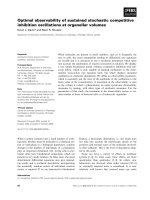
Fig. 1. Schematic representation of the connected sterol and steroid pathway in yeast. C, cytosol; ER, endoplamic reticulum; LP, lipid particles; mat,
mature form of the proteins; PM, plasma membrane. Steroids are shown in green. Ncp1p, NADPH P450 reductase; Adxp, adrenodoxin; Adrp,
adrenodoxin reductase; Are1p, Are2p, Atf2p, alcohol O-acetyltransferase (acetyl pregnenolone acetyl transferase); CYP11A1, P450 side chain
cleaving; Erg2p, sterol C8-C7 isomerase; Erg3p, C-5 sterol desaturase; Erg5p, D 22(23) sterol desaturase; Erg6p, S-adenosyl methionine D-24-sterol-
C-methyl-transferase; 3b-HSD, 3b-hydroxy steroid dehydrogenase.
1506 C. Duport et al. (Eur. J. Biochem. 270) Ó FEBS 2003
activity of CYP11A1. This suggests that the availability of
the CYP11A1 substrate is not limiting in the experimental
conditions used, but it might be limiting for higher levels of
CYP11A1 activity or expression.
Tgl1p, a putative ester hydrolase, regulates
SCC activity
In wild-type yeast, more than 90% of the predominant
sterol is stored as esters [37]. The balance between the levels
of free and esterified sterols is regulated by esterification and
hydrolysis and is modified in strains disrupted for the ester-
synthase genes ARE 1 and ARE 2 ([20,28]), or in which
steryl ester hydrolase activity is altered [40]. To further
investigate the potential limiting effect of sterol availability
on CYP11A1 activity, we cloned and expressed the TGL1
gene, that is predicted to code for a 46-kDa protein with a
potential steryl ester hydrolase catalytic domain [41]. Tgl1p
function was first evaluated in vitro by monitoring the
cholesteryl hydrolase activities of cell-free extracts prepared
from the wild-type strain, FY1679/pCD69 and an isogenic
are1D are2D double mutant, CDS04/pCD69. Surprisingly,
ester hydrolase activity in extracts of the control strain,
FY1679/pYeDP60 was higher than in extracts of FY1679/
pCD69, which overexpresses Tgl1p (Fig. 4A). In contrast,
although control CDS04/pYeDP60 extracts exhibited the
same activity as FY1679/pYeDP60 extracts, CDS04/
pCD69 cell-free extracts exhibited a twofold increase in
cholesteryl ester hydrolase activity. These apparently con-
tradictory results can be rationalized if cholesteryl esterase
activity can be monitored in vitro only in cellular extracts
devoid of endogenous sterol esters, that likely compete with
the labeled cholesteryl oleate used as a substrate. Inhibition
in FY1679/pCD69 extracts also suggested a strong prefer-
ence of Tgl1p for yeast sterol esters compared to cholesteryl
oleate. The hydrolytic activity of Tgl1p was further analyzed
in vivo (Fig 4B,C). Similar twofold, increases in the ratios of
free vs. total ergosterol and ergosta-5-eneol were observed in
Tgl1p-overexpressing FY-1679 and CA10 cells, when com-
pared to their respective control strains (Fig. 4B). This effect
was not observed in the corresponding are1D are2D double
mutant strains CDS04 and CA23, consistent with the
disruption of ester synthase genes (data not shown). As
established for ergosterol in wild-type strains [40], the
highest free ergosta-5-eneol to total protein ratio was
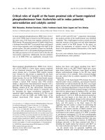
Fig. 2. The efficient production of pregnenolone and progesterone (A) in
the atf2D recombinant strains CA14/pCD63 (C) (Derg5, expressing
CYP11A1) and CA19/pCD63 (Derg5, expressing CYP11A1 and
3bHSD) is accompanied by an accumulation of ergosta-5-eneol precur-
sors (B). Gas chromatography (GC) profiles were obtained from cel-
lular lysates prepared from cultures harvested after 100 h of induction
with galactose. The sterol extraction procedure allows free sterol to be
detected (B). The ATF2 strain CA10/pCD63 was used as a control.
Relative retention times to cholesterol under our conditions are shown
between brackets. Steroids are: P, pregnenolone (0.598); Pr, pro-
gesterone (0.685), PA, pregnenolone acetate (0.714). Atypical sterols
detected in CA14/pCD63 and CA19/pCD63 cells are the following: D,
desmosterol (1.04); E5, ergosta-5-enel (1.13); E5,7, ergosta-5,7-dieneol
(1.16);F,fecosterol(1.11);U,unknownsterolwithaMWof384which
might be cholesta-7,24-dieneol (1.17); Z, zymosterol (1.06).
Ó FEBS 2003 CYP11A1 localizes to the yeast plasma membrane (Eur. J. Biochem. 270) 1507
observed in the PM of steroid-producing cells (Fig. 4C). In
contrast, internal membranes (ER, lipid particles and
mitochondrial membranes) exhibited only residual levels
of ergosta-5-eneol. Tgl1p overexpression did not modify this
subcellular distribution, and the influence of Tgl1p was
observed only in the PM fraction. In summary, we showed
that Tgl1p containing extracts have a steryl ester hydrolytic
activity and that these extracts could mediate the release of
free ergosta-5-eneol from esterified forms in steroid-produ-
cing cells with the same efficiency as observed for ergosterol
in wild-type yeast cells. In addition, Tgl1p overproduction
leads to an increase of free ergosta-5-eneol in tested
organelles, especially in the PM, which contains the highest
concentration of substrate.
To determine whether the Tgl1p-dependent increase in
free ergosta-5-eneol content could affect the SCC reaction,
the concentrations of accumulated pregnenolone were
measured in both the wild-type strain CA10/pCD63 and
in the are1D are2D double mutant CA23/pCD63, in the
absence or presence of the Tgl1p overexpression construct
(Fig. 5). As expected, CA23/pCD63, that is devoid of ester
synthase activity [42], contained higher amounts of free
ergosta-5-eneol than CA10/pCD63 (data not shown). This
phenomenon could explain the limited enhancement of
CYP11A1 activity observed in the are1D are2D mutant
(Fig. 5). When CA10 and CA23 were cotransformed with
pCD69, only the free ergosta-5-eneol content of the former
strain was increased (Fig. 4B and data not shown), whereas
pregnenolone production was enhanced in both strains,
with similar final concentrations of the steroid (Fig. 5).
Therefore, these results reveal two levels of complexity of
the CYP11A1 reaction in yeast. On one hand, the content
of free ergosta-5-eneol poorly correlates with the extent of
pregnenolone production, suggesting that SCC activity is
only partially limited by substrate concentration (Fig. 1).
On the other, an artificial increase in the level of free ergosta-
5-eneol improves the yield of steroid (Fig. 5). This latter
effect may involve steryl hydrolase activity potentially
encoded by TGL1. Moreover, the effects of Tgl1p on
CYP11A1 activity is observed both in the presence and
absence of ester synthase activity.
In recombinant yeast, the concentration of Adxp,
but not of Adrp, controls SCC activity
To determine whether electron transfer from NADPH to
CYP11A1 via Adrp and Adxp could regulate the extent of
synthesis, we built two CA10 derivatives, CA15 and CA17.
Like CA10, CA15 carries a unique ADR expression cassette
integrated in the LEU2-SPL1 intergenic region but in CA15
the mature ADR ORF is under the control of the constitu-
tive TEF1 promoter instead of the inducible GAL10/CYC1
promoter. CA17 also carries a cassette with the mature ver-
sion of ADX integrated in the HIS3-DED1 intergenic
region. (Table 1). The accumulation of steroids in the strains
CA15/pCD63 (that carries a multicopy plasmid bearing
CYP11A1 and ADX) and CA17/pV13sccm (that carries a
multicopy plasmid for CYP11A1 and has a single integrated
copy of ADX) was compared to that in CA10/pCD63. The
level of expression of mature Adrp was found to be lower in
CA15/pCD63 as compared to CA10/pCD63, as judged by
Western blot analysis (data not shown and [43]). In contrast,
CA17 exhibited the same level of Adrp as CA15 but a lower
level of Adxp as Adxp was expressed from a single genomic
copy (data not shown and Table 1). Similar concentra-
tions of pregnenolone (2.9 ± 0.5 mgÆL
)1
A
600
units) were
observed for CA10/pCD63 and CA15/pCD63, whereas a
36-fold decrease was observed for CA17/pCD63. These
results suggest that CYP11A1 activity depends strongly on
the levels of expression of Adxp but not Adrp and therefore
that the concentration of Adxp is the major factor
controlling pregnenolone synthesis in recombinant yeast.
In conclusion, the SCC reaction appears to be regulated
similarlyinyeastandinadrenalcells;inthelatter,Adxpand
not Adrp limits the activity of CYP11A1 and hence controls
the extent of pregnenolone production [14].
Protein partners involved in the yeast SCC system
localize to three distinct subcellular compartments
To gain a deeper insight into how the artificial steroid path-
way is coordinated with the endogenous ergosta-5-eneol
pathway, we determined the subcellular localizations of
Adrp, Adxp, D7-Red and 3b-HSD. A total cell extract
from the progesterone-producing strain CA19/pCD63 was
subjected to sucrose gradient fractionation (Fig. 6). Char-
acterization of each fraction with anti-Pma1p (PM), anti-
3-PGKp (cytosol), anti-40 kDa protein (ER) and anti-porin
(mitochondrial membranes) antisera revealed that Adrp,
Fig. 3. The final levels of accumulated steroids are independent of Datf2
genetic background (A) and do not correlate with the content of free
ergosta-5-eneol (B). Steroid-producing cells were grown as described in
Material and methods. Accumulated steroid contents are the sum of
pregnenolone and pregnenolone acetate (CA10/pCD63) (Derg5,
expressing CYP11A1), pregnenolone (CA14/pCD63) (Fig. 2), preg-
nenolone and progesterone (CA19/pCD63) (Fig. 2). Statistical signi-
ficance by paired t-tests was performed using the
STATVIEW
program.
**P < 0.05 when a strain is compared to the two others (B).
1508 C. Duport et al. (Eur. J. Biochem. 270) Ó FEBS 2003
D7-Red and 3b-HSD clearly localize to the ER whereas
Adxp is a soluble protein, as shown previously [19]. In
contrast to the other enzymes, recombinant CYP11A1 was
not restricted to a single subcellular compartment but
instead was found distributed broadly throughout the
gradient. Either this experiment reflects a broad intracellular
distribution of the protein, or a cell surface transport of the
protein as described [44,45]. To distinguish between these
two hypotheses, indirect immunofluorescence studies were
performed with polyclonal Igs raised against markers for
each of the different compartments (Fig 7. A,D,G, red
fluorescence, CY-2
TM
conjugated secondary Ig). The PM
(Fig. 7B), ER (Fig. 7E) and mitochondrial membranes
(Fig. 7H) were simultaneously visualized in the same cells
with Igs to Gpa1p, Dpm1p and porin, respectively (green
fluorescence, FITC-conjugated secondary antibodies).
Fluorescence was not detected in control experiments
performed in the absence of the primary anti-CYP11A1
and anti-marker Igs (data not shown). Confocal microscopy
was used to evaluate the degree of CYP11A1 colocalization
with each of these organelle markers, as seen in merged
images (Fig 7. C,F,I). CYP11A1 was found to colocalize
with the PM marker Gpa1p (Fig. 7C; yellow areas corres-
pond to regions where the red and green signals are
superimposed), suggesting that most of the CYP11A1
antigen resides in the PM. There was no overlap between the
red and green signals corresponding to the CYP11A1 and
porin mitochondrial markers, respectively, but some yellow
signals could be seen in cells labeled with both the CYP11A1
and the ER marker Ig (Fig. 7). However, the best corres-
pondence was observed for the CYP11A1-derived signal
and the PM-derived signal. In conclusion, the CYP11A1
antigen appears to be excluded from mitochondria in vivo,
and most of the antigen is detected in the plasma membrane,
with a minor fraction localizing to the ER.
To determine whether the SCC reaction occurs in the
PM, where most CYP11A1 is found, or in the ER, we
performed an analysis of free ergosta 5-eneol distribution in
the steroid-producing strain, CA19/pCD63 and in the
control strain, CA19/pDP10037. Figure 8 shows that the
level of free sterol in the PM is significantly depleted upon
the expression of SCC activity, whereas no decrease is
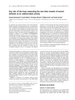
Fig. 4. Tgl1p has a steryl ester hydrolase activity. Tgl1p activity was
illustrated by comparing the steryl esterase cell free extract activities
of Tgl1p-overexpressing cells (strains transformed with pCD69,
stripped bars) vs. the controls (strains transformed with the vector
pYeDP60, gray bars). The CDS04 strain is an Dare1, Dare2 isogenic
derivative of FY1679-28 C and was generated by disruption of both
ARE 1 and ARE 2 genes, that encode two sterol ester-transferases
that catalyze the synthesis of steryl ester in yeast. (A) The cholesteryl
esterase activity of Tgl1p is detected only in the sterol esterification
deficient strain, CDS04 (Dare1, Dare2). Experiments used choleste-
ryl[4-
14
C]oleate as a substrate [37]. Specific activities were measured
in whole cell extracts prepared from lysed spheroplasts of FY1679/
pYeDP60, FY1679/pCD69 (expressing Tgl1p), CDS04/pCD69 (see
above), and CDS04/pYeDP60 cells. Data are mean values ± SEM
from three independent experiments with a maximum deviation of
5%. (B) The free ergosterol and ergosta-5-eneol contents are
increased in the Tgl1p-overproducing strains FY1679, CA10 (Derg5)
and CA10/pCD63 (Derg5 expressing CYP11A1), respectively. Sterols
were extracted from cellular lysates and analyzed by gas chroma-
tography with or without preliminary saponification for detection of
total sterols and free sterols, respectively. Data are expressed as ratio
of free vs. total sterol. Data are mean values ± SEM obtained from
three independent experiments. (C) The subcellular partitioning of
free ergosta-5-eneol is not changed by TGL1 overexpression. Whole
cell lysates were subjected to fractionation to isolate subcellular
organelles, mitochondria, lipid particles and ER as described [29]
and PM as described [30]. Data are expressed as the sterol : protein
ratio and are mean values from three independent experiments with
a maximum deviation of 5%. The ratio obtained in lipid particles
were 6.69 · 10
)4
and 6.78 · 10
)4
mgÆmg protein
)1
in CA10/
pYeDP60 and CA10/pCD69, respectively.
Ó FEBS 2003 CYP11A1 localizes to the yeast plasma membrane (Eur. J. Biochem. 270) 1509
observed in the ER or mitochondrial fractions. These data
are consistent with ergosta-5-eneol bioconversion at the
PM level. In conclusion, the mature form of recombinant
CYP11A1 (e.g., the protein without the mitochondrial
targeting sequence and with an extra methionine at the
NH
2
terminus) is primarily in the PM in yeast, and the
SCC system depends on electron transfer from ER-
localized Adrp to cytosolic Adxp and finally to CYP11A1
in the PM.
Discussion
The reconstruction in S. cerevisiae of the steroidogenic
pathway allows the self-sufficient production of pregneno-
lone and progesterone, as reported previously [19] (Fig. 1).
Like mammals, S. cerevisiae possesses a system that
efficiently protects against the toxic accumulation of preg-
nenolone. In mammals, pregnenolone is present as a
biologically active sulfo-conjugate [46], whereas in yeast,
APAT activity converts pregnenolone into the correspond-
ing acetate ester [38]. Acetylation or sulfatation at position 3
of pregnenolone prevents further metabolism by 3b-HSD.
Yeast strains devoid of APAT activity allow high-yield
biosynthesis of free pregnenolone or progesterone but
accumulate ergosta-5-eneol biosynthesis intermediates, such
as desmosterol, fecosterol and zymosterol (Fig. 1). This
phenomenon likely results from the inhibition of S-adenosyl
sterol 24-methyl transferase (Erg6p [47]), and to a lesser
extent of sterol C8–C7 isomerase (Erg2p [48]). Erg6p
permits the transformation of cholesta-derivatives into
ergosta-derivatives. Thus, inhibition of Erg6p allows the
accumulation of zymosterol that is sequentially transformed
by Erg2p and Erg3p (the C-5 desaturase [49]), into cholesta-
7,24-dieneol and cholesta-5,7,24-trieneol, respectively
(Fig. 1). In the presence of D7-reductase, the latter is
modified into cholesta-5,24-dieneol (desmosterol) that is
detected in the membranes of CA14/pCD63 (Fig. 2B).
Accumulation of zymosterol indicates that Erg2p might
also be inhibited. A similar effect was observed in mamma-
lian cells, in that progesterone [50] and pregnenolone [51]
inhibit cholesterol biosynthesis, resulting in the accumula-
tion of cholesterol precursors. Finally, in yeast strains
devoid of APAT activity, there is almost no accumulation of
pregnenolone when 3b-HSD activity is present, and the rate
of progesterone biosynthesis is only marginally reduced
compared to the rate observed for pregnenolone alone.
Therefore, an efficient coupling of the two first steps of
steroidogenesis is possible, and as reported for mammalian
cells [9], the SCC reaction is the rate-determining step in
progesterone synthesis in yeast.
The yeast SCC system offers the possibility of increasing
or decreasing the availability of the endogenous substrate.
Ergosterol or related yeast sterols exist in the free form or as
esters conjugated to fatty acids. Conversion between free
sterols and steryl esters is thus a critical homeostatic
determinant for membrane function in yeast as in all
eukaryotic cells ([52,53]). The PM is the major subcellular
location of free sterol [40]. Steryl esters are synthesized in the
ER by the ACAT-related ARE 1 and ARE 2 gene products
[20,42], stored in lipid droplets and mobilized by a process
involving steryl ester hydrolases in the PM [37].
Our work shows that Tgllp over-expressing cells and
extracts exhibit steryl ester hydrolase activities in vivo and
in vitro, respectively. As it has been shown previously that
Tgl1p had significant homologies to triglyceride lipase, it is
rather likely that Tgl1p is a steryl ester hydrolase [41]. In
the steroid-producing yeast strain, CA10/pCD63 free
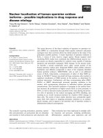
Fig. 6. Subcellular localization of heterologous CYP11A1, Adrp, Adxp,
D7-Red, 3b-HSD and organelle membrane after sucrose density frac-
tionation in recombinant yeast. The heterologous Adrp, D7-Red and
3b-HSD proteins cofractionate with the ER marker (40 kDa protein)
and Adxp cofractionates with the cytosol marker 3-PGK on a sucrose
continuous gradient. No clear subcellular localization in the PM, ER
or mitochondrial membranes was observed for CYP11A1. CA19/
pCD63 (Datf2, Derg5, expressing CYP11A1 and 3bHSD) cells were
grown as described in Materials and methods, converted to sphero-
plasts, lysed and fractionated on a continuous sucrose gradient (0.7–
1.6
M
) from the bottom (fraction 1) to the top (fraction 25). Fractions
were subjected to Western blot analysis with Igs against the following
organelle marker proteins: 3-PGK for cytosol, Pma1p for the PM, the
40-kDa protein for the ER and porin for mitochondria. The distri-
bution of heterologous proteins was similarly detected with Igs against
CYP11A1, Adrp, Adxp, 3b-HSD and D7-Red.
Fig. 5. Pregnenolone biosynthesis is increased in recombinant yeast cells
overproducing Tgl1p. Pregnenolone was extracted from cellular lysates
prepared from cultures harvested after 100 h of induction by galactose
[19]. The Tgl1p-overproducing effect was analyzed in ARE 1 ARE 2
strains (CA10/pCD63 + pCD69) (Derg5, expressing CYP11A1
and Tgl1p) and (CA10/pCD63 + pYeDP60) (Derg5, expressing
CYP11A1) and in Dare 1 Dare 2 strains (CA23/pCD63 + pCD69)
(Derg5, expressing CYP11A1 and Tgl1p) and CA23/pCD63 +
pYeDP60) (Derg5, expressing CYP11A1).
1510 C. Duport et al. (Eur. J. Biochem. 270) Ó FEBS 2003
ergosta-5-eneol represents, as expected, only a minor
fraction of the cellular pool of sterols, and this fraction
increases when Tgl1p is overexpressed or when both the
ARE 1 and ARE 2 genes are deleted. Thus, the two ester
synthases, Arelp and Are2p and the probable steryl ester
hydrolase Tgllp, play complementary roles in maintaining
free ergosta-5-enol at appropriate levels, reminiscent of the
mechanism reported for cholesterol in adrenal cells ([40,54]).
In addition to normally cycling between the PM and other
cellular organelles, free ergosta-5-eneol also must be avail-
able in the yeast cell to serve as a substrate for CYP11A1.
While SCC driven formation of pregnenolone likely occurs
at the PM, ergosta-5-enol biosynthesis occurs mainly in the
ER, consistent with D7-Red localization (Fig. 6). Are1p and
Are2p are localized in the ER [42] while Tgl1p could be
localized in lipid particles, and activated with the supply of
sterol from lipid particles to PM. Therefore, de novo
synthesis and transport processes are critical for continued
activity.
In theory, the sterol pool used for steroidogenesis must be
constantly supplied from cellular sterol stores while the
membrane structural pool is more static, at least for cells in
stationary phase, in which CYP11A1-dependent conversion
is active but cell growth has ceased. Thus, free and esterified
sterol pools are exchanged rapidly. Indeed, Tgllp overpro-
duction results in an increased level of free sterol (Fig. 4B),
and it would also be expected to increase the cycling of
sterols by esterification and hydrolysis, resulting in an
increase in intracellular sterol trafficking. It is unclear, then,
whether the corresponding increase in pregnenolone pro-
duction results from an increase in free sterol concentration
or from an enhancement of sterol trafficking.
Fig. 7. CYP11A1 colocalizes with the endo-
genous Gpa1p plasma membrane protein.
Spheroplasts from CA19/pCD63 (Datf2,
Derg5, expressing CYP11A1 and 3b)HSD)
were fixed, permeabilized and then incubated
with primary polyclonal CYP11A1 Igs. A
CY2
TM
conjugated secondary Ig (red fluores-
cence in A, D, G) was used to visualize the
CYP11A1 protein. PM (B), ER (E) and
mitochondrial (H) membranes were detected
in the same cells with anti-Gpa1p (guanine
nucleotide-binding protein alpha subunit),
Dpm1p (dolichol-phosphate mannosyltrans-
ferase) and porin monoclonal Igs coupled to
FITC secondary Igs (green fluorescence). The
merged CY2
TM
and FITC images are shown
in C, F and I.
Fig. 8. Distribution of the CYP11A1 substrate. Whole cell lysates were
fractionated to isolate the ER and mitochondrial membranes as des-
cribed [29] and PM as described [30]. Data are expressed as percent of
the total activity measured for the whole cellular extract. Relative to
CA10/pDP10037 (gray bars, Derg5), the pregnenolone-producing
strain CA10/pCD63 (stripped bars, Derg5 expressing CYP11A1)
shows a significant depletion of the levels of free ergosta 5-eneol in the
PM fraction and increased overall levels in the ER and mitochondrial
fractions.
Ó FEBS 2003 CYP11A1 localizes to the yeast plasma membrane (Eur. J. Biochem. 270) 1511
In conclusion, the recombinant yeast strain described in
this report mimics mammalian steroid-producing cells with
respect to the coupling of the SCC and 3b-HSD reactions;
the inhibitory effect of steroid products on CYP11A1
substrate biosynthesis; the regulation of intracellular free
sterol concentration by the ACAT-related enzymes Are1p
and Are2p; the steryl ester hydrolase Tgl1p and finally, the
modulation of substrate availability by Tgl1p.
To determine if the delivery of reducing equivalents to
CYP11A1 could contribute to limit SCC activity in yeast,
the importance of the concentrations of Adxp and Adrp was
evaluated. The recombinant SCC system in yeast is sensitive
to the levels of expression of these components, similar to
that found for mammalian adrenal cells [14] or for in vitro
assay systems [55]. In yeast, as in transfected mammalian
cells [56], a decrease in the level of expression of Adxp has
dramatic consequences for SCC activity. Alterations in the
level of expression of Adrp in yeast have no effect as
described above. This contrasts with a recent paper where,
in placenta, the Adrp concentration is limiting, giving an
alternative regulation of the SCC reaction [57].
It was of interest to characterize the localization of the
different proteins of the reaction – namely, CYP11A1,
Adxp and Adrp in yeast cells that exhibit mammalian
steroidogenic activity. In order to avoid the difficulty of
transporting the CYP11A1 substrate to yeast mitochondria,
we chose to express CYP11A1, Adxp and Adrp outside the
mitochondrion by simply replacing their respective mito-
chondrial targeting sequences with a methionine residue.
Adrp and CYP11A1 were expected to be found in the ER,
while Adxp was expected to be in the cytosol. Classical
differential centrifugation (data not shown) or sucrose
gradient fractionation (Fig. 6) confirmed the localization of
Adxp and Adrp, but CYP11A1 could not be definitively
localized using these biochemical fractionation techniques.
Finally, indirect immunofluorescence with polyclonal anti-
CYP11A1 antibodies together with confocal microscopy
was the only technique that reliably and reproducibly
showed that CYP11A1 is mainly localized to the PM.
We cannot exclude the possibility that the deletion of the
mitochondrial targeting sequence from CYP11A1 reveals a
cryptic recognition motif that allows targeting to the PM.
For example, the mature forms of the human, bovine and
pig CYP11A1 have two positively charged residues at their
NH
2
termini that could favor targeting to the PM [58].
The presence of CYP11A1 in the yeast PM is rather
puzzling considering that mammalian CYP11A1 is a
specialized enzyme localized to the inner mitochondrial
membrane of adrenal or placenta cells. Moreover, the
protein is apparently catalytically competent in the yeast
PM, based on the observation of the specific depletion of
ergosta-5-eneol in the PM ([19], and this work). Interest-
ingly, both Adrp and Adxp are absent from the PM, as
evidenced by Western blot analyses, and their respective ER
and cytosolic localizations were confirmed by immuno-
fluorescence and differential centrifugation (data not shown,
and [59]). This result indicates that Adxp functions as a
soluble transporter of electrons from the ER associated
Adrp to the PM-bound CYP11A1. Therefore, our work
supports a shuttle mechanism of electron transport in which
Adxp dissociates from the ER (where Adrp is located)
before delivering electrons to CYP11A1 at the PM. In
conclusion, these findings demonstrate that the ÔclusterÕ
model proposed for the mitochondrial system ([17,60]), is
not obligatory and that the mitochondrial environment is
not absolutely required for CYP11A1 function.
The results of this work provide convincing evidence that
recombinant CYP11A1 associates functionally with the PM
in yeast but do not allow us to exclude the possibility that it
has a second association with ER membranes (Fig. 7).
Ectopic localization at the PM of a functional microsomal
cytochrome P450 protein, has been reported for CYP2D6
expressed heterologously in yeast [30] and endogenously in
rat hepatocytes [61]. Dual localization has been also
reported for CYP2E1 and other P450 enzymes, and these
enzymes can exhibit different substrate specificities in the
mitochondrion and in microsomes. Whereas CYP2D6
requires NADPH-P450 reductase as an electron carrier at
both sites, CYP2E1 is bifunctional: in the mitochondrion it
receives electrons from Adxp and Adrp, and in microsomes
it receives electrons from NADPH-P450 reductase.
CYP11A1 cannot receive electrons from NADPH-P450
reductase [62] and the existence of alternate electron carriers
has not been established. However, cytochrome b5 has been
shown to interact specifically with CYP11A1, but this
interaction is unproductive in the absence of Adxp and
Adrp [63].
In conclusion, we have used steroid-producing S. cere-
visiae strains to study the different factors involved in the
SCC of ergosta-5-eneol into pregnenolone, including the
effect of the steroid products on the yeast sterol synthesis
pathway, acetylation of the product, availability of the
substrate, electron flow and localization of the different
protein partners. We find that yeast mimics mammalian
adrenal cells in these respects. The quantity of steroid
produced is controlled by the availability and mobility of
the substrate together with the concentration of Adxp.
Unexpectedly, Adxp, Adrp and CYP11A1 are localized in
the cytosol, ER and PM, respectively, without impairing the
SCC reaction and its coupling to the ER-associated
3b-HSD activity. Nonetheless, Adxp can shuttle electrons
from ADR to CYP11A1 in a productive fashion. The
unusual presence of the mitochondrial CYP11A1 in the PM
may reflect a possible alternative localization of this enzyme
in mammalian cells, suggestive of an alternative way of
producing pregnenolone.
Acknowledgements
The anti-PmaIp Igs were kindly supplied by R. Hagenauer-Tsapis. We
thank J. Loeper and N. Chaverot for their helpful assistance in confocal
scanning microscopy. We also thank C. Roubal for continuous
support. This work was supported by Aventis Pharma.
References
1. Fannon, S.A., Vidaver, R.M. & Marts, S.A. (2001) An abridged
history of sex steroid hormone receptor action. J. Appl. Physiol.
91, 1854–1859.
2. Miller, W.L. (1988) Molecular biology of steroid hormone syn-
thesis. Endocr. Rev. 9, 295–318.
3. Sugano, S., Miura, R. & Morishima, N. (1996) Identification of
intermediates in the conversion of cholesterol to pregnenolone
with a reconstituted cytochrome p-450scc system: accumulation of
1512 C. Duport et al. (Eur. J. Biochem. 270) Ó FEBS 2003
the intermediate modulated by the adrenodoxin level. J. Biochem.
(Tokyo). 120, 780–787.
4. Morohashi, K., Fujii-Kuriyama, Y., Okada, Y., Sogawa, K.,
Hirose, T., Inayama, S. & Omura, T. (1984) Molecular cloning
and nucleotide sequence of cDNA for mRNA of mitochondrial
cytochrome P-450 (SCC) of bovine adrenal cortex. Proc. Natl
Acad. Sci. USA. 81, 4647–4651.
5. Zuber, M.X., Mason, J.I., Simpson, E.R. & Waterman, M.R.
(1988) Simultaneous transfection of COS-1 cells with mitochon-
drial and microsomal steroid hydroxylases: incorporation of a
steroidogenic pathway into nonsteroidogenic cells. Proc. Natl
Acad. Sci. USA. 85, 699–703.
6. Nelson, D.R., Koymans, L., Kamataki, T., Stegeman, J.J.,
Feyereisen, R., Waxman, D.J., Waterman, M.R., Gotoh, O.,
Coon, M.J., Estabrook, R.W., Gunsalus, I.C. & Nebert, D.W.
(1996) P450 superfamily: update on new sequences, gene mapping,
accession numbers and nomenclature. Pharmacogenetics. 6, 1–42.
7. Vickery, L.E. (1997) Molecular recognition and electron transfer
in mitochondrial steroid hydroxylase systems. Steroids. 62,124–
127.
8. Grinberg, A.V., Hannemann, F., Schiffler, B., Muller, J., Heine-
mann, U. & Bernhardt, R. (2000) Adrenodoxin: structure,
stability, and electron transfer properties. Proteins. 40, 590–612.
9. Miller, W.L. (1995) Mitochondrial specificity of the early steps in
steroidogenesis. J. Steroid Biochem. Mol. Biol. 55, 607–616.
10. Lin, D., Gitelman, S.E., Saenger, P. & Miller, W.L. (1991) Normal
genes for the cholesterol side chain cleavage enzyme, P450scc, in
congenital lipoid adrenal hyperplasia, J. Clin. Invest. 88, 1955–
1962.
11. Bose, H., Lingappa, V.R. & Miller, W.L. (2002) Rapid regulation
of steroidogenesis by mitochondrial protein import. Nature. 417,
87–91.
12. Stocco, D.M. & Clark, B.J. (1996) Regulation of the acute pro-
duction of steroids in steroidogenic cells. Endocr. Rev. 17, 221–244.
13. Yang, H., Cromley, D., Wang, H., Billheimer, J.T. & Sturley, S.L.
(1997) Functional expression of a cDNA to human acyl-coenzyme
A: cholesterol acyltransferase in yeast. Species-dependent sub-
strate specificity and inhibitor sensitivity. J. Biol. Chem. 272, 3980–
3985.
14. Hanukoglu, I. & Hanukoglu, Z. (1986) Stoichiometry of mito-
chondrial cytochromes P-450, adrenodoxin and adrenodoxin
reductase in adrenal cortex and corpus luteum. Implications for
membrane organization and gene regulation. Eur. J. Biochem. 157,
27–31.
15. Tuckey, R.C. & Sadleir, J. (1999) The concentration of adreno-
doxin reductase limits cytochrome p450scc activity in the human
placenta. Eur. J. Biochem. 263, 319–325.
16. Tuckey, R.C., McKinley, A.J. & Headlam, M.J. (2001) Oxidized
adrenodoxin acts as a competitive inhibitor of cytochrome
P450scc in mitochondria from the human placenta. Eur. J. Bio-
chem. 268, 2338–2343.
17. Muller, E.C., Lapko, A., Otto, A., Muller, J.J., Ruckpaul, K. &
Heinemann, U. (2001) Covalently crosslinked complexes of
bovine adrenodoxin with adrenodoxin reductase and cytochrome
P450scc. Mass spectrometry and Edman degradation of com-
plexes of the steroidogenic hydroxylase system. Eur. J. Biochem.
268, 1837–1843.
18. Cauet, G., Balbuena, D., Achstetter, T. & Dumas, B. (2001)
CYP11A1 stimulates the hydroxylase activity of CYP11B1 in
mitochondria of recombinant yeast in vivo and in vitro. Eur. J.
Biochem. 268, 4054–4062.
19. Duport, C., Spagnoli, R., Degryse, E. & Pompon, D. (1998) Self-
sufficient biosynthesis of pregnenolone and progesterone in
engineered yeast. Nature Biotechnol. 16, 186–189.
20. Yang, H., Bard, M., Bruner, D.A., Gleeson, A., Deckelbaum,
R.J., Aljinovic, G., Pohl, T.M., Rothstein, R. & Sturley, S.L.
(1996) Sterol esterification in yeast: a two-gene process. Science.
272, 1353–1356.
21. Gietz, R.D., Schiestl, R.H., Willems, A.R. & Woods, R.A. (1995)
Studies on the transformation of intact yeast cells by the LiAc/
SS-DNA/PEG procedure. Yeast. 11, 355–360.
22. Adams, A., Gottsching, D., Kaiser, C. & Stearns, T. (1997)
Methods in Yeast Genetics: a Laboratory Course Manual,Cold
Spring Harbor Laboratory Press, Cold Spring Harbor, New York.
23. Baudin, A., Ozier-Kalogeropoulos, O., Denouel, A., Lacroute, F.
& Cullin, C. (1993) A simple and efficient method for direct
gene deletion in Saccharomyces cerevisiae. Nucleic Acids Res. 21,
3329–3330.
24. Dumas, B., Cauet, G., Lacour, T., Degryse, E., Laruelle, L.,
Ledoux, C., Spagnoli, R. & Achstetter, T. (1996) 11 Beta-hydro-
xylase activity in recombinant yeast mitochondria.Invivocon-
version of 11-deoxycortisol to hydrocortisone. Eur. J. Biochem.
238, 495–504.
25. Degryse,E.,Dumas,B.,Dietrich,M.,Laruelle,L.&Achstetter,T.
(1995) In vivo cloning by homologous recombination in yeast
using a two- plasmid-based system. Yeast 11, 629–640.
26. Cullin, C. & Pompon, D. (1988) Synthesis of functional mouse
cytochromes P-450 P1 and chimeric P-450 P3–1 in the yeast
Saccharomyces cerevisiae. Gene 65, 203–217.
27. Wach, A., Brachat, A., Pohlmann, R. & Philippsen, P. (1994) New
heterologous modules for classical or PCR-based gene disruptions
in Saccharomyces cerevisiae. Yeast 10, 1793–1808.
28. Ness, F., Achstetter, T., Duport, C., Karst, F., Spagnoli, R. &
Degryse, E. (1998) Sterol uptake in Saccharomyces cerevisiae heme
auxotrophic mutants is affected by ergosterol and oleate but not
by palmitoleate or by sterol esterification. J. Bacteriol. 180, 1913–
1919.
29. Zinser, E. & Daum, G. (1995) Isolation and biochemical char-
acterization of organelles from the yeast, Saccharomyces cerevi-
siae. Yeast 11, 493–536.
30. Loeper, J., Louerat-Oriou, B., Duport, C. & Pompon, D. (1998)
Yeast expressed cytochrome P450 2D6 (CYP2D6) exposed on the
external face of plasma membrane is functionally competent.
Mol. Pharmacol. 54, 8–13.
31. Mihara, K., Blobel, G. & Sato, R. (1982) In vitro synthesis and
integration into mitochondria of porin, a major protein of the
outer mitochondrial membrane of Saccharomyces cerevisiae. Proc.
NatlAcad.Sci.USA. 79, 7102–7106.
32. Daum, G., Bohni, P.C. & Schatz, G. (1982) Import of proteins
into mitochondria. Cytochrome b
2
and cytochrome c peroxidase
are located in the intermembrane space of yeast mitochondria.
J. Biol. Chem. 257, 13028–13033.
33. Hermann, G.J., Thatcher, J.W., Mills, J.P., Hales, K.G., Fuller,
M.T.,Nunnari,J.&Shaw,J.M.(1998)Mitochondrialfusionin
yeast requires the transmembrane GTPase Fzo1p. J. Cell Biol.
143, 359–373.
34. Serrano, R., Kielland-Brandt, M.C. & Fink, G.R. (1986) Yeast
plasma membrane ATPase is essential for growth and has
homology with (Na
+
+K
+
), K
+
-andCa
2+
-ATPases. Nature.
319, 689–693.
35. Hirschman, J.E., De Zutter, G.S., Simonds, W.F. & Jenness, D.D.
(1997) The G beta gamma complex of the yeast pheromone
response pathway. Subcellular fractionation and protein–protein
interactions. J. Biol. Chem. 272, 240–248.
36. Jackson, D.D. & Stevens, T.H. (1997) VMA12 encodes a yeast
endoplasmic reticulum protein required for vacuolar H
+
-ATPase
assembly. J. Biol. Chem. 272, 25928–25934.
37. Zinser, E., Paltauf, F. & Daum, G. (1993) Sterol composition of
yeast organelle membranes and subcellular distribution of enzymes
involved in sterol metabolism. J. Bacteriol. 175, 2853–2858.
38. Cauet, G., Degryse, E., Ledoux, C., Spagnoli, R. & Achstetter, T.
(1999) Pregnenolone esterification in Saccharomyces cerevisiae.
Ó FEBS 2003 CYP11A1 localizes to the yeast plasma membrane (Eur. J. Biochem. 270) 1513
A potential detoxification mechanism. Eur. J. Biochem. 261, 317–
324.
39. Gachotte, D., Sen, S., Eckstein, J., Barbuch, R., Krieger, M., Ray,
B. & Bard, M. (1999) Characterization of the Saccharomyces
cerevisiae ERG27 gene encoding the 3-keto reductase involved in
C-4 sterol demethylation. Proc. Natl Acad. Sci. USA. 96, 12655–
12660.
40. Zinser, E., Sperka-Gottlieb, C.D., Fasch, E.V., Kohlwein, S.D.,
Paltauf, F. & Daum, G. (1991) Phospholipid synthesis
and lipid composition of subcellular membranes in the uni-
cellular eukaryote Saccharomyces cerevisiae. J. Bacteriol. 173,
2026–2034.
41. Anderson, R.A. & Sando, G.N. (1991) Cloning and expression of
cDNA encoding human lysosomal acid lipase/cholesteryl ester
hydrolase. Similarities to gastric and lingual lipases. J. Biol. Chem.
266, 22479–22484.
42. Zweytick, D., Leitner, E. & Kohlwein, S.D., Yu, C., Rothblatt, J.
& Daum, G. (2000) Contribution of Are1p and Are2p to steryl
ester synthesis in the yeast Saccharomyces cerevisiae. Eur. J. Bio-
chem. 267, 1075–1082.
43. Nacken, V., Achstetter, T. & Degryse, E. (1996) Probing the limits
of expression levels by varying promoter strength and plasmid
copy number in Saccharomyces cerevisiae. Gene. 175, 253–260.
44. Kolling, R. & Hollenberg, C.P. (1994) The ABC-transporter Ste6
accumulates in the plasma membrane in a ubiquitinated form in
endocytosis mutants. EMBO J. 13, 3261–3271.
45. Chen, C., Ingram, M.F., Rosal, P.H. & Graham, T.R. (1999) Role
for Drs2p, a P-Type ATPase and potential aminophospholipid
translocase, in yeast late golgi function. J. Cell Biol. 147, 1223–
1236.
46. Akwa, Y., Ladurelle, N., Covey, D.F. & Baulieu, E.E. (2001) The
synthetic enantiomer of pregnenolone sulfate is very active on
memory in rats and mice, even more so than its physiological
neurosteroid counterpart: distinct mechanisms? Proc. Natl Acad.
Sci. USA. 98, 14033–14037.
47. Venkatramesh, M., Guo, D.A., Harman, J.G. & Nes, W.D. (1996)
Sterol specificity of the Saccharomyces cerevisiae ERG6 gene
product expressed in Escherichia coli. Lipids 31, 373–377.
48. Moebius, F.F., Bermoser, K., Reiter, R.J., Hanner, M. & Gloss-
mann, H. (1996) Yeast sterol C8–C7 isomerase: identification and
characterization of a high-affinity binding site for enzyme inhibi-
tors. Biochemistry. 35, 16871–16878.
49. Lees, N.D., Skaggs, B., Kirsch, D.R. & Bard, M. (1995) Cloning
of the late genes in the ergosterol biosynthetic pathway of
Saccharomyces cerevisiae –areview.Lipids. 30, 221–226.
50. Metherall, J.E., Waugh, K. & Li, H. (1996) Progesterone inhibits
cholesterol biosynthesis in cultured cells. Accumulation of cho-
lesterol precursors. J. Biol. Chem. 271, 2627–2633.
51. Metherall, J.E., Li, H. & Waugh, K. (1996) Role of multidrug
resistance P-glycoproteins in cholesterol biosynthesis. J. Biol.
Chem. 271, 2634–2640.
52. Lewis, T.A., Rodriguez, R.J. & Parks, L.W. (1987) Relationship
between intracellular sterol content and sterol esterification and
hydrolysis in Saccharomyces cerevisiae. Biochim. Biophys. Acta.
921, 205–212.
53. Sturley, S.L. (2000) Conservation of eukaryotic sterol homeo-
stasis: new insights from studies in budding yeast, Biochim.
Biophys. Acta. 1529, 155–163.
54. Yeaman, S.J. (1990) Hormone-sensitive lipase – a multipurpose
enzyme in lipid metabolism. Biochim. Biophys. Acta. 1052, 128–
132.
55. Beckert, V. & Bernhardt, R. (1997) Specific aspects of electron
transfer from adrenodoxin to cytochromes p450scc and
p45011beta. J. Biol. Chem. 272, 4883–4888.
56. Cao, P.R. & Bernhardt, R. (1999) Interaction of CYP11B1
(cytochrome P-45011 beta) with CYP11A1 (cytochrome P-450scc)
in COS-1 cells. Eur. J. Biochem. 262, 720–726.
57. Tuckey, R.C. & H.M. (2002) Placental cytochrome P450scc
(CYP11A1): comparison of catalytic properties between condi-
tions of limiting and saturating adrenodoxin reductase. J. Steroid
Biochem. Mol. Biol. 81, 153–158.
58. Neve, E.P. & Ingelman-Sundberg, M. (2000) Molecular basis for
the transport of cytochrome P450 2E1 to the plasma membrane.
J. Biol. Chem. 275, 17130–17135.
59. Akiyoshi-Shibata, M., Sakaki, T., Yabusaki, Y., Murakami, H. &
Ohkawa, H. (1991) Expression of bovine adrenodoxin and
NADPH-adrenodoxin reductase cDNAs in Saccharomyces cere-
visiae. DNA Cell Biol. 10, 613–621.
60. Hara, T., Koba, C., Takeshima, M. & Sagara, Y. (2000) Evidence
for the cluster model of mitochondrial steroid hydroxylase system
derived from dissociation constants of the complex between
adrenodoxin reductase and adrenodoxin. Biochem. Biophys. Res.
Commun. 276, 210–215.
61. Wu, D. & Cederbaum, A.I. (1992) Presence of functionally active
cytochrome P-450IIE1 in the plasma membrane of rat hepato-
cytes. Hepatology. 15, 515–524.
62. Sakaki, T., Akiyoshi-Shibata, M., Yabusaki, Y. & Ohkawa, H.
(1992) Organella-targeted expression of rat liver cytochrome
P450c27 in yeast. Genetically engineered alteration of mitochon-
drial P450 into a microsomal form creates a novel functional
electron transport chain. J. Biol. Chem. 267, 16497–16502.
63. Chudaev, M.V., Gilep, A.A. & Usanov, S.A. (2001) Site-directed
mutagenesis of cytochrome b5 for studies of its interaction with
cytochrome P450. Biochemistry (Mosc). 66, 667–681.
64. Thierry, A., Fairhead, C. & Dujon, B. (1990) The complete
sequence of the 8.2 kb segment left of MAT on chromosome III
reveals five ORFs, including a gene for a yeast ribokinase. Yeast. 6,
521–534.
65. Lecain, E., Chenivesse, X., Spagnoli, R. & Pompon, D. (1996)
Cloning by metabolic interference in yeast and enzymatic char-
acterization of Arabidopsis thaliana sterol delta 7-reductase. J. Biol.
Chem. 271, 10866–10873.
1514 C. Duport et al. (Eur. J. Biochem. 270) Ó FEBS 2003