Báo cáo khoa học: Unique features of the hemoglobin system of the Antarctic notothenioid fish Gobionotothen gibberifrons ppt
Bạn đang xem bản rút gọn của tài liệu. Xem và tải ngay bản đầy đủ của tài liệu tại đây (433.98 KB, 7 trang )
Unique features of the hemoglobin system of the Antarctic
notothenioid fish
Gobionotothen gibberifrons
Panagiotis Marinakis, Maurizio Tamburrini, Vito Carratore and Guido di Prisco
Institute of Protein Biochemistry, CNR, Naples, Italy
ThehemolysateoftheAntarcticteleostGobionotothen
gibberifrons (family Nototheniidae) contains two hemoglo-
bins (Hb 1 and Hb 2). The concentration of Hb 2 (15–20%
of the total hemoglobin content) is higher than that found
in most cold-adapted Notothenioidei. Unlike the other
Antarctic species so far examined having two hemoglobins,
Hb 1 and Hb 2 do not have globin chains in common.
Therefore this hemoglobin system is made of four globins
(two a-andtwob-chains). The complete amino-acid
sequence of the two hemoglobins (Hb 1, a
1
2
b
1
2
;Hb2,a
2
2
b
2
2
)
has been established. The two hemoglobins have different
functional properties. Hb 2 has lower oxygen affinity than
Hb 1, and higher sensitivity to the modulatory effect of
organophosphates. They also differ thermodynamically,
as shown by the effects on the oxygen-binding properties
brought about by temperature variations. The oxygen-
transport system of G. gibberifrons, with two functionally
distinct hemoglobins, suggests that the two components may
have distinct physiological roles, in relation with life style
and the environmental conditions which the fish may have to
face. The unique features of the oxygen-transport system of
this species are reflected in the phylogeny of the hemoglobin
amino-acid sequences, which are intermediate between those
of other fish of the family Nototheniidae and of species of the
more advanced family Bathydraconidae.
Keywords: fish; Antarctic; hemoglobin; amino-acid
sequence; oxygen binding.
Organisms living in extreme environments, such as the
Arctic and Antarctic sea waters, are exposed to strong
constraints, among which temperature is often a driving
factor [1–4]. Hemoglobin (Hb), a direct link between the
exterior and body requirements, has thus experienced a
major evolutionary pressure in these organisms to adapt
its functional features at molecular/functional level. The
search for correlation between fish hematology and the
extreme conditions of the Antarctic environment leads to
a study on the biochemistry of oxygen transport, centred
on Hb molecular structure and oxygen-binding
properties, taking the ecological constraints under con-
sideration. In view of the role of temperature in
modifying the oxygenation-deoxygenation cycle in respir-
ing tissues, thermodynamic analysis deserves special
attention.
The largely dominant Antarctic suborder Notothenioidei
is by far the most thoroughly characterized group of fish in
the world. Thirty-five species (all bottom dwellers) of the 38
so far investigated were shown to have a single major Hb
(Hb 1) and often a minor one (Hb 2, 5% of the total Hb
content, generally having the b-chain in common with
Hb 1) both displaying, with some exceptions, strong Bohr
and Root effects [2,5]. Each of the remaining three species
(Trematomus newnesi and Pagothenia borchgrevinki,two
active cryopelagic species; Pleuragramma antarcticum,a
pelagic, sluggish but migratory fish) all belonging to the
family Nototheniidae, have a unique oxygen-transport
system, and each system appears adjusted to the fish specific
life style, substantially different from that of the sluggish
benthic species.
Compared with other Notothenioidei, the Antarctic
teleost, Gobionotothen gibberifrons (family Nototheniidae),
is endowed with novel hematological features. A detailed
study of the oxygen-transport system is herewith reported.
A preliminary communication on the Hb system of this
species has appeared previously [6]. G. gibberifrons lives at a
depth range between 5 and 750 m in the waters of northern
Antarctic Peninsula and of the islands located north-east.
G. gibberifrons has Hb 1 and Hb 2. The complete amino-
acid sequence of the two components has been established,
and the regulation of oxygen binding by pH, allosteric
effectors (chloride and organophosphates) and temperature
has been investigated.
Materials and methods
Toyopearl Super Q-650S was from TosoHaas (Laboratory
Service Analytical); trypsin (EC 3.4.21.4) treated with
L
-1-tosylamide-2-phenylethylchloromethylketone from
Cooper Biomedical; dithiothreitol from Fluka; Tris, bis-
Tris, Hepes, Mes, 4-vinylpyridine and IHP from Sigma;
sequanal-grade reagents from Applied Biosystems;
Correspondence to G. di Prisco, Institute of Protein Biochemistry,
CNR, Via Marconi 12, I-80125 Naples, Italy.
Fax: + 39 0815936689; Tel.: + 39 0817257242;
E-mail:
Abbreviations: Hb, haemoglobin; OPA, o-phthalaldehyde; IHP,
inositol hexakisphosphate; P
50
, partial pressure of oxygen required to
achieve Hb half saturation.
Note: The protein sequence data reported in this paper will appear in
the SWISS-PROT and TrEMBL knowledgebase under the accession
numbers P83611 (a
1
chain), P83612 (b
1
chain), P83613 (a
2
chain) and
P83614 (b
2
chain).
(Received 7 July 2003, revised 1 August 2003,
accepted 7 August 2003)
Eur. J. Biochem. 270, 3981–3987 (2003) Ó FEBS 2003 doi:10.1046/j.1432-1033.2003.03786.x
HPLC-grade acetonitrile from Laboratory-Scan Analytical.
All other reagents were of the highest purity commercially
available.
Specimens of G. gibberifrons were caught at Dallmann
Bay and Low Island (63°25¢S, 62°15¢W), onboard the
research vessels R/V Hero and R/S Polar Duke. Blood
samples were drawn from the caudal vein by means of
heparinized syringes. Hemolysates were prepared as des-
cribed [7]. Stripping of endogenous ligands was carried out
by running aliquots through a small column of Dowex AG
501 X8 (D), a mixed bed ion-exchange resin.
Separation of Hbs was achieved by FPLC anion-
exchange chromatography on a Toyopearl Super Q-650S
column (1.5 · 10 cm). Elution was carried out with a
gradient from 0 to 50% of buffer B (500 m
M
Tris/HCl,
pH 7.6) in buffer A (10 m
M
Tris/HCl, pH 7.6) in 75 min.
The flow rate was 0.5 mLÆmin
)1
;theabsorbancewas
measured at 546 nm. The Hb-containing pooled fractions
were dialysed against 10 m
M
Hepes pH 7.7. All steps were
carried out at 0–5 °C. No oxidation was spectrophoto-
metrically detectable. Hb solutions were stored in small
aliquots at )80 °C until use.
Globin separation was accomplished by reverse-phase
HPLC of purified Hbs on a lBondapak C
18
column
(Waters, 3.9 · 300 mm). Elution was carried out with a
gradient from 0 to 100% of eluent B (60% acetonitrile) in
eluent A (45% acetonitrile, containing 0.3% trifluoroacetic
acid) in 32.5 min. The flow rate was 1 mLÆmin
)1
;the
absorbance was followed at 280 nm.
Alkylation of the sulfhydryl groups with 4-vinylpyridine,
deacetylation of the a-chain N-terminus and tryptic diges-
tions were carried out as described [8,9].
Tryptic peptides were purified by reverse-phase HPLC on
a lBondapak-C
18
column (Waters, 3.9 · 300 mm). Clea-
vage of Asp-Pro bonds was performed on polybrene-coated
glass-fibre filters in 70% (v/v) formic acid, for 24 h at 42 °C
[10]. Asp-Pro-cleaved globins were treated with OPA before
sequencing [11] in order to block the non-Pro N-terminus
and reduce the background.
Sequencing was performed with an Applied Biosystems
Procise 492 automatic sequencer, equipped with on-line
detection of phenylthiohydantoin amino acids.
ThemolecularmassoftheS-pyridylethylated a-and
b-chains and of peptides of less than 10 kDa was
measured by MALDI-TOF mass spectrometry on a
PerSeptive Biosystems Voyager-DE Biospectrometry
Workstation. Analyses were performed on premixed
solutions prepared by diluting samples (final concentra-
tion, 5 pmolÆlL
)1
) in four volumes of matrix, namely (a)
10 mgÆmL
)1
sinapinic acid in 30% acetonitrile containing
0.3% trifluoroacetic acid (v/v/v; for globin analysis), and
(b) 10 mgÆmL
)1
a-cyano-4-hydroxycinnamic acid in 60%
acetonitrile containing 0.3% trifluoroacetic acid (v/v/v;
for peptide analysis).
Oxygen-saturation curves were determined as described
[8]. Oxygen equilibria were measured at 5 °Cand10°C, in
100 m
M
Hepes buffer (pH range 6.0–8.0) prepared at the
temperature of the oxygen-binding measurements. The final
Hb concentration was 0.5–1.0 m
M
on a heme basis. An
average standard deviation of ± 3% for values of P
50
was
calculated. Experiments were performed in duplicate. To
measure stepwise oxygen saturation, a modified gas diffu-
sion chamber (Eschweiler) was used, coupled to cascaded
Wo
¨
sthoff pumps for mixing pure nitrogen with air [12,13].
Absorbance variations between deoxygenated and oxygen-
ated Hb were measured at 436 nm with an Eppendorf
spectrophotometer model 1101 M. pH values were meas-
ured at the end of each experiment with a Radiometer
BMS Mk2 thermostatted electrode. Sensitivity to chloride
was assessed by adding NaCl to a final concentration of
100 m
M
. The effects of IHP were measured at a final ligand
concentration of 3 m
M
, namely a large excess over the
concentration of tetrameric Hb. Oxygen affinity (denoted
by P
50
) and cooperativity (n
H
)werecalculatedfromthe
linearized Hill plot of log S/(1-S) vs. log P
O2
at half
saturation; S denotes fractional oxygen saturation.
The amplitude of the Bohr effect is given by the Bohr
coefficient, / ¼ Dlog P
50
/DpH.
The overall oxygenation enthalpy change DH
(kcalÆmol
)1
;1kcal¼ 4.184 kJ), corrected for the heat of
oxygen solubilization ()3kcalÆmol
)1
), was calculated by the
integrated van’t Hoff equation, DH ¼ ) 4.574[(T
1
T
2
)/
(T
1
–T
2
)]Dlogp
50
/1000.
Results
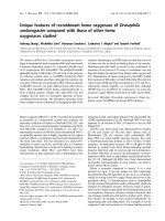
Hb and globin purification
Cellulose acetate electrophoresis showed that the hemo-
lysate of G. gibberifrons contains two Hbs (Hb 1 and Hb 2),
accounting for 80–85% and 15–20%, respectively, of the
total Hb content. The two Hbs were purified by ion-
exchange chromatography on a Super Q ToyoPearl column
(Fig. 1). Hb 2 often appeared to be contaminated by Hb 1;
a second chromatography on the same column yielded pure
Hb 2 (not shown).
Reverse-phase HPLC of the hemolysate showed two
major and two minor peaks (Fig. 2). HPLC of Hb 1
showed two peaks, whose elution times corresponded to
those of the major peaks of the hemolysate; Hb 2 showed
two peaks having elution times corresponding to those of
the minor peaks. The molecular mass values (Da), obtained
by MALDI-TOF mass spectrometry, were 15 597 and
Fig. 1. Ion-exchange chromatography of G. gibberifrons hemolysate on
a Toyopearl column. Details are given in Materials and methods.
3982 P. Marinakis et al.(Eur. J. Biochem. 270) Ó FEBS 2003
16 097 (a
1
and b
1
chains, respectively, of Hb 1), and 15 800
and 16 420 (a
2
and b
2
of Hb 2).
Amino-acid sequencing
The primary structure of the globins was established by
sequencing intact proteins, internal regions obtained after
specific hydrolysis of Asp-Pro bonds, and tryptic peptides
purified by reverse-phase HPLC.
a-Chains
Direct sequencing of intact a-chains was unsuccessful,
suggesting that (as in all Antarctic fish Hbs examined so far)
the N-terminal residue is blocked, therefore not available
to direct Edman degradation. The molecular masses of the
N-terminal tryptic peptides of a
1
and a
2
, measured by
MALDI-TOF mass spectrometry, were 43 Da higher than
those found after sequencing the unblocked peptides, thus
confirming that the a-chain N-terminus is acetylated.
Following cleavage of the Asp-Pro bonds, sequencing
proceeded from Pro96 to Asp127 in a
1
, and from Pro96
to Lys140 in a
2
.
In a
1
, cleavage at Lys5 and at Arg93 by trypsin was not
complete, therefore peptides T1–T2 and T12–T13 were also
found. The peptide bond after Lys116 was not cleaved at all.
Also the peptide bonds after Lys61 and Lys62 were not
completely cleaved, generating peptides T10a and T10b
which coeluted. Four additional peptide pairs (T1 and T1–
T2; T3 and T16; T6 and T13; T7 and T12–T13) coeluted
from the column; however, sequences were unambiguosly
established on the basis of their different amounts.
In a
2
, trypsin failed to cleave the peptide bond after Lys7.
Two peptide pairs (T1 and T10; T5 and T9) coeluted; again,
sequences were established on the basis of their different
amounts. Sequence 101–140 was established only after Asp-
Pro cleavage.
Figure 3A,B shows the complete amino-acid sequences
of the a chains. Tryptic peptides were aligned on the basis of
sequence homologies with known globin sequences, and
with the sequences obtained following Asp-Pro cleavage.
Each chain is made of 142 residues. The molecular masses,
calculated from the sequence, are 15 605 and 15,790, for a
1
and a
2
, respectively.
b-Chains
Direct sequencing proceeded for 20 and 31 residues for the
b-chain of Hb 1 (b
1
)andHb2(b
2
), respectively. After
cleavage of the Asp–Pro bond, the internal sequences from
Pro100 to Leu134 in b
1
, and from Pro100 to Lys132 in
b
2
, were established.
In b
1
, T2 and T11 were not found, and their sequence was
directly established from the N-terminus of the intact globin
(T2) and from the internal sequence obtained after cleavage
of the Asp-Pro bond (T11). T10 and T12 coeluted in the
same chromatographic peak, and their sequence was
established on the basis of their different amount.
In b
2
, T2 and T6 coeluted in the same peak. Being the
sequence of T2 already known from the N-terminus, the
sequence of T6 was established by difference analysis.
Figure 3C,D reports the complete sequences of the
b chains. Tryptic peptides were aligned as described for
the a chains, and with the sequences obtained from the
N-terminus. Each chain is made of 146 residues.
The molecular masses, calculated from the sequence, are
16 081 and 16 400 for b
1
and b
2
, respectively.
Oxygen binding
Functional studies were carried out on Hb 1 and Hb 2,
determining the oxygen-binding curves as a function of pH
in the temperature range 5–10 °C, in the absence and
presence of allosteric effectors (Fig. 4 and Table 1). In the
pH range examined, the oxygen affinity of Hb 2 was lower
than that of Hb 1. All samples displayed the alkaline Bohr
effect, slightly enhanced by chloride and, to a higher extent,
by organophosphate. The latter had a very strong effect at
alkaline pH values, especially in Hb 2, which, although
apparently reducing the amplitude of the Bohr coefficient
(/) in the pH range examined, is indicative of a stronger
overall Bohr effect. In all samples oxygen binding was
cooperative above pH 6.5 in the absence of effectors.
Chloride and, to a higher extent, phosphate, enhanced the
decrease in oxygen-binding cooperativity brought about by
increasing proton concentration. In fact, IHP virtually
abolished cooperativity from pH 7.5 downwards.
All samples displayed the Root effect, which was
maximal in the pH range 6.5–7.5 (Fig. 5). Oxygen-satura-
tion curves were determined at atmospheric pressure. In the
Fig. 2. Reverse-phase HPLC of G. gibberifrons hemolysate, Hb 1 and
Hb 2 (A, B and C, respectively). Details are given in Materials and
methods.
Ó FEBS 2003 The hemoglobins of G. gibberifrons (Eur. J. Biochem. 270) 3983
Fig. 3. Amino-acid sequences of a
1
, a
2
, b
1
and b
2
globin chains (A, B, C and D, respectively). The tryptic peptides (T) and the sequence portions
elucidated by automated Edman degradation from the N-terminus and after cleavage of an Asp-Pro bond are indicated below the sequences.
3984 P. Marinakis et al.(Eur. J. Biochem. 270) Ó FEBS 2003
absence of IHP, complete saturation was achieved at pH 7.5
with Hb 1 and Hb 2. At pH 6.0, the saturation of Hb 1 and
Hb 2 was 73 and 56%, respectively. In the presence of IHP,
maximal saturation was achieved at pH 8.0 in Hb 2, and at
pH 7.5 in Hb 1. At pH 6.0, the saturation of Hb 1 and
Hb 2 was 58 and 47%, respectively. In all samples, IHP
shifted the inflexion of the curve (corresponding to maximal
Root effect) towards more alkaline pH.
Temperature variations had different effects on the
oxygen binding of Hb 1 and Hb 2, both in the absence
and presence of allosteric effectors (Fig. 6). Compared to
Hb 1, Hb 2 showed more exothermic DH values at acidic
pH, whereas lower values were measured at pH 8.0. In the
presence of chloride, the DH values of Hb 2 were higher (in
absolute value) than those of Hb 1 in the entire pH range.
Oxygen-binding studies were also carried out on intact
erythrocytes and stripped hemolysate (not shown), which
showed intermediate functional properties between those of
Hb 1 and Hb 2. Erythrocytes contain endogenous organo-
phosphates; consequently, the curves are similar to those
obtained with the stripped hemolysate in the presence of
effectors.
Discussion
In the Antarctic suborder Notothenioidei, most species of
the family Nototheniidae have one major and one minor Hb
(Hb 1 and Hb 2, 95% and 5% of the total, respectively)
[2, 4,14]. The two Hbs have the b-chain in common, with the
exception of those of Cygnodraco mawsoni [15] which share
the a-chain. Therefore, Nototheniidae (and all Notothe-
nioidei) are generally characterized by reduced Hb multi-
plicity compared to many teleosts of temperate waters [2]. In
T. newnesi, P. antarcticum and P. borchgrevinki higher
multiplicity was observed [16–18], but these species are
pelagic and migratory, differing in life style from the other
notothenioids, which are in general sluggish, benthic fish.
G. gibberifrons is also a sluggish, benthic nototheniid.
Not much more is known about its life style. However, in
comparison with all other benthic nototheniids, it has
distinct and novel hematological features. The blood has the
highest level (approx. 15–20%) of the minor component
Hb 2 ever found in Antarctic Notothenioidei; unlike other
nototheniids, in which Hb 2 tends to disappear in adults
(e.g. T. bernacchii and D. mawsoni only have Hb 2 in
juveniles), the level of Hb 2 does not decrease in adult fish.
Moreover, unlike all other species, the two Hbs of
G. gibberifrons do not have any globin in common. An
Hb system where two components are made by four chains
is a unique case among Notothenioidei. Four chains instead
of three might well be a feature of speciation in the pathway
of evolution of the suborder.
Fig. 4. Oxygen-equilibrium isotherms (Bohr effect) and subunit coop-
erativity, as a function of pH, of Hb 1 (A, B, respectively) and Hb 2 (C,
D, respectively). Experiments were carried out at 5 °C in 100 m
M
Hepes or Mes buffers, in the absence of effectors (s), in the presence of
100 m
M
NaCl (m), and of 100 m
M
NaCl, 3 m
M
IHP (j).
Table 1. Bohr coefficients (/), calculated from the oxygen-binding
curves determined in the absence of effectors and in the presence of
100 m
M
NaCl or 100 m
M
NaCl and 3 m
M
IHP.
No effectors NaCl (100 m
M
) NaCl/IHP (100/3 m
M
)
Hb 1
5 °C )0.64 )0.69 )0.62
10 °C )0.57 )0.68 )0.61
Hb 2
5 °C )0.75 )0.77 )0.43
10 °C )0.89 )0.77 )0.61
Fig. 5. Oxygen-saturation curves at atmospheric pressure (Root effect)
ofHb1(A)andHb2(B).Experiments were carried out at 2 °C, in
100 m
M
Tris/HCl or bisTris/HCl buffers, in the absence (s)and
presence (d)of3m
M
IHP.
Ó FEBS 2003 The hemoglobins of G. gibberifrons (Eur. J. Biochem. 270) 3985
Although from a morphological point of view G. gibber-
ifrons evidently belongs to the family Nototheniidae [19–21],
Tokita et al. [22] have reported dendrograms based on
genetic distances obtained from two-dimensional gel elec-
trophoresis of total protein constituents of cardiac muscle.
In these dendrograms the nototheniid G. gibberifrons
appears more closely related to species belonging to
the most phyletically advanced notothenioid families
Bathydraconidae and Channichthyidae, than to the clade
composed by three other nototheniid species. As far as red-
blooded species are concerned, this interesting conclusion
is not fully supported by our results on Hb sequences
(Table 2). In fact, the sequence identities do not indicate
clear grouping of G. gibberifrons either with species of the
same family (Trematomus bernacchii and T. newnesi)orof
Bathydraconidae (Gymnodraco acuticeps and C. mawsoni).
However, the lack of a clear relationship with the Hb
sequences of other nototheniid species suggests that in this
case the evolution of the oxygen-transport system has
occurred in response to special needs of this species, as
shown by the intermediate position taken by G. gibberifrons
Hbs in phylogenetic trees [23].
In line with other Antarctic Hbs, the sequence identity
between the a chains of the two Hbs of G. gibberifrons is
68%; it is 70% between the b-chains. In summary, the
a-chain and the extra b-chain of Hb 2 have much higher
sequence identity with minor than with major Hbs of other
Antarctic species. Thus the latter extra chain also groups
with minor Hbs.
In Antarctic fish Hbs, Aspb94, which in human HbA
establishes a strong ionic bond with Hisb146, important for
the Bohr-effect mechanism [24], is generally conservatively
replaced by Glu. Moreover, Vala1 is always replaced by Ac-
Ser, which cancels the contribution of the N-terminus to the
Bohr effect. These substitutions are also found in the two
Hbs of G. gibberifrons, characterized by the Bohr and Root
effects. However, these Hbs show significantly distinct Bohr
coefficients and amplitude of Root effect; in particular,
Hb 2 has lower oxygen affinity than Hb 1 in the pH range
examined, and phosphate modulation of the affinity is
stronger in Hb 2 than Hb 1. These differences might be due
to other substitutions in the primary structure. For instance,
in Hb 2 it is worth noting that position b82, which is part of
the phosphate binding site [25,26], is occupied by Lys,
whereas in Hb 1 the latter residue is replaced by Ala. This
substitution may well account for the lower effect of
organophosphates in the latter Hb.
Temperature dependence, which is governed by the
associated overall enthalpy change, is an important feature
of the reaction of Hbs with oxygen. Heat absorption and
release can be considered physiologically relevant modula-
ting factors, similar to hetero and homotropic ligands. The
two Hbs of G. gibberifrons also differ in thermodynamic
behaviour. Hb 1 is less sensitive to temperature variations
than Hb 2 which, in turn, shows strong variations of
enthalpy change especially at pH below 7.5, depending on
chloride and/or phosphate. In particular, Hb 2 shows a
progressive increase of the exothermic enthalphy change as
a function of proton concentration. This feature has never
been reported in fish Hbs. Chloride virtually abolishes this
exothermic change by providing a strong endothermic
contribution to oxygen binding. Although a molecular inter-
pretation is hard to find, this differential thermodynamic
Fig. 6. Oxygenation enthalpy of Hb 1 (A) and Hb 2 (B). DH values
were calculated in the temperature range 5–10 °C from the oxygen-
binding data reported in Fig. 4 and Table 1. Experimental conditions
were: 100 m
M
Hepes or Mes buffers, in the absence of effectors (s), in
the presence of 100 m
M
NaCl (m), and of 100 m
M
NaCl, 3 m
M
IHP
(j).
Table 2. Sequence identity (%) between a- and b-chains of G. gibb erifrons and of some other Antarctic fish Hbs. T. bernacchii Hb C, P. borchgrevinki
Hb 0 and C. mawsoni Hb 2 are minor components. P. borchgrevinki Hb 0 and Hb 1, in addition to C. mawsoni Hb 1 and Hb 2, share the a-chain.
T. bern. Hb 1 T. bern. Hb C P. borch. Hb 1 P. borch. Hb 0 G. acut. Hb C. maws. Hb 1 C. maws. Hb 2
a-chains
G. gibberifrons Hb 1 94 – 90 90 89 90 90
G. gibberifrons Hb 2 69 – 65 65 66 68 68
b-chains
G. gibberifrons Hb196719171838867
G. gibberifrons Hb268927091666587
3986 P. Marinakis et al.(Eur. J. Biochem. 270) Ó FEBS 2003
regulation of the oxygenation/deoxygenation cycle (that
may play a significant role in keeping the internal tempera-
ture constant) may be an important adaptive tool.
This paper reports some novel features of the oxygen
transport of a notothenioid fish species. The results suggest
that G. gibberifrons Hb 2 cannot merely be considered an
evolutionary remnant, as in other Antarctic Notothenioidei
[27]. The functional differences suggest that Hb 2, rather
than being a vestigial or larval remnant, may indeed have a
physiological role; the two Hbs of this cold-adapted teleost
might be used alternatively to face special needs in relation
with life style and different environmental conditions (e.g.
temperature fluctuations during migration) requiring fine
regulation of oxygen binding. Finally, although in an
organism biosynthesis of higher amounts of an additional
Hb can be easily accomplished and may be considered a
short-time response to environmental changes, preservation
of the role of the gene duplication which has produced an
additional chain is a physiologically complex long-term
response, and may well be considered an evolutionarily
important adaptation.
Acknowledgements
This study is in the framework of the Italian National Programme for
Antarctic Research.
References
1. di Prisco, G. & Tamburrini, M. (1992) The hemoglobins of marine
and freshwater fish: the search for correlations with physiological
adaptation. Comp. Biochem. Physiol. 102B, 661–671.
2. di Prisco, G. (1998) Molecular adaptations in Antarctic fish
hemoglobins. In Fishes of Antarctica. A Biological Overview (di
Prisco, G., Pisano, E. & Clarke, A., eds), pp. 339–353. Springer,
New York.
3. di Prisco, G., Tamburrini, M. & D’Avino, R. (1998) Oxygen-
transport systems in extreme environments: multiplicity and
structure/function relationship in hemoglobins of Antarctic fish.
In Cold Ocean Physiology (Po
¨
rtner, H.O. & Playle, R., eds), pp.
143–165. Cambridge University Press, Cambridge, UK.
4. Tamburrini, M. & di Prisco, G. (2000) Oxygen-transport system
and mode of life in Antarctic fish. In Hemoglobin Function in
Vertebrates. Molecular Adaptation in Extreme and Temperate
Environments (di Prisco, G., Giardina, B. & Weber, R.E., eds), pp.
51–59. Springer-Verlag, Italy.
5. di Prisco, G., D’Avino, R. & Tamburrini, M. (1999) Structure and
function of hemoglobins from Antarctic organisms: the search for
correlations with adaptive evolution. In Cold-Adapted Organisms.
Ecology, Physiology, Enzymology and Molecular Biology
(Margesin, R. & Schinner, F., eds), pp. 239–253. Springer-Verlag,
Berlin, Germany.
6. di Prisco, G. (1988) A study of hemoglobin in Antarctic fishes:
purification and characterisation of hemoglobin from four species.
Comp. Biochem. Phisiol. 90B, 631–637.
7. D’Avino, R. & di Prisco, G. (1988) Antarctic fish hemoglobin: an
outline of the molecular structure and oxygen binding properties.
I. Molecular structure. Comp.Biochem.Physiol.90B, 579–584.
8. D’Avino, R. & di Prisco, G. (1989) Hemoglobin from the Ant-
arctic fish Notothenia coriiceps neglecta. 1. Purification and char-
acterisation. Eur. J. Biochem. 179, 699–705.
9. Tamburrini, M., Brancaccio, A., Ippoliti, R. & di Prisco, G. (1992)
The amino acid sequence and oxygen-binding properties of single
hemoglobin of the cold-adapted Antarctic teleost Gymnodraco
acuticeps. Arch. Biochem. Biophys. 292, 295–302.
10. Landon, M. (1977) Cleavage at aspartyl-prolyl bonds. In Methods
Enzymology,Vol.47(Hirs,C.H.W.&Timasheff,S.N.,eds),pp.
145–149. Academic Press, New York, USA.
11. Brauer, A.W., Oman, C.L. & Margolies, M.N. (1984) Use of
o-phthalaldehyde to reduce background during automated
Edman degradation. Anal. Biochem. 137, 134–142.
12. Sick, H. & Gersonde, K. (1969) Method for continuous registra-
tion of O
2
-binding curves of hemoproteins by means of a diffusion
chamber. Anal. Biochem. 32, 362–376.
13. Lykkeboe, G., Johansen, K. & Maloy, G.M.O. (1975) Functional
properties of hemoglobin in the teleost Tilapia grahami. J. Comp.
Physiol. 104, 1–11.
14. di Prisco, G. (1997) Physiological and biochemical adaptations
in fish to a cold marine environment. In Proceedings of the SCAR
6th Biol. Symposium, Venice (Antarctic Communities: Species,
Structure and Survival) (Battaglia, B., Valencia, J. & Walton,
D.W.H., eds), pp. 251–260. Cambridge University Press,
Cambridge, UK.
15. Caruso,C.,Rutigliano,B.,Romano,M.&diPrisco,G.(1991)
The hemoglobins of the cold-adapted Antarctic teleost Cygno-
draco mawsoni. Biochim. Biophys. Acta 1078, 273–282.
16. D’Avino, R., Caruso, C., Tamburrini, M., Romano, M., Ruti-
gliano, B., Polverino de Laureto, P., Camardella, L., Carratore,
V. & di Prisco, G. (1994) Molecular characterization of the
functionally distinct hemoglobins of the Antarctic fish Tremato-
mus newnesi. J. Biol. Chem. 269, 9675–9681.
17. Tamburrini, M., D’Avino, R., Fago, A., Carratore, V., Kunz-
mann, A. & di Prisco, G. (1996) The unique hemoglobin system of
Pleuragramma antarcticum, an Antarctic migratory teleost.
Structure and function of the three components. J. Biol. Chem.
271, 23780–23785.
18. Riccio, A., Tamburrini, M., Carratore, V. & di Prisco, G. (2000)
Functionally distinct haemoglobins of the cryopelagic Antarctic
teleost Pagothenia borchgrevinki. J. Fish Biol. 57A, 20–32.
19. Iwami, T. (1985) Osteology and relationships of the family
Channichthyidae. Mem. Natl. Institute Polar Res. Tokyo Series E.
36, 1–69.
20. Gon, O. & Heemstra, P.C., eds (1990) Fishes of the Southern
Ocean. Smith Institute of Ichthyology, South Africa.
21. Balushkin, A.V. (1992) Classification, phylogenetic relationships,
and origins of the families of the suborder Notothenioidei
(Perciformes). J. Ichthyol. 32, 90–110.
22. Tokita, M., Ishii, S., Iwami, T. & Miyazaki, J I. (2002) Phylo-
genetic analysis of Antarctic notothenioid fishes based on two-
dimensional gel electrophoresis. Polar Biol. 25, 163–168.
23. Verde, C., Carratore, V., Riccio, A., Tamburrini, M., Parisi, E. &
di Prisco, G. (2002) The functionally distinct hemoglobins of the
Arctic spotted wolffish Anarhichas minor. J. Biol. Chem. 277,
36312–36320.
24. Perutz, M.F. & Brunori, M. (1982) Stereochemistry of cooperative
effects in fish and amphibian haemoglobins. Nature 299, 421–426.
25. Arnone, A. (1972) X-ray diffraction study of binding of 2,3-
diphosphoglycerate to human deoxyhaemoglobin. Nature 237,
146–149.
26. Arnone, A. & Perutz, M.F. (1974) Structure of inositol hexa-
phosphate-human deoxyhaemoglobin complex. Nature 249,
34–36.
27. di Prisco, G., D’Avino, R., Caruso, C., Tamburrini, M., Cam-
ardella, L., Rutigliano, B., Carratore, V. & Romano, M. (1991)
The biochemistry of oxygen transport in red-blooded Antarctic
fish. In Biology of Antarctic Fish (diPrisco,G.,Maresca,B.&
Tota, B., eds), pp. 263–281. Springer-Verlag, New York, USA.
Ó FEBS 2003 The hemoglobins of G. gibberifrons (Eur. J. Biochem. 270) 3987