Báo cáo khoa học: Protective effect of dietary curcumin and capsaicin on induced oxidation of low-density lipoprotein, iron-induced hepatotoxicity and carrageenan-induced inflammation in experimental rats ppt
Bạn đang xem bản rút gọn của tài liệu. Xem và tải ngay bản đầy đủ của tài liệu tại đây (594.11 KB, 10 trang )
Protective effect of dietary curcumin and capsaicin on
induced oxidation of low-density lipoprotein, iron-induced
hepatotoxicity and carrageenan-induced inflammation
in experimental rats
Hanumanthappa Manjunatha and Krishnapura Srinivasan
Department of Biochemistry and Nutrition, Central Food Technological Research Institute, Mysore, India
Oxidative damage at the cellular and subcellular level
is now considered to be an important event in disease
processes like cardiovascular disease, inflammatory dis-
ease, carcinogenesis and aging. In humans, plasma
low-density lipoprotein (LDL) is the major transport
vehicle for cholesterol and its elevation is regarded as
one of the principal risk factors for the development of
atherosclerotic vascular disease [1,2]. A relatively large
amount of cholesterol in the LDL fraction is athero-
genic, whereas that in high-density lipoprotein fraction
appears protective [3]. Oxidation of LDL has been sug-
gested to play an important role in the development
of atherosclerosis [4]. Inhibition of LDL oxidation
can reduce the risk of atherosclerosis independent of
Keywords
anti-inflammatory effect; capsaicin;
curcumin; hepatoprotective effect; low-
density lipoprotein oxidation
Correspondence
K. Srinivasan, Department of Biochemistry
and Nutrition, Central Food Technological
Research Institute, Mysore 570020, India
Fax: +91 0821 2517233
Tel: +91 0821 2514876
E-mail:
(Received 4 June 2006, revised 7 August
2006, accepted 10 August 2006)
doi:10.1111/j.1742-4658.2006.05458.x
The beneficial influence of dietary curcumin, capsaicin and their combina-
tion on the susceptibility of low-density lipoprotein (LDL) to oxidation
was examined in an animal study. Individually, both dietary curcumin and
capsaicin significantly inhibited the in vivo iron-induced LDL oxidation, as
well as copper-induced oxidation of LDL in vitro. The protective effect of
the combination of curcumin and capsaicin on LDL oxidation was greater
than that of individual compounds. This protective influence of spice prin-
ciples was also indicated by the relative anodic electrophoretic mobility of
oxidized LDL on agarose gel. In another study, rats injected with iron
showed hepatic toxicity as measured by an increase in lipid peroxides and
elevated serum enzymes, alanine aminotransferase, aspartate aminotrans-
ferase and lactate dehydrogenase. Dietary curcumin, capsaicin and their
combination reduced the activities of these enzymes, and lowered the liver
lipid peroxide level, indicating amelioration of the severity of iron-induced
hepatotoxicity. In yet another study, a comparison of the extent of carrage-
enan-induced paw inflammation showed that both dietary curcumin and
capsaicin moderately lowered inflammation, while the spice principles in
combination were more effective. Dietary curcumin and capsaicin signifi-
cantly decreased the activity of 5¢-lipoxygenase activity in the polymorpho-
nuclear lymphocytes in carrageenan-injected rats, the decrease being even
higher in the case of combination of these two spice principles. Results sug-
gest that dietary curcumin and capsaicin individually are protective to
LDL oxidation both in vivo and in vitro, to iron-induced hepatotoxicity
and to carrageenan-induced inflammation. This beneficial effect was higher
when the two compounds were fed in combination.
Abbreviations
AlAT, alanine aminotransferase; AsAT, aspartate aminotransferase; LDH, lactate dehydrogenase; LDL, low-density lipoprotein; NF-jB,
nuclear factor-kappa B; PMNL, polymorphonuclear lymphocytes; TBARS, thiobarbituric acid reactive substances.
4528 FEBS Journal 273 (2006) 4528–4537 ª 2006 The Authors Journal compilation ª 2006 FEBS
lowering plasma cholesterol levels. The effectiveness of
antioxidant vitamins C and E in the prevention of
LDL oxidation has been well demonstrated [5]. Phe-
nolic compounds of red wine have been shown to inhi-
bit oxidation of LDL both in vitro and in vivo [6,7].
The antioxidant properties of several spice principles
have been evidenced in rats both in vivo and in vitro.
While curcumin (turmeric), capsaicin (red pepper) and
eugenol (clove) were found to be more effective anti-
oxidants, piperine (black pepper), zingerone (ginger),
linalool (coriander) and cuminaldehyde (cumin) were
only marginally inhibitory to lipid peroxidation [8].
These compounds inhibited lipid peroxidation by
quenching oxygen free radicals [9] and by enhancing
the activity of endogenous antioxidant enzymes, super-
oxide dismutase, catalase, glutathione peroxidase and
glutathione transferase [10]. Spice active principles, i.e.
curcumin (turmeric), capsaicin (red pepper), piperine
(black pepper), eugenol (cloves) and allyl sulfide (gar-
lic), have been shown to have a protective effect on
oxidation of human LDL in vitro [11]. Dietary spice
principles curcumin, capsaicin and garlic were found
to be antioxidative by enhancing the antioxidant mole-
cules and antioxidant enzymes in erythrocytes and liver
of hyperlipidemic ⁄ hypercholesterolemic rats [12,13].
The toxic effects of iron overloading leads to chronic
liver disease, impaired cardiac function, endocrinopa-
thies, skin pigmentation and orthropathy [14,15].
Hepatotoxicity is the most common finding in patients
with iron overloading. The massive deposition of iron
in hepatic parenchymal cells eventually produces fibro-
sis and ultimately results in cirrhosis [16]. Spice princi-
ples, curcumin and capsaicin, can effectively inhibit
lipid peroxidation in rat liver by enhancing the anti-
oxidant enzyme activities (current authors’ unpublished
work). Curcumin has been shown to scavenge reactive
oxygen species and also prevent the oxidation of
iron(II) by hydrogen peroxide in the Fenton reactions
[9], which generates hydroxyl radicals involved in the
initiation of lipid peroxidation [17]. Hence it would be
relevant to examine if these two antioxidant spice prin-
ciples could also have a protective role in iron-induced
hepatotoxicity.
Spice principles, curcumin (of turmeric), capsaicin
(of red pepper) and eugenol (of cloves), have been
understood to possess health beneficial anti-inflamma-
tory properties [9,18]. Curcumin and the volatile oil
from turmeric have been shown to reduce edema in rats
[19], and to moderately reduce the clinical symptoms in
rheumatoid arthritis patients [20]. Curcumin inhibits
the formation of proinflammatory compounds like pro-
staglandins and leukotrienes [21]. Dietary curcumin
and capsaicin have been shown to lower the generation
of proinflammatory mediators such as reactive oxygen
species and nitric oxide released by macrophages [22].
The current study examines the beneficial antioxid-
ant influence, if any, of dietary curcumin, capsaicin,
and their combination in terms of protecting LDL
from oxidation in experimental rats. In the present
investigation, the protective role, if any, of dietary cur-
cumin, capsaicin and their combination on the damage
caused to liver by iron overloading measured in terms
of lipid peroxidation and elevation of plasma alanine
aminotransferase (AlAT), aspartate aminotransferase
(AsAT) and LDH was assessed. The present study also
investigates the anti-inflammatory property of curcu-
min and capsaicin when fed in combination on car-
rageenan-induced inflammatory responses in rats.
Results and Discussion
The dietary levels of curcumin and capsaicin employed
in this animal study, i.e. 0.2 g% and 0.015 g%,
respectively, corresponds to about 10 times the average
dietary intake of the corresponding parent spices (tur-
meric and red pepper) in the Indian population. At
these dietary levels, the feed intake was essentially sim-
ilar in various groups fed spice principles and the cor-
responding control group. Similarly, the gain in body
weight during the 8 weeks of spice compound treat-
ment was comparable to the corresponding controls.
Protective effect of dietary curcumin, capsaicin
and their combination on iron-induced LDL
oxidation in vivo and copper-induced LDL
oxidation in vitro
Oxidation of LDL observed in iron(II) sulfate-injected
rats as measured by thiobarbituric acid reactive sub-
stance (TBARS) values is presented in Table 1. Dietary
curcumin and dietary capsaicin significantly inhibited
the oxidation of LDL, as indicated by TBARS values
which were 71 and 62% of control rats. Extent of
iron-induced oxidation of LDL was considerably lower
in curcumin + capsaicin-fed groups when compared
with curcumin-fed animals (TBARS value 56% of con-
trol). Extensive oxidation of LDL in vitro from control
rats was noticed in the presence of copper(II) sulfate,
as measured by the time-dependent increase in TBARS
values over the period of 12 h (Table 1). The extent of
copper-induced oxidation of LDL in vitro was signifi-
cantly less in the case of LDL isolated from curcumin
fed rats; 17 and 18% less TBARS formation at 3 and
12 h of LDL oxidation, respectively, was seen in this
case. The extent of copper-induced oxidation of LDL
in vitro was also significantly less in the case of LDL
H. Manjunatha and K. Srinivasan Health protective effects of curcumin and capsaicin
FEBS Journal 273 (2006) 4528–4537 ª 2006 The Authors Journal compilation ª 2006 FEBS 4529
isolated from capsaicin-fed animals. The decrease in
TBARS formation was 29 and 21% at 3 and 12 h of
LDL oxidation, respectively, in this case. A decrease in
TBARS formation of 37 and 24% was seen at 3 and
12 h of copper-induced LDL oxidation in the case
of LDL isolated from animals fed the combination of
curcumin and capsaicin. Thus, the protective effect of
the combination of dietary curcumin and capsaicin on
LDL oxidation both in vivo and in vitro was greater
than that of the individual spice principles.
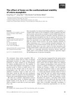
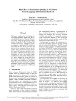
Agarose gel electrophoresis revealed that LDL oxi-
dation induced in vivo by the iron(II) ion caused an
increase in the anodic mobility of LDL in the case of
control rats (Fig. 1). In the case of animals maintained
on curcumin, capsaicin or curcumin + capsaicin, the
anodic mobility of LDL oxidized in vivo by the iron(II)
ion was slower compared with the control animals.
The decreased anodic mobility of oxidized LDL in the
case of spice principles-fed animals is thus consistent
with the observed protective influence on LDL oxida-
tion by these compounds.
In humans, plasma LDL is a major transport vehicle
for cholesterol and its elevation is regarded as one of
the principle risk factors for the development of
atherosclerotic vascular disease [1,2]. A relatively large
amount of cholesterol in LDL fraction is atherogenic,
whereas that in the high-density lipoprotein fraction
appears protective [3]. Oxidation of LDL has been sug-
gested to play an important role in the development of
atherosclerosis [4]. It is also known that dietary factors
influence plasma lipid levels and lipoprotein metabo-
lism, altering the atherogenicity of lipoprotein profile
[39]. The hypothesis states that the oxidative modifica-
tion of LDL or other lipoproteins is central, if not
obligatory, to the atherogenic process. The important
corollary is that inhibition of such oxidation should
reduce the progression of atherosclerosis, independent
of reduction of other factors, such as elevated LDL
levels [40,41].
It is universally accepted that hypercholesterolemia
is an important independent risk factor for atheroscler-
osis [42]. Pathogenesis of atherosclerosis is most likely
to involve a free radical-mediated process. Oxidative
modifications of LDL, which dysregulate the homeo-
stasis between blood and vascular cells and alteration
in endothelial function, are considered among the early
events in the pathogenesis of atherosclerosis [43]. The
alteration of oxidant ⁄ antioxidant balance may affect
the susceptibility of LDL to oxidation. LDL oxidation
can lead to its subsequent aggregation, which fur-
ther increases cellular cholesterol accumulation [44].
Table 1. Effect of dietary curcumin and capsaicin on iron(II)-induced (in vivo) and copper-induced (in vitro) LDL oxidation in rats. Rats were
injected with 1 mL saline or FeSO
4
in 1 mL saline (30 mg per kg body weight) 1 h before death. Values expressed as nanomoles
TBARSÆmg
)1
protein are mean ± SEM of eight rats in each group. Values in parenthesis represent percentage decrease as compared with
respective control.
Diet group In vivo Fe
2+
induced
In vitro Cu
2+
induced
3 h 12 h
Control 1.036 ± 0.059 11.0 ± 0.45 19.6 ± 0.47
Curcumin 0.731 ± 0.022* 9.08 ± 0.40* 16.l ± 0.52*
(29.4%) (17.5%) (17.9%)
Capsaicin 0.640 ± 0.050* 7.84 ± 0.61* 15.5 ± 0.61*
(38.7%) (28.7%) (20.9%)
Curcumin + capsaicin 0.578 ± 0.055* 6.88 ± 0.16* 14.9 ± 0.47*
(44.2%) (37.5%) (24.0%)
*Significantly different from control group.
12345678
Fig. 1. Agarose gel electrophoresis of LDL in different diet groups
oxidized in vivo by iron(II). 1, Control (Fe
2+
-injected); 2, control (sal-
ine-injected); 3, curcumin (saline-injected); 4, curcumin (Fe
2+
-injec-
ted); 5, capsaicin (saline-injected); 6, capsaicin (Fe
2+
-injected);
7, curcumin + capsaicin (saline-injected); 8, curcumin + capsaicin
(Fe
2+
-injected).
Health protective effects of curcumin and capsaicin H. Manjunatha and K. Srinivasan
4530 FEBS Journal 273 (2006) 4528–4537 ª 2006 The Authors Journal compilation ª 2006 FEBS
Factors that have been reported to affect the suscepti-
bility of LDL to oxidation include antioxidant content,
particle size and fatty acid composition. a-Tocopherol
is the most abundant antioxidant in LDL [45] and
LDL isolated after individuals have been given a-toco-
pherol supplementation has been reported to exhibit
increased resistance to oxidative modification [46,47].
Supplementing corn oil- and beef tallow-enriched diets
with moderate amounts of dietary cholesterol increased
the susceptibility of LDL to oxidation, but LDL
a-tocopherol levels tended to be higher after consu-
ming the diets with cholesterol supplementation [48].
However, although the LDL a-tocopherol content
increased in the beef tallow diet supplemented with
cholesterol, no significant relationship was observed
between the a-tocopherol concentration of the LDL
particles and the susceptibility of LDL to oxidation.
Active principles of spices such as curcumin, capsaicin,
piperine, eugenol and allyl sulfide have been shown to
have protective effects on the oxidation of human
LDL in vitro [11].
Protective effect of dietary curcumin, capsaicin
and their combination on iron-induced
hepatotoxicity
One of the mechanisms by which iron induces toxicity
is by increasing oxidative stress and lipid peroxidation.
Lipid peroxidation of membranes is the major dam-
aging factor in iron toxicity [49]. The ability of iron to
accelerate lipid peroxidation is well established [50].
The primary mechanism for this acceleration is
believed to be the iron-catalyzed decomposition of
lipid peroxides. The role of iron in in vivo and in vitro
lipid peroxidation has been well studied [50]. Iron
overload increased formation of urinary malondialde-
hyde, tissue thiobarbituric acid reactive substances and
conjugated dienes [51]. In experimental animals, iron
overload can be effected by intraperitoneal injection of
iron salts [51].
Effects of dietary curcumin, capsaicin and their
combination on iron-induced lipid peroxidation in rat
serum and liver are presented in Table 2. The results
of the present study demonstrated that excess iron
introduced by intraperitoneal injection induced oxida-
tive stress by increasing lipid peroxide levels in liver
as well as in serum. The intraperitoneal injection of
iron significantly elevated the hepatic lipid peroxides
(418% increase in control group). The levels of
TBARS in liver were lower in animals fed curcumin,
capsaicin or their combination; these decreases were
28, 26 and 22% in the respective diet groups. Dietary
curcumin, capsaicin and their combination signifi-
cantly reduced the severity of iron-induced lipid per-
oxidation in liver. The decreases brought about by
dietary curcumin, capsaicin and their combination in
liver TBARS in iron(II)-injected rats were 26, 28 and
37%, respectively. Intraperitoneal injection of iron(II)
to rats also resulted in higher lipid peroxides in
serum (Table 2). The increase in serum TBARS value
in control rats as a result of iron(II) injection was
76%. Dietary curcumin, capsaicin and their combi-
nation lowered serum lipid peroxide levels by 24, 33
and 29%, respectively, in iron(II)-treated rats. These
dietary spice principles, however, did not influence
the basal TBARS values in serum in saline-injected
rats.
The serum enzymes are very important adjuncts to
clinical diagnosis of diseases affecting specific organs
and tissue damage. Liver damage by iron toxicity can
be assessed by leakage of enzymes such as alanine
aminotransferase (AlAT), aspartate aminotransferase
(AsAT) and lactate dehydrogenase into blood [52,53].
Higher activities of all these three enzymes in blood
have been found in response to iron-induced oxidative
stress in the present study (Table 3). The intraperito-
neal injection of iron significantly elevated the serum
AlAT, AsAT and LDH; the increases were 150, 172
and 215%, respectively. Dietary curcumin, capsaicin
and their combination reduced activities of serum
Table 2. Effect of dietary curcumin and capsaicin on iron-induced lipid peroxidation in rat serum and liver. Rats were injected with 1 mL saline
or FeSO
4
in 1 mL saline (30 mg per kg body weight) 1 h before death. Values are expressed as mean ± SEM of six rats in each group.
Diet group
Serum
(lmol TBARSÆdL
)1
)
Liver
(nmol TBARSÆmg
)1
protein)
Saline-injected Fe
2+
-injected Saline-injected Fe
2+
-injected
Control 77.0 ± 6.32 135.2 ± 10.2 4.52 ± 0.31 23.4 ± 1.50
Curcumin 63.8 ± 4.22 102.4 ± 9.24* 3.25 ± 0.10* 17.2 ± 2.12*
Capsaicin 60.1 ± 6.74 91.1 ± 5.80* 3.33 ± 0.07* 16.8 ± 1.93*
Curcumin + capsaicin 68.4 ± 4.76 95.4 ± 7.40* 3.52 ± 0.32* 14.7 ± 2.45*
*Significantly different from control group.
H. Manjunatha and K. Srinivasan Health protective effects of curcumin and capsaicin
FEBS Journal 273 (2006) 4528–4537 ª 2006 The Authors Journal compilation ª 2006 FEBS 4531
enzymes, AlAT, AsAT and LDH, indicating that these
spice principles reduce the severity of iron-induced
hepatotoxicity by lowering lipid peroxidation. Dietary
curcumin, capsaicin and their combination lowered
serum AlAT by 28, 37 and 34%, respectively, in
iron(II)-injected animals (Table 3). Dietary curcumin,
capsaicin and their combination lowered serum AsAT
activity by 18, 28 and 38%, respectively, in iron-injec-
ted rats. Similarly, the increase in serum LDH as a
result of iron(II) administration was countered by 21,
31 and 41% by dietary curcumin, capsaicin and their
combination, respectively. Thus, the combination of
the two spice principles brought about greater protect-
ive effect against iron(II)-induced hepatotoxicity when
viewed in terms of the beneficial influence on serum
AsAT and LDH. This is consistent with a greater
countering influence of the spice combination on
iron(II)-induced liver lipid peroxides described above.
There was no change in the activities of these enzymes
as a result of curcumin, capsaicin or their combination
in the saline-injected animals (Table 3). Among the
activities of alkaline and acid phosphatases measured
in the serum of iron(II)-injected rats, only the latter
was elevated by about 20% as a result of iron over-
loading. While individual dietary spice principles did
not influence the activity of serum alkaline phospha-
tase and acid phosphatase in iron(II)-injected rats, only
the combination of spice principles significantly coun-
tered the elevated serum acid phosphatase in iron(II)-
injected animals (Table 3).
In general, iron-induced liver injury resulted in a
marked elevation in the activity of these enzymes. The
extent of elevation in the activities of these enzymes,
which are indicators of hepatic injury, was generally
lower in various spice principles-fed animal groups.
Combination of the two spice principles was found to
be more protective to liver in iron-induced hepatotox-
icity, when compared with the two individual spice
principles.
Effect of dietary curcumin, capsaicin and their
combination on carrageenan-induced paw
inflammation in rats
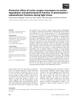
In control rats, greatest swelling was observed 5 h after
carrageenan injection (Fig. 2). A comparison of the
extent of carrageenan-induced paw inflammation at
5 h in various spice principles-fed animals is shown in
Fig. 2. Dietary curcumin lowered inflammation to an
extent of 12%, while dietary capsaicin reduced the
inflammation to an extent of 9%. Spice principles in
combination were more effective in countering the
extent of paw inflammation compared with the two
individual spice principles, where the paw inflamma-
tion at 5 h was 84% of the control. An earlier study
Table 3. Effect of dietary curcumin and capsaicin on serum enzymes in rats injected with iron(II) salt. Values are mean ± SEM of six
animals in each group. Rats were injected with 1 mL saline or FeSO
4
in 1 mL saline (30 mg per kg body weight) 1 h before death.
Treatment
Alanine aminotransferase
a
Aspartate aminotransferase
a
Lactate dehydrogenase
b
Acid phosphatase
c
Saline-injected Fe
2+
-injected Saline-injected Fe
2+
-injected Saline-injected Fe
2+
-injected Saline-injected Fe
2+
-injected
Control 108.3 ± 5.28 270.8 ± 9.80 30.9 ± 2.18 84.2 ± 24.4 65.3 ± 5.78 205.7 ± 9.67 340.6 ± 24.4 410.5 ± 14.8
Curcumin 112.4 ± 6.32 195.4 ± 11.7* 28.6 ± 3.62 69.3 ± 26.0* 74.4 ± 6.38 163.1 ± 7.40* 334.6 ± 20.94 377 ± 18.0
Capsaicin 120.3 ± 7.41 169.8 ± 10.2* 35.6 ± 4.10 60.6 ± 18.5* 69.6 ± 7.24 142.7 ± 14.4* 324.8 ± 23.1 448.6 ± 22.9
Curcumin +
capsaicin
102.2 ± 4.80 179.8 ± 13.2* 26.4 ± 2.83 51.9 ± 14.1* 60.6 ± 4.30 120.4 ± 5.59* 330.6 ± 12.6 319.9 ± 16.4*
Specific activity units:
a
lmol pyruvateÆmin
)1
ÆdL
)1
;
b
lmol NADHÆmin
)1
ÆdL
)1
;
c
lmol p-nitrophenolÆmin
)1
ÆdL
)1
. *Significantly different from
control group.
Fig. 2. Carrageenan-induced paw inflammation in rats fed spice
principles. 1, saline-injected control; 2, control; 3, dietary curcumin;
4, dietary capsaicin; 5, dietary curcumin + capsaicin. Values in
groups 3, 4 and 5 were significantly lower compared with the value
in group 2 (P<0.05).
Health protective effects of curcumin and capsaicin H. Manjunatha and K. Srinivasan
4532 FEBS Journal 273 (2006) 4528–4537 ª 2006 The Authors Journal compilation ª 2006 FEBS
has reported that supplementation of diets with 1%
curcumin for 10 weeks did not affect the inflammatory
responses of animals to carrageenan injection [54].
However, curcumin administered by gavage (15, 30
and 45 mgÆkg
)1
body weight) 3 h prior to carrageenan
injection did show anti-inflammatory property [54].
Similarly, capsaicin has previously been shown to pos-
sess anti-inflammatory properties against carrageenan-
induced inflammation when given as a single oral dose
(0.5 and 1.0 mgÆkg
)1
body weight) 3 h before carrage-
enan injection [54].
The influence of dietary curcumin, capsaicin, and
their combination on 5¢-lipoxygenase activity in the
polymorphonuclear lymphocytes (PMNL) cells in car-
rageenan-injected rats is presented in Table 4. Dietary
curcumin decreased the activity of 5¢-lipoxygenase
activity in the PMNL cells by 39% in carrageenan-
injected rats while dietary capsaicin produced 48%
decrease in the enzyme activity. The decrease in the
enzyme activity was even higher in the case of the
combination of these two spice principles (60%). Thus,
the combination of spice principles curcumin and cap-
saicin had greater effect in countering the 5¢-lipoxyge-
nase activity in the PMNL cells as a result of
carrageenan administration. Activity of 5¢-lipoxygenase
in the PMNL cells was also lower in saline-injected
rats as a result of dietary spice principles, the decreases
being 48, 26 and 49%, respectively, in curcumin, cap-
saicin and curcumin + capsaicin groups. 5¢-Lipoxyge-
nase is known to be regulated by the transcription
factor nuclear factor-kappa B (NF-jB) [55]. Curcumin
and capsaicin have been shown to inhibit NF-jB acti-
vation [56,57]. Hence, the inhibitory influence of these
two spice compounds on 5¢-lipoxygenase enzyme in
carrageenan-injected animals is probably mediated
through their effect on NF-jB.
Histamine concentration in serum was lower under
the influence of dietary curcumin, capsaicin or their
combination (Table 5); the decrease in serum hista-
mine was 30, 37 and 21% lower in the respective
groups among saline-injected rats. Serum histamine
content was lower only in dietary capsaicin group
among carrageenan-injected rats compared with
respective controls (23% decrease). The low serum
histamine titers in animals treated with dietary spice
principles is consistent with their protective influence
in response to carrageenan administration. There was
no gross difference in the serum protein profile
among rats of various diet groups injected with car-
rageenan, as revealed by native PAGE (figure not
shown).
Conclusions
Results of this study suggest that dietary curcumin and
capsaicin individually are protective to LDL oxidation
both in vivo and in vitro, to iron-induced hepatotoxi-
city and to carrageenan-induced inflammation. These
beneficial effects generally appeared to be higher when
the two compounds were fed in combination.
Experimental procedures
Curcumin, the yellow principle of turmeric (Curcuma
longa) and capsaicin, the pungent principle of red pepper
(Capsicum annuum) were procured from M ⁄ s Fluka
Chemie (Buchs, Switzerland). Thiobarbituric acid, agarose,
Sudan black B and dialysis tubing were purchased from
Sigma Chemical Co. (St Louis, MO, USA). Iron(II) sulfate
(FeSO
4
Æ7H
2
O) was obtained from Qualigen Fine Chemicals
Ltd (Mumbai, India). Other chemicals used were of analyt-
ical grade.
The animal experiments were carried out with approval
from the Institutional Animal Ethic Committee. Appropri-
ate measures were taken to minimize pain or discomfort to
the experimental animals and all experiments were carried
out in accordance with the guidelines laid down by the
National Institutes of Health in the USA regarding the care
and use of animals for experimental procedures.
Table 4. Effect of dietary curcumin and capsaicin on 5¢-lipoxyge-
nase activity in polymorphonuclear lymphocytes of carraageenan-
injected rats. Values are expressed as mean ± SEM of six rats in
each group.
Animal
group
Saline-injected
(nmolÆmin
)1
Æmg
)1
protein)
Carrageenan-injected
(nmolÆmin
)1
Æmg
)1
protein)
Control 2.988 ± 0.247 4.410 ± 0.205
Curcumin 1.550 ± 0.210* 2.700 ± 0.371*
Capsaicin 2.210 ± 0.165* 2.310 ± 0.187*
Curcumin +
capsaicin
1.520 ± 0.197* 1.770 ± 0.235*
*Significantly different from control group.
Table 5. Effect of dietary curcumin and capsaicin on serum hista-
mine content in carrageenan injected rats. Values are expressed as
mean ± SEM of six rats in each group.
Animal
group
Saline injected
(ngÆdL
)1
serum)
Carrageenan injected
(ngÆdL
)1
serum)
Control 236.2 ± 15.4 286.1 ± 16.7
Curcumin 164.4 ± 11.8* 260.7 ± 17.8
Capsaicin 148.5 ± 14.1* 220.9 ± 20.0*
Curcumin +
capsaicin
187.0 ± 12.5* 299.2 ± 31.9
*Significantly different from control group.
H. Manjunatha and K. Srinivasan Health protective effects of curcumin and capsaicin
FEBS Journal 273 (2006) 4528–4537 ª 2006 The Authors Journal compilation ª 2006 FEBS 4533
Protective effect of dietary curcumin, capsaicin
and their combination on iron-induced LDL
oxidation in vivo and copper-induced LDL
oxidation in vitro
Male Wistar rats (eight per group), weighing 100–105 g,
housed in individual stainless steel cages, were maintained
on various experimental diets, i.e. 0.2% curcumin ⁄ 0.015%
capsaicin ⁄ 0.2% curcumin + 0.015% capsaicin ad libitum
for 8 weeks. The animals had free access to water. The
basal diet consisted of (%): casein, 21; cane sugar, 10; corn
starch, 54; NRC vitamin mixture, 1; Bernhart-Tommarelli
modified NRC salt mixture, 4; and refined peanut oil, 10.
The spice principles were incorporated into the basal diet,
replacing an equivalent amount of corn starch. At the end
of the feeding period, the rats were starved for 16 h and
killed under light ether anesthesia. Blood was drawn from
the heart into tubes containing 0.1% EDTA.
In vivo induction of LDL oxidation
For the in vivo LDL oxidation study, at the end of feeding
period, rats were fasted overnight (16 h) and were injected
intraperitoneally with 30 mg of iron in the form of iron(II)
sulfate in 1 mL saline ⁄ kg body weight [23], 1 h before ani-
mals were killed. Control animals were injected with the
same volume of saline. Rats were killed by cardiac punc-
ture; blood was drawn from the heart into the tubes con-
taining 0.1% EDTA and liver was excised quickly, perfused
with saline and used for lipid peroxidation measurement.
LDL isolation
Plasma was separated by centrifugation at 600 g for 15 min
and adjusted to a density of 1.3 gÆmL
)1
with potassium
bromide. A discontinuous sodium chloride ⁄ potassium bro-
mide gradient was prepared by layering 1.5 mL of the
adjusted plasma under 3.5 mL of normal saline (density ¼
1.006 ⁄ mL), in 5 mL Ultra clear quick seal tubes (Beckman
Instruments Inc.). The tubes were centrifuged in Beckman
L7 Ultracentrifuge at 4 °C using Beckman vertical rotor
NVT65 at 125 000 g for 2 h. Lipoprotein fractions were
collected with the aid of a peristaltic pump and the LDL
fractions with a density range of 1.020–1.080 gÆmL
)1
were
pooled and dialyzed extensively for 48 h against NaCl ⁄ P
i
to remove potassium bromide and EDTA. LDL fraction
(100 lgÆmL
)1
) suspended in 50 mm NaCl ⁄ P
i
buffer, pH 7.4
in a total volume of 4.0 mL. The purity of LDL fraction
was tested by agarose gel electrophoresis.
Induction of LDL oxidation in vitro
LDL fraction (100 lg proteinÆmL
)1
) was suspended in
50 mm NaCl ⁄ P
i
buffer pH 7.4 in a total volume of 4 mL.
The reaction was initiated with the addition of 10 lm
CuSO
4
and 0.5 mL of aliquots were drawn at 3 and 12 h
and the lipid peroxidation products were measured as
TBARS according to the method described by Fairclough
and Haschemyer [24]. To 0.5 mL of aliquots were added
0.25 mL of 2.5% trichloroacetic acid and 0.25 mL of 1.0%
(w ⁄ v) 2-thiobarbituric acid; mixtures were vortexed and
kept in a boiling water bath for 45 min. After cooling to
room temperature, the fluorescent chromogen that had
developed was extracted into 2 mL n-butanol and its fluo-
rescence intensity was measured spectrofluorimetrically at
515 nm excitation and 553 nm emission wavelengths.
LDL oxidation was measured in LDL isolated from iro-
n(II)-injected rats by taking aliquots containing 400 lg pro-
tein in a total volume of 0.5 mL and fluorescence intensity
was measured after developing the fluorescent chromogen
as above. TBARS concentration was calculated using
1,1,3,3-tetraethoxypropane as standard and expressed as
nanomoles of melondialdehyde ⁄ mg protein of LDL.
Agarose gel electrophoresis
Electrophoretic mobility of LDL was examined by agarose
gel electrophoresis according to the method of Noble [25].
Ten microliters of LDL (200 lg of protein) was incubated
in phosphate-buffered saline (pH 7.4) and oxidation was
initiated by 10 lm of copper(II). After 12 h, the oxidized
samples and LDL isolated from iron(II)-injected rats sam-
ples were electrophoresed in 1% agarose gel with Tris-bar-
bital buffer, pH 8.6, for 2 h at 50 V. The gels were fixed
for 30 min in 5% acetic acid and 75% ethanol and stained
with Sudan Black B.
Protective effect of dietary curcumin, capsaicin
and their combination on iron-induced
hepatotoxicity
In a parallel set of male Wistar rats (weighing 100–105 g)
fed curcumin (0.2%) and capsaicin (0.015%) individually
and in combination for 8 weeks as described earlier,
iron(II) was injected intraperitoneally (30 mg per kg body
weight as solution in saline) 1 h prior to death. The animals
were killed by cardiac puncture after being anaesthetizing
lightly with diethyl ether. The serum was separated by cen-
trifuging blood and was used for analysis of lipid peroxides
and activities of various plasma nonspecific enzymes. The
livers were perfused with saline and homogenized in
10 volumes of 0.15 m KCl.
Lipid peroxides
Lipid peroxides in liver homogenates were measured as
TBARS by the method described by Buege and Aust [26].
An aliquot of tissue homogenate in 1.54 mm potassium
chloride solution was mixed with an equal volume of 8%
Health protective effects of curcumin and capsaicin H. Manjunatha and K. Srinivasan
4534 FEBS Journal 273 (2006) 4528–4537 ª 2006 The Authors Journal compilation ª 2006 FEBS
sodium lauryl sulfate in a test tube. To this was added
1.5 mL of 20% acetic acid (pH 3.5), and the solution
mixed well. Two milliliters of 8% aqueous thiobarbituric
acid was also added, mixed well, boiled for 1 h, and then
cooled. Five milliliters of n-butanol was added and mixed
well; the resulting mixture was centrifuged at 2500 g for
10 min. Absorbance of butanol extract was measured at
532 nm. Values were compared with similarly treated
1,1,3,3-tetraethoxypropane, which was used as standard.
Serum lipid peroxides were determined fluorimetrically as
described by Yagi [27], using 1,1,3,3-tetraethoxypropane as
reference.
Serum enzymes
Plasma-nonspecific enzymes, aspartate aminotransaminase
(AsAT, EC.2.6.1.1) and alanine aminotransaminase (AlAT,
EC.2.6.1.2), were determined by the colorimetric methods
described by Bergmeyer and Bernt [28,29]. Lactate dehy-
drogenase (LDH, EC.1.1.27) was assayed by the method of
Kornberg [30] following the rate of oxidation of NADH.
Alkaline phosphatase and acid phosphatase activities in
serum were determined by the method described by Walter
and Schutt [31] using p-nitrophenyl phosphate as the sub-
strate. Protein concentration of liver homogenate was meas-
ured according to Lowry’s procedure using bovine serum
albumin as reference [32].
Protective effect of dietary curcumin, capsaicin
and their combination on carrageenan induced
inflammation
To examine the postlocal anti-inflammatory potential of
the combination of spice principles curcumin and capsaicin
as compared with these individual compounds in rat mod-
els, groups of male Wistar rats (100–110 g) were main-
tained ad libitum on semisynthetic diets containing 0.2%
curcumin, 0.015% capsaicin and 0.2% curcumin +
0.015% capsaicin, as described earlier, for 10 weeks. At
the end of the feeding period, inflammatory responses in
the rats were followed by measuring the increase in paw
volume after injecting carrageenan [33]. Paw inflammation
was induced by injecting v-carrageenan (2.5 mgÆkg
)1
body
weight) as a suspension in 200 lL sterile saline into the
right hind paw under plantar aponeurosis. An equal vol-
ume of saline was similarly injected into the left hind paw
of the same animal, which served as parallel control. The
extent of paw inflammation was measured by the mercury
displacement method [34] at 1-h intervals up to 5 h and at
multiples of 5 h thereafter. Simultaneously, the volume of
the saline-injected left paw was also measured. After 20 h
of carrageenan injection, rats were killed under light ether
anesthesia. Blood was collected and centrifuged to obtain
serum for further analysis.
5¢-Lipoxygenase activity in PMNL cells
PMNL were isolated from rat blood collected in tubes
containing 10% EDTA solution by centrifugation at 1500 g
for 60 min using a sterile Ficoll histoplaque gradient (1 : 1,
v ⁄ v) as described by Boyum [35]. The middle opaque layer
dense with PMNLs was taken in NaCl ⁄ P
i
for further purifi-
cation and sonicated for 20–30 s at 20 kHz to release the
cytosolic 5¢-lipoxygenase enzyme into solution. The suspen-
sion was centrifuged at 100 000 g for 30 min at 4 °C and
the supernatant was used as source of lipoxygenase enzyme.
5¢-Lipoxygenase was assayed according to the method of
Aharony and Stein [36]. The reaction mixture for the assay
contained 100 mm phosphate buffer, pH 7.4, 300 lm CaCl
2
,
50 lm dithiotritol, 200 lm ATP, 150 lm arachidonic and
the enzyme source. 5¢-Lipoxygenase was measured as
5-hydroperoxy eicosatetraeonoic acid formed at 234 nm.
The molar extinction coefficient of 28 000 m
)1
Æcm
)1
was
used to calculate the activity of the enzyme. Lipoxygenase
activity is expressed as the number of micromoles of
hydroperoxy eicosatetraeonoic acid formed per minute
per milligram of protein.
Histamine determination
Histamine content in serum was measured according to
Siegel et al. [37] by reacting with o-phthalaldehyde. Pro-
teins were precipitated by mixing serum with an equal
volume of 10% trichloroacetic acid (TCA) followed by
centrifugation. To 1 mL of the supernatant was added
300 mg of NaCl and 0.75 mL of butanol. The supernatant
was made alkaline by the addition of 0.1 mL of 10 m
NaOH with simultaneous mixing. The mixture was vort-
exed for 1 min with intermittent vigorous shaking, and
0.5 mL of the butanol was recovered following centrifuga-
tion at 1000 g for 5 min. A second 0.5 mL of butanol was
added and the process repeated. Butanol extracts were
pooled (1.0 mL) and placed in a tube containing 1.9 mL
of heptane and 0.85 mL of 0.12 HCl. This mixture was
vortexed for 1 min and 0.75 mL of the aqueous phase
containing histamine was recovered after centrifugation
and stored at 4 °C until derivatization. The histamine
extract (0.5 mL) was placed in an ice bath, and 0.09 mL
of a 0.05% solution of o-phthalaldehyde in methanol and
0.3 mL of 0.75 m NaOH were added. After 40-min incu-
bation, the reaction was stopped by the addition of
0.15 mL of 1 m o-phosphoric acid. The reaction mixture
was brought to room temperature in a water bath and the
fluorescence was measured at excitation ⁄ emission filters of
360 ⁄ 450 nm, respectively.
Results are expressed as mean ± SEM and comparisons
between groups were made by means of an unpaired Stu-
dent’s t-test [38]. Differences were considered significant
when P < 0.05.
H. Manjunatha and K. Srinivasan Health protective effects of curcumin and capsaicin
FEBS Journal 273 (2006) 4528–4537 ª 2006 The Authors Journal compilation ª 2006 FEBS 4535
Acknowledgements
The first author (HM) is grateful to Council of Scienti-
fic and Industrial Research, New Delhi for the award
of research fellowship.
References
1 Goldstein JL & Brown MS (1977) The low-density lipo-
protein pathway and its relation to atherosclerosis. Annu
Rev Biochem 46, 897–930.
2 Grundy SM (1986) Cholesterol and coronary heart dis-
ease. J Am Med Assoc 256, 2849–2858.
3 Kannel WB, Castlli WP & Gordon T (1979) Cholesterol
in the prediction of atherosclerotic disease: new perspec-
tives based on the Framingham study. Ann Intern Med
90, 85–91.
4 Stamler J, Wentworth D & Neaton JD (1986) Is rela-
tionship between serum cholesterol and risk of prema-
ture death from coronary heart disease continuous and
graded? J Am Med Assoc 256, 2823–2828.
5 Sato K, Niki E & Shimasaki H (1990) Free radical
mediated chain oxidation of low-density lipoprotein and
its synergistic inhibition by vitamin E and vitamin C.
Arch Biochem Biophys 279, 402–405.
6 Frankel EN, Kanner J, German JB, Parks E & Kinsella
JE (1993) Inhibition of oxidation of human low-density
lipoprotein by phenolic substances in red wine. Lancet
341, 544–547.
7 Miyagi Y, Miwa K & Inoue H (1997) Inhibition of low-
density lipoprotein oxidation by flavonoids in red wine
and grape juice. Am J Cardiol 80, 1627–1631.
8 Reddy ACP & Lokesh BR (1992) Studies on spice
principles as antioxidants in the inhibition of lipid peroxi-
dation of rat liver microsomes. Mol Cell Biochem 111,
117–124.
9 Reddy ACP & Lokesh BR (1994) Studies on the inhibi-
tory effects of curcumin and eugenol on the formation
of reactive oxygen species and the oxidation of ferrous
ion. Mol Cell Biochem 137, 1–8.
10 Reddy ACP & Lokesh BR (1994) Alterations in lipid
peroxidation in rat liver by dietary n)3 fatty acids:
modulation of antioxidant enzymes by curcumin, euge-
nol and vitamin E. J Nutr Biochem 5, 181–188.
11 Naidu KA & Thippeswamy NB (2002) Inhibition of
human low-density lipoprotein oxidation by active prin-
ciples from spices. Mol Cell Biochem 229, 19–23.
12 Kempaiah RK & Srinivasan K (2004) Antioxidant sta-
tus of red blood cells and liver in hypercholesterolemic
rats fed hypolipidemic spices. Int J Vitam Nutr Res 74,
199–208.
13 Kempaiah RK & Srinivasan K (2004a) Influence of
dietary curcumin, capsaicin and garlic on the antioxi-
dant status of red blood cells and liver in high fat fed
rats. Ann Nutr Metab 48, 314–320.
14 Niederau C, Fischer R, Sonnenberg A, Stremmel W,
Trampisch HJ & Strohmeyer G (1985) Survival and
causes of death in cirrhotic and in non-cirrhotic patients
with primary hemochromatosis. New Engl J Med 313,
1256–1262.
15 Stremmel W, Kley HK, Kruskemper HL & Strohmeyer
G (1985) Differing abnormalities in estrogen and andro-
gen and insulin metabolism in idiopathic hemochroma-
tosis versus alcoholic liver disease. Semin Liver Dis 5,
84–93.
16 Weintraub LR, Gorel A, Grasso J, Franzblau C, Sullivan
A & Sullivan S (1985) Pathogenesis of hepatic fibrosis in
experimental iron overload. Br J Haematol 59, 321–331.
17 Girrotti AW & Thomas JP (1984) Damaging effects of
oxygen radicals on resealed erythrocyte ghosts. J Biol
Chem 259, 1744–1752.
18 Joe B & Lokesh BRL (1997) Prophylactic and therapeu-
tic effects of n)3 polyunsaturated fatty acids, capsaicin
and curcumin on adjuvant induced arthritis in rats.
J Nutr Biochem 8, 397–407.
19 Chandra D & Gupta SS (1972) Anti-inflammatory and
antiarthritic activity of volatile oil of Curcuma longa
(Haldi). Indian J Med Res 60, 138–142.
20 Deodhar SD, Sethi R & Srimal RC (1980) Preliminary
study on anti-rheumatic activity of curcumin (diferuloyl
methane). Indian J Med Res 71, 632–634.
21 Huang MT, Lysz T, Ferraro T, Abidi TF, Laskin JD &
Conney AH (1991) Inhibitory effects of curcumin on
in vitro lipoxygenase and cyclooxygenase activities in
mouse epidermis. Cancer Res 51, 813–819.
22 Joe B & Lokesh BRL (1994) Role of capsaicin, curcu-
min and dietary n)3 fatty acids in lowering the genera-
tion of reactive oxygen species in rat peritoneal
macrophages. Biochim Biophys Acta 1224, 255–263.
23 Hu M, Frankel EN & Tappel AL (1990) Effect of diet-
ary menhaden oil and vitamin E on in vivo lipid peroxi-
dation induced by iron. Lipids 25, 194–198.
24 Fairclough GF & Haschemeyer RH (1978) Oxygen
mediated heterogeneity of apo-low-density lipoprotein.
Proc Natl Acad Soc 75, 3173–3177.
25 Noble RP (1968) Electrophoretic separation of plasma
lipoproteins in agarose gel. J Lipid Res 9, 693–700.
26 Buege JA & Aust SD (1978) Microsomal lipid peroxida-
tion. Methods Enzymol 52, 302–310.
27 Yagi K (1984) Lipid peroxides in blood plasma or
serum. Methods Enzymol 105, 328–331.
28 Bergmeyer HU & Bernt E (1974) Aminotransferases. In
Methods of Enzymatic Analysis, Vol. 2 (Bergmeyer, HU,
ed.), pp. 727–733. Academic Press, New York.
29 Bergmeyer HU & Bernt E (1974) Aminotransferases. In
Methods of Enzymatic Analysis, Vol. 2 (Bergmeyer, HU,
ed.), pp. 760–764. Academic Press, New York.
30 Kornberg (1974) Lactate dehydrogenase. In Methods of
Enzymatic Analysis, Vol. 2 (Bergmeyer, HU, ed.), pp.
574–576. Academic Press, New York.
Health protective effects of curcumin and capsaicin H. Manjunatha and K. Srinivasan
4536 FEBS Journal 273 (2006) 4528–4537 ª 2006 The Authors Journal compilation ª 2006 FEBS
31 Walter K & Schutt G (1974) In Methods of Enzymatic
Analysis, Vol. 2 (Bergmeyer, HU, eds), pp. 856–858.
Academic Press, New York.
32 Lowry OH, Rosebrough NJ, Farr AL & Randall RJ
(1951) Protein measurement with the folin phenol
reagent. J Biol Chem 193, 265–275.
33 Singh RH & Mourya SP (1972) Development and stan-
dardization of a new apparatus for accurate measure-
ment of swelling in paw of small laboratory animals.
Indian J Med Res 60, 488–490.
34 Otterness IG & Moore PF (1988) Carrageenan foot
edema test. Methods Enzymol 162, 320–327.
35 Boyum A (1976) Isolation of lymphocytes, granulocytes
and macrophages. Scand J Immunol 5, 9–15.
36 Aharony D & Stein RL (1986) Kinetic mechanism of
guinea pig neutrophil 5-lipoxygenase. J Biol Chem 261 ,
11512–11519.
37 Siegel PD, Lewis DM, Peterson M & Olenchock SA
(1990) Observations on the use of o-phthalaldehyde con-
densation for the measurement of histamine. Analyst
115, 1029–1032.
38 Snedecor GW & Cochran WG (1976) Statistical
Methods, 6th edn, p. 298. Iowa State University Press,
Ames.
39 Grundy SM & Denke MA (1990) Dietary influences on
serum lipids and lipoproteins. J Lipid Res 31, 1149–1172.
40 Chisolm GM & Steinberg D (2000) The oxidative modi-
fication hypothesis of atherogenesis: an overview. Free
Radic Biol Med 28, 1815–1826.
41 Steinberg D & Witztum JC (1990) Lipoproteins and
atherogenesis. Current concepts. J Am Med Assoc 264,
3047–3052.
42 Lipid Research Clinics Program (1984) The lipid
research clinics coronary primary prevention trial
results: Reduction in incidence of coronary heart dis-
ease. J Am Med Assoc 51, 351–364.
43 Banti C, Camera M, Giandomenico G, Toschi V, Arpia
M, Mussoni L, Tremoli E & Colli S (2003) Vascular
thrombogenecity induced by progressive LDL oxidation:
protection by antioxidants. Thrombo Haematol 89, 549–
553.
44 Aviram M & Fuhrman B (1998) LDL oxidation by
arterial wall macrophages depends on the oxidative sta-
tus in the lipoprotein and in the cells: role of pro-oxida-
tive v ⁄ s antioxidants. Mol Cell Biochem 188, 149–159.
45 Esterbauer H, Dieber-Rotheneder M, Striegl G & Waeg
G (1991) Role of vitamin E in preventing the oxidation
of low-density lipoprotein. Am J Clin Nutr 53, 314S–
321S.
46 Jialal I & Grundy SM (1992) Effect of dietary supple-
mentation with alpha-tocopherol on the oxidative modi-
fication of low-density lipoprotein. J Lipid Res 33, 899–
906.
47 Jialal I, Fuller CJ & Huet BA (1995) The effect of
a-tocopherol supplementation on LDL oxidation. A
dose–response study. Arterioscler Thromb Vasc Biol 15,
190–198.
48 Schwab US, Ausman LM, Vogel S, Li Z, Lammi-Keefe
CJ, Goldin BR, Ordovas JM, Schaefer EJ & Lichten-
stein AH (2000) Dietary cholesterol increases the sus-
ceptibility of low-density lipoprotein to oxidative
modification. Atherosclerosis 149, 83–90.
49 Jacob SA (1980) In Iron Biochemistry and Medicine,
Vol. 11 (Jacob SA & Worwooden M, eds), pp. 427–459.
Academic Press, New York.
50 Ryan TP & Aust SD (1992) The role of iron in oxygen
mediated toxicities. Crit Rev Toxicol 22, 119–141.
51 Dillard CJ, Downey JE & Tappel AL (1984) Effects of
antioxidants on lipid peroxidation in iron-loaded rats.
Lipids 19, 127–133.
52 Shimuzu M, Morito S, Mayano Y & Yamada A (1989)
Relationship between hepatic glutathione content and
carbon tetrachloride induced hepatotoxicity in vivo.
Toxicol Leb (Austria) 47, 95–102.
53 Ozaki M, Fuchinoue S, Teraoda S & Ota K (1995)
The in vivo cytoprotection of ascorbic acid against
ischemia ⁄ reoxygenation injury of rat liver. Arch
Biochem Biophys 318, 439–445.
54 Reddy ACP & Lokesh BR (1994) Studies on anti-
inflammatory activity of spice principles and dietary
n)3 polyunsaturated fatty acids on carrageenan-induced
inflammation in rats. Ann Nutr Metab 38, 349–358.
55 Surh YJ (2002) Anti-tumor promoting potential of
selected spice ingredients with antioxidative and anti-
inflammatory activities: a short review. Food Chem
Toxicol 40, 1091–1097.
56 Aggarwal BB, Kumar A & Bharti AC (2003) Anticancer
potential of curcumin: preclinical and clinical studies.
Anticancer Res 23, 363–398.
57 Mori A, Lehmann S, O’Kelly J, Kumagai T, Desmond
JC, Pervan M, McBride WH, Kizaki M & Koeffler HP
(2006) Capsaicin, a component of red peppers, inhibits
the growth of androgen-independent, p53 mutant pros-
tate cancer cells. Cancer Res 66, 3222–3229.
H. Manjunatha and K. Srinivasan Health protective effects of curcumin and capsaicin
FEBS Journal 273 (2006) 4528–4537 ª 2006 The Authors Journal compilation ª 2006 FEBS 4537