Báo cáo Y học: Activation of transcription of the human presenilin 1 gene by 12-O-tetradecanoylphorbol 13-acetate pdf
Bạn đang xem bản rút gọn của tài liệu. Xem và tải ngay bản đầy đủ của tài liệu tại đây (268 KB, 7 trang )
Activation of transcription of the human presenilin 1 gene
by 12-
O
-tetradecanoylphorbol 13-acetate
Martine Pastorcic
1
and Hriday K. Das
1,2
1
Department of Pharmacology & Neuroscience and
2
Department of Molecular Biology & Immunology, and Institute of Cancer
Research University of North Texas Health Science Center at Fort Worth, Fort Worth, TX, USA
We have recently identified an Ets element controlling over
90% of the basal expression of the human presenilin 1 (PS1)
gene. We have also shown that Ets1 and Ets2 act as trans-
activators of the PS1 gene by cotransfection experiments in
SK-N-SH neuronal cells. The PS1 gene is widely but dif-
ferentially expressed across tissues and the expression in
brain appears to be restricted to neurons. To gain further
insight into the regulation of the gene we have examined the
regulation of PS1 by 12-O-tetradecanoylphorbol 13-acetate
(TPA). SK-N-SH neuronal cells were treated with 0.2 l
M
TPA for 30 min to 24 h and the level of expression of the
endogenous PS1 gene was measured by Northern blot ana-
lysis. A two- to threefold increase in the level of PS1 mRNA
appeared 4–8 h after the addition of TPA. A similar increase
in transcription activity was observed in nuclear run off
experiments, indicating that the increased mRNA level
results from an activation in the initiation of transcription of
PS1. Consistently, TPA also increased the level of PS1 pro-
tein. No activation of the PS1 endogenous gene by TPA was
observed in hepatoma HepG2 cells. Next we tested the effect
of TPA on the expression of the PS1 promoter by trans-
fecting fusion genes including various fragments of the PS1
promoter linked to a CAT reporter into SK-N-SH cells.
TPA also stimulated the expression of the PS1CAT con-
structs. Generally wild type constructs )687/+178, )118/
+178, )22/+178 including the short )35/+6 fragment
showed a minor two- to threefold activation by TPA. Point
mutations eliminating the )10 Ets motif or the )6 CREB/
AP1 motif did not decrease the stimulation by TPA. Thus
TPA appears to activate the transcription of the PS1 gene by
a mechanism which does not require these elements.
Keywords: presenilin; transcription; TPA; SK-N-SH; PKC.
Mutations in the presenilin 1 (PS1) gene are the cause of a
majority of familial early onset Alzheimer’s disease (FAD)
cases [1,2]. PS1 is an integral membrane protein involved in
the regulation of gamma secretase cleavage generating
amyloid beta protein [3] and appears to play a crucial role in
the normal metabolism of beta amyloid precursor protein as
well as in the pathological increase of the Ab42 cleavage
product [4]. Furthermore, the global phenotype of PS1
knockout mice indicates that PS1 function is also required
for mammalian embryogenesis, including CNS and skeletal
development [5,6]. Hence the identification of the mecha-
nisms controlling the expression of the PS1 gene should
relate directly to understanding further the development
and differentiation pathways and the pathogenesis of FAD.
PS1 is differentially expressed in a variety of tissues [2] and
brain expression is restricted to neurons [7–11]. We have
previously identified the promoter sequences controlling the
basal expression of the PS1 gene [12]. In particular we have
identified at position )10 an Ets element which controls
over 90% of the basal expression. Typically Ets factors act
in conjunction with other transcription factors binding at
adjacent sites [13,14]. A Ca
2+
/cAMP response element
binding protein (CREB) as well as an AP1 consensus
homology are located immediately downstream from the
Ets motif. Recent data has shown that the )5CREB
homology is required for activation of PS1 by N-methyl-
D
-
aspartate (NMDA) in SK-N-SH cells [15]. TPA (12-O-
tetradecanoylphorbol 13-acetate) is a known activator of
protein kinase C- (PKC) and AP1-dependent transcription.
Prolonged treatment by TPA induces morphological and
functional differentiation in cultured neurons including
SH-SY5Y human neuroblastoma cells and the parental cell
line SK-N-SH [16–19]. We have examined the regulation of
PS1 during short (< 24 h) exposure to 0.2 l
M
TPA in
SK-N-SH cells.
EXPERIMENTAL PROCEDURES
Northern blot analysis
SK-N-SH and HepG2 cells were grown to 75% confluency
in MEM Eagle’s culture medium containing 12.5% (v/v)
fetal bovine serum. The TPA treatment was started by
replacing the culture medium with serum-free medium
containing 0.2 l
M
TPA. After various incubation times
(from 30 min to 48 h) cells were harvested and total RNA
was prepared by guanidine thiocyanate extraction [20].
RNA samples (15 lg) were resolved on denaturing 1%
Correspondence to H. K. Das, University of North Texas Health
Science Center at Fort Worth, 3500 Camp Bowie Boulevard,
Fort Worth, Texas 76107, USA.
Fax: + 1 817 735 2091, Tel.: + 1 817 735 5448,
E-mail:
Abbreviations: EMSA, Electrophoretic mobility shift assays; FAD,
familial early onset Alzheimer’s disease; GAPDH, glyceraldehyde-3
phosphate dehydrogenase; JNK, c-Jun N-terminal kinase; PKC,
protein kinase C; PS1, presenilin 1; TPA, 12-O-tetradecanoylphorbol
13-acetate; wt, wild type.
(Received 9 August 2002, revised 11 October 2002,
accepted 22 October 2002)
Eur. J. Biochem. 269, 5956–5962 (2002) Ó FEBS 2002 doi:10.1046/j.1432-1033.2002.03320.x
(w/v) agarose gels containing formaldehyde, blotted onto
MSI nylon filters (Micron Separation Inc., Westboro, MA,
USA), UV cross-linked and hybridized sequentially with
DNA probes. Prehybridizations were for 2 h, and hybrid-
izations were for 20 h, in 50% (v/v) formamide, 1
M
NaCl,
10% (w/v) dextran sulfate, 1· Denhardt’s solution, 2% (w/
v) SDS and 0.1 mgÆmL
)1
salmon sperm DNA at 42 °C.
After hybridizations, filters were washed three times with 1·
NaCl/Cit for 10 min at 24 °C and once for 10 min at 55 °C.
The DNA probes used were labeled by random priming
with [a-
32
P]dCTP to specific activity > 2 · 10
9
cpmÆlg
)1
.
The PS1 probe was the 1115 bp fragment from 429–1543 of
the human presenilin 1 cDNA sequence clone cc44 (acces-
sion number L76517) obtained by PCR amplification of the
cDNA with the forward primer 5¢-GGAGCCTGCAAGT
GACAACAGC-3¢ and the reverse primer 5¢-GCCATCAT
CATTCTCTGCAACAG-3¢. The human glyceraldehyde-
3-phosphate dehydrogenase (GAPDH) probe included the
entire cDNA.
Nuclear run off analysis of transcripts initiated
during TPA treatment
At the end of treatment with TPA, SK-N-SH cells were
washed with NaCl/P
i
and harvested. Aliquots of 10
7
cells
were resuspended into 1 mL of 10 m
M
Tris, pH 7.4, 10 m
M
NaCl, 3 m
M
MgCl
2
and 0.5% Igepal CA-630 (Sigma). The
cells were allowed to lyze on ice for 5 min. Nuclei were then
pelleted for 5 min at 500 g, washed once with same buffer
and resuspended into 50 lLof50m
M
Tris, pH 8.3, 5 m
M
MgCl
2
,0.1 m
M
EDTA and 40% (v/v) glycerol and stored at
)70 °C. Transcription reactions were started by adding an
equal volume of 10 m
M
Tris, pH 8, 5 m
M
MgCl
2
,0.3
M
KCl, 1 m
M
ATP, 1 m
M
CTP, 1 m
M
GTP and 5 m
M
dithiothreitol to the nuclei suspension with 10 lCi
[a-
32
P]UTP. Mixtures were incubated for 30 min at 30 °C
with agitation at 150 r.p.m. Reactions were stopped by
adding 150 lL of buffer containing 0.5
M
NaCl, 50 m
M
MgCl
2
,2m
M
CaCl
2
,10m
M
Tris, pH 7.4, and 40 lgÆmL
)1
of RNase-free DNase I. DNase I treatment was stopped by
adding 50 lLof5%(w/v)SDS,0.5
M
Tris, pH 7.4, 0.125
M
EDTA and 50 lg of proteinase K. After 30 min incubation
at 42 °C, samples were extracted with phenol : chloro-
form : isoamyl alcohol (25 : 24 : 1, v/v/v). RNA was pre-
cipitated by adding 2 mL ice cold H
2
O containing 100 lg
tRNA and 2.5 mL of 10% (v/v) trichloroacetic acid. After
incubation on ice for 40 min the precipitates were collected
by filtration onto 0.45 lm Milllipore HA filters. Filters were
washed three times with 10 lL of 5% (v/v) TCA, 30 m
M
sodium pyrophosphate and transferred to vials containing
2mLof20m
M
Hepes, pH 7.5, 5 m
M
MgCl
2
,1m
M
CaCl
2
and 20 lgÆmL
)1
DNase I. After 30 min treatment at 37 °C
reactions were stopped with 50 lLof0.5
M
EDTA and
70 lL of 20% (w/v) SDS, and heat-treated at 65 °Cfor
10 min. Samples were then treated with proteinase K for
30 min at 37 °C and extracted with an equal volume of
phenol. RNA was precipitated with 0.3
M
sodium acetate.
RNA pellets were resuspended in 1 mL 10 m
M
Tes, pH 7.4,
0.2% (w/v) SDS and 10 m
M
EDTA. An equal volume of
the same buffer containing 0.6
M
NaCl was added and
nitrocellulose strips bearing DNA samples to be tested were
added to the vials and incubated at 65 °C for 48 h.
Membraneswerewashedwith2· NaCl/Cit, 1% (w/v)
SDS at 24 °C for 30 min and at 65 °C for 15 min Filters
were exposed for 24 h. DNA probes for presenilin 1,
GAPDH and 18S RNA were the same DNA fragments
used in Northern blotting. DNA was denatured in 50 lLof
0.1
M
NaOH for 30 min at 24 °C. Solutions were neutral-
ized by addition of 450 lL6· NaCl/Cit and applied to
nitrocellulose membrane.
Transfection assays
SK-N-SH cells were transfected with PS1CAT fusion genes
containing various fragments of PS1 sequences flanking the
transcription initiation site [12]. Cells were seeded at a
density of 10
4
Æcm
)2
2 days before transfection. Transfection
by calcium phosphate precipitation and glycerol shock were
as described previously [12]. After glycerol shock cells were
treatedwith0.2l
M
TPA or dimethylsulfoxide for 16–18 h
in serum-free MEM. Promoter activity in different samples
was compared using the amount of protein present in the
cellular extracts as an internal control. Each experiment was
repeated three times, with a minimum of triplicate tests of
each construct and treatment. The ()118, +178) m6
PS1CAT construct contains a mutation within the )6
CREB motif from AATGACGA (wt) to AATcgaGA (m6).
It was generated by PCR-based site-directed mutagenesis
using the QuickChange kit from Stratagene and the
complementary primers 5¢-CAGAGCCGGAAATCGAG
ACAACGGTGAG-3¢ and 5¢-CTCACCGTTGTCTCGA
TTTCCGGCTCTG-3¢ including the mutant CREB site
with PS1CAT ()118, +178) as a template.
Electrophoretic mobility shift assays
Nuclear extracts from SK-N-SH cells treated with 0.2 l
M
TPA or dimethylsulfoxide for 5 h in serum–free MEM were
prepared as described previously [12]. Electrophoretic
mobility shift assays (EMSAs) included either a
32
P-labeled
probe containing the wild type sequences () 22, + 6) or a
mutation of the )10 Ets motif from GGAAA to ttAAA.
Reactions were carried out by incubating 0.1–0.2 ng of
probe with 2–5 lg of nuclear extracts in the presence of
1–2 lg of poly(dI-dC)Æpoly(dI-dC) in 10 m
M
Hepes,
pH 7.9, 50 m
M
NaCl, 0.75 m
M
MgCl
2
,0.1m
M
EDTA,
1m
M
dithiothreitol, 1% Igepal CA-630 (Sigma) and 10%
(v/v) glycerol for 30 min at 4 °C. DNAÆprotein complexes
formed were then analyzed by electrophoresis on nondena-
turing 6% polyacrylamide gels containing 0.5% Igepal CA-
630. The electrophoresis buffer was 0.25 · TBE (89 m
M
Tris, 89 m
M
boric acid and 1 m
M
EDTA). The gels were
prerun for 20 min, and sample electrophoresis was for
90 min at 10 V cm
)1
at 4 °C.
Western blotting
SK-N-SH cells were washed twice with NaCl/P
i
and
harvestedin2· sample buffer [0.1
M
Tris/HCl, pH 6.8,
4% (w/v) SDS, 5% (v/v) 2-mercaptoethanol, 20% (v/v)
glycerol containing 200 lgÆmL
)1
aprotinin, 100 lgÆmL
)1
pepstatin, 50 lgÆmL
)1
leupeptin and 10 m
M
benzamidine]
[21]. The DNA was sheared with a 22-gauge needle and
extracts were centrifuged at 14 000 g for 30 min at 4 °C.
The supernatant was stored at )70 °C. Aliquots (25 lg)
were fractionated by electrophoresis on 12% polyacrylamide
Ó FEBS 2002 Regulation of the presenilin 1 gene (Eur. J. Biochem. 269) 5957
gels. Proteins were transferred to poly(vinylidene difluoride)
(PVDF) membranes (Millipore). Membranes were blocked
with 1% (w/v) BSA for 60 min at 24 °C, and incubated with
a 1 : 1000 dilution of the primary antibody aPS1-N [21] in
1% (w/v) BSA for 60 min, and with 1 : 2000 dilution of the
secondary antibody for 45 min. Blots were stained with
ECL reagent (Amersham). The same blots were stripped in
60 m
M
Tris, pH 6.8, 2% (w/v) SDS, and 100 m
M
b-mercaptoethanol at 75 °C for 30 min and retested for
the level of actin protein with 1 : 1000 aActin (sc-8432,
Santa Cruz Biotechnology, CA, USA).
RESULTS
TPA treatment increases the level of PS1 mRNA
in SK-N-SH cells
SK-N-SH cells were treated with 0.2 l
M
TPA for increasing
amounts of time from 30 min to 24 h. Total cellular RNA
samples (15 lg) from each time point were analyzed by
Northern blotting (Fig. 1A). The PS1 cDNA probe revealed
a major transcript at about 3 kb (Fig. 1A) and a lesser
amount of a larger mRNA of about 7 kb was also visible
only in the samples with higher expression of PS1 (not
shown). This is consistent with the size of 3 kb reported for
PS1 mRNA and 7 kb for a minor transcript initiating at an
alternative site [22]. No significant difference in mRNA level
between control and TPA treated samples was observed
before the 1 h time point as displayed in the histogram
quantification of the Northern data (Fig. 1B). By 2 and 4 h
TPA treatment increased PS1 mRNA level by twofold to
threefold. Over longer treatment time (24 h) no significant
difference was observed between TPA treated and control
samples. The GAPDH mRNA level used as an internal
control showed no difference over time or with TPA
treatment. In the same experiment carried out with hepa-
toma HepG2 cells the level of PS1 mRNA remained
unchanged over time or in the presence of TPA (Fig. 1C,
Table 1). Therefore treatment of SK-N-SH cells by 0.2 l
M
TPA results in a transient increase in the level of PS1
mRNA, showing a maximum at 4–8 h.
TPA increases the rate of transcription initiation
of the PS1 gene in SK-N-SH cells
To determine whether the increase in the level of PS1
mRNA results from the activation of the transcription of
the gene we have performed nuclear run-off assays (Fig. 2).
We prepared nuclei from SK-N-SH cells treated with
0.2 l
M
TPA for 5 h. The transcripts already initiated within
the nuclei at the time of harvest were allowed to elongate
in vitro in the presence of
32
P-labeled ribonucleotides. The
labeled RNAs were then purified and the level of specific
mRNAs was quantified by hybridization to DNA probes
for the human PS1 cDNA and 18S RNA immobilized onto
nitrocellulose filters. The level of 18S transcription remained
unchanged after TPA treatment and was used as an internal
control to quantify the changes in PS1 transcription. The
rate of PS1 transcription appeared to increase by 2.5– to
threefold in the presence of TPA. Thus the increase in the
level of PS1 mRNA observed by Northern blotting of total
cellular RNA results from an increase in the rate of
initiation of transcription of PS1.
PS1 protein level increases with TPA
To confirm and extend the previous observations we have
examined the level of PS1 protein in SK-N-SH cells.
Cellular proteins were fractionated by electrophoresis on
12% (w/v) polyacrylamide gels and analyzed by Western
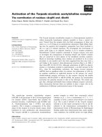
Fig. 1. TPA increases the level of PS1 mRNA in SK-N-SH cells. (A)
SK-N-SH cells were incubated in the presence of 0.2 l
M
TPA (T) or
dimethylsulfoxide (C) for increasing amounts of time from 30 min to
24 h as indicated above the lanes. RNA (15 lg) was fractionated on
denaturing 1.4% (w/v) agarose gels and analyzed by Northern blot-
ting. Membranes were sequentially hybridized with cDNA probes for
the human PS1 and glyceraldehyde-3 phosphate dehydrogenase
(GAPDH) genes. (B) The level of transcription at each time point was
quantified by laser scanning of the autoradiograms. The level of the
PS1 3 kb transcript in each lane was expressed as its ratio to GAPDH
mRNA in the same sample. The average level of the normalized PS1
mRNA at each time point was estimated with n ¼ 4orn ¼ 5ineach
of three experiments. The histogram displays the ratio between the
average level of PS1 mRNA in the TPA-treated samples and the
average levelin dimethylsulfoxide controlat each time point. (C) HepG2
cells were incubated with TPA or dimethylsulfoxide and total RNA
was analyzed by Northern blotting as described for SK-N-SH cells in
(A). The average level of the normalized PS1 mRNA at each time point
was estimated with n ¼ 3orn ¼ 4 in each of two experiments. In the
2 h control lane the PS1 band is partially masked by a gel artefact.
5958 M. Pastorcic and H. K. Das (Eur. J. Biochem. 269) Ó FEBS 2002
blotting using an antibody recognizing specifically the N
terminus of the PS1 protein. Three species were detected: the
full length PS1 appearing as a 45 kDa polypeptide, as well
as a larger aggregated form and the 30 kDa N-terminal
fragment (Fig. 3). After a 17-h TPA treatment the level of
the full length 45 kDa species and aggregated form
increased by 1.5– twofold. No significant increase in the
30 kDa N-terminal fragment protein was observed. Thus
TPA treatment increases the level of the PS1 protein. The
full length PS1 has a relatively short half-life, and it is
normally cleaved by endoproteolysis into a 30 kDa
N-terminal fragment and 17 kDa C-terminal fragment
which are considerably more stable [23]. It is possible that
any increase in newly synthesized PS1 in the presence of
TPA does not appear against the background of the larger
cellular pool of the stable 30 kDa form. Hence we observe
an increase in the level of the PS1 protein by TPA treatment
which is consistent with the increased mRNA level.
DNA sequences required to confer activation
of transcription of PS1 by TPA
We have recently identified a promoter area required for
efficient expression of the PS1 gene in SK-N-SH cells and
HepG2 cells including DNA sequences from )35 to +178
flanking the transcription initiation site [12]. We have
transfected SK-N-SH cells with PS1CAT fusion gene
constructs containing various fragments of PS1 sequences
from )687 to +178 inserted upstream from the CAT
reporter gene. With constructs including sequences from
Table 1. TPA does not alter the level of the PS1 mRNA in HepG2 cells. HepG2 cells were incubated with TPA or DMSO and total RNA was
analyzed by Northern blotting as described for SK-N-SH cells in (A). The level of PS1 mRNA was quantified by laser scanning of the auto-
radiograms and normalized with the level of GAPDH mRNA in the same samples. The average level of PS1 mRNA at each time point was
estimated with n ¼ 3 or 4 in each of 2 experiments.
30¢ 1h 2h 4h 8h 24h
Dimethylsulfoxide 3 ± 0.4 1.4 ± 0.4 2.7 ± 1 3.9 ± 0.8 1.06 ± 0.34 1.3 ± 0.02
TPA 2.5 ± 0.4 1.9 ± 0.6 2.6 ± 0.6 3.3 ± 0.8 0.97 ± 0.06 1.4 ± 0.3
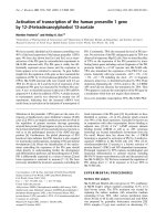
Fig. 3. TPA increases the level of PS1 protein in SK-N-SH cells. SK-N-
SH cells were treated with 0.2 l
M
TPA for 17 h and cell extracts were
fractionated by electrophoresis on 12% (w/v) polyacrylamide gels and
analyzed by Western blotting as described in Experimental procedures.
Control extract (C) and TPA-treated extract (T) (25 lg) were loaded in
lanes 1 and 2, respectively. The size of molecular mass markers is
indicated in kDa alongside the gel. Arrows mark the position of the full
length 45 kDa, the aggregated form and the 30 kDa N-terminal
fragment. The same blot was stripped and the level of actin protein was
analyzed as a control. Bands were quantified by laser scanning of the
autoradiograms. The level of PS1 was normalized to actin and was
determined in three distinct experiments. Values were analyzed by the
paired t-test/
ANOVA
method, and a value of P < 0.05 was considered
significant. The average level of the aggregated form was 1.7 ± 0.36
(P < 0.05) in TPA-treated samples and 0.88 ± 0.15 in control sam-
ples. The full length PS1 was 1.74 ± 0.2 in TPA samples and the
control level was 0.94 ± 0.2 (P < 0.05). The 30 kDa species was
1.2 ± 0.28 in the TPA-treated samples and 0.98 ± 0.4 in the controls.
All averages were derived from n ¼ 3.
Fig. 2. Nuclear run-off analysis of the transcription of PS1 in the
presence of TPA. SK-N-SH cells were incubated in the presence of
0.2 l
M
TPAfor5h.Nucleiwerethenpurifiedandusedintranscrip-
tion run-off analysis to quantify the RNAs being actively transcribed
at the time of harvest as described in Experimental procedures.
Transcription was quantified by laser scanning of the autoradiograms.
The changes in level of PS1 transcripts were quantified after normal-
ization with 18S RNA. TPA increased transcription of PS1 by 2.8
(± 0.8) with n ¼ 3 in two independent experiments.
Ó FEBS 2002 Regulation of the presenilin 1 gene (Eur. J. Biochem. 269) 5959
)687 to +178, )118 to +178, )22 to +178, )22 to +42 or
the minimal promoter )35 to +6 the activation by TPA
was two- to threefold (Fig. 4). Thus the minimal promoter
)35 to +6 is sufficient to confer activation by TPA. This
sequence interval contains an Ets element at )10 (Fig. 5)
which is crucial for the expression of PS1. It also contains a
sequence element sharing homology with the consensus
CREB/AP1 binding motif immediately adjacent to the Ets
site [24]. The effects of TPA on transcription are commonly
mediated by AP1. Furthermore, Ets factors are known to
act in conjunction with a number of other regulatory
proteins including AP1. Thus we have tested the effect of a
point mutation eliminating the AP1 homology (m6) as well
as a point mutation abolishing the )10 Ets site (m1) (Fig. 5).
M6 reduced the activity of the )118 to +178 construct by
about twofold; however, the mutant promoter retained two-
to threefold stimulation by TPA, similar to the )118 to
+178 wild type construct (Fig. 4). Thus the )6 CREB/AP1
homology is not required for TPA activation. Similarly, the
point mutation m1 eliminating the )10 Ets binding site did
not abolish induction by TPA. This may indicate that
neither the )10 Ets element, nor the )6CREB/AP1motif
are required for stimulation by TPA.
Changes in DNAÆprotein interactions over the )22/+6
region of the PS1 promoter in nuclear extracts
from SK-N-SH cells treated with TPA
We have used EMSAs to detect changes in the binding
activity of the proteins recognizing specifically the )10
region of the PS1 promoter in nuclear extracts of SK-N-SH
cells treated with TPA (Fig. 6). In dimethylsulfoxide-treated
Fig. 4. DNA sequences required for activation of PS1 transcription by
TPA. PS1CAT fusion genes containing various fragments of the PS1
promoter linked to the CAT reporter gene were transfected into SK-N-
SH cells. The end-points of the promoter fragments used in each of the
constructs are indicated below the graph. m1 is a mutation from
CCGGAAATGACGA to CC
ttAAATGACGA eliminating the )10
Ets site. In m6 the mutation to CCGGAAAT
cgaGA eliminates the
adjacent CREB and AP1 homologies (underlined) [24].
Fig. 5. PS1 promoter sequence. PS1 promoter sequence from )118 to
+178. The endpoint of the 3¢ and 5¢deletions used in this study are
indicated by arrows. The transcription initiation site is shown (+ 1).
The position of the Ets, CREB and Sp1 binding sites are underlined.
Fig. 6. Changes in DNAÆprotein interactions over the )22 to +6 region
of the PS1 promoter induced by TPA treatment of SK-N-SH cells.
(A) Nuclear extracts from SK-N-SH cells were prepared from cells
treated with 0.2 l
M
TPA for 5 h as well as from cultures where the
same dilution of dimethylsulfoxide was added (D). DNAÆprotein
interactions over the ()22 to +6) region of the PS1 promoter were
examined by EMSAs. The positions of the specific complexes are
indicated. Extracts were preincubated with aEts1/2 (aElanes),an
antibody recognizing specifically Ets1 and Ets2, for 45 min at 24 °Cin
the absence of DNA probe. An antibody unrelated to Ets factors (anti-
PS1 sc-1245, from Santa Cruz Biotechnology, CA, USA) was included
in control lanes (C). The probe was added and incubation was con-
tinued for another 20 min. Reactions were analyzed by electrophoresis
on 6% (w/v) native polyacrylamide gels at 4 °C. Lanes 1–5 include the
wild type probe, lanes 6–10 display binding to the probe containing a
mutation (GGAA fi ttAA) within the )10 Ets motif. (B) Low
exposure of the region of the gel including complex B.
5960 M. Pastorcic and H. K. Das (Eur. J. Biochem. 269) Ó FEBS 2002
nuclear extracts (D) the pattern of DNAÆprotein complexes
observed with the PS1 probe produced the specific com-
plexes A, B, C, D, E, F, G and H. These specific
proteinÆDNA complexes (A–H) appear to be generated by
proteinÆprotein interaction with Ets factors and other
proteins [12]. These complexes (A–H) are found to be
absent in assays with the Ets motif mutant probe similar to
the data described previously [12]. TPA treatment appears
to result in the loss of the specific complexes F and H, a
decrease in complex B, as well as an increase in complexes
A, C, D, E and G. Complexes A and G are eliminated by
preincubation of the control or TPA treated nuclear extracts
with anti-Ets1/2 Ig, indicating that at least these complexes
involve interactions with Ets1/2. Therefore TPA treatment
generally increased the specific interactions of the )10
region of the PS1 promoter with nuclear factors, including
the amount of complexes involving Ets1/2 factors.
DISCUSSION
The loss of PKC is a prognostic element in the severity of
neuronal damage resulting from ischemia in vivo [25]. The
activation of PKC by TPA inhibits cell death in vitro
through a complex set of pathways where different PKC
isozymes appear to play opposite roles [26]. TPA increases
the level of the expression of the endogenous PS1 gene in
SK-N-SH cells at the level of initiation of transcription.
TPA had no effect on the mRNA level in HepG2 cells (data
not shown), thus the regulation pathway implicated here
may be somewhat cell specific. Most of the known
biological effects of TPA are attributed to its ability to
activate PKC. The effect(s) of TPA observed here are likely
to result from the activation of PKC because the increase in
PS1 mRNA appears to be abolished by bisindolylmalei-
mide, a specific inhibitor of PKC [27,28], in preliminary data
(not shown). Furthermore, the time course of activation of
PS1 indicates that the maximum increase in the level of PS1
mRNA is reached by 4 h and that a longer exposure to TPA
(24 h) no longer activates PS1 expression. This is consistent
with the down-regulation of protein kinase C with long
exposure to TPA observed in many cell types [29].
In order to analyze further the mechanism of activation
we have tested the effect of TPA on the activity of the PS1
promoter in transient infection assays in SK-N-SH cells.
TPA treatment activated similarly by two- to 2.5-fold the
transcriptional activity of all promoter fragments tested.
The minimal promoter including sequences )35/+6
appears to retain TPA activation. Mutations eliminating
the )10 Ets binding or the )6 AP1/CREB motifs did not
reduce activation by TPA. This suggests that induction
results from the modification of protein(s) of the initiation
complex which do not bind directly to DNA. They may
however, interact with factors recognizing specific motifs,
such as Ets, and promote changes in proteinÆDNA interac-
tions within complexes including Ets. For example there is a
significant increase in the amount of the larger complexes A
and B (Fig. 6) in the TPA treated extracts. Complex A is
likely to contain Ets1 or Ets2, as it is eliminated by the
addition of anti-Ets1/2 Ig. It is possibly converted into
complex B (which increases from lane 4 to lane 5). This may
indicate that Ets 1/2 is not required for the formation of
complex B. The identity of the protein recognizing specif-
ically the PS1 promoter within B is not known. However its
ability to interact directly or indirectly with Ets1/2 should
enable its identification by the 2-hybrid selection technique.
Members of the AP1 protein complex have been impli-
cated in the onset of apoptosis. Induction by c-fos is an early
event in apoptosis [30], the overexpression of c-jun domin-
ant negative mutants protects sympathetic neurons against
programmed cell death induced by the withdrawal of nerve
growth factor whereas overexpression of wild type c-jun
appears to trigger apoptosis [31]. Retinoic acid-induced
apoptosis in F9 cells also induces c-jun, and the reduction of
c-jun levels by antisense reduces apoptosis [32]. In contrast,
the same F9 cells stably transformed with wild type PS1
show a significantly reduced level of apoptosis after retinoic
acid treatment, whereas mutant PS1 suppresses apoptosis
only weakly. This indicates that PS1 may play a protective
role in the development of c-jun-mediated apoptosis. Thus
the induction of PS1 gene expression after treatment with
TPA in the experiments described here is consistent with a
role of PS1 in the c-jun cascade leading to apoptosis.
However, the role of the c-jun N-terminal kinase (JNK)/
c-jun cascade for in vivo apoptosis and particularly in
Alzheimer’s disease is still unclear [33]. Growing evidence
implicates JNK-dependent pathways in Ab-dependent
apoptosis [34]. A role of PS1 in the development of
Ab-induced apoptosis has previously been suggested [35].
Overexpression of mutant PS1 increased the susceptibility of
PC12 cells to apoptosis induced by Ab or the withdrawal of
trophic factors. In contrast with this proapoptotic effect of
PS1 mutants, the wild type PS1 suppresses apoptosis
induced by the activation of p53 [36], which is a target of
JNK. Therefore, increasing evidence is indicating the
importance of the JNK/c-jun pathway in the neuronal
death in Alzheimer’s disease, and its differential interaction
with mutant and wild type PS1 suggests further its
importance in the development of the disease. Thus it
should be important to understand how the regulation of
the genes in both pathways interface. It is also important to
note that neuron specific activation of PS1 may increase
Notch-1 processing which could lead to neurite outgrowth
and decrease the risk of Alzheimer’s disease [37,38].
ACKNOWLEDGEMENT
We wish to thank Dr B. Yankner for his very generous gift of the aPS1
N-terminal antibody. This research was supported by a grant from the
National Institute of Health (AG18452) to H.K.D.
REFERENCES
1. Tanzi,R.E.,Kowacs,D.M.,Kim,T W.,Moir,R.,Guenette,S.Y.
& Wasco, W. (1996) The gene defects responsible for familial
Alzheimer’s disease. Neurobiol. Dis. 3, 159–168.
2. Sherrington. R., Rogaev, E.I., Liang, Y., Rogaeva, E.A.,
Levesque,G.,Ikeda,M.,Chi,H.,Lin,G.,Holman,K.,Tsuda,T.,
et al. (1995) Cloning of a gene bearing missense mutations in early-
onset familial Alzheimer’s disease. Nature 375, 754–760.
3. Wolfe, M.S., Xia, W., Ostaszewski, B.L., Diehl, T.S., Kimberly,
W.T. & Selkoe, D.J. (1999) Two transmembrane aspartates in
presenilin-1 required for presenilin endoproteolysis and gamma-
secretase activity. Nature 398, 513–517.
4. De Strooper, B., Saftig, P., Craessaerts, K., Vanderstichele, H.,
Guhge, G., Annaert, W., Von Figura, K. & Van Leuven, F. (1998)
Deficiency of presenilin-1 inhibits the normal cleavage of amyloid
precursor protein. Nature 391, 387–390.
Ó FEBS 2002 Regulation of the presenilin 1 gene (Eur. J. Biochem. 269) 5961
5. Wong, P.C., Zeng, H., Chen, H., Becher, M.W., Sirinathsinghji,
D.J.S., Trumbauer, M.E., Chen, H.Y., Price, D.L., Van der Ploeg,
L.H.T. & Sisodia, S.S. (1997) Presenilin 1 is required for Notch1
and DII1 expression in the paraxial mesoderm. Nature 387,
288–292.
6. Shen, J., Bronson, R.T., Chen, D.F., Xia, W., Selkoe, D.J. &
Tonegawa, S. (1997) Skeletal and CNS defects in presenilin-1-
deficient mice. Cell 89, 629–639.
7. Kovacs, D.M., Fausett, H.J., Page, K.J., Kim, T., Moir, R.D.,
Merriam, D.E., Hollister, R.D., Hallmark, O.G., Mancini, R.,
Felsenstein, K.M., et al. (1996) Alzheimer-associated presenilins
1 and 2: neuronal expression in brain and localization to
intracellular membranes in mammalian cells. Nat. Med. 2,
224–229.
8. Cribbs, D.H., Chen, L.S., Bende, S.M. & LaFerla, F.M. (1996)
Widespread neuronal expression of the presenilin-1 early-onset
Alzheimer’s disease gene in the murine brain. Am. J. Pathol. 148,
1797–1806.
9. Lee, M.K., Slunt, H.H., Martin, L.J., Thinakaran, G., Kim, G.,
Gandy, S.E., Seeger, M., Koo, E., Price, D.L. & Sisodia, S.S.
(1996) Expression of presenilin 1 and 2 (PS1 and PS2) in human
and murine tissues. J. Neurosci. 16, 7513–7525.
10. Quarteronet,D.,Pradier,L.,Czech,C.,Delalonde,L.,Burgevin,
M.C., Doble, A. & Petitet, F. (1996) Localization of presenilin-1
mRNA in rat brain. Neuroreport 7, 2587–2591.
11. Takami, K., Terai, K., Matsuo, A., Walker, D.G. & McGeer, P.L.
(1997) Expression of presenilin-1 and -2 mRNAs in rat and
Alzheimer’s disease brains. Brain Res. 748, 122–130.
12. Pastorcic, M. & Das, H.K. (1999) An upstream element contain-
ing an ETS binding site is crucial for transcription of the human
presenilin-1 gene. J. Biol. Chem. 274, 24297–24307.
13. Wasylyk, B., Hahn, S.L. & Giovane, A. (1993) The Ets family of
transcription factors. Eur. J. Biochem. 211, 7–18.
14. Janknecht, R. & Nordheim, A. (1993) Gene regulation by Ets
proteins. Biochim. Biophys. Acta 1155, 346–356.
15. Mitsuda, N., Ohkubo, N., Tamatani, M., Lee, Y.D., Taniguchi,
M., Namikawa, K., Kiyama, H., Yamaguchi, A., Sato, N.,
Sakata, K., et al. (2001) Activated cAMP-response element-
binding protein regulates neuronal expression of presenilin-1.
J. Biol. Chem. 276, 9688–9698.
16. Pahlman, S., Odelstad, L., Larsson, E., Grotte, G. & Nilsson, K.
(1981) Phenotypic changes of human neuroblastoma cells in cul-
ture induced by 12-O-tetradecanoyl-phorbol-13-acetate. Int. J.
Cancer 28, 583–589.
17. Pahlman, S., Ruusala, A.I., Abrahamsson, L., Odelstad, L. &
Nilsson, K. (1983) Kinetics and concentration effects of TPA-
induced differentiation of cultured human neuroblastoma cells.
Cell Differ. 12, 165–170.
18. Pahlman, S., Ruusala, A.I., Abrahamsson, L., Mattsson, M.E. &
Esscher, T. (1984) Retinoic acid-induced differentiation of cul-
tured human neuroblastoma cells: a comparison with phorbo-
lester-induced differentiation. Cell Differ. 14, 135–144.
19. Pahlman, S., Hoehner, J.C., Nanberg, E., Hedborg, F.,
Fagerstrom, S., Gestblom, C., Johansson, I., Larsson, U.,
Lavenius, E., Ortoft, E. et al. (1995) Differentiation and survival
influences of growth factors in human neuroblastoma. Eur J.
Cancer 31A, 453–458.
20. Chomczynski, P. & Sacchi, N. (1987) Single-step method of RNA
isolation by acid guanidinium thiocyanate-phenol-chloroform
extraction. Anal Biochem. 162, 156–159.
21. Hartmann, H., Busciglio, J., Baumann, K H., Staufenbiel, M. &
Yankner, B.A. (1997) Developmental regulation of presenilin-1
processing in the brain suggests a role in neuronal differentiation.
J. Biol. Chem. 272, 14505–14508.
22. Rogaev, E.I., Sherrington, R., Wu, C., Levesque, G., Liang, Y.,
Rogaeva, E.A., Ikeda, M., Holman, K., Lin, C., Lukiw, W.J.,
et al. (1997) Analysis of the 5¢ sequence, genomic structure, and
alternative splicing of the presenilin-1 gene (PSEN1) associated
with early onset Alzheimer disease. Genomics 40, 415–424.
23. Ratovitski, T., Slunt, H.H., Thinakaran, G., Price, D.L., Sisodia,
S.S. & Borchelt, D.R. (1997) Endoproteolytic processing and
stabilization of wild-type and mutant presenilin. J. Biol. Chem.
272, 24536–24541.
24. Wingender,E.,Chen,X.,Hehl,R.,Karas,H.,Liebich,I.,Matys,
V., Meinhardt, T., Pru
¨
ß, M., Reuter, I. & Schacherer, F. (2000)
TRANSFAC: an integrated system for gene expression regulation.
Nucleic Acids Res. 28, 316–319.
25. Durkin, J.P., Tremblay, R., Chakravarthy, B., Mealing, G.,
Morley, P., Small, D. & Song, D. (1997) Evidence that the early
loss of membrane protein kinase C is a necessary step in the
excitatory amino acid-induced death of primary cortical neurons.
J. Neurochem. 68, 1400–1412.
26. Maher, P. (2001) How protein kinase C activation protects nerve
cells from oxidative stress-induced cell death. J. Neurosci. 21,
2929–2938.
27. Toullec,D.,Pianetti,P.,Coste,H.,Bellevergue,P.,Grand-Perret,
T., Ajakane, M., Baudet, V., Boissin, P., Boursier, E., Loriolle, F.
et al. (1991) The bisindolylmaleimide GF 109203X is a potent and
selective inhibitor of protein kinase C. J. Biol. Chem. 266, 15771–
15781.
28. Heikkila, J., Jalava, A. & Eriksson, K. (1993) The selective protein
kinase C inhibitor GF 109203X inhibits phorbol ester-induced
morphological and functional differentiation of SH-SY5Y human
neuroblastoma cells. Biochem. Biophys Res. Commun. 197, 1185–
1193.
29. Parrow,V.,Nnberg,E.,Heikkila,J.,Hammerling,U.&Pa
˚
hlman,
S. (1992) Protein kinase C remains functionally active during TPA
induced neuronal differentiation of SH-SY5Y human neuro-
blastoma cells. J. Cell. Physiol. 152, 536–544.
30. Smeyne, R.J., Vendrell, M., Hayward, M., Baker, S.J., Miao,
G.G., Schilling, K., Robertson, L.M., Curran, T. & Morgan, J.I.
(1993) Continuous c-fos expression precedes programmed cell
death in vivo. Nature 363, 166–169.
31.Ham,J.,Babij,C.,Whitfield,J.,Pfarr,C.M.,Lallemand,D.,
Yaniv, M. & Rubin, L.L. (1995) A c-Jun dominant negative
mutant protects sympathetic neurons against programmed cell
death. Neuron 14, 927–939.
32. Imafuku, I., Masaki, T., Waragai, M., Takeuchi, S., Kawabata,
M., Hirai, S., Ohno, S., Nee, L.E., Lippa, C.F., Kanazawa, I.,
et al. (1999) Presenilin 1 suppresses the function of c-Jun homo-
dimers via interaction with QM/Jif-1. J. Cell Biol. 147, 121–134.
33. Marx, J. (2001) Neuroscience. New leads on the ÔhowÕ of Alzhei-
mer’s. Science 293, 2192–2194.
34.Morishima,Y.,Gotoh,Y.,Zieg,J.,Barrett,T.,Takano,H.,
Flavell, R., Davis, R.J., Shirasaki, Y. & Greenberg, M.E. (2001)
Beta-amyloid induces neuronal apoptosis via a mechanism that
involves the c-Jun N-terminal kinase pathway and the induction of
Fas ligand. J. Neurosci. 21, 7551–7560.
35. Guo, Q., Sopher, B.L., Furukawa, K., Pham, D.G., Robinson, N.,
George, M., Martin, G.M. & Mattson, M.P. (1997) Alzheimer’s
presenilin mutation sensitizes neural cells to apoptosis induced by
trophic factor withdrawal and amyloid beta-peptide: involvement
of calcium and oxyradicals. J. Neurosci. 17, 4212–4222.
36. Roperch, J.P., Alvaro, V., Prieur, S., Tuynder, M., Nemani, M.,
Lethrosne, F., Piouffre, L., Gendron, M.C., Israeli, D., Dausset,
J.,Oren,M.,Amson,R.&Telerman,A.(1998)Inhibitionof
presenilin 1 expression is promoted by p53 and p21WAF-1 and
results in apoptosis and tumor suppression. Nat. Med. 4, 835–838.
37. Sestan, N., Artavanis-Tsakonas, S. & Rakic, P. (1999) Contact-
dependent inhibition of cortical neurite growth mediated by Notch
signaling. Science 286, 741–746.
38. Redmond, L., Oh, S.R., Hicks, C., Weinmaster, G. & Ghosh, A.
(2000) Nuclear Notch1 signaling and the regulation of dendritic
development. Nature Neurosci. 3, 30–40.
5962 M. Pastorcic and H. K. Das (Eur. J. Biochem. 269) Ó FEBS 2002