Báo cáo khoa học: NBR1 interacts with fasciculation and elongation protein zeta-1 (FEZ1) and calcium and integrin binding protein (CIB) and shows developmentally restricted expression in the neural tube pptx
Bạn đang xem bản rút gọn của tài liệu. Xem và tải ngay bản đầy đủ của tài liệu tại đây (303.5 KB, 8 trang )
NBR1 interacts with fasciculation and elongation protein zeta-1 (FEZ1)
and calcium and integrin binding protein (CIB) and shows
developmentally restricted expression in the neural tube
Caroline Whitehouse
1
, Julie Chambers
1,
*, Kathy Howe
1
, Martyn Cobourne
2
, Paul Sharpe
2
and Ellen Solomon
1
1
Division of Medical and Molecular Genetics, and
2
Department of Craniofacial Development, GKT School of Medicine,
Guy's Hospital, London, UK
NBR1 (named as next to BRCA1) was originally cloned as a
candidate gene for the ovarian cancer antigen CA125, using
expression cloning with the anti-CA125 Ig, OC125. NBR1
has been of interest due to its position close to BRCA1,
although no involvement in breast or ovarian cancer has
been demonstrated. Recently, the a ntigen CA125 has been
cloned, and identi®ed as a new mucin, MUC16, entirely
dierent from NBR1. The function of NBR1 remains un-
known. To investigate its function, a yeast two-hybrid study
was performed to identify interacting protein partners that
may re¯ect a biological role for t his protein. H ere, we show
that NBR1 interacts with two proteins; fasciculation and
elongation protein zeta-1 (FEZ1), a P KCf interacting pro -
tein, and calcium a nd integrin binding p rotein (CIB), which
is associated with polo-like kinases Fnk/Snk and t he
Alzheimer's disease presenilin 2 p rotein. Co-transfection of
FEZ1 and NBR1 showed overlapping localization in the
cytoplasm, whereas c oexpression of N BR1 a nd CIB resulted
in a shift of CIB protein expression from the nucleus to the
perinuclear compartment. FEZ1 i s highly expressed in t he
brain and in situ hybridization analysis of Nbr1 showed that
its expression is also regulated in t he murine brain d uring
development. These data suggest th at NBR1 m ay function,
through interaction with CIB and FEZ1 in cell signalling
pathways, w ith a developmentally restricted expression
suggesting a possible role in neural development.
Keywords: ZZ zinc binding domain; OPR domain; UBA
domain; CIB; FEZ1.
The human NBR1 gene (named for its location, next to
BRCA1), originally named 1A1.3B, was cloned as a
candidate g ene for the ovarian cancer antigen CA125 [1].
A serum assay for CA125 using the monoclonal a ntibody
OC125 showed that levels of this antigen are elevated in
> 80% of patients with epithe lial o varian cancer [2].
Recently a new mucin, MUC16 has been cloned as
CA125 [3] whose biochemical characteristics as a high
molecular mass, heavily O-glycosylated protein c orrelate
with the expected p roperties of the polypeptide carrying the
CA125 epitope [4].
NBR1 shares little h omology with other k nown proteins,
but does contain a number of protein motifs (see Fig. 1A).
These domains include two putative metal binding regions;
a zinc binding domain originally thought to belong to the
B-box family of zinc binding domains and a n octicosapep-
tide sequence (OPR) also thought to be involved in divalent
cation binding. The octicosapeptide domain is a 28-residue
motif also present in protein kinase C isoforms iota, lambda
and zeta. The B-box domain, however, more closely
resembles the ZZ zinc-binding domain present in dystro-
phin-like proteins, and CREB-binding proteins/p300
homologues. The ZZ domain in dystrophin is t hought to
bind calmodulin, and a missense mutation i n one of the
conserved cysteine residues in dystrophin was described in a
patient with Duchene muscular dystrophy [5]. NBR1 also
contains a coiled-coil domain that i s often implicated in
protein±protein interactions [6]. An ubiquitin-associated
domain (UBA) has also been predicted at the C-terminus of
NBR1 [7]. This domain is thought not to bind ubiquitin
domains directly but is postulated to be involved in
conferring target speci®city to multiple enzymes of the
ubiquitination system [8]. The BRCA1 protein has recently
also been shown to be involved i n the ubiquitination
pathway, where cancer-associated mutations in the
N-terminal RING ®nger domain of BRCA1 results in loss
of ubiquitin protein ligase (E3) activity [ 9]. The related
RBCC family of proteins which contain a RING ®nger
domain i n addition to a B -box and coiled-coil domain, and
Correspondence to C. W h itehouse, Division of Medical and Molecular
Genetics, GKT School of Medicine, Guy's Hospital, London SE1
9RT. Fax: + 020 79558762, Tel.: + 020 79555000 ext. 5585.
E-mail:
Abbreviations: NBR1, next to BRCA1; CIB, calcium and integrin
binding protein; FEZ1, fasciculation and elongation protein zeta-1;
PKC f, protein kinase C zeta isoform; d.p.c., days post coitum; RBCC,
RING, B- box and c oiled-coil protein; PML, pr omyelocytic leukaemia
gene; HA-, haemaglutinin epitope; EGFP, e nhanced gree n ¯uorescent
protein; OPR, octicosapeptide sequence; UBA domain, ubiquitin-
associated domain; rfp, ret ®nger protein; BDH, acetic anhydride;
TESPA, 3-aminopropyltriethoxysilane.
Note: further information o n the proteins in this paper i s available
from GenBank under the following accession n umb ers: NBR1
(X76952), CIB (U82226), FEZ1 (XM006241), PKCf, (XM001533),
PML (XM007642).
*Present address: A stra Zeneca, Alderley Park, Maccles®eld, UK.
(Received 24 September 2001, revised 14 November 2001, accepted 16
November 2 001)
Eur. J. Biochem. 269, 538±545 (2002) Ó FEBS 2002
includes members such as PML and the ret ®n ger protein
(rfp), have been shown to have transforming activities when
inappropriately expressed [10].
The NBR1 protein is highly conserved, with 87% sequence
similarity with the m urine homologue. The murine Nbr1 gene
liesheadtoheadwiththeBrca1 gene, with the intragenic
region of 289 bp suggesting the possibility of co-ordinated
expression of the two genes. Mutations in the human BRCA1
gene were found in 81% of breast±ovarian c ancer families i n
a large study [11] and are thought to be responsible for 45%
of cases of familial e arly onset breast cancer [12].
This study describes the results of a yeast two-hybrid
experiment to identify interact ing partners of NBR1, and in
situ hybridization analysis of the murine Nbr1 embryonic
expression pattern. Two interacting partners were identi®ed,
CIB and FEZ, which w ere also shown to i nteract directly,
and the interacting domains were delineated. Nbr1 expres-
sion in the d eveloping mouse embryo showed an early
uniform pattern of expression, which th en becomes restric-
ted around 10.5±13.5 d .p.c. (days post coitum) to the neural
tube, and then showed a wide expression pattern in the adult
mouse.
MATERIALS AND METHODS
Yeast two-hybrid library screening
The full-length cDNA of human NBR1 was c loned in frame
into the GAL4 DNA-binding domain (BD) vector p GBT9
(Clontech). The yeast strain HF7c was transformed with
this construct (pGBT9NBR1) and a human placenta
MATCHMAKER cDNA library fused with the GAL4
AD (Clontech). Transformants were selected on plates
lacking leucine, tryptophan and histidine, and containing
3m
M
3-amino-1,2,4-triazole (Sigma) for 5 days. Putative
positive colonies were restreaked onto fresh master plates
for b-galactosidase assays. These were performed following
colony-lifts onto H ybond-N nylon membranes and assayed
as described previously [13]. The pACT2 library plasmid
DNA from candidate clones was recovered and the cDNA
inserts a nalysed by nucleotide sequencing to ensure the
inserts were in frame with the GAL4 activation domain.
Any candidate clone s were screened for false-positives by
cotransformation with the empty pGBT9 vector or control
plasmid pLAM5¢ (Clontech).
Delineation of interacting regions
Sections of the N BR1 cDNA were PCR a mpli®ed and
subcloned into the pGBT9 vector EcoRI site to give the
following constructs: pGBT9NBR1
216
(amino acids 1±216),
pGBT9NBR1
333
(amino acids 1±333), pGBT9NBR1c1
(amino acids 340±453), pGBT9NBR1c2 (amino acids
451±597), pGBT9NBR1c3 (amino acids 592±754) and
pGBT9NBR1c4 (amino acids 748±910). A BclIfragment
of pGBT9NBR1 was subcloned into the BamHI site of
pGBT9 to give a C-terminal c onstruct, pGBT9NBR1
Cterm
(amino acids 494±849). All constructs were subsequently
sequenced before testing in the yeast two-hybrid a ssay.
Interactions were scored as +++ (strongly po sitive), ++
(moderately positive), + (weakly positive) or ± (negative),
where +++ denoted growth on ± leu-trp-his plates in
three days and a positive colony lift b-galactosidase assay in
4 h . A weakly positive result denoted growth on ± leu-
trp-his plates in ®ve days and a positive colony lift
b-galactosidase assay i n 8 h.
Northern blot analysis
CIB and FEZ1 probes were excised from the pACT2 vector
with BamHI/BglII and EcoRI/XhoI, respectively. The
NBR1 probe used for Northern blot analysis w as as
described p reviously [1]. Human multiple tissue Northern
blots (Clontech) were hybridized at 42 °C for 18±24 h in 5 ´
NaCl/P
i
/EDTA, 10 ´ Denhardts, 2% SDS and 50%
formamide. Filters were washed twice in 2 ´ NaCl/Cit/
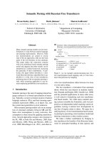
Fig. 1. Schematic representation of the protein
domains of NBR1. (A) (OPR) octicosapeptide
repeat (ZZ) zinc ®nger (CC) c oiled-coil and
(UBA) ubiquitin-associated domain. The
amino acids encoding each domain are shown
below and the ® gure is not drawn to s cale.
(B) Yeast two-hybrid assay analysis identi®es
CIB and F EZ1 as interacting partners of
NBR1. HF7c cells were cotransformed w ith
either pACT2FEZ1 (Y214), pACT2FEZ1
(Y156), pACT2FEZ1 (Y163), pACT2CIB
(Y198) or pGAD424NBR1 and
(i) pGBT9NBR1 (ii) pGBT9NBR1
216
(iii) pGBT9NBR1
333
(iv) pGBT9NBR1
Cterm
(v) pGBT9NBR1c1 (vi) pGBT9NBR1c2
(vii) pGBT9NBR1c3, viii) pGBT9NBR1c4 or
(ix) pGBKT7CIB and tested for p ro tein±pro-
tein intera ction by a colony lift b-galactosidase
assay and growth on ± leu-trp-his m edium.
(+++, strongly positive; ++, moderately
positive; + , weakly positive; ±, negative; n/d,
not determined; j,GAL4BD).
Ó FEBS 2002 Interactions between proteins NBR1, CIB and FEZ1 (Eur. J. Biochem. 269) 539
0.1%SDS at room temperature for 15 min and then twice in
1 ´ NaCl/Cit/0.1% SDS and 0.5 ´ NaCl/Cit/0.1%SDS at
50 °C for 20 min.
In situ
hybridization analysis on foetal sections
35
S-Radiolabelled in situ hybridization was carried out as
follows: mouse embryos were sectioned at 8 lm and ¯oated
onto TESPA (3-aminopropyltriethoxysilane) (Sigma)
coated slides. The slides were pretreated with 5 lgámL
)1
proteinase K (Sigma) and 2 mgámL
)1
glycine (Sigma) in
NaCl/P
i
and then re-®xed in 4% paraformaldehyde
(Sigma). Following treatment with 0 .25% acetic anhydride
(BDH), hybridization was carried out overnight in a
humidi®ed chamber at 55 °C. The s lides were then washed
at high stringency (20 min at 55 °Cin2´ NaCl/Cit, 50%
formamide,10 m
M
dithiothreitol) and then treated with
40 lgámL
)1
RNAse A for 30 min at 37 °C. The high
stringency washes were repeated at 65 °C, followed by a
furtherwashin0.1´ NaCl/Cit/10 m
M
dithiothreitol, also a t
65 °C. The s lides were then washed in 0.1´ NaCl/Cit at
room temperature and dehydrated through 300 m
M
ammonium acetate in 70% ethanol, 95% ethanol and then
100% ethanol. Following air-drying, the slides were dipped
in Ilford K.5 photographic emulsion. Autoradiography was
performed by e xposing the sections in a light-proof box at
4 °C for 14 days. The slides were then developed using
Kodak D19 developer, ®xed with Kodak UNIFIX and
counter stained w ith malachite green. Sections were photo -
graphed under dark ®eld with an Olympus BH-2 micro-
scope and photographed with an Olympus camera using
Fujichrome 64T Tungsten ®lm.
Cloning of eukaryotic expression constructs
Full length CIB cDNA was P CR ampli®ed from the yeast
GAL4 library pACT2 plasmid (Y198) and s ubcloned into
the pcDNA3.1 vector (Invitrogen) to produce the
C- terminal myc-tagged CIB construct C IB-myc. The full-
length cDNA of human FEZ1 was obtained as an IMAGE
clone from the UK HGMP Resource Centre and subcloned
into the pEGFP-N2 expression vector (Clontech) to produce
a C-terminal E GFP tagged construct (pFEZ1-EGFP). The
full-length NBR1 cDNA was s ubcloned into the pHM6
expression vector (Roche) to produce an N-terminal HA
epitope tagged construct (pHA-NBR1). All p lasmids crea-
ted b y P CR during the cloning steps were also sequenced.
Immunoprecipitation and Western blotting
COS-7 cells were cultured in DMEM supplemented w ith
10% fetal calf serum ( Life Technologies). Transient trans-
fection o f eukaryotic expression vector constructs was
performed using the FuGENE
TM
reagent (Roche). Typi-
cally 1±2 lg of each c onstruct w as used to transfect COS-7
cells at 70% con¯uency in a six-well dish. Sixteen hours
post-transfection, cells were washed o nce in ice cold NaCl/P
i
and lysed at 4 °C in 0.5 mL Tris/NaCl/P
i
lysis buffer
containing 137 m
M
NaCl, 20 m
M
Tris pH 8.0, 0.5% Tween
20, including a protease inhibitor cocktail (Complete
TM
,
Roche). After lysis, cell homogenates were centrifuged at
16 000 g at 4 °C for 15 min and the supernatant collected.
To con®rm protein expression, 10 lL s amples were sepa-
rated by SDS/PAGE and analysed by Western blotting.
Immunoprecipitations were carried out from 200 to 400 lL
precleared cell l ysate at 4 °Cfor2hwith2.5lgoftheanti-
GFP Living C olors A.v.Ò peptide antibody (Clontech) and
collected by protein LA agarose beads (Clontech). Beads
were washed ®ve times with lysis buffer and bound pr oteins
were eluted by boiling. Proteins were separated by SDS/
PAGE, transferred to an Immobilon-P membrane
(Millipore) and i mmunoblotted with t he appropriate
antibody according to manufacturer's instructions. Western
blots were developed by enhanced chemiluminescence
(Amersham Pharmacia).
Cell transfection and immuno¯uorescence analysis
COS-7 cells were transfected as described above and
cultured for 16 h before analysis for ¯uorescence as
described previo usly [14]. NBR1 was detected using either
HA mAb 12CA5 (Roche) and TRITC or FITC c onjugated
goat anti-(mouse Ig) Ig (DAKO) or HA polyclonal
antibody sc805 (Santa Cruz Biotechnology, Inc) followed
by TRITC or FITC conjugated swine anti-(rabbit Ig) Ig
(DAKO) as appropriate. CIB-myc was detected u sing the
c-myc mAb 9E10 (Sigma) followed by TRITC or FITC
conjugated goat anti-(mouse Ig) Ig (DAKO). Cell immu-
no¯uorescence was analysed using a L SM510 laser scanning
confocal microscope (Zeiss).
RESULTS
NBR1 interacts with the PKC zeta interacting protein
FEZ1 and the calcium and integrin binding protein CIB
In order to identify proteins that interact with NBR1, a
yeast two-hybrid study was performed using full length
human NBR1 as bait. A human placental GAL4 AD fusion
cDNA library was screened and a total of 2 ´ 10
6
clones
were analysed. Four positive clones were isolated, Y156,
Y198, Y163 and Y214, which were positive by both the
b-galactosidase assay a nd nutritional selection assays.
Sequence analysis of these four cDNA library clones
showed that they represented two proteins, Y156, Y163 and
Y214 all encoded p artial cDNAs of FEZ1 and were all in
frame with the GAL4 act ivation domain (GAL4 AD). The
FEZ1 clones could be subdivided into clones encoding
overlapping regions of amino acids 248±360 and 248±349
(Y214 and Y156, respectively), and the C-terminal amino
acids 370±392 (Y163). Clone Y198 encoded the full-length
cDNA of the calcium and integrin binding protein (CIB) in
frame with the GA L4 AD.
Mapping of regions involved in the interaction
show that CIB and FEZ1 bind in the same domain
of NBR1
To identify which region of NBR1 interacts with CIB or
FEZ1, C-terminal d eletions of NBR1 wer e made and
analysed in the GAL4 yeast two-hybrid system with Y 214,
Y156, Y163 and Y198. The results of this analysis are
shown in Fig. 1B. These constructs included the N-terminal
216 a mino acids only (pGBT9NBR1
216
), the N-terminus,
ZZ and coiled-coil domain (pGBT9NBR1
333
)anda
C-terminal f ragment encoding amino acids 494 ±849 which
540 C. Whitehouse et al. (Eur. J. Biochem. 269) Ó FEBS 2002
contains none of these domains (pGBT9NBR1
Cterm
). As
shown in Fig. 1B, both CIB and FEZ1 interacted within the
C-terminus of NBR1. This region was then subdivid ed into
approximately 150 amino-acid fragments and further t ested
by the y east two-hybrid method (see Fig 1B, v±viii). B oth
CIB and FEZ1 interact strongly within the same region of
NBR1, encoding amino acids 451±597 and weakly with
amino acids 592±754. This r egion of NBR1 does n ot contain
any known o r predicted functional domains. The full
length NBR1 coding region was also subcloned into the
GAL4-AD vector pGAD424 to produce the construct
pGAD424NBR1. When this construct was tested in the
yeast two-hybrid assay, NBR1 was also s hown to form
homodimers. We can conclude that this interaction occurs
between two domains, as protein products of both
pGBT9NBR1
333
and pGBT9NBR1
Cterm
were able to
interact with full-length NBR1 (see Fig. 1B, i,iii,iv and vi).
FEZ1 and CIB interact with each other
To con®rm the interactions between N BR1, CIB and FEZ1,
the full-length FEZ1 and CIB cDNAs w ere subcloned into
the GAL4-BD vector pGBKT7 (Clontech) and tested in the
yeast interaction assay with pGAD424NBR1. The
pGBKT7±FEZ1 construct showed transcriptional a ctiva-
tion function, and thus could not be used to verify the
NBR1/FEZ1 interaction, but the interactio n between CIB
and NBR1 was con®rmed. Using the yeast two-hybrid
assay, full length CIB and FEZ1 were also shown to bind
to each other. This interaction w as delineated to t he
C-terminus of FEZ1 as Y163 (encoding amino acids
370±392) but not Y214 (encoding amino acids 248±360) of
FEZ1 were shown to interact with full length CIB (data not
shown). FEZ1 was cloned as a mammalian homologue of
the C. elegans UNC-76 protein involved in axonal out-
growth. The two regions of FEZ1, which we have shown
can interact with NBR1, are part of domains that are
conserved between UNC-76 and rat FEZ1 [15].
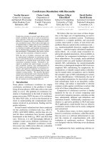
Co-immunoprecipitation experiments con®rm
that NBR1 interacts with FEZ1
in vivo
Due to the lack of antibodies that could detect NBR1 and
either FEZ1 or CIB, w e were unable to perform immuno-
precipitation or colocalization s tudies of endogenous
proteins. Thus we exogenously expressed epitope-tagged
constructs, w here COS-7 cells were either singly transfected
with pHA-NBR1, pFEZ1-EGFP or cotransfected with
pHA-NBR1 and either pFEZ1-EGFP or the empty vector
pEGFP-N2 that expresses EGFP only. Immunoprecipitates
from cell lysates using a GFP polyclonal antibody were
separated by S DS/PAGE and Western blotted before
immunodetection using the HA mAb 12CA5. Figure 2D
shows that HA-NBR1 is coimmunoprecipitated only in the
presence of FEZ1-EGFP (lanes 4 ) and no t when expressed
with EGFP (lane 5). Panels A, B a nd C show Western
analysis of cell lysates to show comparable expression of
HA-NBR1, FEZ1-EGFP a nd EGFP where expected. These
results con®rm that NBR1 and FEZ1 interact in v ivo.The
interactionbetweenNBR1andCIBwasalsotestedby
immunoprecipitation with GST fusion proteins, in vitro
translated proteins and over-expressed proteins from trans-
fected COS-7 cells under several different buffer conditions,
but we were unable to con®rm binding of CIB a nd NBR1
by other m ethods. Nevertheless, given the data described i n
later sections of this paper, it is still likely that CIB and
NBR1 interact directly.
Northern analysis of NBR1, CIB and FEZ1 expression
in human tissues
To address the question of whether the three proteins
NBR1, CIB and FEZ1 are expressed in the same tissues,
Northern analysis was performed using human multiple
tissue RNA blots (see Fig. 3). Expression of NBR1 and CIB
was shown to be widespread, whereas FEZ1, although
expressed w eakly i n most of t he tissues examined was most
highly expressed in the brain. As well as the more common
4.4-kb NBR1 transcript observed in all tissues analysed, a
smaller 4-kb RNA transcript of NBR1 was a lso present in
testes. This smaller transcript corresponds to the alternati-
vely spliced Nbr1(1a) transcript, which has been suggested
to be a testes-speci®c isoform [16] but was detected by R T-
PCR in several other m ouse tissues analysed (C. Whitehouse,
unpublished results). The ubiquitously expressed CIB
transcript was1.2 kb in size and a n additional transcript of
approximately 1.5 kb was also observed in the testes which
agrees with previous results [17]. A 2.4-kb transcript of
FEZ1 was also present in testes compared to the more
common 2-kb mRNA, but the highest level of expression of
FEZ1 was observed i n the brain where a smaller transcript
of less than 1 k b was also present. Thus NBR1 and CIB
appear to be ubiquitously expressed in adult human tissues
whereas FEZ1 e xpression is m ore restricted, although not
exclusively, to the brain.
Developmental pattern of expression of murine Nbr1
Using an a ntisense cDNA probe to murine Nbr1 encoding
exons 16±3¢ UTR, the developmental e xpression pattern of
Fig. 2. Co-immunoprecipitation experiments con®rm that NBR1 and
FEZ1 interact in vivo. COS-7 c ells (lane 1), COS-7 c ells transfected with
pHA-NBR1 (lane 2), pFEZ1-EGFP (lane 3), pHA-NBR1 and
pFEZ1-EGFP (lane 4) and pHA-NBR1 and p EGFP-N 2 (lan e 5) were
assayed by Western blotting for expression of H A-NBR1(panel A),
FEZ1-EGFP (panel B) and EGFP (panel C). Cell lysates were
immunoprecipitated with the GFP polyclonal antibody and immuno-
blotted with the HA antibo dy 12CA5 (panel D ).
Ó FEBS 2002 Interactions between proteins NBR1, CIB and FEZ1 (Eur. J. Biochem. 269) 541
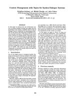
Nbr1 was analysed in mouse embryos by in situ hybridiza-
tion (see Fig. 4). The earliest sections analysed were at
embryonic stage 9 d.p.c. and show that Nbr1 is widely
expressed in all tiss ues. However at embryonic stage 10.5±
13.5 d.p.c., the distribution of Nbr1 RNA is shown to be
largely restricted to the neural tube. Northern analysis of
Nbr1 expression in the adult mouse shows that expression is
restored in all of the tissues analysed [18].
Development of the nervous system of the mouse embryo
is thought to begin a t 7 d.p.c. with the formation of the
neural plate and is completed by 17 d.p.c. The pattern of
expression of Nbr1 in the developing embryo thus suggests
that this gene may h ave a general role in early and late stages
of mouse development, but the restricted expression pro®le
at 10.5±13.5 d.p.c. suggests a more speci®c role for Nbr1 in
neural development. This pattern of expression overlaps
with the altered transcript pro®le o f its interacting partner
FEZ1, p roviding further evidence for the possible involve-
ment of these two genes in a common cellular pathway, and
more speci®cally in neuronal t issues.
Subcellular colocalization of CIB, NBR1
and FEZ1 protein
The subcellular localization of NBR1, CIB and FEZ1 was
analysed by transfection of C OS-7 cells with epitope-tagged
constructs. pHA-NBR1, pcDNA3.1 CIB-myc and pFEZ1-
EGFP expression plasmids were singly or cotransfected into
COS-7 cells and assayed 1 6 h later (see Fig. 5 ). NBR1 was
predominantly localized to the cytoplasm and was restricted
to a p articulate, perinuclear fraction (Fig. 5A). FEZ1-
EGFP was also mainly localized to the cytoplasm, but
Fig. 4. In situ hybridization analysis of Nbr1
expression during mouse embryonic develop-
ment. A N br1 antisense p robe covering e xons
16±3¢ UTR was labelled with [
35
S]dUTP and
incubated with embryonic mouse sections at
(A) 9 d.p.c. (B) 10.5 d .p.c. (C) 11.5 d.p.c. and
(D) 13.5 d.p.c. N, neural tube; M, mandible;
B, brain; BV, b rain ventricles
Fig. 3. Northern blot anal ysis of expression patte rns of CIB (panel A),
NBR1 (panel B) and FEZ1 (panel C ) in a panel of human adult tissues.
Northern blots containing 2 lg per lane of poly(A)
+
RNA from
various adult human tissues were hybridized w ith cDNA probes
labelled with [a-
32
P]dCTP. The position of RNA size markers is indi-
cated on the left sid e of each blot. Comparable loading of RN A in e ach
lane was shown by hybridization of a b-actin probe t o the same blots
(panel d).
542 C. Whitehouse et al. (Eur. J. Biochem. 269) Ó FEBS 2002
showed a more diffuse pattern of expression than NBR1,
with some p lasma membrane staining also present
(Fig. 5B,D). Upon cotransfection of pFEZ1-EGFP and
pHA-NBR1, both FEZ1-EGFP ( Fig. 5D) a nd HA-NBR1
(Fig. 5E) were shown to localize in the cytoplasm, and
showed very strong coexpression in the perinuclear com-
partment (yellow) (Fig. 5F). Overexpression of HA-NBR1
and FEZ1-EGFP in the same cell also often resulted in a
shift in HA-NBR1 expression to a more diffuse cytoplasmic
compartment similar to that observed for FEZ1-EGFP
alone (Fig. 5F).
CIB-myc showed a more c omplex pattern of expression,
with protein detected chie¯y in the nucleus, but expression
was also observed in t he cytoplasm or both nucleus and
cytoplasm (Fig. 5C). This agrees with previous data that
shows CIB expression in both cellular compartments and is
consistent with the hypothesis that CIB is involved in
dynamic processes such as cell signalling pathways [19]. In a
proportion of cells that coexpressed CIB and NBR1
however, the localization of CIB was drastically altered s o
that it now completely colocalized with the perinuclear
pattern observed for NBR1 (Fig. 5 I). This was not observed
however, in all cells that coexpressed CIB and NBR1, which
suggests that this m ay be a transient, dynamic association,
possibly linked to the stage of the cell cycle or signalling
status of the cell. NBR1, when coexpressed with nuclear
CIB however, retained its perinuclear, cytoplasmic location.
A s imilar shift in CIB e xpression pattern to the ER
compartment was ob served with the presenilin protein PS2
[20] and to t he cytoplasm when coexpressed w ith Snk [19].
Mutations in the presenilin genes PS1 and PS2 cause the
majority of cases of early onset Alzheimer's disease [21].
Stimuli t hat induce synaptic plasticity result in increased
expression of Fnk and Snk and lead to targeting of these
proteins to the dendrites of activated n eurons [19]. The shift
in localization o f CIB when coexpressed with N BR1 does
however, provide persuasive evidence t hat NBR1 and CIB
do interact in vivo.
DISCUSSION
Using a yeast two-hybrid approach, these experiments
describe the identi®cation of two proteins that interact with
NBR1; CIB and FEZ1, which were also shown to interact
with each other. CIB was itself cloned by a yeast two-hybrid
study using the integrin a
IIb
subunit as bait [22]. Sub-
sequently, CIB has been shown to interact with a number of
other proteins, including the polo-like kinases Fnk and Snk
[19], DNA dependent kinase [17], and the Alzheimer's
disease presenilin 2 p rotein [20]. CIB sho ws 58% amino-acid
similarity with calcineurin B, the regulatory subunit of
calcineurin ( phosphatase 2B) and also shares 56% protein
sequence similarity to calmodulin. The protein contains two
EF-hand motifs that bind Ca
2+
and thus it has been
suggested that the protein may act as a r egulatory subunit of
interacting partners [17]. Northern analysis of CIB expres-
sion showed that it is widely expressed in all tissues
examined. The subcellular localization of exogenously
expressed CIB described herein agrees with previous studies,
which h ave shown the accumulation in the nucleus and the
cytoplasm of both t ransfected and endogenous CIB protein
[19]. The presence of CIB in different cellular c ompartments,
and its ability to interact with proteins in the nucleus
(DNA-PK), and cytoplasmi c compartments ( NBR1)
Fig. 5. Intracellular l ocalization o f NBR1, CIB
and FEZ1 pro teins in COS-7 c ells. Cells were
transfected either s ingly (a,b and c) or
cotransfected (d±i) and processed for i mmu-
no¯uorescence 16 h later. (a) H A-NBR1 (b)
FEZ1-EGFP (c) CIB-myc protein expression,
(d±f) coexpression of HA-NBR1 and F EZ1-
EGFP, and (g±i) coexpression of HA-NBR1
and CIB-myc. T he pattern of C IB staining
observed in (g) w as never observed in cells
expressing CIB-myc only. Bar, 20 lm.
Ó FEBS 2002 Interactions between proteins NBR1, CIB and FEZ1 (Eur. J. Biochem. 269) 543
suggests that t his protein may be involved in dynamic
processes o r s ubcellular targeting of other proteins.
Co-expression of NBR1 and CIB did not lead to targeting
of NBR1 to the nucleus but did result in accumulation of
CIB in a proportio n of cells to the perinuclear compartment.
The functional s igni®cance of this change in localization is
now being studied.
The interaction of NBR1 with FEZ1 that was ®rst
identi®ed by the yeast two-hybrid assay was con®rmed
in vivo by coimmunoprecipitation s tudies. FEZ1 was also
identi®ed by a yeast two-hybrid assay as a PKC zeta
interacting protein [15]. PKC zeta is a member of the
atypical PKC family of serine/threonine protein k inases. It
has been shown to be i nvolved in a wide variety o f
cellular processes, including signal transduction pathways
regulating cell proliferation, differentiation and apoptosis.
FEZ1, by its interaction with PKC f via its regulatory
domain, may be involved in in¯uencing or determining
the subcellular l ocalization or activity o f this e nzyme.
Another protein, RBCK1, which as a member of the
RBCC family of proteins has structural similarities to
NBR1, was identi®ed as a PKC beta I and PKC zeta-
interacting protein [23]. This suggests the possibility that
similar members o f the family of RBCC proteins may act
as downstream modi®ers of PKC signalling pathways,
and the possibility that NBR1 is involved in PKC zeta
signalling will be an alysed.
Several other proteins, including NBR1, the Drosophila
ref(2)p protein, rat PKC-zeta interacting p rotein (ZIP), a
novel interleukin-12 p40-related protein and a phos-
photyrosine-independent ligand of the p56lck SH2
domain share a common domain composition and
organization, con sisting of an octicosapeptide r epeat
domain, a ZZ zinc ®nger and a ubiquitin-associated
domain, and t hus have been suggested to be members of
a novel protein family [7]. Further analysis of the
function of these proteins may demonstrate if there is a
common functional r ole f or these family members i n
signal transduction pathways.
The changing pattern of expression of Nbr1 during
murine development suggests that t he protein may have a
more speci®c function during e arly development of the
murine neuronal tissues, and this is re¯ected in the
expression pattern of FEZ1. To further investigate the func-
tion of NBR1, a mouse knock-out model has been
produced, where the possible developmental effect of lack
of any Nbr1 protein on neuronal development and function,
as well as susceptibility to c ancer, is being m onitored.
ACKNOWLEDGEMENTS
We would like t o acknowledge Chris Healy for interpretation o f the
in si tu results. This work was supported by an MRC Program me G rant
No. G6900577.
REFERENCES
1. Campbell, I .G., Nicolai, H.M., Foulkes, W.D., Senger, G., Stamp,
G.W., Allan, G., Boyer, C., Jones, K., B ast, R.C. Jr & Solomon,
E. (1994) A novel gene e ncoding a B-box protein within the
BRCA1 region at 17q21.1. Hum. Mo l. Genet. 3, 589±594.
2. Bast,R.C.Jr,,Klug,T.L.,StJohn,E.,Jenison,E.,Nilo,J.M.,
Lazarus, H., B erkowitz, R.S., Leavitt, T., Griths, C .T., Parker,
L., Z urawski, V.R . Jr & Knapp, R.C. ( 1983) A radioimmunoassay
using a monoclonal ant ibody to monitor the course of epithelial
ovarian cancer. New E ngl. J. Med. 309, 883±887.
3. Yin, B.W. & L loyd, K.O. ( 2001) Molecular cloning of the ca125
ovarian cancer antigen. I denti®cation as a new mucin, muc16.
J. Biol. C hem. 276, 27371±27375.
4. Nustad, K., Onsrud, M., Jansson, B. & Warren, D. (1998) CA
125 ± epitopes and molecular size. Int. J. Biol. Markers. 13,
196±199.
5. Lenk, U ., Oexle, K., Voit, T., Ancker, U., Hellner, K.A., Speer, A.
& Hubner, C. (1996) A cy steine 3340 substitution in the dystro-
glycan-binding domain of dystrophin associated with Duchenne
muscular dystrophy, mental retardation and absence of the ERG
b-wave. Hum. Mo l. Genet. 5, 973±975.
6. Beck,K.&Brodsky,B.(1998)Supercoiledproteinmotifs:the
collagen triple-helix and the alpha-helical coiled coil. J. Struct.
Biol. 122, 1 7±29.
7. Dimitrov,S.D.,Matouskova,E.&Forejt,J.(2001)Expressionof
BRCA1, NBR1 and NB R2 genes in hu man breast cancer cells.
Folia Biol. 47, 120±127.
8. Hofmann, K. & Bucher, P. (1 996) The U BA domain: a sequence
motif present in multiple enzyme classes of the ubiquitination
pathway. Trends Biochem. Sci. 21, 172±173.
9. Runer, H ., J oazeiro, C.A., Hemmati, D., H unter, T. & V erma,
I.M. (2001) C ancer-predisposing mutations within th e R ING
domain of BRCA1: loss of u biquitin protein ligase activity and
protection from radiation h yp ersensitivity. Proc. N atl Acad. Sci.
USA 98, 5134 ±5139.
10. Cao, T., Borden, K.L., Freemont, P.S. & E tkin, L.D. (1997)
Involvement of the rfp t ripartite motif in protein±protein
interactions and subcellular distribution. J. Cell Sci. 110, 1563±
1571.
11. Ford, D., Easton, D.F., Stratton, M., Narod, S., Goldgar, D .,
Devilee, P., Bishop, D.T., Weber, B., Lenoir, G., Chang-Claude
et al. (1998) Genetic heterogeneity and penetrance analysis of
the BRCA 1 and BRCA2 genes i n breast cancer f amilies.
The B reast C ancer Linkage Consortium. Am.J.HumGenet.62,
676±689.
12. Easton, D .F., Bishop, D.T ., F ord, D . & Crockford, G .P. ( 1993)
Genetic linkage analysis in familial breast and ovarian cancer:
results from 214 families. The Breast C ancer L inkage Consortium.
Am. J. Hum Genet. 52, 678±701.
13. Breeden, L. & Nasmyth, K. (1985) Regulation of the yeast HO
gene. Cold Spring Ha rb. Symp. Quant. Biol. 50, 6 43±650.
14. Whitehouse, C., Burchell, J., Gschmeissner, S., Brockhausen, I.,
Lloyd, K.O. & Taylor-Papadimitriou, J. (1997) A transfected
sialyltransferase that is elevated in bre ast cancer and localizes to
the medial/trans-Golgi apparatus inhibits t he development of
core-2-based O-glycans. J. Cell Biol. 137, 122 9±1241.
15. Kuroda, S., Nakagawa, N., Tokunaga, C., Tatem atsu, K. &
Tanizawa, K. (1999) Mammalian homologue of the Caeno-
rhabditis elegans UNC-76 protein involved in axonal outgrowth is
a protein kinase C zeta-interacting protein. J. Cell Biol. 14 4,
403±411.
16.Dimitrov,S.,Brennerova,M.&Forejt,J.(2001)Expression
pro®les and intergenic structure of head-to-head oriented Brca1
and Nbr1 genes. Gene 262, 89±98.
17. Wu, X. & Lieber, M .R. (1997) Interaction between DNA-depen-
dent protein kinase a nd a novel protein, K IP. Mutat. Res. 385,
13±20.
18. Chambers, J.A. & Solomon, E. (1996) Isolation of the murine
Nbr1 gene adjacent to the murine Brca1 gene. Genomics 38,
305±313.
19. Kauselmann,G.,Weiler,M.,Wul,P.,Jessberger,S.,Konietzko,
U.,Sca®di,J.,Staubli,U.,Bereiter-Hahn,J.,Strebhardt,K.&
Kuhl, D. (1999) The polo-like protein kina ses Fnk and Snk
associate with a Ca(2+)- and integrin-binding protein and are
544 C. Whitehouse et al. (Eur. J. Biochem. 269) Ó FEBS 2002
regulated dynamically with synaptic plasticity. EMBO J. 18,
5528±5539.
20. Stabler, S.M., Ostrowski, L.L., Janicki, S.M. & M onteiro, M.J.
(1999) A m yristoylated calcium-bi nding p rotein that preferentially
interacts with the Alzheimer's disease presenilin 2 protein. J. Cell
Biol. 145, 1277 ±1292.
21. Cruts, M. & Van Broeckhoven, C. (1998) Presenilin mutations in
Alzheimer's disease. Hum. Mutat. 11, 183±190.
22. Naik, U.P., Patel, P.M. & Parise, L.V. (1997) Identi®cation of a
novel calcium-binding protein that interacts with t he i ntegrin
alphaIIb cytoplasmic domain. J. Biol. Chem. 27 2 , 4651±4654.
23. Tokunaga, C., Kuroda, S., Tatematsu, K. , Nakagawa, N., Ono,
Y. & Kikkawa, U. (1998) Molecular cloning and characterization
of a novel protein kinase C- interacting protein with structural
motifs related to R BCC fam ily proteins. Biochem. Biophys. Res.
Commun. 244, 353±359.
Ó FEBS 2002 Interactions between proteins NBR1, CIB and FEZ1 (Eur. J. Biochem. 269) 545