Báo cáo khoa học: Inactivation of annexin II tetramer by S-nitrosoglutathione pot
Bạn đang xem bản rút gọn của tài liệu. Xem và tải ngay bản đầy đủ của tài liệu tại đây (349.02 KB, 10 trang )
Inactivation of annexin II tetramer by
S
-nitrosoglutathione
Lin Liu
1,2
, Edward Enright
2
, Peng Sun
1
, Shwu Yar Tsai
1
, Pragna Mehta
2
, David L. Beckman
2
and David M. Terrian
3
1
Department of Physiological Sciences, Oklahoma State University, USA; Departments of
2
Physiology, and
3
Anatomy
and Cell Biology, East Carolina University, USA
We investigated the effect of nitric oxide (NO) donors on the
activities of annexin II tetramer (AIIt), a member of the
Ca
2+
- dependent phospholipid-binding protein family.
Incubation of purified AIIt with S-nitrosoglutathione
(GSNO) led to the inhibition of AIIt-mediated liposome
aggregation. This effect was dose-dependent with an IC
50
of
approximately 100 l
M
. Sodium nitroprusside, another NO
donor also inhibited AIIt-mediated liposome aggregation,
whereas reduced glutathione, nitrate, or nitrite had no
effects. GSNO also inhibited AIIt-mediated membrane
fusion, but not the binding of AIIt to the membrane. GSNO
only has a modest effect on liposome aggregation mediated
by annexins I, III or IV. The binding of AIIt to the mem-
brane protected the reactive sites of GSNO on AIIt. GSNO
did not inhibit AIIt-mediated liposome aggregation in the
presence of dithiothreitol. Taken together, our results sug-
gest that GSNO inactivates AIIt possibly via S-nitrosylation
and/or the formation of disulfide bonds.
Keywords: annexin; nitric oxide; S-nitrosoglutathione; lipo-
some aggregation; membrane fusion.
Annexins are a multigene family of Ca
2+
-dependent
phospholipid-binding proteins and plays roles in many
membrane-associated events including exocytosis, endocy-
tosis, ion transport, inflammation, anticoagulation, inhi-
bition of phospholipases, signal transduction, Ca
2+
homeostasis, cell-matrix, cell-cell or cell–virus interaction,
etc. [1–7]. However, most studies were carried out in vitro.
Physiological functions of annexins are still unclear
although progress has been made in the past several years.
Annexin VI and VII knock-out mice [8,9], and annexin VI
over expression transgenic mice [10] have been generated
and annexin-related diseases (annexinopathies) have
recently been recognized [11].
Annexins share common structural features, i.e. a con-
served core domain of four or eight repeats of approxi-
mately 70 amino acids and a short variable N-terminal
segment. The C-terminal core domain contains Ca
2+
- and
phospholipid-binding sites. N-termini of annexins are
regulatory and are subjected to various post-translational
modifications including proteolysis and phosphorylation.
Annexin II, a member of this family, exists as a monomer
(p36) or a heterotetramer [(p36)
2
(p11)
2
]. The latter consists
of two annexin II monomers; each associated with p11
protein, member of S100 family of Ca
2+
-binding proteins.
Annexin II binds to acidic phospholipids or biological
membranes and causes them to aggregate and fuse [12–14].
The formation of annexin II tetramers (AIIt) markedly
reduces the Ca
2+
requirement for its membrane aggregation
activity compared to annexin II monomers [12,14]. How-
ever, the N-terminal phosphorylation of annexin II tetra-
mer by protein kinase C (PKC) or protein tyrosine kinase
pp60
c–src
inhibits its membrane aggregation activity without
affecting its membrane binding activity [15,16]. In vitro
incubation of annexin II tetramers with plasma membrane
vesicles and chromaffin granules results in the formation of
a plasma membrane vesicle-annexin II tetramer-chromaffin
granule complex [16]. An annexin II bridge between the
plasma membrane and secretory granules has been observed
in chromaffin cells and anterior pituitary secretory cells
using electron microscopy [17,18]. Reconstitution experi-
ments have demonstrated that annexin II can enhance
secretory activity from permeabilized chromaffin cells [19].
A role of annexin II in regulated exocytosis in pulmonary
artery endothelial cells has been documented [20]. We have
previously shown that annexin II tetramer promotes in vitro
fusion of lamellar bodies with liposomes. This process is
enhanced by arachidonic acid, a lung surfactant secreta-
gogue and is inhibited by 4,4¢-diisothiocyanatostilbene-2, 2¢-
disulfonic acid (DIDS) and phenothiazines, inhibitors of
lung surfactant secretion [14,21]. Annexin II also partially
restores surfactant secretion from permeabilized type II cells
[22]. Furthermore, annexin II translocates from cytoplasm
to the plasma membrane of type II cells upon stimulation
[23]. These results suggest that annexin II is involved in
membrane fusion during surfactant secretion.
In addition to its well-studied membrane fusion activity
in exocytosis and endocytosis, biological activities of
annexin II extend to both intracellular and extracellular
compartments. Annexin II may regulate the organization of
cytoskeleton by binding to F-actin [24]. Heterodimer
formation between annexin II and DNA polymerase a
Correspondence to L. Liu, Department of Physiological Sciences,
Oklahoma State University, 264 McElroy Hall, Stillwater, OK 74078,
USA. Fax: + 1 405 744 8263, Tel.: + 1 405 744 4526,
E-mail:
Abbreviations: AIIt, annexin II tetramer; GSH, reduced glutathione;
GSNO, S-nitrosoglutathione; NBD-PtdEtn, N-(7-nitro-2-1,3-ben-
zoxadiazol-4-yl) diacyl PtdEtn; NO, nitric oxide; PtdEtn, phospha-
tidylethanolamine; PtdSer, phosphatidylserine; Rh-PtdEtn,
N-(lissamine rhodamine B sulfonyl) diacyl PtdEtn;
SNP, sodium nitroprusside.
(Received 13 May 2002, revised 11 July 2002, accepted 16 July 2002)
Eur. J. Biochem. 269, 4277–4286 (2002) Ó FEBS 2002 doi:10.1046/j.1432-1033.2002.03118.x
indicates a role for annexin II in DNA replication [4].
Partitioning of annexin II between nuclei and cytoplasm is
controlled by a nuclear export signal and p11 [25].
Annexin II also exists in extracellular cell surface and acts
as a receptor for cytomegalovirus [26], tenascin C [27], and
tissue plasminogen activator [28]. Annexin II tetramer has
been identified as a plasmin reductase [29] and may be
involved in cancer [30].
Nitric oxide (NO) is a membrane-permeable intracellular
and intercellular messenger and plays an important role in
vascular tone, neurotransmission and pulmonary functions.
However, it can be toxic when generated in excess. Alveolar
epithelium is constantly exposed to NO from two sources:
inhaled air and endogenous production from lung cells inclu-
ding macrophages, endothelial cells, vascular smooth muscle
cells, and epithelial cells. NO is generated from
L
-arginine by
NO synthase (NOS). Two types of NOS have been described.
One is a Ca
2+
-dependent and constitutive form (cNOS),
which is stimulated by agents that increase intracellular
Ca
2+
. Another is a Ca
2+
-independent and inducible form
(iNOS), which is induced by cytokines and/or endotoxins
and is transcriptionally regulated. Both types of NOS are
present in alveolar type II cells [31]. NO has been shown to
alter lung surfactant metabolism [31]. We have previously
shown that NO donors inhibit lung surfactant secretion from
cultured type II cells at high concentrations [32].
There are five cysteine residues in each annexin II
mononer (four in human) and two in the p11 subunit.
However, the role of cysteine residues in AIIt’s functions has
not been appreciated. We have previously shown that
treatment of AIIt by N-ethylmaleimide resulted in the loss
of its activity [33]. NO and its derivatives have been reported
to react with the sulfhydryl groups of several cellular
proteins including calpain [34], protein kinase C [35], low
molecular weight phosphotyrosine protein phosphatase [36]
and glyceraldehyde-3-phosphate dehydrogenase [37], and
inactivate these proteins. We reasoned that NO donors
might also inhibit AIIt’s activity, and this could be another
mechanism of NO-mediated inhibition of lung surfactant
secretion, in addition to the nitration of annexin II by
peroxynitrite [38]. As nitrosothiols occur naturally in human
airways [39], we chosen S-nitrosoglutathione (GSNO) as a
NO donor. In this report, we determined: (a) whether
GSNO influences annexin II’s activities including mem-
brane aggregation, membrane fusion, and membrane bind-
ing; (b) whether the GSNO effect is specific to annexin II;
(c) whether Ca
2+
and phospholipid alter the GSNO effect
on annexin II; and (d) whether the GSNO effect is due to
the modification of cysteine residues of annexin II.
MATERIALS AND METHODS
Materials
S-Nitrosoglutathione (GSNO) was purchased from Cayman
(Ann Arbor, MI, USA). Dithiothreitol, reduced glutathione
(GSH), sodium nitrate, sodium nitrite and sodium nitro-
prusside (SNP) were from Sigma (St Louis, MO, USA).
Phosphatidylserine (PtdSer), phosphatidylethanolamine
(PtdEtn), N-(7-nitro-2-1,3-benzoxadiazol-4-yl) diacyl
PtdEtn (NBD-PtdEtn) and N-(lissamine rhodamine B
sulfonyl) diacyl PtdEtn (Rh-PtdEtn) were from Avanti Polar
Lipids (Alabaster, AL, USA). DEAE-Sepharose CL 6B,
Sephacryl S-300, Mono S and Mono Q columns were from
Amersham Biosciences Corp. (Piscataway, NJ, USA). 1,1¢-
bis(4-anillino) naphthalene-5, 5¢-disulfonic acid (bis-ANS)
was from Molecular Probes (Eugene, OR, USA). Biospin 6
column was from Bio-Rad (Melville, NY, USA). Anti-
annexin I, II and IV antibodies were from Zymed (San
Francisco, CA, USA). Anti-annexin III antibodies were
kindly provided by Dr J. D. Ernst (University of California
San Francisco, USA).
Purification of annexins I–IV
Annexins were isolated from bovine lung tissue according
to Khanna et al. [40] as previously described in detail [22].
The bovine lung tissue (300 g) was powdered in a blender
at slightly above liquid nitrogen temperature. One litre of
buffer A (10 m
M
imidazole, pH 7.4, 150 m
M
NaCl, 1 m
M
dithiothreitol, 100 lgÆmL
)1
soybean trypsin, 1 m
M
PMSF,
5 lgÆmL
)1
leupeptin and 2 m
M
EGTA) was added to the
powder. Once dissolved, the mixture was centrifuged at
650 g for 10 min. Ca
2+
concentration in the supernatant
was then adjusted to 2 m
M
by the addition of 0.1
M
Ca
2+
stock solution. The membrane fraction was collected by
centrifugation at 24 000 g for 40 min and washed three
timesinbufferB(10m
M
imidazole, pH 7.4, 150 m
M
NaCl, 1 m
M
dithiothreitol and 1 m
M
Ca
2+
). The final
pellet was resuspended in buffer C (buffer B plus 5 m
M
EGTA) and centrifuged at 100 000 g for 1 h. The
supernatant containing all annexins was dialyzed against
buffer D (10 imidazole, pH 7.4, 0.5 m
M
dithiothreitol and
1m
M
EGTA) for 2 days with three changes of buffer D.
The dialyzate was centrifuged at 100 000 g for 1 h. The
supernatant (the crude annexin preparation) was loaded
on a DEAE-sepharose column (2.5 · 20 cm) and eluted
using a linear salt gradient (0–0.3
M
NaCl in buffer D).
Three peaks were resolved: peak A (10–35 m
M
NaCl)
contained annexins I and II; peak B (45–60 m
M
NaCl)
contained annexins III and IV and peak C (160–190 m
M
NaCl) contained annexins V and VI. Annexins were
identified by Western blot using specific antibodies.
Fraction A was concentrated and applied to a Sephacryl
S-300 column (1.5 · 150 cm) equilibrated with buffer E
(40 m
M
Tris/HCl, pH 7.4, 150 m
M
NaCl, 0.5 m
M
dithio-
threitol and 1 m
M
EGTA). Two peaks containing
annexin I plus annnexin II monomer, and annexin II
tetramer were collected separately. The low molecular
weight peak was dialyzed against buffer F (25 m
M
Mes,
pH 6.0 and 0.5 m
M
dithiothreitol) and applied to an
FPLC Mono S column at a flow rate of 1 mLÆmin
)1
.The
column was developed with a gradient of 0–0.4
M
NaCl in
buffer F. Annexin I and annexin II monomer were eluted at
0.125
M
and 0.225
M
NaCl, respectively. The higher
molecular weight peak (annexin II tetramer) was also
purified by Mono S column chromatography as decribed
above. Similarly, peak B, from the DEAE column, was
concentrated and chromatographed on a Sephacryl S 300
column. The major peak containing annexins III and IV was
dialyzed against buffer G (40 m
M
Tris, pH 8.5 and 0.5 m
M
dithiothreitol) and applied to an FPLC Mono Q column.
Annexins III and IV were eluted at 0.114
M
and 0.077
M
NaCl when a gradient of 0–0.15
M
NaCl in buffer G was
applied. All annexins were homogenous as revealed by SDS/
PAGE and staining with coommassie Brilliant Blue.
4278 L. Liu et al.(Eur. J. Biochem. 269) Ó FEBS 2002
Preparation of liposomes
Liposomes were prepared by the extrusion method [41].
Phospholipids dissolved in chloroform were dried in a test
tube under a stream of nitrogen gas. The lipid film was
hydrated with liposome buffer (40 m
M
Hepes, pH 7.0,
100 m
M
KCl) by vigorously vortexing. The resulting
suspension was passed through a 0.1-lm-filter membrane
three times using an Extruder (Lipex Biomembrane,
Vancouver, Canada).
Preparation of lamellar bodies
Lamellar bodies were isolated from male Sprague–Dawley
rat lung tissue by upward flotation [42] on a discontinuous
sucrose gradient (1.0, 0.8, 0.7, 0.6, 0.5, 0.4, 0.3, and 0.2
M
).
The lamellar bodies enriched at the interface between 0.4
and 0.5
M
sucrose were collected and resuspended in 0.2
M
sucrose, 10 m
M
Hepes/Tris buffer (pH 7.4).
Treatment of annexin with NO donors
The following standard procedure was used unless stated
otherwise. Annexin II tetramer (5 lg) was mixed in 50 lL
of 40 m
M
Hepes, pH 7.4 with GSNO or other agents. After
a 30 min incubation at room temperature, the treated
annexin was then tested for its ability to aggregate and fuse
membrane. In some experiments, the unreacted GSNO was
removed by gel filtration using a Biospin 6 chromatography
column according to the manufacture’s instructions. In this
case, in order to improve the recovery of annexin protein,
1mgÆmL
)1
of BSA was added to the reaction mixture
before loading on the column.
Liposome aggregation and binding assays
Liposome aggregation activity was determined by monit-
oring the changes in turbidity as previously described [14].
Liposomes (PtdSer, 100 lg of lipid) were mixed in 1 mL of
Ca
2+
-EGTA buffer (40 m
M
Hepes, pH.7.0, 100 m
M
KCl,
2m
M
MgCl
2
,1m
M
EGTA and various concentrations of
Ca
2+
). After recording the zero time value (A°
540nm
)of
absorbance at 540 nm, annexin was added to initiate
liposome aggregation and incubation continued for
30 min. At the end of incubation, the absorbance at
540 nm (A
30
540nm
) was read again. The aggregation activity
was expressed as (A
30
540nm
) A°
540nm
). For the time-depend-
ence of AIIt-mediated liposome aggregation, the absorb-
ance at 540 nm was read every 2 min. At the end of the
liposome aggregation assay, the sample was centrifuged at
100 000 g for 1 h. The pellet was analyzed on 10% SDS/
PAGE to determine the amount of AIIt bound to
liposomes. The bands were quantitated by densitometry
(GS-710 Calibrated Imaging Densitometer, Bio-Rad,
Hercules,CA).Ca
2+
-EGTA buffer was prepared according
to the method of Bers [43] and the free Ca
2+
concentration
was verified using a Ca
2+
-selective electrophode (Orion
Research, Inc, Boston, MA).
Membrane fusion assay
Membrane fusion between lamellar bodies and liposomes
mediated by AIIt was measured, as described previously
[14], according to the method of Struck et al.[44].Fusion
was monitored by following the decrease in the efficiency of
resonance energy transfer between two fluorescent-labeled
phospholipid probes: NBD-PtdEtn (donor) and Rh-PtdEtn
(acceptor), due to the dilution of the probes upon
membrane fusion. Liposomes were composed of PtdSer/
PtdEtn/NBD-PtdEtn/Rh-PtdEtn (24.5 : 74 : 0.75 : 0.75).
Labeled liposomes (4 l
M
in lipid) were mixed with lamellar
bodies (20 lgÆmL
)1
) in 0.5 mL of the assay buffer (40 m
M
Hepes, pH 7.0, 100 m
M
KCl, 2 m
M
MgCl
2
,1m
M
EGTA
and 2 m
M
CaCl
2
). After a 1 min incubation, AIIt was
added to initiate the reaction. NBD-PtdEtn fluorescence
(lambda Ex ¼ 450 nm and lambda Em ¼ 530 nm) was
monitored as a function of time. Fusion was expressed as a
percentage of the maximal NBD-PtdEtn fluorescence,
which was determined after disrupting the membrane with
0.1% Triton X-100. Because Trition X-100 causes the
fluorescence quenching, the maximal fluorescence was
corrected by a factor of 1.3 [13].
Other methods
Protein concentration was determined by the method of
Bradford [45], using bovine plasma gamma globulin as a
standard. SDS/PAGE was carried out according to
Laemmli [46], using a Bio-Rad mini-protean II apparatus.
RESULTS
Effect of NO donors on AIIt-mediated liposome
aggregation
To determine whether NO donors affect AIIt’s functions,
we exposed AIIt to GSNO and measured AIIt-mediated
liposome aggregation activity as assessed by monitoring the
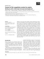
changes in absorbance at 540 nm. Figure 1A shows the time
course of AIIt-mediated liposome aggregation in the
presence of various concentrations of GSNO. Figure 1B
depicts a dose-dependence of GSNO inhibition of AIIt-
mediated liposome aggregation. The concentration effecting
50% inhibition (IC
50
) was approximately 100 l
M
.Ca
2+
(1 m
M
) and/or GSNO (2 m
M
) did not cause liposome
aggregation in the absence of AIIt under our assay
conditions. Sodium nitropresside (SNP), another NO donor
structurally different from GSNO, also inhibited AIIt-
mediated liposome aggregation (Fig. 1B) although the IC
50
(approximately 2 m
M
) was higher than that of GSNO. To
exclude the effect of the unreactive GSNO on liposome
aggregation, we removed these small molecules from
annexin II protein by gel filtration using a Biospin 6
chromatography column at the end of preincubation and
measured its liposome aggregation. We observed a similar
inhibition to these without column purification (data not
shown). Furthermore, GSH, nitrite and nitrate had no
effects (Fig. 2). GSNO inhibited AIIt-mediated liposome
aggregation at all the AIIt concentrations and all the Ca
2+
concentrations tested (Figs 3 and 4). At a higher concen-
tration of AIIt (10 lg) less inhibition was observed.
Effect of GSNO on AIIt-mediated membrane fusion
Although the mechanisms by which AIIt mediates mem-
brane fusion are still unclear, at least three steps are
Ó FEBS 2002 Annexin II and nitric oxide (Eur. J. Biochem. 269) 4279
involved: (a) the binding of AIIt to membrane; (b)
membrane aggregation and (c) membrane fusion. We have
previously shown that AIIt promotes the fusion of
liposomes with lamellar bodies, the secretory granules of
lung alveolar type II cells [14,22]. We therefore examined
whether GSNO also blocks this process. Membrane fusion
was monitored by a lipid mixing assay [44]. The addition of
AIIt caused a rapid fusion of lamellar bodies with
liposomes. Pre-treatment of AIIt with 0.1 and 1 m
M
GSNO
resulted in 70 ± 4% and 83 ± 10% inhibition of AIIt-
mediated membrane fusion, respectively (Fig. 5). It was
noted that AIIt-mediated membrane fusion was more
sensitive to GSNO compared to the membrane aggregation.
This is probably because GSNO not only affects the
membrane aggregation step, but also the membrane fusion
step.
Fig. 1. NO donors inhibit annexin II tetramer (AIIt)–mediated liposome aggregation in a dose-dependent fashion. Purified AIIt (5 lg) was incubated in
50 lLof40m
M
Hepes (pH 7.4) buffer containing varying concentrations of S-nitrosoglutathione (GSNO) or sodium nitroprusside (SNP) at room
temperature for 30 min. Liposome aggregation activity was measured by monitoring the turbidity change (A
540nm
). The aggregation assay was carried
out in 1 mL of Ca
2+
-EGTA buffer (40 m
M
Hepes, pH 7.0, 100 m
M
KCl, 1 m
M
EGTA, and 2 m
M
Ca
2+
) containing 100 lg phosphatidylserine
liposomes. AIIt was used to initiate liposome aggregation. (A) A representative time course curve of AIIt-mediated liposme aggregation in the
presence of various concentrations of GSNO. (d)0m
M
(j) 0.001 m
M
(m)0.1 m
M
(.)1m
M
(r)10m
M
. (B) Dose-dependence of NO donor-
mediated inhibition of AIIt-mediated liposome aggregation. The activity was expressed as the increase in absorbance at 540 nm after a 30 min
incubation over the initial value. The results were expressed as percentage control. The control was treated the same way as other samples except that
no other reagents were added. The data shown are mean ± SE from three experiments (GSNO, d) or mean from two experiments (SNP, j).
Fig. 2. Sodium nitroprusside and S-nitrosoglutathione inhibit AIIt-
mediated liposome aggregation, whereas reduced glutathione, nitrate or
nitrite has little effect. AIIt (5 lg) was incubated with sodium nitro-
prusside (SNP, 2 m
M
), S-nitrosoglutathione (GSNO, 2 m
M
), reduced
glutathione (GSH, 2 m
M
), nitrate (2 m
M
) or nitrite (2 m
M
)in50lLof
40 m
M
Hepes buffer (pH 7.4) for 30 min. The treated AIIt was tested
for its ability to mediate liposome aggregation. The results were
expressed as a percentage of the control. The control was treated the
same way as other samples except that no other reagents were added.
The data shown are mean ± SE from three experiments. wP <0.05
vs. control.
Fig. 3. A dose-dependence of AIIt-mediated liposome aggregation in the
presence or absence of GSNO. Various concentrations of AIIt
(0–10 lg) were incubated with or without 2 m
M
GSNO in 50 lLof
40 m
M
Hepes buffer (pH 7.4) for 30 min. Liposome aggregation was
determined and expressed as the increase in absorbance at 540 nm
after a 30 min incubation over the initial value.
4280 L. Liu et al.(Eur. J. Biochem. 269) Ó FEBS 2002
Effect of GSNO on the binding of AIIt to membrane
As the binding of AIIt to membrane would be the
first step of AIIt-mediated membrane fusion, we also
investigated whether GSNO inhibits the binding of AIIt
to liposomes. We treated AIIt with GSNO for 30 min
and mixed the samples with liposomes and 1 m
M
Ca
2+
.
After a 30 min incubation, we pelleted liposomes by
centrifugation and analyzed AIIt associated with lipo-
somes on 10% SDS/PAGE. As shown in Fig. 6A,
GSNO had no effect on the amount of AIIt associated
with liposomes. In the absence of Ca
2+
, little AIIt was
bound to liposomes. The results indicate that the
modification of annexin II by GSNO does not affect
the binding of AIIt to membrane. When different
amounts of AIIt were treated with GSNO, no inhibition
were observed for the binding of AIIt to liposmes at all
the AIIt concentrations tested (Fig. 6B).
Effect of GSNO on liposome aggregation mediated
by annexins I, III and IV
Annexins are a large gene family. In mammals, so far, 12
members have been identified and in other organisms
more than 60 and over 200 isoforms [47]. Annexins share
common structural features and some biochemical pro-
perties. All annexins bind to phospholipids in the
presence of Ca
2+
. Some annexins (I, II, III, IV and
VII) are able to mediate liposome aggregation although
their Ca
2+
sensitivities differ [22]. We therefore examined
whether GSNO inhibits liposome aggregation mediated
by various annexins. As shown in Fig. 7, GSNO only
has a modest effect on liposome aggregation mediated by
other annexins.
Effect of Ca
2+
and phospholipid on GSNO inhibition
Ca
2+
causes protein conformational changes in annexin II
[48] and may alter the environment of reactive sites of
annexin II by GSNO. We tested whether such changes
influence the GSNO inhibition of the activity of
annexin II. AIIt (5 lg)waspreincubatedin50lLof
buffer containing 1 m
M
Ca
2+
for 30 min to induce
protein conformational changes. The mixture was then
added to 1 mL of the assay buffer containing 100 lg
liposome for measuring liposome aggregation. As shown
in Fig. 8 (two bars with minus liposome during the
preincubation), a similar inhibition was observed when
AIIt was pretreated with EGTA or Ca
2+
, suggesting that
the conformational changes caused by Ca
2+
had no effect
on the GSNO inhibition.
After binding to the membrane, some residues in AIIt
may be hidden due to the polymerization of AIIt on the
membrane or a protein conformational change, and are
no longer accessible to GSNO. To test this possibility,
AIIt (5 lg) was preincubated with 50 lg liposomes in
50 lL of buffer containing 1 m
M
Ca
2+
to allow AIIt
binding to liposomes and then treated with 1 m
M
GSNO.
At the end of preincubation, the mixture was added to
1 mL of assay buffer containing 50 lg liposomes for
measuring liposome aggregation. As expected, GSNO still
inhibited AIIt-mediated liposome aggregation when no
Ca
2+
existed and thus AIIt did not bind to the membrane
during the preincubation. However, in the presence of
Ca
2+
and liposomes during the preincubation, AIIt was
bound to the membrane before the addition of GSNO. In
this case, no significant inhibition was observed (Fig. 8).
Fig. 4. Ca
2+
-dependence of AIIt-mediated liposome aggregation in the
presence or absence of GSNO. AIIt (5 lg) was incubated with or
without 2 m
M
GSNO in 50 lLof40m
M
Hepes buffer (pH 7.4) for
30 min. Liposome aggregation was measured in various concentra-
tions of Ca
2+
-EGTA buffer and expressed as the increase in absorb-
ance at 540 nm after a 30 min incubation over the initial value.
Fig. 5. GSNO inhibits AIIt-mediated fusion of lamellar bodies with
liposomes. AIIt (5 lg) was incubated with 0.1 m
M
or 1 m
M
GSNO in
50 lLof40m
M
Hepes(pH7.4)for30minandAIIt-mediated
membrane fusion was measured. Lipid (4 l
M
) in labeled liposomes
(PtdSer/PtdEtn/NBD-PtdEtn/Rh-PtdEtn, 24.5 : 74 : 0.75 : 0.75)
were mixed with 20 lgÆmL
)1
lamellar bodies in 0.5 mL Ca
2+
-EGTA
buffer (1 m
M
free Ca
2+
). After a stable baseline was established,
AIIt was added to initiate the reaction. Fusion was monitored by
following the increase in NBD-PtdEtn fluorescence (Ex ¼ 450 nm,
Em ¼ 530 nm). The data shown are a representative from three
experiments.
Ó FEBS 2002 Annexin II and nitric oxide (Eur. J. Biochem. 269) 4281
In an additional experiment, GSNO (1 m
M
)wasdirectly
added to liposome aggregation assay medium before the
addition of AIIt. Under these conditions, a 48% inhibition
was observed. However, if GSNO was added 5 min or
10 min after the addition of AIIt, less inhibition (26% or
17%) was seen (data not shown). Presumably, this is due
to the binding of AIIt to liposomes. These results indicate
that the reactive sites on AIIt were protected by the
binding of AIIt to the membrane.
Fig. 6. GSNO does not affect the binding of AIIt to liposomes. (A)AIItwasincubatedwithGSNO(1 m
M
) for 30 min. At the end of incubation, AIIt
was mixed with liposomes in the presence of 1 m
M
EGTA or Ca
2+
. After a 30 min incubation, liposomes were sedimented by centrifugation and
AIIt associated with liposomes was analyzed by 10% SDS/PAGE. The data shown are a representative from three experiments. (B) A dose
dependence of AIIt binding to liposomes in the presence or absence of GSNO. The conditions were the same as in the figure legend of Fig. 3. At the
end of the aggregation assay, liposomes were sedimented by centrifugation. AIIt associated with liposomes were analyzed by SDS/PAGE and
quantitated by densitometry. The results were expressed as the percentage of the maximal binding (i.e. 10 lg AIIt without GSNO).
Fig. 7. A dose-dependence of liposome aggregation mediated by ann-
exin I, III and IV in the presence or absence of GSNO. Various amounts
of annexins I, III and IV (0–10 lg) were incubated with or without
2m
M
GSNO for 30 min. Liposome aggregation was determined and
expressed as the increase in absorbance at 540 nm after a 30 min
incubation over the initial value. For the comparison, the results were
expressed as percentages of the maximal activity (i.e. 10 lg of annexins
without GSNO treatment). The latter values were 0.28, 0.32, and 0.23
for annexins I, III, and IV, respectively. (d)AI(s) AI + GSNO (.)
AIII (,) AIII + GSNO (j)AIV(h)AIV+GSNO.
Fig. 8. Ca
2+
-induced protein conformational change in AIIt has no
effect on GSNO inhibition of AIIt-mediated liposome aggregation.
However, GSNO does not inhibit AIIt-mediated liposome aggregation
once AIIt binds to membrane. For the first two bars, AIIt (5 lg) was
incubated with or without 1 m
M
GSNO in 50 lLof40m
M
Hepes
(pH 7.4) containing 1 m
M
EGTA or 1 m
M
Ca
2+
for 30 min. The
samples were added to 1 mL of the assay buffer containing 100 lg
liposome for aggregation activity determination. For the last two bars,
AIIt (5 lg)waspreincubatedwith50lg liposome in the presence or
absence of 1 m
M
GSNO in 50 lLof40 m
M
Hepes (pH 7.4) containing
1m
M
EGTA or Ca
2+
for30min.Thesampleswerethenaddedto
1 mL of assay buffer containing 50 lg liposome for aggregation
activity determination. The zero time absorbance was recorded sepa-
rately using 1 mL of the assay buffer containing 100 lg liposomes. The
results were expressed as percentage control (i.e. activity of GSNO-
treated AIIt/activity of untreated AIIt · 100%). The data shown are
mean ± SE from three experiments.
4282 L. Liu et al.(Eur. J. Biochem. 269) Ó FEBS 2002
Effect of dithiothreitol on GSNO inhibition
of AIIt-mediated liposome aggregation
To evaluate whether the GSNO inhibition of AIIt-mediated
liposome aggregation is involved in the formation of
disulfide bonds, we incubated purified AIIt (5 lg) with
GSNO (1 m
M
) in the presence of the reducing agent,
dithiothreitol. As shown in Fig. 9, when dithiothreitol
(0.5 m
M
) was included in the incubation medium, the
inhibition of AIIt-mediated liposome aggregation by
GSNO was no longer observed, suggesting that the
inactivation of AIIt may be due to the formation of
disulfide bridges. However, no intermolecular disulfide
bonds between annexin molecules were formed, because
when GSNO-treated AIIt was resolved on nonreduced
SDS/PAGE, no extra-bands were seen (data not shown).
However, we cannot rule out the possibility of disulfide
bond formation between AIIt and glutathione because of
the resolution of SDS/PAGE.
Conformational changes
To detect possible conformational changes of AIIt treated
with GSNO, we used the hydrophobic fluorescent probe,
bis-ANS. This dye binds to hydrophobic sites of proteins
and causes an increase of intensity in fluorescence with a
concomitant shift to the lower wavelength [49]. As expected,
the addition of AIIt to the bis-ANS aqueous solution, the
fluorescence increased and maximal emission wavelength
was shifted from 510 nm to 490 nm (data not shown).
Those changes are less, compared to annexin I [50]. GSNO-
treated AIIt had a similar increase of fluorescence and
wavelength shifts. GSNO itself had no effect on either
fluorescence or maximum wavelength. The results suggest
that GSNO does not cause a major conformational change
of AIIt as detected by the fluorescent dye, bis-ANS.
However, it is possible that the method used here may not
be able to detect small conformational changes.
DISCUSSION
Annexins are subjected to various post-translational modi-
fications. Although the phosphorylation of tyrosine or
serine/threonine residues in annexin has been extensively
studied, the relationship between other residues and annex-
in’s activity attracted less attention. When AIIt was treated
with N-ethylmaleimide, a sulfhydryl agent, AIIt’s activity
was reduced [33]. However, modification of annexin by
reactive nitrogen species has not been reported. Our recent
study has shown that AIIt can be nitrated by peroxynitrite
to form nitrotyrosine and such modification inhibited AIIt-
mediated liposome aggregation [38]. In the present study,
we, for the first time, showed that NO donors, GSNO and
SNP, also inhibit annexin II’s activities including membrane
aggregation and fusion. This modification was abolished in
the presence of dithiothreitol. Although physiological
significance of this in vitro observation remains to be
determined, it might imply a new post-translational modi-
fication and possibly a regulatory mechanism for annexin II
in cells. Recently, Fas-induced caspase-3 de-nitrosylation
was observed in lymphocyte cells, but the factors responsible
for the de- nitrosylation was not identified [51]. Because NO
inhibits surfactant secretion from alveoar type II cells [32]
and AIIt is a criticial component for the secretion of
lung surfactant in type II cells [14,22], NO inhibition of
AIIt’s activity may provide an alternative mechanism
of NO-mediated reduction of lung surfactant secretion.
Annexins including annexin II have also shown to be
associated with oxidative stress [52–56], NO modification
may also have implications in this process as well as other
biological activities of annexin II.
NO and its derivatives can attack protein targets involved
in many physiological processes and thus modifies their
functions. Interaction of NO with the heme or nonheme
iron of proteins leads to activation of soluble guanylyl
cyclase [57] and inactivation of cyclooxygenase [58] or
mitochondrial complexes I and II [59]. NO also regulates
protein functions by covalent attachment of the NO group
to cysteine residues in proteins via S-nitrosylation, which
may involve other nitrogen species such as NO
+
.Increasing
amounts of evidence demonstrate that this post-transla-
tional modification may represent an important cellular
regulatory mechanism [34–37]. Depending on different
proteins, S-nitrosylation may be followed by secondary
modification. For example, for glyceraldehyde-3-phosphate
dehydrogenase, S-nitrosylation of four active site cysteines
in the tetramer ultimately results in S-ADP-ribosylation and
inactivation [37]. If vicinal thiols in the protein are
S-nitrosylated, a more stable disulfide may be formed.
One of the examples is the N-methyl-
D
-asparate receptor
[60]. Our present study has shown that the GSNO inhibition
of annexin II-mediated liposome aggregation is no longer
observed in the presence of dithiothreitol, suggesting that,
most likely, the modification of annexin II’s activity by
Fig. 9. Effect of ditiothreitol on the inhibition of AIIt-mediated liposome
aggregation caused by GSNO. AIIt (5 lg) was incubated with 1 m
M
GSNO in the presence of the reducing agent, dithiothreitol (0, 0.1, 0.5,
1.0 m
M
). After a 30 min incubation, liposome aggregation activity was
determined. The results were expressed as a percentage of the control.
The control was treated as the same way as other samples except that
no GSNO and dithiothreitol was added. The data shown are
mean ± SE from three experiments.
Ó FEBS 2002 Annexin II and nitric oxide (Eur. J. Biochem. 269) 4283
GSNO is through S-transnitrosylation and the formation of
disulfide bond(s). As no dimers or polymers in GSNO-
treated AIIt were observed on nonreduced SDS/PAGE, the
disulfide bonds could be formed either within the AIIt
molecules or between annexin II thiol and GSNO [61].
UV and fluorescence studies of annexin II revealed a
Ca
2+
-induced conformational change in which the aroma-
tic amino acids, tyrosine and tryptophan, expose more to
the aqueous phase [48]. For annexin V, Ca
2+
causes
conformational changes in domain III that leads to the
formation of an additional Ca
2+
-binding site and exposure
of Trp187 to the solvent [62,63]. These conformational
changes appear not to affect the GSNO reaction with
annexin II, as a similar inhibition of AIIt-mediated lipo-
some aggregation by GSNO was observed whether AIIt
was pretreated with Ca
2+
or not prior to the liposome
aggregation assay. Probably, because Ca
2+
only induced a
modest conformational change circular dichroism studies
failed to detect major changes in secondary structure of
Ca
2+
-bound annexin II [48].
The present study indicated that GSNO no longer inhibits
AIIt-mediated liposome aggregation once the protein binds
to the membrane. This is consistent with the finding that
some annexins are accessible to quenchers in the solution
more than in the membrane-bound state [64]. There are
several possibilities: (a) after the binding of annexin II, the
reactive sites were hidden by membrane; (b) the binding of
AIIt to membrane causes a conformational change [65], such
changes may bury the reactive sites of GSNO more deeply in
the protein matrix therefore rendering them inaccessible to
GSNO; (c) annexins V and XII has been shown to form
trimers or hexamers on membrane [66,67]. We have previ-
ously shown that AIIt can self-associate in the presence of
Ca
2+
[23]. Therefore, it is possible that AIIt forms polymers
on membrane, thus hiding the reactive sites.
Nitrosothiols occur naturally in human plasma mainly as
the nitrosothiol of human serum albumin [68]. S-nitroso-
glutathione has been identified on normal airways [39] and
in neutrophils [69]. S-nitrothiol concentrations in inflamed
and transplanted lungs were much higher than normal
subjects. The half-life of GSNO in the lavage fluid is
approximately 3 h, much longer than NO [39]. Therefore,
GSNO may contribute to physiological and pathological
processes in the lung and GSNO regulation of annexin II
activity may be physiologically relevant.
ACKNOWLEDGEMENTS
This work was supported by US Public Health Service Grant NHLBI
HL-52146, OCAST HR01-093 and OAES (to L. L.). We thank Ms.
Dierra Davis and Ms Krista J. Schone for secretarial assistance.
REFERENCES
1. Creutz, C.E. (1992) The annexins and exocytosis. Science 258,
924–931.
2. Gerke, V. & Moss, S.E. (1997) Annexins and membrane
dynamics. Biochim. Biophys. Acta 1357, 129–154.
3. Raynal, P. & Pollard, H.B. (1994) Annexins: the problem of
assessing the biological role for a gene family of multifunctional
calcium- and phospholipid-binding proteins. Biochim. Biophys.
Acta 1197, 63–93.
4. Waisman, D.M. (1995) Annexin II tetramer: structure and func-
tion. Mol. Cell. Biochem. 149/150, 301–322.
5. Bandorowicz-Pikula, J., Buchet, R. & Pikula, S. (2001) Annexins
as nucleotide-binding proteins: facts and speculations. Bioessays
23, 170–178.
6. Kourie, J.I. & Wood, H.B. (2000) Biophysical and molecular
properties of annexin-formed channels. Prog. Biophys. Mol. Biol.
73, 91–134.
7. Gerke, V. & Moss, S.E. (2002) Annexins: from structure to
function. Physiol. Rev. 82, 331–371.
8. Hawkins, T.E., Roes, J., Rees, D., Monkhouse, J. & Moss, S.E.
(1999) Immunological development and cardiovascular function
are normal in annexin VI null mutant mice. Mol. Cell Biol. 19,
8028–8032.
9. Srivastava, M., Atwater, I., Glasman, M., Leighton, X., Goping,
G., Caohuy, H., Miller, G., Pichel, J., Westphal, H., Mears, D.,
Rojas, E. & Pollard, H.B. (1999) Defects in inositol 1,4,5-tripho-
sphate receptor expression, Ca(2+) signaling, and insulin secre-
tion in the anx7 (+/-) knockout mouse. Proc. Natl Acad. Sci. USA
96, 13783–13788.
10. Gunteski-Hamblin, A.M., Song, G., Walsh, R.A., Frenzke, M.,
Boivin, G.P., Dorn, G.W., Kaetzel, M.A., Horseman, N.D. &
Dedman, J.R. (1996) Annexin VI overexpression targeted to heart
alters cardiomyocyte function in transgenic mice. Am.J.Physiol.
270, H1091–H1100.
11. Rand, J.H. (2000) The annexinopathies: a new category of dis-
eases. Biochim. Biophys. Acta 1498, 169–173.
12. Drust, D.S. & Creutz, C.E. (1988) Aggregation of chromaffin
granules by calpactin at micromolar levels of calcium. Nature 331,
88–91.
13. Blackwood, R.A. & Ernst, J.D. (1990) Characterization of Ca
2+
-
dependent phospholipid binding, vesicle aggregation and mem-
brane fusion by annexins. Biochem. J. 266, 195–200.
14. Liu, L., Fisher, A.B. & Zimmerman, U.J.P. (1995) Lung annexin
II promotes fusion of isolated lamellar bodies with liposomes.
Biochim. Biophys. Acta 1259, 166–172.
15. Johnstone, S.A., Hubaishy, I. & Waisman, D.M. (1992) Phos-
phorylation of annexin II tetramer by protein kinase C inhibits
aggregation of lipid vesicles by the protein. J. Biol. Chem. 267,
25976–25981.
16. Hubaishy, I., Jones, P.G., Bjorge, J., Bellagamba, C., Fitzpatrick,
S., Fujita, D.J.X. & Waisman, D.M. (1995) Modulation of
annexin II tetramer by tyrosine phosphorylation. Biochemistry
34, 14527–14534.
17. Nakata, T., Sobue, K. & Hirokawa, N. (1990) Conformational
change and localization of calpactin I complex involved in exo-
cytosis as revealed by quick-freeze, deep-etch electron microscopy
and immunocytochemistry. J. Cell Biol. 110, 13–25.
18. Senda, T., Okabe, T., Matsuda, M. & Fujita, H. (1994) Quick-
freeze, deep-etch visualization of exocytosis in anterior pituitary
secretory cells: localization and possible roles of actin and annexin
II. Cell Tissue Res. 277, 51–60.
19. Ali, S.M., Geisow, M.J. & Burgoyne, R.D. (1989) A role for
calpactin in calcium-dependent exocytosis in adrenal chromaffin
cells. Nature 340, 313–315.
20. Konig, J., Prenen, J., Nilius, B. & Gerke, V. (1998) The
annexin II-p11 complex is involved in regulated exocytosis in
bovine pulmonary artery endothelial cells. J. Biol. Chem. 273,
19679–19684.
21. Liu, L., Tao, J.Q., Li, H.L. & Zimmerman, U.J.P. (1997) Inhibi-
tion of lung surfactant secretion from alveolar type II cells and
annexin II tetramer-mediated membrane fusion by phenothia-
zines. Arch. Biochem. Biophys. 342, 332–328.
22. Liu, L., Wang, M., Fisher, A.B. & Zimmerman, U.J.P. (1996)
Involvement of annexin II in exocytosis of lamellar bodies
from alveolar epithelial type II cells. Am.J.Physiol.270, L668–
L676.
23. Liu, L. (1999) Calcium-dependent self-association of annexin II:
a possible implication in exocytosis. Cell Signal 11, 317–324.
4284 L. Liu et al.(Eur. J. Biochem. 269) Ó FEBS 2002
24. Filipenko, N.R. & Waisman, D.M. (2001) The C-terminus of
annexin II mediates binding to F-actin. J. Biol. Chem. 276, 5310–
5315.
25. Eberhard, D.A., Karns, L.R., Vandenberg, S.R. & Creutz, C.E.
(2001) Control of the nuclear-cytoplasmic partitioning of annexin
II by a nuclear export signal and by p11 binding. J. Cell Sci. 114,
3155–3166.
26. Wright, J.F., Kurosky, A. & Wasi, S. (1994) An endothelial cell-
surface form of annexin II binds human cytomegalovirus.
Biochem. Biophys. Res. Commun. 198, 983–989.
27. Chung, C.Y. & Erickson, H.P. (1994) Cell surface annexin II is a
high affinity receptor for the alternatively spliced segment of
tenascin-C. J. Cell Biol. 126, 539–548.
28. Tsao, F.H., Chen, X. & Vu, V.X. (1994) Immunocharacterization
and developmental regulation of rabbit lung calcium-dependent
phospholipid-binding proteins. Biochim. Biophys. Acta 1213, 91–
99.
29. Kwon, M., Caplan, J.F., Filipenko, N.R., Choi, K.S., Fitzpatrick,
S.L., Zhang, L. & Waisman, D.M. (2002) Identification of annexin
II heterotetramer as a plasmin reductase. JBiolChem.277, 10903–
10911.
30. Bastian, B.C. (1997) Annexins in cancer and autoimmune diseases.
Cell Mol. Life Sci. 53, 554–556.
31. Hallman, M. & Bry, K. (1996) Nitric oxide and lung surfactant.
Semin. Perinatol. 20, 173–185.
32. Beckman, D.L., Mehta, P., Enright, E. & Liu, L. (1998) Nitric
oxide donor, spermine NONOate, modulate lung surfactant
secretion from cultured alveolar type II cells. FASEB J. 12, A491.
33. Singh, T.K. & Liu, L. (2000) Modification of cysteine residues by
N-ethylmaleimide inhibits annexin II tetramer mediated liposome
aggregation. Arch. Biochem. Biophys. 381, 235–240.
34. Michetti, M., Salamino, F., Melloni, E. & Pontremoli, S. (1995)
Reversible inactivation of calpain isoforms by nitric oxide.
Biochem. Biophys. Res. Commun. 207, 1009–1014.
35. Gopalakrishna, R., Chen, Z.H. & Gundimeda, U. (1993) Nitric
oxide and nitric oxide-generating agents induce a reversible
inactivation of protein kinase C activity and phorbol ester binding.
J. Biol. Chem. 268, 27180–27185.
36. Caselli, A., Camici, G., Manao, G., Moneti, G., Pazzagli, L.,
Cappugi, G. & Ramponi, G. (1994) Nitric oxide causes inactiva-
tion of the low molecular weight phosphotyrosine protein phos-
phatase. J. Biol. Chem. 269, 24878–24882.
37. Mohr, S., Stamler, J.S. & Brune, B. (1996) Post-translational
modification of glyceraldehyde-3-phosphate dehydrogenase by
S-nitrosylation and subsequent NADH attachment. J. Biol. Chem.
271, 4209–4214.
38. Rowan, W.H., Sun, P. & Liu, L. (2002) Nitration of annexin II
tetramer. Biochemistry 41, 1409–1420.
39. Gaston, B., Reilly, J., Drazen, J.M., Fackler, J., Ramdev, P.,
Arnelle, D., Mullins, M.E., Sugarbaker, D.J., Chee, C., Singel,
D.J., Loscalzo, J. & Stamler, J.S. (1993) Endogenous nitrogen
oxides and bronchodilator S-nitrosothiols in human airways.
Proc. Natl Acad. Sci. USA 90, 10957–10961.
40. Khanna, N.C., Helwig, E.D., Ikebuchi, N.W., Fitzpatrick, S.,
Bajwa, R.X. &. Waisman, D.M. (1990) Purification and char-
acterization of annexin proteins from bovine lung. Biochemistry
29, 4852–4862.
41. Hope, M.J., Bally, M.B., Webb, G. & Cullis, P.R. (1985) Pro-
duction of large unilamellar vesicles by a rapid extrusion proce-
dure, characterization of size distribution, trapped volume and
ability to maintain a membrane potential. Biochim. Biophys. Acta
812, 55–65.
42. Chander, A., Dodia, C.R., Gil, J. & Fisher, A.B. (1983) Isolation
of lamellar bodies from rat granular pneumocytes in primary
culture. Biochim. Biophys. Acta 753, 119–129.
43. Bers, D.M. (1982) A simple method for the accurate determination
of free [Ca] in Ca-EGTA solutions. Am.J.Physiol.242, C404–
C408.
44. Struck, D.K., Hoekstra, D. & Pagano, R.E. (1981) Use of
resonance energy transfer to monitor membrane fusion. Bio-
chemistry 20, 4093–4099.
45. Bradford, M.M. (1976) A rapid and sensitive method for the
quantitation of microgram quantities of protein utilizing the
principle of protein-dye binding. Anal. Biochem. 72, 248–254.
46. Laemmli, U.K. (1970) Cleavage of structural proteins during
the assembly of the head of bacteriophage T4. Nature 227, 680–
685.
47. Morgan, R.O. & Fernandez, M.P. (1997) Annexin gene structures
and molecular evolutionary genetics. Cell Mol. Life Sci. 53, 508–
515.
48. Gerke, V. & Weber, K. (1985) Calcium-dependent conformational
changes in the 36-kDa subunit of intestinal protein I related to the
cellular 36-kDa target of Rous sarcoma virus tyrosine kinase.
J. Biol. Chem. 260, 1688–1695.
49. Rosen, C.G. & Weber, G. (1969) Dimer formation from 1-amino-
8-naphthalenesulfonate catalyzed by bovine serum albumin. A
new fluorescent molecule with exceptional binding properties.
Biochemistry 8, 3915–3920.
50. Liu, L. & Zimmerman, U.J. (1995) An intramolecular disulfide
bond is essential for annexin I- mediated liposome aggregation.
Biochem. Mol. Biol. Int. 35, 345–350.
51. Mannick, J.B., Hausladen, A., Liu, L., Hess, D.T., Zeng, M.,
Miao, Q.X., Kane, L.S., Gow, A.J. & Stamler, J.S. (1999) Fas-
induced caspase denitrosylation. Science 284, 651–654.
52. Ammendola, R., Fiore, F., Esposito, F., Caserta, G., Mesuraca,
M., Russo, T. & Cimino, F. (1995) Differentially expressed
mRNAs as a consequence of oxidative stress in intact cells. FEBS
Lett. 371, 209–213.
53. Li, Y.D., Patel, J.M., Zhang, J. & Block, E.R. (1997) Over-
expression of plasma membrane annexin II in NO
2
-exposed
pulmonary artery endothelial cells. Free Radic. Biol. Med. 23,
120–126.
54. Hoyal, C.R., Thomas, A.P. & Forman, H.J. (1996) Hydroper-
oxide-induced increases in intracellular calcium due to annexin VI
translocation and inactivation of plasma membrane Ca
2+
-
ATPase. J. Biol. Chem. 271, 29205–29210.
55. Polla, B.S., Jacquier-Sarlin, M.R., Kantengwa, S., Mariethoz, E.,
Hennet, T., Russo-Marie, F. & Cossarizza, A. (1996) TNF alpha
alters mitochondrial membrane potential in L929 but not in TNF
alpha-resistant L929.12 cells: relationship with the expression of
stress proteins, annexin 1 and superoxide dismutase activity. Free
Radic. Res. 25, 125–131.
56. Sacre, S.M. & Moss, S.E. (2002) Intracellular localization of
endothelial cell annexins is differentially regulated by oxidative
stress. Exp. Cell Res. 274, 254–263.
57. Ignarro, L.J. (1989) Heme-dependent activation of soluble
guanylate cyclase by nitric oxide: regulation of enzyme activity by
porphyrins and metalloporphyrins. Semin. Hematol. 26, 63–76.
58. Kanner, J., Harel, S. & Granit, R. (1992) Nitric oxide, an inhibitor
of lipid oxidation by lipoxygenase, cyclooxygenase and
hemoglobin. Lipids 27, 46–49.
59. Drapier, J.C. & Hibbs, J.B.J. (1988) Differentiation of murine
macrophages to express nonspecific cytotoxicity for tumor cells
results in 1-arginine-dependent inhibition of mitochondrial iron-
sulfur enzymes in the macrophage effector cells. J. Immunol. 140,
2829–2838.
60. Lei, S.Z., Pan, Z.H., Aggarwal, S.K., Chen, H.S., Hartman, J.,
Sucher, N.J. & Lipton, S.A. (1992) Effect of nitric oxide produc-
tion on the redox modulatory site of the NMDA receptor-channel
complex. Neuron 8, 1087–1099.
Ó FEBS 2002 Annexin II and nitric oxide (Eur. J. Biochem. 269) 4285
61. Xian, M., Chen, X., Liu, Z., Wang, K. & Wang, P.G. (2000)
Inhibition of papain by S-nitrosothiols: formation of mixed
disulfides. J. Biol. Chem. 275, 20467–20473.
62. Concha, N.O., Head, J.F., Kaetzel, M.A., Dedman, J.R. &
Seaton, B.A. (1993) Rat annexin V crystal structure: Ca
2+
-
induced conformational changes. Science 261, 1321–1324.
63. Meers, P. & Mealy, T. (1993) Relationship between annexin V
tryptophan exposure, calcium, and phospholipid binding.
Biochemistry 32, 5411–5418.
64. Hofmann, A., Escherich, A., Lewit-Bentley, A., Benz, J.,
Raguenes-Nicol, C., Russo-Marie, F., Gerke, V., Moroder, L. &
Huber, R. (1998) Interactions of benzodiazepine derivatives with
annexins. J. Biol. Chem. 273, 2885–2894.
65. Pigault,C.,Follenius-Wund,A.Lux,B.&Gerard,D.(1990)A
fluorescence spectroscopy study of the calpactin I complex and its
subunits p11 and p36: calcium-dependent conformation changes.
Biochim. Biophys. Acta 1037, 106–114.
66. Concha, N.O., Head, J.F., Kaetzel, M.A., Dedman, J.R. &
Seaton, B.A. (1992) Annexin V forms calcium-dependent trimeric
units on phospholipid vesicles. FEBS Lett. 314, 159–162.
67. Luecke, H., Chang, B.T., Mailliard, W.S., Schlaepfer, D.D. &
Haigler, H.T. (1995) Crystal structure of the annexin XII hexamer
and implications for bilayer insertion. Nature 378, 512–515.
68. Stamler, J.S., Jaraki, O., Osborne, J., Simon, D.I., Keaney, J.,
Vita, J., Singel, D., Valeri, C.R. & Loscalzo, J. (1992) Nitric
oxide circulates in mammalian plasma primarily as an S-nitroso
adduct of serum albumin. Proc. Natl Acad. Sci. USA 89, 7674–
7677.
69. Clancy, R.M., Levartovsky, D., Leszczynska-Piziak, J., Yegu-
din, J. & Abramson, S.B. (1994) Nitric oxide reacts with
intracellular glutathione and activates the hexose monophosphate
shunt in human neutrophils: evidence for S-nitrosoglutathione as
a bioactive intermediary. Proc. Natl Acad. Sci. USA 91, 3680–
3684.
4286 L. Liu et al.(Eur. J. Biochem. 269) Ó FEBS 2002