Báo cáo khoa học: Supramolecular calsequestrin complex Protein–protein interactions in chronic low-frequency stimulated muscle, postnatal development and ageing pot
Bạn đang xem bản rút gọn của tài liệu. Xem và tải ngay bản đầy đủ của tài liệu tại đây (331.01 KB, 10 trang )
Supramolecular calsequestrin complex
Protein–protein interactions in chronic low-frequency stimulated muscle, postnatal
development and ageing
Louise Glover
1
, Sandra Quinn
1
, Michelle Ryan
1
, Dirk Pette
2
and Kay Ohlendieck
3
1
Department of Pharmacology, University College Dublin, Belfield, Ireland;
2
Fachbereich Biologie, Universita
¨
t Konstanz, Germany;
3
Department of Biology, National University of Ireland, Maynooth, Co. Kildare, Ireland
As recently demonstrated by overlay assays using calseque-
strin-peroxidase conjugates, the major 63 kDa Ca
2+
-bind-
ing protein of the sarcoplasmic reticulum forms complexes
with itself, and with junctin (26 kDa), triadin (94 kDa) and
the ryanodine receptor (560 kDa) [Glover, L., Culligan, K.,
Cala, S., Mulvey, C. & Ohlendieck, K. (2001) Biochim.
Biophys. Acta 1515, 120–132]. Here, we show that variations
in the relative abundance of these four central elements of
excitation–contraction coupling in different fiber types, and
during chronic electrostimulation-induced fiber type transi-
tions, are reflected by distinct alterations in the calsequestrin
overlay binding patterns. Comparative immunoblotting
with antibodies to markers of the junctional sarcoplasmic
reticulum, in combination with the calsequestrin overlay
binding patterns, confirmed a lower ryanodine receptor
expression in slow soleus muscle compared to fast fibers, and
revealed a drastic reduction of the RyR1 isoform in chronic
low-frequency stimulated tibialis anterior muscle. The fast-
to-slow transition process included a distinct reduction in
fast calsequestrin and triadin and a concomitant reduction in
calsequestrin binding to these sarcoplasmic reticulum ele-
ments. The calsequestrin-binding protein junctin was not
affected by the muscle transformation process. The increase
in calsequestrin and decrease in junctin expression during
postnatal development resulted in similar changes in the
intensity of binding of the calsequestrin conjugate to these
sarcoplasmic reticulum components. Aged skeletal muscle
fibers tended towards reduced protein interactions within the
calsequestrin complex. This agrees with the physiological
concept that the key regulators of Ca
2+
homeostasis exist in
a supramolecular membrane assembly and that protein–
protein interactions are affected by isoform shifting under-
lying the finely tuned adaptation of muscle fibers to changed
functional demands.
Keywords: calsequestrin; calcium homeostasis; chronic low-
frequency stimulation; excitation–contraction coupling;
ryanodine receptor.
The physiological importance of direct protein–protein
interactions being involved in Ca
2+
-regulatory processes is
exemplified by a supramolecular triad membrane complex
mediating between sarcolemmal excitation and muscular
contraction [1]. It is well established that physical coupling
between the voltage-sensing dihydropyridine receptor and
the Ca
2+
-release channel provides the signal transduction
mechanism between the transverse tubules and the junc-
tional sarcoplasmic reticulum (SR) in mature skeletal
muscle fibers [2]. Conversely, it has not yet been determined
how many SR elements are involved in the regulation of the
contraction-inducing efflux of Ca
2+
-ions from the SR
lumen through the ryanodine receptor (RyR) complex, and
which components prevent passive disintegration of these
large heterogeneous SR membrane assemblies. Previous
studies on excitation–contraction coupling have established
that the RyR1 isoform of the Ca
2+
-release channel exists in
a close neighborhood relationship with various potential
regulators, such as triadin (TRI), junctin (JUN), JP-45,
JP-90, the histidine-rich Ca
2+
-binding protein, calsequestrin
(CSQ) and CSQ-like proteins [3].
Domain binding experiments [4], differential coimmuno
precipitation studies [5] and chemical crosslinking analysis
[6] indicate that the RyR of 560 kDa, TRI of 94 kDa, JUN
of 28 kDa and CSQ of 63 kDa form a tightly associated
complex in skeletal muscle membranes. TRI and CSQ
appear to function as endogenous regulators of the Ca
2+
-
release channel [7]. Thus, the high-capacity, low-affinity
Ca
2+
-binding element CSQ [8] and its larger isoforms of
150–220 kDa, termed CSQ-like proteins (CLPs) [9], do not
only represent the major Ca
2+
-reservoir complex within the
terminal cisternae region [10], but are also directly involved
in regulating ion fluxes [11]. The existence of a subpopula-
tion of CSQ within supramolecular SR complexes from
mature skeletal muscle fibers has recently been shown using
an optimized overlay technique [5]. Peroxidase-conjugated
CSQ clearly labelled itself [12] and its binding-protein JUN,
TRI and the RyR [5]. Protein-protein coupling between
CSQ and the other junctional elements could be modified by
detergent treatment, changes in Ca
2+
concentration, anti-
body adsorption and purified CSQ binding [5]. Based on
these findings showing a tightly associated junctional SR
complex providing the physiological basis of regulating
Correspondence to K. Ohlendieck, Department of Biology, National
University of Ireland, Maynooth, Co. Kildare, Ireland.
Fax: + 353 1 708 3845, Tel.: + 353 1 708 3842,
E-mail:
Abbreviations: CSQ, calsequestrin; CLP, calsequestrin-like protein;
IB, immuno blot; JUN, junctin; mAb, monoclonal antibody;
POD, peroxidase; RyR, ryanodine receptor; SR, sarcoplasmic
reticulum; TRI, triadin.
(Received 2 May 2002, revised 16 July 2002, accepted 1 August 2002)
Eur. J. Biochem. 269, 4607–4616 (2002) Ó FEBS 2002 doi:10.1046/j.1432-1033.2002.03160.x
excitation–contraction coupling, we extended our investi-
gation of CSQ complex formation on muscle tissues under
varying physiological conditions. A high degree of adapta-
bility to changed functional demands and a large regener-
ative capacity are intrinsic properties of differentiated
skeletal muscle fibers [13]. In addition, muscle fibers
undergo major molecular changes during development
[14] and ageing [15]. Major alterations in the relative
abundance and/or isoform expression pattern of Ca
2+
-
regulatory membrane proteins involved in excitation–con-
traction coupling are associated with these cell biological
changes. We therefore applied the CSQ overlay technique to
study complex formation in developing, transforming and
ageing skeletal muscle fibers.
Blot overlays are a technically challenging approach to
studying complex protein–protein interactions between
different elements of a heterogeneous membrane assembly.
Recently, we enhanced the sensitivity of detection [5,12],
which overcame the main obstacle of a previously unsuc-
cessful approach to determining high-molecular-mass CSQ-
binding elements [16]. However, due to the many steps
involved in this analytical procedure, the visualization of SR
proteins via a peroxidase (POD)-CSQ conjugate does not
achieve the same degree of linear signaling achieved by
Western blotting for example. At the current state of
optimization, the blot overlay technique represents a
semiquantitative tool, similar to immuno precipitation
analysis. Nevertheless, this does not limit the range of
potential biochemical applications of the blot overlay
method in determining protein linkage. Its greatest advant-
age is the direct visualization of protein–protein interactions
under controlled conditions. In contrast, other established
protein biochemical methods for the analysis of large
membrane complexes such as chemical crosslinking analysis
might introduce artifacts by random protein linkage.
Although gel filtration chromatography, domain binding
studies with recombinant or isolated peptide domains,
differential coimmuno precipitation or analytical ultra
centrifugation supply sophisticated data, they do not
directly illustrate protein interactions within supramolecular
complexes. In this regard, the analyses using the optimized
CSQ-POD overlay procedure presented in this study are an
excellent example of applying a direct decoration method to
studying heterogeneous membrane assemblies under differ-
ing biological conditions.
EXPERIMENTAL PROCEDURES
Animals
Skeletal muscle from young, adult and ageing New
Zealand white rabbits were obtained from the Biomedical
Facility, National University of Ireland, Dublin. The
relevant ages of the animals used were (d, days; y, years):
14d, 21d, 28d, 41d, 44d, 1.0y, and 2.4y after birth, whereby
the last age group represents the oldest rabbits commer-
cially available in Ireland. For evaluating potential varia-
tions in CSQ complex formation in different skeletal
muscle fiber types, psoas, gastrocnemius and soleus mus-
cles were dissected and separately prepared for the isolation
of microsomal membranes [17]. Chronic low-frequency
stimulated muscles were produced by tele-stimulation for 0,
5 and 78 days through the peroneal nerve of the left hind
limb of adult male rabbits in the Animal Facility of the
University of Konstanz [18].
Materials
Protease inhibitors, peroxidase-conjugated secondary anti-
bodies, and acrylamide stock solutions were obtained from
Boehringer Mannheim (Lewis, East Sussex, UK). Primary
antibodies were purchased from Affinity Bioreagents,
Golden, CO, USA (mAb VIIID1
2
to fast calsequestrin;
pAb to slow calsequestrin; mAb IIH11 to the fast SERCA1
isoform of the Ca
2+
-ATPase; mAb IID8 against the slow
SERCA2 isoform of the Ca
2+
-ATPase, and mAb IIG12 to
muscle triadin) and Upstate Biotechnology, Lake Placid,
NY, USA (pAB to the RyR1 isoform of the ryanodine
receptor Ca
2+
-release channel). Immobilon-P nitrocellulose
membranes were from Millipore Corporation (Bedford,
MA, USA). An affinity-purified polyclonal antibody to
junctin was a generous gift from Steve Cala (Wayne State
University, Detroit, MI, USA). The EZ-Link-Plus activated
peroxidase kits, Slide-A-Lyzer dialysis cassettes and chemi-
luminescence substrates were purchased from Perbio Sci-
ence UK Ltd. (Tattenhall, Cheshire, UK). All other
chemicals used were of analytical grade and purchased
from Sigma Chemical Company (Poole, Dorset, UK).
Membrane preparation
Microsomal membrane vesicles were isolated from rabbit
skeletal muscle homogenates by an established protocol at
0–4 °C in the presence of a protease inhibitor cocktail
(0.2 m
M
Pefabloc, 1.4 l
M
pepstatin A, 0.3 l
M
E-64, 1 l
M
leupeptin, 1 m
M
EDTA, and 0.5 l
M
soybean trypsin
inhibitor) [19]. Using bovine serum albumin as a standard,
the protein concentration of isolated membrane vesicles was
determined by the method of Bradford [20]. Following
isolation, membrane vesicles were immediately used for
electrophoretic separation, blot overlay assays and immu-
noblot analysis.
Gel electrophoresis and immunoblot analysis
SDS/PAGE under reducing conditions was carried out by
standard methodology [21] using 7% gels and 20 lgprotein
per lane [22]. Protein band patterns were visualized by
Coomassie Brilliant Blue or Silver staining. For blotting
experiments, separated microsomal muscle proteins were
electrophoretically transferred for 1 h at 100 V onto nitro-
cellulose membranes by the method of Towbin et al.[23].
Membrane blocking, incubation with primary antibodies,
washing steps, incubation with peroxidase-conjugated sec-
ondary antibodies, visualization of immuno-decorated pro-
tein bands and densitometric scanning of developed
immunoblots was carried out as described previously [24].
Calsequestrin blot overlay
Recently established optimum conditions were used for
CSQ blot overlay assays [5,12]. Skeletal muscle CSQ was
purified to homogeneity by Phenyl-Sepharose chromato-
graphy as described by Cala & Jones [25]. The purified SR
protein was conjugated to an amine-reactive marker enzyme
as described in the manufacturer’s instructions of the
4608 L. Glover et al. (Eur. J. Biochem. 269) Ó FEBS 2002
EZ-Link-Plus activated peroxidase kit. A Pierce Slide-A-
Lyzer dialysis cassette system was employed to remove
contaminates from the CSQ-POD conjugate. As previously
documented [5], homogeneity of the CSQ preparation and
successful POD conjugation was evaluated by silver staining
and immunoblotting of electrophoretically separated pro-
teins. Nitrocellulose replicas of protein gels were incubated
with the CSQ-POD complex overnight at room tempera-
ture. After several washes, decorated protein bands were
visualized by the enhanced chemiluminescence technique.
Densitometric scanning of developed overlay blots was
performed on a Molecular Dynamics 300S computing
densitometer (Sunnyvale, CA, USA) with
IMAGE QUANT
v3.0 software [22]. Protein identification by mass spectro-
scopy was performed with trypsin-digested protein samples
by J. Coffey (Micromass UK Ltd, Manchester, UK) using a
Q-Tof Ultima API/CapLC system and sequence similarities
determined with the
EXPASY
-
PEPTIDENT
program. N-Ter-
minal sequencing for protein band identification was carried
out by J. Fox, Alta Bioscience (School of Biosciences,
University of Birmingham, Edgbaston, UK) and sequence
similarities determined with the
BLAST
-
P
program.
RESULTS
To complement previous domain binding studies and
chemical crosslinking analyses of triadic protein–protein
interactions, we present here the analysis of CSQ interac-
tions with electrophoretically separated microsomal pro-
teins derived from developing, transforming and ageing
skeletal muscle fibers. Because CSQ represents the luminal
protein backbone regulating SR Ca
2+
buffering during the
excitation–contraction–relaxation cycle, a POD-conjugated
CSQ probe was employed to determine potential differences
in the overlay pattern under varying physiological condi-
tions. Following the identification of the purified SR protein
of apparent 63 kDa as calsequestrin (Fig. 1), the results
from our CSQ overlay analysis are presented with respect to
fiber types (Fig. 2), chronic low-frequency stimulation-
induced fiber transitions (Fig. 3), developing (Fig. 4) and
ageing skeletal muscles (Fig. 5).
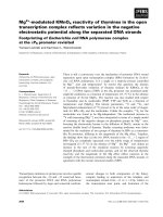
Identification of purified calsequestrin
To determine the CSQ-binding proteins, the purified status
of CSQ for usage as a POD-conjugated probe had to be
properly established. To unequivocally identify the protein
species purified by Phenyl-Sepharose chromatography from
the alkaline extracted microsomal fraction (Fig. 1A), three
independent methods were employed, i.e. immunoblotting
with monoclonal antibody VIIID1
2
to fast CSQ (Fig. 1B),
N-terminal sequencing (Fig. 1C) and mass spectroscopy of
trypsinated fragments (Fig. 1C). All three methods clearly
identified the 63 kDa SR protein species used for POD-
conjugation as rabbit fast skeletal muscle CSQ. Immuno-
blotting with a polyclonal antibody to the slow CSQ
isoform did not reveal a signal above background labeling
(Fig. 1B) demonstrating that the purified protein species
represents almost exclusively the fast isoform. Mass spec-
troscopical analysis revealed that the sequence of 11 tryspin
fragments of the protein band of approximately 63 kDa
matched 29% of the entire CSQ sequence (SwissProt
P07221) (Fig. 1C). Using N-terminal sequencing, this
finding was confirmed by a match of a 20 amino acid
stretch of sequence (EGLDFPEYDGVDRVINVNA) with
the primary structure of CSQ [26] (Fig. 1C). Successful
Fig. 1. Identification and conjugation of puri-
fied calsequestrin from rabbit skeletal muscle.
Shown is a silver-stained gel (A) of micro-
somes (MIC) (lane 1) and purified calseque-
strin (CSQ) (lane 2), and an immunoblot (B)
of purified CSQ prior (lanes 3 and 4) and after
(lane 5) conjugation to a peroxidase (POD)
marker. The blots have been immuno-
decorated with a polyclonal antibody to slow
CSQ (lane 3) and mAb VIIID1
2
to the fast
isoform of CSQ (lanes 4 and 5). The relative
positions of CSQ and CSQ-POD are marked
by arrow heads. Molecular mass standards
(in kDa) are indicated on the left. In (C) is
shown the primary sequence of CSQ with
capital letters marking the sequence deter-
mined by mass spectroscopy and the under-
lined sequence showing the peptide domain
determined by N-terminal sequencing.
Ó FEBS 2002 Supramolecular calsequestrin complex (Eur. J. Biochem. 269) 4609
conjugation of purified CSQ to the POD-marker was
demonstrated by a shift to a higher relative molecular mass,
as illustrated by the immunoblot analysis in Fig. 1B.
Sequence information by peptide sequencing or mass
spectroscopy for bands recognized by blot overlay did not
reveal sufficient data for proper databank searches (not
shown). Therefore, the identification of CSQ-decorated
bands described below was performed by immunoblotting
with established antibodies to triad markers.
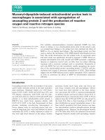
Calsequestrin complex formation in fast and slow
muscle fibers
In order to determine potential difference in CSQ complex
formation in slow vs. fast skeletal muscle fibers, the
electrophoretically separated protein complement of the
microsomal fraction derived from soleus, gastrocnemius
and psoas muscle homogenates was analysed by blot
overlay. Prior to comparative immunoblotting with junc-
tional SR markers and CSQ-POD binding, the fiber type-
specific differences of the preparations were established.
Although the Coomassie-stained gel representing the three
different muscles did not show any major differences in the
overall protein band pattern (with the exception of a low-
molecular-mass species in soleus) (Fig. 2A), immuno-dec-
oration with mAb IIH11 to the fast SERCA1 isoform of the
SR Ca
2+
-ATPase demonstrated the well established differ-
ence in slow vs. fast fiber distribution in soleus vs.
gastrocnemius and psoas muscles (Fig. 2B). The CSQ
overlay binding pattern showed a highly specific binding
pattern to four major protein species of 28, 63, 94 and
560 kDa in predominantly fast-twitching muscle (Fig. 2C).
The specificity of our newly developed CSQ-POD overlay
assay has been documented previously [5]. Incubation with
antibodies to CSQ, the ionic detergent SDS or the nonionic
detergent Triton X-100 eliminates these interactions (not
shown). Interestingly, the CSQ-POD probe exhibited only
very weak labeling of the 560 kDa band in soleus muscle
microsomes (Fig. 2C). This agrees with the reduced expres-
sion of the RyR1 isoform in slow-twitching muscle as
illustrated in the immunoblot analysis of Fig. 2D.
Due to the heterogeneous self-aggregation of triadin [27],
the 94 kDa band of the fast isoform is often accompanied
by high-molecular-mass bands in fast-twitch muscle
(Fig. 2E). The decreased relative density of fast triadin in
soleus muscle preparations (Fig. 2E) is partially reflected by
a reduced CSQ overlay signal (Fig. 2C). This result shows
both the strength and limitations of the overlay technique.
On the one hand, the CSQ-POD probe clearly labels the
main triad components forming the SR Ca
2+
-binding and
-release complex, but on the other hand changes in protein
concentration are only semiquantitatively revealed. Immu-
noblotting of fast CSQ and its binding-protein JUN showed
relatively similar levels in microsomal preparations derived
from predominantly fast- and slow-twitching muscle fibers
(Fig. 2F,G) and this is also reflected by the CSQ-POD
overlay pattern of these two SR proteins (Fig. 2C).
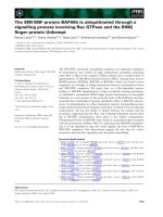
Calsequestrin complex formation in chronic low-
frequency stimulated muscle fibers
The isoform-specific expression of many SR proteins is
affected during fast-to-slow fiber transitions, including CSQ
[28]. We therefore studied the complex formation of this
terminal cisternae Ca
2+
-binding protein in chronic low-
frequency stimulated muscle fibers. During the fast-to-slow
transition process, a drastic decrease in the 110 kDa
protein band region was illustrated by Coomassie staining
of the electrophoretically separated microsomal fraction
(Fig. 3A). This protein species mostly represents the fast
Fig. 2. Calsequestrin complex formation in fast
and slow muscle fibers. Shown is a Coomassie-
stained gel (A) of microsomal preparations
and identical blots (B–G) labeled with anti-
bodies to the fast SERCA1 isoform of the
Ca
2+
-ATPase (B), the RyR1 isoform of the
Ca
2+
-release channel (D), triadin (TRI) (E),
fast calsequestrin (fCSQ) (F) and junctin
(JUN)(G).In(C)isshownablotoverlay
using a CSQ-POD probe. Lanes 1–3 represent
membrane vesicles derived from soleus (S),
gastrocnemius (G) and psoas (P) muscle
homogenates, respectively. The relative posi-
tions of immuno-decorated proteins are
marked by closed arrow heads and the protein
species recognized by the CSQ-POD overlay
technique are indicated by open arrow heads.
Molecular mass standards (in kDa) are indi-
cated on the left.
4610 L. Glover et al. (Eur. J. Biochem. 269) Ó FEBS 2002
SERCA1 isoform of the SR Ca
2+
-ATPase as revealed by
the drastic reduction of this fast-twitch marker following
chronic electro-stimulation (Fig. 3B). The switch between
the fast SERCA1 and slow SERCA2 (Fig. 3B,C), and the
exchange of the fast CSQ with the slow CSQ isoform
(Fig. 3E,F) agrees with previous studies [28] and clearly
documents a successful fiber transition. The CSQ-POD
overlay pattern showed some changes in the labeling
intensity of the apparent 94 and 560 kDa bands after
5 days of electro-stimulation, and a drastic decrease in the
decoration of the 63, 94 and 560 kDa bands after 78 days of
chronic low-frequency stimulation (Fig. 3D). The latter
finding agrees with the stimulation-induced reduction in the
fast isoforms of CSQ, TRI and the RyR1 (Fig. 3E,G,H).
The disproportionate weakening of immuno labeling of
the RyR1 band in stimulated muscle fibers is probably
due to a combination of factors, i.e. the existence of
proteolytic degradation products, heterogeneous aggregates
and/or an electrophoretic separation artifact often seen with
very large membrane proteins such as the Ca
2+
-release
channel. At high abundance the antibody to the RyR1
recognizes all separated RyR species (Fig. 3G, lane 1).
However, at reduced density, major RyR bands are
recognized (Fig. 3G, lane 2), but molecular species of lower
relative concentration are covered by other SR proteins with
a similar electrophoretic mobility and are thus not properly
recognized by the antibody. The appearance of a double
band pattern of immuno decorated TRI (Fig. 3H) is
probably due to the tight aggregation of this triadic
component. As has been previously documented [27], native
triadin exists as a disulfide-linked polymer and even under
reducing conditions these complexes do not completely
disintegrate. In Fig. 3H, the major protein band of apparent
94 kDa represents the monomeric TRI unit and this
molecule exhibits a dramatic reduction in its relative density
following electro-stimulation. In contrast, both the CSQ
binding to JUN and the relative concentration of JUN did
not decrease after 78 days of muscle fiber transformation
(Fig. 3D,I). A very interesting observation was the appar-
ent lack of interaction between the fast CSQ-containing
overlay probe and the slow CSQ band in 78 day stimulated
tibialis anterior microsomes (Fig. 3D,F). Possibly, fast and
slow CSQ isoforms exhibit different degrees of self-aggre-
gation and heterogeneous protein–protein interactions.
Slow CSQ might be involved in a more indirect type of
physiological coupling process in transformed fibers, while
fast CSQ appears to be a directly interacting endogenous
regulator of the Ca
2+
-release and Ca
2+
-cycling process in
fast muscle.
Calsequestrin complex formation in developing and
ageing muscle fibers
Because many Ca
2+
-regulatory proteins exhibit changes in
their isoform expression pattern and/or relative abundance
during postnatal myogenesis [29], we performed CSQ blot
overlay of 14- to 41-day-old-muscle preparations. Due to
the limited degree of differentiation during early myogenesis
it was not possible to prepare fiber-type specific microsomal
vesicles from developing muscle specimens. These analyses
were performed with mixed fiber populations. Blotting of
electrophoretically separated microsomes from 1, 3 and
7-day-old-rabbits did not reveal a sufficient signal-to-noise
ratio for proper comparative overlay and immunoblot
analysis (not shown). Shown are the data obtained with
muscle preparations from young animals before (14 day
old) and after (41 day old) maturation of the excitation–
contraction coupling mechanism [29]. Although the overall
protein band pattern is relatively similar during postnatal
development (Fig. 4A), immunoblotting clearly showed an
increase in the relative expression of RyR1, fast CSQ and
the fast SERCA1 isoform of the Ca
2+
-pump (Fig. 4C,E,F).
The double band pattern of the RyR1 protein species
(Fig. 4C) is probably due to the proteolytic degradation of
the Ca
2+
-release channel during membrane preparation.
Fig. 3. Calsequestrin complex formation in chronic low-frequency
stimulated muscle fibers. Shown is a Coomassie-stained gel (A) of
microsomal preparations and identical blots (B–I) labeled with anti-
bodies to the fast SERCA1 isoform of the Ca
2+
-ATPase (B), the slow
SERCA2 isoform of the Ca
2+
-ATPase (C), the fast CSQ isoform (E),
the slow/cardiac CSQ isoform (F), the RyR1 isoform of the Ca
2+
-
release channel (G), triadin (TRI) (H), and junctin (JUN) (I). In (D) is
shown a blot overlay using a CSQ-POD probe. Lanes 1–3 represent
membrane vesicles derived from unstimulated control, 5 day and
78 day chronic low-frequency (10 Hz) stimulated muscle, respectively.
The relative positions of a 110 kDa Coomassie-stained band (A) and
immuno-decorated proteins (B–I) are marked by closed arrow heads
and the protein species recognized by the CSQ-POD overlay technique
are indicated by open arrow heads. Molecular mass standards (in kDa)
are indicated on the left.
Ó FEBS 2002 Supramolecular calsequestrin complex (Eur. J. Biochem. 269) 4611
Even in the presence of a protease inhibitor cocktail, a
certain degree of degradation occurs with large proteins,
probably because of the high Ca
2+
levels in muscle
homogenates. In contrast to the other triad markers, the
expression of JUN was greatly reduced during myogenesis
(Fig. 4D). The changes in the relative density of the four SR
elements studied were reflected by a modified CSQ-POD
overlay pattern, which is especially striking for the reduced
interactions between JUN and CSQ (Fig. 4B).
One of the key elements of excitation–contraction
coupling, the voltage-sensing dihydropyridine receptor, is
believed to play a key pathophysiological role in sarcopenia,
the age-related functional decline of skeletal muscle [30]. It
was therefore of interest to determine whether CSQ complex
formation is modified during pathophysiological down-
stream events of muscle ageing. Silver staining of electroph-
oretically separated microsomal proteins from aged muscle
showed an increase of the 110 kDa SR protein band
(Fig. 5A), but otherwise exhibited no major changes in the
protein band pattern. Although the immunoblot analysis of
the RyR1, CSQ and JUN did not reveal drastic changes in
their expression during ageing (Fig. 5C–E), the CSQ-POD
overlay showed a tendency of reduced linkage of CSQ to
TRI, JUN and the RyR1 in senescent fibers (Fig. 5B).
Immunoblotting of TRI in both ageing and developing
microsomes showed weak and broad labeling patterns (not
shown), probably due to high-molecular-mass isoforms [27],
and the analysis of this triad marker could thus not be
further pursued.
DISCUSSION
CSQ of apparent 63 kDa and its isoforms of higher relative
molecular mass play a central role in Ca
2+
-cycling through
the SR lumen [10]. The results of our CSQ overlay analysis
of microsomal membrane proteins isolated from varying
Fig. 4. Calsequestrin complex formation in
developing muscle fibers. Shown is a silver-
stained gel (A) of microsomal preparations
and identical blots (B–F) labeled with anti-
bodies to the RyR1 isoform of the Ca
2+
-
release channel (C), junctin (JUN) (D), fast
calsequestrin (fCSQ) (E), and the fast SER-
CA1 isoform of the Ca
2+
-ATPase (F). In (B)
is shown a blot overlay using a CSQ-POD
probe. Lanes 1–4 represent membrane vesicles
derived from 14-, 21-, 28- and 41-day-old
postnatal muscle, respectively. The relative
positions of immuno-decorated proteins are
marked by closed arrow heads and the protein
species recognized by the CSQ-POD overlay
technique are indicated by open arrow heads.
Molecular mass standards (in kDa) are indi-
cated on the left.
Fig. 5. Calsequestrin complex formation in ageing muscle fibers. Shown
is a silver-stained gel (A) of microsomal preparations and identical
blots (B–E) labeled with antibodies to the RyR1 isoform of the Ca
2+
-
release channel (C), fast calsequestrin (fCSQ) (D), and junctin (JUN)
(E). In (B) is shown a blot overlay using a CSQ-POD probe. Lanes 1–3
represent membrane vesicles derived from 44-day-, 1-year- and 2.4-
year-old-muscle, respectively. The relative positions of immuno-dec-
orated proteins are marked by closed arrow heads and the protein
species recognized by the CSQ-POD overlay technique are indicated
by open arrow heads. Molecular mass standards (in kDa) are indicated
on the left.
4612 L. Glover et al. (Eur. J. Biochem. 269) Ó FEBS 2002
fiber types, developing muscle, transforming fibers and
ageing muscle (as summarized in Fig. 6) agrees with the
concept that this ion-buffering SR element exists in a
supramolecular complex. Clusters of negatively charged
residues in the carboxy-terminal region of CSQ represent
Ca
2+
-binding domains [8,26], whereby CSQ oligomeriza-
tion is associated with positive co-operativity with respect to
high capacity Ca
2+
-binding [31]. CSQ aggregation and
solubilization cycles seem to be intrinsically linked to the
Ca
2+
-uptake and -release mechanism of the skeletal muscle
SR [32]. The results presented here suggest that protein–
protein interactions between CSQ and the RyR, TRI, JUN
and itself are important for regulating overall SR Ca
2+
-
handling. A similar complex has previously been described
to exist in cardiac muscle fibers [33].
CSQ functions as the major Ca
2+
-reservoir element of
the SR lumen, but also acts as an endogenous regulator of
the RyR Ca
2+
-release units [11]. Many luminal proteins are
retained in the SR by expressing the carboxy-terminal
retrieval signal KDEL, but CSQ remains associated with the
terminal cisternae region without this mechanism [34].
Interestingly, deletion of its carboxy-terminal domain,
phosphorylation sites or post-translational glycosylation
does not affect the proper targeting of CSQ [35–37]. Thus
self-aggregation and tight anchoring to other SR elements,
as demonstrated in this study by blot overlay analysis,
possibly prevent a high degree of heterogeneous CSQ
distribution and mechanisms other than the KDEL signal
are responsible for continuous recycling from the Golgi
complex [34].
That mature motor units retain a high capacity of
plasticity and that the neuron-specific impulse pattern exerts
a critical phenotypic influence on fibers are generally
accepted concepts of modern muscle biology [38]. Adult
skeletal muscle fibers are not static entities with inalterable
contractile properties, but represent extremely versatile
biological entities with a high capacity to transform into
faster or slower twitching units. Terminally differentiated
skeletal muscle fibers may undergo fast-to-slow transitions
induced by changes in mechanical loading, neuromuscular
activity or hormonal influence. Especially well established
are changes in elements of the contractile apparatus such as
troponin isoforms, and myosin light and heavy chains [38].
However, the enormous functional, metabolic and struc-
tural diversity of muscle fibers is not only reflected on the
molecular level by the diversity in myosin isoforms, but also
encompasses many ion-regulatory proteins.
Because fiber type-specific isoform expression patterns
exist for key Ca
2+
-regulatory proteins [39], it is not
surprising that changes in fiber type composition also
influences the abundance and/or isoform expression of
excitation–contraction coupling elements as demonstrated
in this study. With respect to understanding the molecular
changes associated with muscle transition, the finding that
SR complex formation is drastically reduced after chronic
low-frequency stimulation is extremely interesting. Com-
pared to the CSQ-POD overlay pattern in soleus micro-
somes, the long-term electro-stimulated muscle preparations
exhibited a much more pronounced decrease in coupling
between CSQ and TRI. This agrees with the physiological
concept that chronic electro-stimulation induces major
adaptive responses of Ca
2+
-handling proteins in muscle
fibers undergoing phenotypic changes and suggests that
transformed fibers might exhibit a more cardiac-like Ca
2+
-
induced Ca
2+
-release mechanism [40].
Numerous muscle proteins proceed through isoform
transitions during myogenesis. Ca
2+
-regulatory membrane
proteins are detectable relatively early in prenatal myogen-
esis [41]. Probably the same myogenic differentiation
program that controls the up-regulation of contractile
proteins [42] is also responsible for the initiation of the
expression of voltage sensors, Ca
2+
-reservoir elements,
Ca
2+
-release units and Ca
2+
-uptake pumps in developing
fibers [43]. During the first weeks after birth, the functional
maturation of the elements regulating the excitation–
contraction–relaxation cycle occurs whereby the transverse
tubular dihydropyridine receptor complex and the SR RyR
units show temporal differences in their developmental
induction during myogenesis [44]. Our immunoblot analysis
of developing fibers agrees with this concept and showed
that the expression of fast isoforms of the Ca
2+
-release and
-reservoir complex clearly increase at later stages of
postnatal myogenesis. Previous biochemical studies on
potential changes in triad components during postnatal
Fig. 6. Calsequestrin complex formation dur-
ing postnatal myogenesis, fiber transitions and
ageing. Summarized are the findings of the
comparative immunoblot and CSQ-POD
overlay analysis presented in this study. A
change in ryanodine receptor (RyR), calse-
questrin (CSQ) and junctin (JUN) expression,
and triad complex formation (TCF) is indi-
cated by the following symbols: m,increase;
, decrease; s, no major change. Listed are
modifications of the CSQ-containing supra-
molecular triad complex during postnatal
myogenesis, stimulation-induced fiber transi-
tions and the ageing process.
Ó FEBS 2002 Supramolecular calsequestrin complex (Eur. J. Biochem. 269) 4613
myogenesis demonstrated increased expression of fast
isoforms of CSQ, sarcalumenin, the Ca
2+
-ATPase and
the a
1
-dihydropyridine receptor and showed a greater
tendency of Ca
2+
-regulatory proteins to oligomerize in
adult muscle fibers as compared to early postnatal stages
[29]. Hence, during postnatal development protein–protein
interactions within triad junctions become more complex
andoligomerizationappearstobeanessentialprerequisite
for proper physiological functioning of key membrane
proteins in matured skeletal muscle fibers.
Takekura et al. [45] suggest that the induction process for
the molecular differentiation and structural organization of
the triad junction can be divided into three main events.
After membrane docking between the transverse tubular
membrane system and the SR, the RyR Ca
2+
-release units
are incorporated into the junctions and membrane cou-
plings are positioned at the I-A band interface, and the
process is completed by the transverse orientation of
dihydropyridine receptor-containing membrane domains
[45]. The CSQ-POD overlay analysis presented in this study
indicates that within 6 weeks of postnatal development the
proper physical coupling within the supramolecular SR
Ca
2+
-release complex units has occurred. Especially inter-
esting is the apparent lack of coupling between the CSQ-
POD probe and slow CSQ after chronic electro-stimulation.
Perhaps cardiac/slow CSQ does not form as tightly a
terminal cisternae aggregate for Ca
2+
-binding in the SR
lumen as is apparently present in fast-twitching fibers.
With the advancement of age, skeletal muscle fibers
undergo many structural and functional changes. Promin-
ent biological features of cellular decline are abnormal
metabolism, impaired bioenergetics and ion homeostasis,
and a loss of muscle mass due to fiber atrophy [46].
Pathophysiological alterations in the capacity to maintain
normal Ca
2+
-homeostasis and the functional impairment
of excitation–contraction coupling appear to be major
factors triggering senescent muscle fiber weakness. Both,
pharmacological binding studies and immunoblotting have
clearly shown a drastic decline in the voltage-sensing
a
1
-subunit of the DHPR complex [30,47]. Here, we can
show that aged muscle fibers also exhibit a tendency
towards reduced SR complex formation. Thus, uncoupling
between the voltage sensor and Ca
2+
-release channel units,
in conjunction with altered turnover of key Ca
2+
-regulatory
SR membrane proteins [48] and reduced protein coupling,
might play an important role in sarcopenia. Abnormal
voltage-sensing leads to a drastic reduction of the amount of
Ca
2+
-ions available for initiating mechanical responses in
aging fibers and therefore results in a reduced Ca
2+
-peak
transient [49]. As shown by our CSQ-POD overlay analysis
of senescent fibers, changes in protein interactions between
other SR Ca
2+
-regulatory proteins might also be involved
in triggering impaired triadic signal transduction resulting in
a progressive functional decline of skeletal muscles.
In conclusion, the four central elements of the signal
transduction mechanism at the junctional SR, the Ca
2+
-
binding protein CSQ, the RyR Ca
2+
-release channel, the
auxiliary triad element TRI and the CSQ-binding element
JUN, show decreased protein–protein interactions during
fiber type shifting and the ageing process. Reduced protein
coupling between the major elements regulating Ca
2+
-
homeostasis in long-term stimulated tibialis anterior fibers is
considerably more pronounced than in slow-twitch soleus
muscle. This supports the biochemical concept that the
Ca
2+
-mediated signal transduction process underlying
excitation–contraction coupling is regulated by tight direct
protein–protein interactions in fast fibers and via a more
cardiac-like Ca
2+
-induced Ca
2+
-release mechanism in
transformed fibers. Molecular interactions between triad
components are probably both of structural and functional
importance. This involves the initial formation of junctional
couplings and the maintenance of peripheral triad structures
by preventing passive disintegration of the Ca
2+
-release
complex. The major physiological function of the triad
complex is in mediating signal transduction at the triad
contact zones and regulating ion flux mechanism from the
SR lumen to the cytosol. It is not known whether only one
molecular hierarchy of successive protein coupling exists
during triad assembly and re-organizing, and whether only
two sets of factors act as positive and negative regulators of
the junctional Ca
2+
-release process. The results from the
blot overlay study presented in this study suggest a
molecular scenario of interdependence between the major
excitation–contraction coupling elements from skeletal
muscle. The initial triggering factor could be a change in
cytosolic Ca
2+
-levels. It has previously been established that
enhanced neuronal stimulation leads to a higher free Ca
2+
-
concentration in slower contracting fibers and that a
calcineurin-dependent transcriptional pathway controls
fiber type-specific expression patterns [50]. Changes in the
relative abundance of one particular triad marker, such as
TRI, might then result in reduced stabilization of the
interactions between the RyR1 isoform and auxiliary or
regulatory elements. This in turn may cause the disintegra-
tion of a tight triad complex and introduce the establish-
ment of a Ca
2+
-induced Ca
2+
-release mechanism lacking
direct physical coupling between the major excitation–
contraction coupling elements.
ACKNOWLEDGEMENTS
The authors thank Drs S. Cala, J. Coffey and J. Fox for providing our
lab with antibodies and protein identification technology. This study
was supported by project grant HRB-01/99 from the Irish Health
Research Board and research network grants from the European
Commission (QLRT-1999-02034; RTN2-2001-00337).
REFERENCES
1. Berchtold, M.W., Brinkmeier, H. & Muntener, M. (2000) Calcium
ion in skeletal muscle: its crucial role for muscle function, plasti-
city, and disease. Physiol. Rev. 80, 1215–1265.
2. Leong, P. & MacLennan, D.H. (1998) Complex interactions
between skeletal muscle ryanodine receptor and dihydropyridine
receptor proteins. Biochem. Cell. Biol. 76, 681–694.
3. Murray, B.E., Froemming, G.R., Maguire, P.B. & Ohlendieck, K.
(1998) Excitation-contraction-relaxation cycle: Role of Ca
2+
-
regulatory membrane proteins in normal, stimulated and patho-
logical skeletal muscle fibres. Int. J. Mol. Med. 1, 677–697.
4. Shin, D.W., Ma, J. & Kim, D.H. (2000) The asp-rich region at the
carboxyl-terminus of calsequestrin binds to Ca
2+
and interacts
with triadin. FEBS Lett. 486, 178–182.
5. Glover, L., Culligan, K., Cala, S., Mulvey, C. & Ohlendieck, K.
(2001) Calsequestrin binds to monomeric and complexed forms of
key calcium-handling proteins in native sarcoplasmic reticulum
membranes from rabbit skeletal muscle. Biochim. Biophys. Acta
1515, 120–132.
4614 L. Glover et al. (Eur. J. Biochem. 269) Ó FEBS 2002
6. Murray, B.E. & Ohlendieck, K. (1998) Complex formation
between calsequestrin and the ryanodine receptor in fast- and
slow-twitch skeltal muscle. FEBS Lett. 429, 317–322.
7. Ohkura, M., Furukawa, K., Fujimori, H., Kuruma, A., Kawano,
S., Hiraoka, M., Kuniyasu, A., Nakayama, H. & Ohizumi, Y.
(1998) Dual regulation of the skeletal muscle ryanodine receptor
by triadin and calsequestrin. Biochemistry 37, 12987–12993.
8. Wang, S., Trumble, W.R., Liao, H., Wesson, C.R., Dunker, A.K.
& Kang, C.H. (1998) Crystal structure of calsequestrin from rabbit
skeletal muscle sarcoplasmic reticulum. Nat. Struct. Biol. 5, 476–
483.
9. Cala, S.E., Scott, B.T. & Jones, L.R. (1990) Intralumenal sarco-
plasmic reticulum Ca
2+
-binding proteins. Sem. Cell Biol. 1,
265–275.
10. MacLennan, D.H. & Reithmeier, R.A. (1998) Ion tamers. Nat.
Struct. Biol. 5, 409–411.
11. Donoso, P., Beltran, M. & Hidalgo, C. (1996) Luminal pH
regulated calcium release kinetics in sarcoplasmic reticulum
vesicles. Biochemistry 35, 13419–13425.
12. Glover, L., Froemming, G. & Ohlendieck, K. (2001) Calsequestrin
blot overlay of two-dimensional electrophoretically separated
microsomal proteins from skeletal muscle. Anal. Biochem. 299,
268–271.
13. Pette, D. (2001) Historical Perspectives: plasticity of mammalian
skeletal muscle. J. Appl. Physiol. 90, 1119–1124.
14. Hauschka, S.A. (1994) The embryonic origin of muscle. In
Myology: Basic and Clinical (Engel, A.G. & Franzini-Armstrong,
C., eds), pp. 3–73. McGraw-Hill, Inc., New York.
15. Carmeli, E., Coleman, R. & Reznick, A.Z. (2002) The biochem-
istry of aging muscle. Exp. Gerontol. 37, 477–489.
16. Damiani, E. & Margreth, A. (1990) Specific protein–protein
interactions of calsequestrin with junctional sarcoplasmic reti-
culum of skeletal muscle. Biochem. Biophys. Res. Commun. 172,
1253–1259.
17. Froemming,G.R.,Pette,D.&Ohlendieck,K.(1999)The90kDa
junctional sarcoplasmic reticulum protein forms an integral part of
a supramolecular triad complex in skeletal muscle. Biochem.
Biophys. Res. Comm. 261, 603–609.
18. Hicks, A., Ohlendieck, K., Go
¨
pel, S.O. & Pette, D. (1997) Early
functional and biochemical adaptations to low-frequency stimu-
lation of rabbit fast-twitch muscle. Am.J.Physiol.273, C297–
C305.
19. Murray, B. & Ohlendieck, K. (1997) Crosslinking analysis of the
ryanodine receptor and a
1
-dihydropyridine receptor in rabbit
skeletal muscle triads. Biochem. J. 324, 689–696.
20. Bradford, M.M. (1976) A rapid and sensitive method for the
quantitation of microgram quantities of protein utilizing the
principle of protein-dye binding. Anal. Biochem. 72, 248–254.
21. Dunn, M.J. & Bradd, S.J. (1993) Separation and analysis of
membrane proteins by SDS-polyacrylamide gel electrophoresis.
Methods Mol. Biol. 19, 203–210.
22. Harmon,S.,Froemming,G.R.,Leisner,E.,Pette,D.&Ohlen-
dieck, K. (2001) Selected contribution: Low-frequency stimulation
of fast muscle affects the abundance of Ca
2+
-ATPase but not its
oligomeric status. J. Appl. Physiol. 90, 371–379.
23. Towbin,H.,Staehelin,T.&Gordon,J.(1979)Electrophoretic
transfer of proteins from polyacrylamide gels to nitrocellulose
sheets: procedure and some applications. Proc. Natl Acad. Sci.
USA 76, 4350–4354.
24. Culligan, K., Banville, N., Dowling, P. & Ohlendieck, K. (2002)
Drastic reduction of calsequestrin-like proteins and impaired cal-
cium binding in dystrophic mdx muscle. J. Appl. Physiol. 92, 435–
445.
25. Cala, S.E. & Jones, L.R. (1983) Rapid purification of calsequestrin
from cardiac and skeletal muscle sarcoplasmic reticulum vesicles
by Ca
2+
-dependent elution from phenyl-sepharose. J. Biol. Chem.
258, 11932–11936.
26. Fliegel, L., Ohnishi, M., Carpenter, M.R., Khanna, V.K.,
Reithmeier, R.A. & MacLennan, D.H. (1987) Amino acid
sequence of rabbit fast-twitch skeletal muscle calsequestrin
deduced from cDNA and peptide sequencing. Proc. Natl Acad.
Sci. USA 84, 1167–1171.
27. Froemming, G.R., Murray, B.E. & Ohlendieck, K. (1999) Self-
aggregation of triadin in the sarcoplasmic reticulum of rabbit
skeletal muscle. Biochim. Biophys. Acta 1418, 197–205.
28. Ohlendieck, K., Froemming, G.R., Murray, B.E., Maguire, P.B.,
Leisner,E.,Traub,I.&Pette,D.(1999)Effectsofchroniclow-
frequency stimulation on Ca
2+
-regulatory membrane proteins in
rabbit fast muscle. Pflu
¨
gers Arch. Eur. J. Physiol. 438, 700–708.
29. Froemming, G.R. & Ohlendieck, K. (1998) Oligomerisation of
Ca
2+
-regulatory membrane components involved in the excita-
tion-contraction-relaxation cycle during postnatal development of
rabbit skeletal muscle. Biochim. Biophys. Acta 1387, 226–238.
30. Ryan, M., Carlson, B.M. & Ohlendieck, K. (2000) Oligomeric
status of the dihydropyridine receptor in aged skeletal muscle.
Mol. Cell Biol. Res. Commun. 4, 224–229.
31. He, Z., Dunker, A.K., Wesson, C.R. & Trumble, W.R. (1993)
Ca
2+
-induced folding and aggregation of skeletal muscle sarco-
plasmic reticulum calsequestrin. The involvement of the trifluop-
erazine-binding site. J. Biol. Chem. 268, 24635–24641.
32. Tanaka,M.,Ozawa,T.,Maurer,A.,Cortese,J.D.&Fleischer,S.
(1986) Apparent cooperativity of Ca
2+
-binding associated with
crystallization of Ca
2+
-binding protein from sarcoplasmic reti-
culum. Arch. Biochem. Biophys. 251, 369–378.
33. Zhang,L.,Kelley,J.,Schmeisser,G.,Kobayashi,Y.M.&Jones,
L.R. (1997) Complex formation between junctin, triadin, calse-
questrin, and the ryanodine receptor. Proteins of the cardiac
junctional sarcoplasmic reticulum membrane. J. Biol. Chem. 272,
23389–23397.
34. Gatti, G., Trifari, S., Mesaeli, N., Parker, J.M., Michalak, M. &
Meldolesi, J. (2001) Head-to-tail oligomerization of calsequestrIn
a novel mechanism for heterogeneous distribution of endoplasmic
reticulum luminal proteins. J. Cell Biol. 154, 525–534.
35. Nori, A., Gola, E., Tosato, S., Cantini, M. & Volpe, P. (1999)
Targeting of calsequestrin to sarcoplasmic reticulum after dele-
tions of its acidic carboxy terminus. Am.J.Physiol.277, C974–
C981.
36. Nori, A., Furlan, S., Patiri, F., Cantini, M. & Volpe, P. (2000) Site-
directed mutagenesis and deletion of three phosphorylation sites of
calsequestrin of skeletal muscle sarcoplasmic reticulum. Effects on
intracellular targeting. Exp. Cell Res. 260, 40–49.
37. Nori, A., Valle, G., Massimino, M.L. & Volpe, P. (2001) Targeting
of calsequestrin to the sarcoplasmic reticulum of skeletal muscle
upon deletion of its glycosylation site. Exp. Cell Res. 265, 104–113.
38. Pette, D. & Staron, R.S. (2001) Transitions of muscle fiber
phenotypic profiles. Histochem. Cell Biol. 115, 359–372.
39.Froemming,G.R.,Murray,B.E.,Harmon,S.,Pette,D.&
Ohlendieck, K. (2000) Comparative analysis of the isoform
expression pattern of Ca
2+
-regulatory membrane proteins in fast-
twitch, slow-twitch, cardiac, neonatal and chronic low-frequency
stimulated muscle fibres. Biochim. Biophys. Acta. 1466, 151–168.
40. Ohlendieck, K. (2000) Changes in Ca
2+
-regulatory muscle
membrane proteins during the chronic low-frequency stimulation
induced fast-to-slow transition process. Bas. Appl. Myol. 10,
99–106.
41. Franzini-Armstrong, C. & Jorgensen, A.O. (1994) Structure and
development of E-C coupling units in skeletal muscle. Annu. Rev.
Physiol. 56, 509–534.
42. Buckingham, M. (2001) Skeletal muscle formation in vertebrates.
Curr. Opin. Genet. Dev. 11, 440–448.
43. Arai, M., Otsu, K., MacLennan, D.H. & Periasamy, M. (1992)
Regulation of sarcoplasmic reticulum gene expression during
cardiac and skeletal muscle development. Am.J.Physiol.262,
C614–C620.
Ó FEBS 2002 Supramolecular calsequestrin complex (Eur. J. Biochem. 269) 4615
44. Kyselovic, J., Leddy, J.J., Ray, A., Wigle, J. & Tuana, B.S. (1994)
Temporal differences in the induction of dihydropyridine receptor
subunits and ryanodine receptors during skeletal muscle devel-
opment. J. Biol. Chem. 269, 21770–21777.
45. Takekura, H., Flucher, B.E. & Franzini-Armstrong, C. (2001)
Sequential docking, molecular differentiation, and positioning of
T-Tubule/SR junctions in developing mouse skeletal muscle. Dev.
Biol. 239, 204–214.
46. Navarro, A., Lopez-Cepero, J.M. & Sanchez del Pino, M.J. (2001)
Skeletal muscle and aging. Front Biosci. 6, D26–D44.
47. Renganathan, M., Messi, M.L. & Delbono, O. (1997) Dihydro-
pyridine receptor-ryanodine receptor uncoupling in aged skeletal
muscle. J. Membr. Biol. 157, 247–253.
48.Ferrington,D.A.,Krainev,A.G.&Bigelow,D.J.(1998)
Altered turnover of calcium regulatory proteins of the sarco-
plasmic reticulum in aged skeletal muscle. J. Biol. Chem. 273,
5885–5891.
49. Delbono, O., O’Rourke, K.S. & Ettinger, W.H. (1995) Excitation-
calcium release uncoupling in aged single human skeletal muscle
fibers. J. Membr. Biol. 148, 211–222.
50. Chin, E.R., Olson, E.N., Richardson, J.A., Yang, Q., Humphries,
C., Shelton, J.M., Wu, H., Zhu, W., Bassel-Duby, R. &
Williams, R.S. (1998) A calcineurin-dependent transcriptional
pathway controls skeletal muscle fiber type. Genes Dev. 12,
2499–2509.
4616 L. Glover et al. (Eur. J. Biochem. 269) Ó FEBS 2002