Báo cáo khoa học: Isolation and enzymatic characterization of lamjapin, the first ribosome-inactivating protein from cryptogamic algal plant (Laminaria japonica A) ppt
Bạn đang xem bản rút gọn của tài liệu. Xem và tải ngay bản đầy đủ của tài liệu tại đây (258.57 KB, 7 trang )
Isolation and enzymatic characterization of lamjapin, the first
ribosome-inactivating protein from cryptogamic algal plant
(
Laminaria japonica
A)
Ren-shui Liu
1
, Jia-hua Yang
2
and Wang-Yi Liu
1
1
State Key Laboratory of Molecular Biology, Institute of Biochemistry and Cell Biology, Shanghai Institutes for
Biological Sciences, Chinese Academy of Sciences, China;
2
Department of Biochemistry, Yantai University, Shandong, China
Lamjapin,anoveltypeI ribosome-inactivating protein, has
been isolated from kelp (Laminaria japonica A), a marine
alga. This protein has been extensively purified through
multiple chromatography columns. With a molecular mass
of 36 kDa, lamjapin is slightly larger than the other
known single-chain ribosome-inactivating proteins from the
higher plants. Lamjapin can inhibit protein synthesis in
rabbit reticulocyte lysate with an IC
50
of 0.69 n
M
.Itcan
depurinate at multiple sites of RNA in rat ribosome and
produce the diagnostic R-fragment and three additional
larger fragments after the aniline reaction. Lamjapin can
deadenylate specifically at the site A20 of the synthetic oli-
goribonucleotide (35-mer) substrate that mimics the sarcin/
ricin domain (SRD) of rat ribosomal 28S RNA. However, it
cannot hydrolyze the N-C glycosidic bond of guanosine,
cytidine or uridine at the corresponding site of the A20 of
three mutant SRD RNAs. Lamjapin exhibits the same base
and position requirement as the ribosome-inactivating pro-
teins from higher plants. We conclude that lamjapin is an
RNA N-glycosidase that belongs to the ribosome-inacti-
vating protein family. This study reports for the first time
that ribosome-inactivating protein exists in the lower cryp-
togamic algal plant.
Keywords: kelp; lamjapin; marine alga; ribosome-inacti-
vating protein; RNA N-glycosidase.
Plant ribosome-inactivating proteins (RIPs) are a group of
toxic proteins with RNA N-glycosidase activity that act on
the eukaryotic and prokaryotic ribosomes. It was shown
that RNA N-glycosidase activity of RIP could remove a
specific adenine from a highly conserved loop (the sarcin/
ricin domain; SRD) of the largest RNA in ribosome and
thus inhibit the protein synthesis [1,2]. Type I RIPs consist
of a single, intact polypeptide chain of about 11–30 kDa.
Type II RIPs are composed of two chains linked by a
disulfide bond. Type III RIP consists of an amino-terminal
domain resembling type I RIP linked to a carboxyl-terminal
domain with unknown function [3].
This class of plant toxins has drawn much attention
because of their antiviral activity and the potential use as a
toxin moiety in immunotoxins for the treatment of several
important human diseases such as cancer and AIDS [4–6].
RIPs also have promising application in crop plant
biotechnology with the aim of increasing resistance to
insects, fungal and viral pathogens [7–9]. In addition, RIPs
are a powerful tool to probe the topographic structures of
ribosomal RNA and the mechanism of protein synthesis
[10]. Moreover, there is no consensus on the physiological
function, distribution and the evolutionary links of RIPs.
For all these reasons, the search for new RIPs is continuing,
and more novel RIPs are being isolated and characterized
from terrestrial flowering plants, while no RIP has been
isolated from cryptogamic plants so far [11–13]. In this
paper, lamjapin is shown to be the first single-chain RIP
from kelp, a lower cryptogamic algal plant. This result will
help to characterize the function, evolution, as well as the
distribution of this class of protein in plant kingdom.
MATERIALS AND METHODS
Materials
The fresh tender leaves of kelp (Laminaria japonica A) were
collected in winter at the shore of Yantai in the Shangdong
Province of China. CM-Cellulose 52 was purchased from
Whatman. Phenyl-Sepharose CL-4B, pI marker, ampho-
lyte, the FPLC system, Superose-12 and Mono Q columns
were obtained from Pharmacia LKB.
L
-[
14
C]leucine was
from Amersham. The protein molecular-mass markers were
provided by Shanghai Lizhu-dongfeng Biotech. Ultrafiltra-
tion membranes and centricons were purchased from
Amicon. T7 RNA polymerase, was obtained from Pro-
mega. [a-
32
P]UTP was from New England Nuclear. Oligo-
deoxynucleotides were synthesized at the Shanghai
GenecoreÒ Biotechnology Company. All the other chemi-
cals and reagents were of analytical grade.
Isolation and purification of lamjapin from kelp
The fresh tender leaves of kelp were freeze-dried and
powdered in liquid nitrogen. Thirty grams of the finely
ground material were extracted in 1000 mL of buffer A
Correspondence to W Y. Liu, State Key Laboratory of Molecular
Biology, Institute of Biochemistry and Cell Biology, Shanghai
Institutes for Biological Sciences, Chinese Academy of Sciences,
320 Yue-Yang Road, Shanghai 200031, China.
Fax: + 86 21 64348357, E-mail:
Abbreviations: RIP, ribosome-inactivating proteins; SRD, sarcin/ricin
domain.
(Received 28 May 2002, revised 22 July 2002, accepted 2 August 2002)
Eur. J. Biochem. 269, 4746–4752 (2002) Ó FEBS 2002 doi:10.1046/j.1432-1033.2002.03165.x
(50 m
M
Tris/HCl, pH 9.2, 0.2
M
NaCl, 10 m
M
ascorbic
acid) by gentle stirring at 8 °C overnight. The homogenate
was centrifuged (8000 g,4°C) for 30 min, then the
supernatant was decanted and filtered through four layers
of gauze. The filtrate was adjusted to pH 8.5 with 1
M
HCl
and solid ammonium sulfate was added to 1
M
. This fluid
was used as the crude extract from which lamjapin was
purified by the following four steps of column chromato-
graphy.
Phenyl-Sepharose CL-4B. The crude extract was applied
to the phenyl-Sepharose CL-4B column (10 · 4cm)pre-
equilibrated with buffer B [50 m
M
Tris/HCl, pH 8.5, 1
M
(NH
4
)
2
SO
4
and 0.2
M
NaCl]. After being washed with the
buffer B until the A
280
fell below 0.1, the column was eluted
with buffer C [50 m
M
Tris/HCl, pH 8.5, 0.4
M
(NH
4
)
2
SO
4
]
and buffer D [50 m
M
Tris/HCl, pH 8.5, 0.1
M
(NH
4
)
2
SO
4
]
sequentially; the protein in peak 2 was collected.
CM-Cellulose 52. The protein solution collected from
peak 2 of the phenyl-Sepharose CL-4B column was
dialyzed exhaustively against buffer E (5 m
M
phosphate
buffer, pH 6.0) and then loaded on a CM-Cellulose column
(10 · 2.4 cm) preequilibrated with the same buffer. After
loading the protein, the column was washed with 60 mL of
buffer E and then eluted with buffer E containing 0.15
M
sodium chloride. The protein in peak 1 was collected.
Superose-12 FPLC. After dialysis against distilled water,
the proteins in peak 1 from the CM-cellulose column were
lyophilized and dissolved in buffer F (50 m
M
phosphate
buffer, pH 7.2, 0.15
M
NaCl). The protein was then loaded
on a Superose12 HR (30 cm · 10 mm) column preequili-
brated with buffer F and eluted the column with buffer F.
Three peaks appeared and the protein in peak 2 was
collected for further purification.
Mono Q FPLC. The protein solution in peak 2 with RIP
activity from the Superose-12 column was desalted and
adjusted to 10 m
M
ethanolamine, pH 9.2 by repeated
concentration in an Amicon concentrator equipped with a
PM10 membrane. Then the protein solution was loaded on
toaFPLCMonoQHR5/5column(50· 1.6 mm)
preequilibrated with the buffer G (10 m
M
ethanolamine,
pH 9.2). A linear gradient elution with 40 mL of NaCl
solution (0–1
M
) in buffer G was performed. The purified
RIP in the peak 2 was collected and was named as lamjapin.
Protein synthesis in the cell-free system
Rabbit reticulocyte lysate was prepared and the protein
synthesis in rabbit reticulocyte lysate was performed
according to the methods of Sambrook et al. [14]. Various
amounts of lamjapin (0.25–8 ng) were added to the 50 lL
of reaction buffer to measure their inhibition of protein
synthesis.
Activity of lamjapin on rat ribosomes
Rat liver ribosomes were isolated as described by Spedding
[15]. One point five A
260
units of ribosomes (27 pmol) were
incubated with 20 or 30 ng of lamjapin in 100 lLofbuffer
H(25m
M
Tris/HCl, pH 7.6, 25 m
M
KCl, 5 m
M
MgCl
2
)at
37 °C for 15 min. After adding 10 lL of 10% SDS solution
to the reaction mixture in an ice-bath, ribosomal RNAs
were extracted with phenol/chloroform and precipitated by
ethanol. After acidic aniline treatment at 60 °C for 10 min,
ribosomal RNAs were separated on 8
M
urea-denatured
polyacrylamide gel (3.5%) for 1 h or on 8
M
urea-denatured
polyacrylamide gel (4.5%) for 3 h. The RNA fragments
were stained by methylene blue.
Fluorescence assay of the adenine released by lamjapin
Adenine was quantitated by the fluorimetric method of
Zamboni et al.[16].OneA
260
(18 pmol) of rat liver
ribosomes was incubated with 0.64 lgoflamjapinin
100 lL of buffer H at 37 °C for 0–40 min. After incubation,
ribosomal RNA was precipitated with 2 vol. of ethanol.
The ethanol-soluble fractions were diluted to 1 mL with
water, and then 0.4 mL of 0.14
M
chloroacetaldehyde
containing 0.1
M
sodium acetate (pH 5.1) was added to
each sample. The samples were incubated at 85 °Cfor1h.
After cooling to room temperature, fluorescence was
measured at an excitation wavelength of 280 nm and an
emission wavelength of 400 nm. Adenine at concentrations
ranging from 1 to 1200 pmol was used as a standard for
calculation of the amount of adenine in the samples.
Assay for the deadenylation of SRD RNA by lamjapin
RNA N-glycosidase activity of lamjapin was assayed
according to the method of Endo et al. [17] with a slight
modification. The radiolabeled wild type SRD RNA and its
three mutant SRD RNAs (G20, C20 and U20 instead of
A20) were prepared by in vitro transcription using T7 RNA
polymerase and synthetic DNA oligomers as template. The
radiolabeled SRD RNA and its three mutants were
incubated, respectively, with 1 l
M
lamjapin in 20 lLof
buffer I (30 m
M
sodium citrate, pH 5.0, 1 m
M
MgCl
2
)at
35 °C for 90 min and then treated with acid aniline. The
RNA fragments produced by lamjapin and aniline treat-
ment were extracted with phenol/chloroform and precipi-
tated by ethanol. After separating the RNA fragments by
electrophoresis on 20% polyacryamide gel containing 8
M
urea, gels were dried and exposed to X-ray film.
Assay of the release of adenine from the SRD RNA
by lamjapin
The preparation of the oligoribonucletides was as described
above except that the synthesis was with [2,8-
3
H] ATP
instead of ATP. The assay conditions were also the same.
The reaction was carried out in the same way except that,
after incubation, 10 lL of a solution of 0.2
M
NaCl
containing 100 lL of carrier tRNA and 40 lL of ethanol
was added to the sample and kept at °C for 60 min. The
mixture was centrifuged (15 000 g for 20 min) and the
radioactivity in a portion of the supernatant was determined
in a liquid scintillation counter.
Other analytical methods
SDS/PAGE analysis was carried out on 12% SDS-poly-
acryamide gels by the method of Laemmli [18] and protein
bands were stained with silver according to the method of
Ó FEBS 2002 Lamjapin, a novel type I RIP from marine alga (Eur. J. Biochem. 269) 4747
Ansorge [19]. The pI was measured by isoelectric focusing
on a polyacrylamide gel in the pH range 3–10 and the
proteins were stained with Coomassie Brilliant Blue by the
method of Neuhoff et al. [20]. The pI was determined by
calculating the linear regression of the marker proteins vs.
the migration distances. The protein concentration was
determined by the method of Bradford [21].
RESULTS
Isolation and purification of lamjapin
In a search for RIP from marine plants, we found that the
crude kelp extract exhibited the RNA N-glycosidase
activity towards rat ribosome, but some inhibitory sub-
stances in the extract could inactivate this enzyme activity.
To overcome this problem, the phenyl-Sepharose CL-4B
column chromatography was chosen as the first step and
it separated efficiently the target proteins from the
inhibitory substances (Table 1). Proteins of peak 2 from
the phenyl-Sepharose CL-4B were then subjected to a
general protocol of RIP purification. As Fig. 1 shown, the
CM-cellulose 52 column retained the most of the RIP
activity and also separated the lamjapin from the other
impurities. The proteins obtained from the CM-cellulose
52 column were resolved into three peaks by Superose-12
FPLC; peak 2 with RIP activity was further fractionated
by Mono Q FPLC from which the pure lamjapin was
eluted in peak 2 with 0.25
M
NaCl. Approximately
30 lg of pure lamjapin could be obtained from 30 g of
dry kelp powder. The low yield of lamjapin was due to its
low abundance in the total proteins and also to the low
efficiency of extraction.
Physical properties of lamjapin
Lamjapin obtained from the Mono Q column migrated as a
single band characterized by both 12% SDS/PAGE and
isoelectric focusing gel electrophoresis, indicating that it is a
homogeneous protein. Furthermore, it appeared as a single
band by SDS/PAGE in the presence or absence of
dithiothreitol, demonstrating that it is a type I RIP com-
posed of a single peptide chain without an intradisulfide
bond (Fig. 2). Lamjapin has an apparent molecular mass of
36 000 Da, which is a little larger than the average
molecular mass ( 30 000 Da) of other known single-chain
RIPs from the higher plant. Like other type I RIPs,
lamjapin is a basic protein with a pI of 8.4, as determined
by isoelectrophoresis (Fig. 2).
Inhibition of protein synthesis in cell-free system
by lamjapin
As shown in Fig. 3A, lamjapin inhibited protein synthesis in
the cell-free system of rabbit reticulocyte. The protein
synthesis decreased gradually with the increment of lamja-
pin in the reaction mixture. The IC
50
(the concentration of
RIP causing 50% inhibition of translation) of lamjapin is
about 0.69 n
M
, a very low value for an inhibitor of protein
Table 1. Purification procedures of lamjapin from 30 g dry powder of L. japonica A. One unit of specific activity is defined as the amount of protein
necessary to inhibit protein synthesis by 50% in 50 lL of reaction mixture including rabbit reticulocyte lysate.
Procedures
Total protein
(mg)
Total activity
(10
5
U)
Specific activity
(10
5
UÆmg
)1
)
Yield
(%)
Crude extract 160.00 6.25 0.04 100
Phenyl-Sepharose CL-4B
a
32.00 9.48 0.29 152
CM-52 cellulose 2.32 6.01 2.59 96
FPLC Superose-12 0.78 4.38 5.61 70
FPLC Mono-Q 0.03 2.42 80.67 39
a
This step can separate lamjapin from the inhibitory substances in the crude extract. As a result, the total activity is larger than that of the
crude extract.
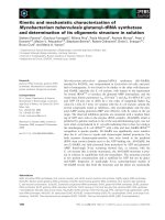
Fig. 1. Purification of lamjapin from L. japonica A by column chro-
matography. (A) Phenyl-Sepharose CL-4B; (B) CM-cellulose; (C)
Superose-12 FPLC and (D) Mono Q. The experimental procedures are
described in Material and methods. The solid lines represent the elu-
tion curves; dash line represents the NaCl gradient. Each fraction was
assayed for the RNA N-glycosidase activity to rat ribosome. Those
fractions that contained the major activity inhibiting the protein syn-
thesis in rabbit reticulocyte lysate were pooled and subjected to further
purification.
4748 R s. Liu et al. (Eur. J. Biochem. 269) Ó FEBS 2002
biosynthesis, and still in the range of the IC
50
(0.03–4 n
M
)of
type I RIPs. The IC
90
(the concentration of RIP causing
90% inhibition of translation) of lamjapin in the cell-free
system of rabbit reticulocyte is 5.56 n
M
.
RNA N-glycosidase activity of lamjapin to rat ribosomes
As shown in Fig. 3B, the RNA N-glycosidase activity of
lamjapin is compared with that of cinnamomin, a type II
RIP purified in our laboratory. At a molar ratio of
lamjapin/ribosome of 1 : 48, 20 ng of lamjapin could
induce rat liver ribosome to produce the ricin/sarcin
fragment (R-fragment) after aniline treatment. The R-frag-
ment did not appear if the ribosome was incubated with
only lamjapin without aniline treatment. Therefore, the
R-fragment was not caused by the RNase contamination in
the purified sample. This demonstrated that lamjapin has an
RNA N-glycosidase activity like other RIPs from higher
plants.
Lamjapin acts on ribosomal RNAs at multiple sites
Besides the predominant R-fragment produced by lamjapin
from rat ribosomal RNAs, three additional RNA fragments
larger than the R-fragment were found when the treated
ribosomal RNAs were separated by 8
M
urea-denatured
polyacrylamide gel (4.5%) electrophoresis for a longer time
(3 h). The R-fragment and three larger fragments did not
appear if the ribosome was incubated with only lamjapin
without aniline treatment. These data demonstrated con-
clusively that the emergence of the larger RNA fragments
by lamjapin was not artifact caused by the nuclease
contaminant. It was also shown that the ratio of fragments
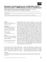
Fig. 3. Activity of lamjapin. (A) Effect of lamjapin on protein synthesis
in rabbit reticulocyte lysate. The protein synthesis system contained the
indicated amount of lamjapin in a final volume of 50 lLofreaction
mixture as described in Material and methods. The control value of
[
14
C]leucine incorporated is 22 000 d.p.m. (B). Activity of lamjapin to
rat liver ribosomes. Ribosomes were treated with lamjapin and acid
aniline. The ribosomal RNAs were extracted and electrophoresed on
3.5% polyacrylamide gel (8
M
urea) at 25 mA for 1 h and ribosomal
RNAs were visualized with methylene blue. The R-fragment was
produced with acid aniline from the treated ribosomes. B indicates
ribosomes were treated with only buffer H; C indicates ribosomes were
treated with cinnamomin (20 ng), L indicates ribosomes were treated
with lamjapin (20 or 30 ng).
Fig. 2. Purity of lamjapin identified by SDS/PAGE and isoelectric
focusing. (A) SDS/PAGE of the purified lamjapin. M, protein markers.
Lane 1, lamjapin (4 lg) without treatment by dithiothreitol; Lane 2,
lamjapin (4 lg) treated with dithiothreitol. SDS/PAGE (12%) was
performed and the protein bands were silver stained as described in
Materials and methods. (B) Isoelectric focusing of the purified lamja-
pin. Lane L, lamjapin (4 lg); M, protein markers. Proteins were
focused and stained with Coomassie Brilliant Blue. Regression analysis
of the migration distance plotted vs. the pI values of the protein
markers was used to calculate the pI value of lamjapin.
Ó FEBS 2002 Lamjapin, a novel type I RIP from marine alga (Eur. J. Biochem. 269) 4749
a, b, c and the R-fragment was constant (0.2 : 0.3 :
0.1 : 1.0), independent of the amount of lamjapin employed.
This indicated that the action of lamjapin on these sites of
ribosomal RNA was specific but the sensitivity of these sites
to lamjapin was much lower than that of A4324 in the S/R
domain.
In order to confirm the multiple sites of depurination of
lamjapin on the rat ribosomal RNA, the adenine base
released from ribosomal RNA by lamjapin were quantita-
tively analyzed by the chloroacetaldehyde method. A time
course study demonstrated that the release of adenine by
lamjapin continued at a linear rate for at least 40 min when
RIP and ribosome are presented at 1 : 1 molar ratio.
Quantitative analysis revealed that lamjapin could release
more than one mole of adenine from each mole of
ribosomes in 10 min and even up to 12 mol of adenines in
40 min (Fig. 4B).
Base and position specificity of lamjapin in depurination
of SRD RNA
Synthetic oligoribonucletide (a 35-mer) that mimics the S/R
domain of rat ribosomal RNA (SRD RNA) is an useful
substrate for studying the mechanism of action of RIP and
for analysis of the chemistry of recognition of RNA by RIP
[22,23]. As shown in Fig. 5, ricin could deadenylate A20 of
SRD RNA, the site corresponding to position A4324 of rat
ribosomal RNA, producing two fragments (20-mer and
15-mer). The SRD RNA treated with lamjapin and acidic
aniline also released two fragments with the same size as
that produced by ricin, while there was no fragment released
when the SRD RNA was treated only with lamjapin. The
activity of lamjapin on the SRD RNA exhibited the base
specificity as demonstrated by the fact that both the
transitional and transversionanl mutants (A20 to G20,
C20 or U20) were insensitive to lamjapin and no fragment
appeared from these mutants treated with lamjapin and acid
aniline.
In the next experiment, SRD RNA and the mutant SRD
RNA (A20 to G20) were labeled with [
3
H]adenine and
treated with lamjapin and aniline. The result revealed that
lamjapin could release the
3
H-labeled adenine from the wild
type SRD RNA (5.3 · 10
4
d.p.m. of
3
H-labeled adenine),
while no adenine was released from the A20 mutant SRD
RNAs treated with lamjapin. This result showed that only
A20 and no other adenines could be released by lamjapin
from SRD RNA. The activity of lamjapin to the SRD RNA
is absolutely dependent on the preservation of adenine at a
proper site. The base- and site-specific RNA N-glycosidase
activity of lamjapin is the same as other RIPs from higher
plants like ricin A-chain [17].
DISCUSSION
Extraction of protein from brow alga is tedious because of
its richness in phenolic compounds, pigments and polyan-
ionic cell wall consisting of alginates [24]. It was difficult to
extract active lamjapin by usual methods of homogenization
in the presence of above inhibitory substances. As an
alternative method, lyophilized kelp was powdered in liquid
nitrogen and then extracted gently at low temperature in the
alkaline solution containing ascorbic acid. These conditions
could efficiently decrease interaction of proteins with
phenolic compounds and alginates, etc. and hence preserved
the enzymatic activity of lamjapin. But the efficiency of
extraction was still low and the proteins could not be
precipitated by ammonium sulfate even up to 85% satura-
tion. Poly(ethylene glycol) partition that has been reported
to improve the efficiency of protein extraction from other
two species of kelp was tried unsuccessfully [25]; this method
resulted in the inactivation of lamjapin. Among several
methods tested, only the phenyl-Sepharose CL-4B column
chromatography could separate efficiently the active lamja-
pin from the inhibitory substances in the crude kelp extract.
Fig. 4. Multiple sites of action of lamjapin at ribosomal RNAs.
(A) Action of lamjapin on ribosomal RNAs. Rat ribosomes were
treated with lamjapin and acid aniline. The ribosomal RNAs were
extracted and electrophoresed on 4.5% polyacrylamide gel (8
M
urea)
at 25 mA for a longer time (3 h) and ribosomal RNAs were visualized
with methylene blue. a, b, c are three additional larger RNA fragments.
(B) Time course of releasing adenine from ribosomes. The experi-
mental conditions are described in the Materials and methods.
4750 R s. Liu et al. (Eur. J. Biochem. 269) Ó FEBS 2002
Four methods are commonly used to assess the RNA-
N-glycosidase activity of RIPs [26]: (a) quantification of the
inhibition of the protein synthesis in cell-free systems, (b)
visualization of the RNA fragment produced by aniline
cleavage at the site of depurination, (c) measurement of the
fluorescent derivative ethenoadenine of released adenine,
and (d) detection of the [
3
H]adenine released. This study
showed that lamjapin exhibited strong inhibitory activity to
protein synthesis in rabbit reticulocyte lysate. It acted on the
rat ribosomal RNA producing the RNA fragments after
aniline treatment. In addition, it could release adenine from
ribosomal RNA and SRD RNA as revealed by the release
of fluorescent derivative ethenoadenine and the [
3
H]adenine.
From these data, we can conclude safely that lamjapin is an
RNA N-glycosidase. It belongs to the ribosome-inactivating
protein family that were previously found only in higher
species of plant kingdom.
Intensive studies by the NMR and X-ray demonstrated
that SRD RNA possessed a tertiary structure similar to the
S/R domain of rat liver ribosomes. It was composed of a
stem and a GAGA tetraloop out of which A20 of SRD
RNA corresponding to the A4324 of rat ribosomal RNA
was flipped [27]. Lamjapin could deadenylate A20 of SRD
RNAandreleasedtheRNAfragmentwiththeexactsizeof
the R-fragment from the ribosomal RNA. It is likely that
lamjapin deadenylates the A4324 of 28S ribosomal RNA
and produces this RNA fragment after the acidic aniline
treatment.
RIPs were originally thought to act exclusively on the
specific A4324 of the S/R domain of rat ribosomes.
However, several RIPs such as saporin-R2 were found to
act on ribosomal RNA at multiple sites [28,29]. In this
study, it was found that lamjapin could deadenylate at
multiple sites in rat ribosomal RNA and produced three
additional RNA fragments in addition to the main R-frag-
ment. The ribosomal RNAs are rich in the stem-loop
structure that is similar to the S/R domain. Perhaps some
adenines of these domains also showed certain sensitivity to
lamjapin and saporin-R2. Lamjapin is one of the few
ribosome-inactivating proteins acting at multiple sites in
ribosomal RNA.
Study on the distribution of this class of protein in lower
plant is still scarce. Most of plant species examined belong
to the class of Angiospermae [30,31]. No RIP has yet been
isolated from the class of Gymnospermae and Cryptogamia.
Lamjapin is the first single-chain RIP isolated from kelp
(L. japonica A) that belongs to the Cryptogamia, the lowest
species in the plant kingdom. This first showed that RIPs
exist outside of the flowering plants. Our group screened
three marine algae and three freshwater algae in Crypto-
gamia and the RNA N-glycosidase was only found in kelp
(L. japonica A). It is very likely that the distribution of RIPs
is sporadic rather than ubiquitous in the plant kingdom as
proposed by Van Damme et al. [3]. The existence of
lamjapin in L. japonica A demonstrates that such sporadic
distribution of RIPs in plant kingdom ranges widely from
the lowest plants to the highest plants.
ACKNOWLEDGEMENTS
This work was supported by one grant of Natural Science Foundation
of China (39970163) and one grant of Academia Sinica (KSCX2-02-
04). The authors thank for Dr Zheng Pu for his technical assistance and
Dr Lee Zou for his critical reading of this manuscript.
REFERENCES
1. Endo. Y., Mitsui, K., Motizuk, M. & Tsurugi, K. (1987) The
mechanism of action of ricin and related toxic lectins on
eukaryotic ribosomes. The site and the characteristics of the
modification in 28 S ribosomal RNA caused by the toxins. J. Biol.
Chem. 263, 5908–5912.
2. Endo, Y. & Tsurugi, K. (1987) RNA N-glycosidase activity of
ricin A-chain: mechanism of action of the toxic lectin ricin on
eukaryotic ribosomes. J. Biol. Chem. 262, 8128–8130.
3. Peumans, W.J., Hao, Q. & Van Damme, E.J.M. (2001) Ribosome
inactivating protein: More than RNA N-glycosidase? FASEB J.
15, 1493–1510.
4. Tazzari, P.L., Polito, L., Bolognesi, A., Pistillo, M.P., Capanni, P.,
Palmisano, G.L., Lemoli, R.M., Curti, A., Biancone, L., Camussi,
G.,Conte,R.,Ferrara,G.B.&Stirpe,F.(2001)Immunotoxins
containing recombinant anti-CTLA-4 single-chain fragment
variable antibodies and saporin: in vitro results and in vivo effects
in an acute rejection model. J. Immunol. 167, 42222–42229.
5. Klinck,R.,Westhof,E.,Walker,S.,Afashar,M.,Collier,A.&
Aboul-Ela, F. (2000) A potential RNA drug target in the hepatitis
C virus internal ribosomal entry site. RNA 6, 1423–1431.
6. Munoz,R.,Arias,Y.,Ferreras,J.M.,Jimenez.P.,Rojo,M.A.&
Girbes, T. (2001) Sensitivity of cancer cell lines to the novel non-
toxic type 2 ribosome-inactivating protein nigrin b. Cancer Lett.
167, 163–169.
7. Girbes, T., Torre, C., Iglesias, R. & Ferreras, J.M. (1996) RIP for
viruses. Nature 379, 777–778.
8. Zoubenko, O., Hudak, K. & Tumer, N.E. (2000) A non-toxic
pokeweed antiviral protein mutant inhibits pathogen infection via
Fig. 5. Depurination of the synthetic SRD RNA by lamjapin. The sus-
ceptibility to lamjapin of mutant oligoribonucleotides having single
base changes at A20 was evaluated. (A) The substrate was the wild
type oligomer; (B) the oligomer has a transition of A20 to G20; (C) it
has a transversion of A20 to C20; and (D) it has a transversion of A20
to U20. R, ricin; L, lamjapin.
Ó FEBS 2002 Lamjapin, a novel type I RIP from marine alga (Eur. J. Biochem. 269) 4751
a novel salicylic acid-independent pathway. Plant Mol. Biol. 44,
219–229.
9. Nielsen, K. & Boston, R.S. (2001) Ribosome-inactivating pro-
teins: a plant perspective. Annu. Rev. Plant Physiol. Plant Mol.
Biol. 52, 785–816.
10. Wool, I.G., Gluck, A. & Endo, Y. (1992) Ribotoxin recognition of
ribosomal RNA and a proposal for the mechanism of transloca-
tion. Trends Biochem. Sci. 17, 266–269.
11. Wang, H.X. & Ng, T.B. (2001) Isolation of pleuturegin, a novel
ribosome-inactivating protein from fresh sclerotia of the edible
mushroom Pleurotus tuber-regium. Biochem. Biophys. Res. Com-
mun. 288, 718–721.
12. Lam, S.K. & Ng, T.B. (2001) Hypsin, a novel thermostable
ribosome-inactivating protein with antifungal and antiprolifera-
tive activities from fruiting bodies of the edible mushroom
Hypsizigus marmoreus. Biochem. Biophys. Res. Commun. 285,
1071–1075.
13. Bolognesi, A., Polito, L., Lubelli, C., Barbierr, L., Parente, A. &
Stirpe, F. (2002) Ribosome-inactivating and adenine polynucleo-
tide glycosylase activity in Mirabilis jalapa L. tissues. J. Biol.
Chem. 277, 13709–13716.
14. Sambrook, J., Fritsch, E.F. & Maniatis, T. (1989) Molecular
Cloning: A Laboratory Manual, 2nd edition, Cold Spring Harbor
Laboratory Press, New York.
15. Spedding, G. (1990) Ribosomes and Protein Synthesis: A Practical
Approach. IRL Press, New York.
16. Zamboni, M., Brigotti, M., Rambelli, F., Montanaro, L. & Sperti,
S. (1989) High-pressure-liquid-chromatographic and fluorimetric
methods for the determination of adenine released from ribosomes
by ricin and gelonin. Biochem. J. 259, 639–643.
17. Endo, Y., Gluck, A. & Wool, I.G. (1991) Ribosomal RNA
identity elements for ricin A-chain recognition and catalysis.
J. Mol. Biol. 221, 193–207.
18. Laemmli, U.K. (1970) Cleavage of structural proteins during the
assembly of the head of bacteriophage T4. Nature 227, 680.
19. Ansorge, W. (1985) Fast and sensitive detection of protein and
DNA bands by treatment with potassium permanganate. J. Bio-
chem. Biophys. Methods 11, 13–20.
20. Neuhoff, V., Stamm. R. & Eibl, H. (1985) Clear background and
highly sensitive protein staining with coomassie blue dyes inpoly-
acrylamide gels: a systematic analysis. Eletrophoresis 6, 427–448.
21. Bradford, M.M. (1976) A rapid and sensitive method for
the quantitation of microgram quantities of protein utilizing
the principle of protein-dye binding. Anal. Biochem. 142,
336–339.
22. Orita, M.N., Ishikawa, F., Shimayama, T., Taira, K., Endo, Y. &
Nishikawa, S. (1993) High-resolution NMR study of a synthetic
oligoribonucleotide with a tetranucleotide GAGA loop that is a
substrate for the cytotoxic protein, ricin. Nucleic Acids Res. 21,
5670–5678.
23. Endo, Y. & Gluck, A. (1990) A new assay to measure RNA
N-glycosidase activity. Nucleic Acids Symp. Series 22, 21–22.
24. Percival, E. & McDowell, H. (1987) Plant Carbohydrates.
Springer-Verlag Press, New York.
25. Jordan, P. & Vilter, H. (1991) Extraction of proteins from material
rich in anionic mucilages: partition and fractionation of vanadate-
dependent bromoperoxidases from the brown algae Laminaria
digitata and L. saccharina in aqueous polymer two-phase systems.
Biochem. Biophy. Acta 1073, 98–106.
26. Brigotti, M., Barbieri, L., Valbonesi, P., Stirpe, F., Montanaro, L.
& Sperti, S. (1998) A rapid and sensitive method to measure the
enzymatic activity of ribosome-inactivating proteins. Nucleic Acids
Res. 18, 4306–4307.
27. Correll, C.C., Munishkin, A., Chan, Y.L., Ren, Z., Wool, I.G. &
Steitz, T.A. (1998) Crystal structure of the ribosomal RNA
domain essential for binding elongation factors. Proc. Natl Acad.
Sci. USA 95, 13436–13441.
28. Barbieri, L., Ferreras, J.M., Barraco, A., Ricci. P. & Stirpe. F.
(1992) Some ribosome-inactivating proteins depurinate ribosomal
RNA at multiple sites. Biochem. J. 286, 1–4.
29. Di Maro, A., Valbonesi, P., Bolognesi, A., Stirpe, F., De Luca, P.,
Gigliano, G., Gaudio, L., Delli Bovi, P., Ferranti, P., Malomi, A.
& Parente, A. (1999) Isolation and characterization of four type-1
ribosome-inactivating proteins, with polynucleotide: adenosine
glycosidase activity, from leaves of Phytolacca Dioica L. Planta
208, 125–131.
30. Barbieri, L., Battelli, M.G. & Stirpe, F. (1993) Ribosome-
inactivating proteins from plants. Biochim. Biophys. Acta 1154,
237–282.
31. Gasperi-Campani, A., Barbieri, L., Battelli, M.G. & Stirpe, F.
(1985) On the distribution of ribosome-inactivating proteins
amongst plants. J. Nat. Prod. 48, 446–454.
4752 R s. Liu et al. (Eur. J. Biochem. 269) Ó FEBS 2002