Báo cáo khoa học: Kinetics of violaxanthin de-epoxidation by violaxanthin de-epoxidase, a xanthophyll cycle enzyme, is regulated by membrane fluidity in model lipid bilayers pptx
Bạn đang xem bản rút gọn của tài liệu. Xem và tải ngay bản đầy đủ của tài liệu tại đây (511.12 KB, 10 trang )
Kinetics of violaxanthin de-epoxidation by violaxanthin de-epoxidase,
a xanthophyll cycle enzyme, is regulated by membrane fluidity
in model lipid bilayers
Dariusz Latowski
1
, Jerzy Kruk
1
, Kvetoslava Burda
2
, Marta Skrzynecka-Jaskier
1
, Anna Kostecka-Gugała
1
and Kazimierz Strzałka
1
1
Department of Plant Physiology and Biochemistry, The Jan Zurzycki Institute of Molecular Biology and Biotechnology,
Jagiellonian University, Krako
´
w, Poland;
2
H. Niewodniczanski Institute of Nuclear Physics, Krako
´
w, Poland
This paper describes violaxanthin de-epoxidation in model
lipid bilayers. Unilamellar egg yolk phosphatidylcholine
(PtdCho) vesicles supplemented with monogalactosyldi-
acylglycerol were found to be a suitable system for studying
this reaction. Such a system resembles more the native
thylakoid membrane and offers better possibilities for
studying kinetics and factors controlling de-epoxidation of
violaxanthin than a system composed only of monogalacto-
syldiacylglycerol and is commonly used in xanthophyll cycle
studies. The activity of violaxanthin de-epoxidase (VDE)
strongly depended on the ratio of monogalactosyldiacyl-
glycerol to PtdCho in liposomes. The mathematical model of
violaxanthin de-epoxidation was applied to calculate the
probability of violaxanthin to zeaxanthin conversion at
different phases of de-epoxidation reactions. Measurements
of deepoxidation rate and EPR-spin label study at different
temperatures revealed that dynamic properties of the
membrane are important factors that might control con-
version of violaxanthin to antheraxanthin. A model of the
molecular mechanism of violaxanthin de-epoxidation where
the reversed hexagonal structures (mainly created by
monogalactosyldiacylglycerol) are assumed to be required
for violaxanthin conversion to zeaxanthin is proposed. The
presence of monogalactosyldiacylglycerol reversed hexa-
gonal phase was detected in the PtdCho/monogalactosyl-
diacylglycerol liposomes membrane by
31
P-NMR studies.
The availability of violaxanthin for de-epoxidation is a dif-
fusion-dependent process controlled by membrane fluidity.
The significance of the presented results for understanding
the mechanism of violaxanthin de-epoxidation in native
thylakoid membranes is discussed.
Keywords: xanthophyll cycle; de-epoxidation; liposomes;
violaxanthin; zeaxanthin
Xanthophyll cycle is a photoprotective mechanism wide-
spread in nature operating in the thylakoid membranes of
all higher plants, ferns, mosses and several algal groups [1].
This cycle involves two reversible reactions, light-dependent
de-epoxidation of violaxanthin to zeaxanthin via anther-
axanthin as an intermediate and light-independent epoxi-
dation of zeaxanthin to anteraxanthin and violaxanthin [2].
The conversion of violaxanthin to zeaxanthin is catalysed by
violaxanthin de-epoxidase (VDE) and the reverse reaction
of violaxanthin formation from zeaxanthin is catalysed by
another enzyme, zeaxanthin epoxidase. VDE has been
isolated from spinach and lettuce chloroplasts and the
molecularmassofthenativeenzymewasestimatedas
43 kDa [3–5]. The gene encoding VDE has been already
isolated and cloned [6]. VDE is located on the lumenal side
of the thylakoid membrane, shows an optimum activity at
pH 4.8 when present in chloroplasts and at 5.2 for the
isolated enzyme [7] and requires ascorbate as a reductant [8].
In the dark, when the pH in thylakoid lumen is neutral or
alkaline, VDE is inactive, whereas under strong light
conditions, pH in the thylakoid lumen decreases, the
enzyme binds to the membrane, becomes active and
converts violaxanthin to zeaxanthin [8,9]. The inhibition of
the enzyme activity by zeaxanthin has been reported [3]. For
optimal activity, VDE requires the presence of monogal-
actosyldiacylglycerol, the major lipid of the thylakoid
membrane [10–12]. With its small head-group area and
critical packing parameter value superior to one, monogal-
actosyldiacylglycerol in water forms reversed hexagonal
phase instead of bilayer structures [13]. It is known that
monogalactosyldiacylglycerol forms hexagonal phases over
a wide temperature range of )15 °Cto80°C at concentra-
tions higher than 50% lipid in water and this process also
depends on the degree of unsaturation of the acyl chains.
Until now, all in vitro studies on the VDE activity have
been carried out using largely undefined systems of buffered
suspension of monogalactosyldiacylglycerol aggregates
containing violaxanthin as substrate. Here, we present a
Correspondence to K. Strzalka, Department of Plant Physiology
and Biochemistry, The Jan Zurzycki Institute of Molecular Biology
and Biotechnology, Jagiellonian University, ul. Gronostajowa 7,
30-387 Krako
´
w, Poland.
Fax: + 48 12 252 69 02, Tel.: + 48 12 252 65 09,
E-mail:
Abbreviations: LHC, light harvesting complex; PSI, photosystem I;
PSII, photosystem II; VDE, violaxanthin de-epoxidase; PtdCho,
phosphatidylcholine; PtdGro, phosphatidylglycerol; VA, probability
of violaxanthin to antheraxanthin conversion; AZ, probability of
antheraxanthin to zeaxanthin conversion; VV, probability that
violaxanthin remains violaxanthin; AA, probability that anthera-
xanthin remains antheraxanthin; ZZ, probability that zeaxanthin
remains zeaxanthin; S
VA
, the constant rate of VA
0
decrease;
S
AZ
, the constant rate of AZ
0
decrease.
(Received 12 April 2002, revised 18 July 2002, accepted 5 August 2002)
Eur. J. Biochem. 269, 4656–4665 (2002) Ó FEBS 2002 doi:10.1046/j.1432-1033.2002.03166.x
new approach in the study of VDE activity employing
violaxanthin-containing liposomes as an experimental sys-
tem, which is a closer to the native thylakoid membrane.
The use of lipid bilayers instead of monogalactosyldiacyl-
glycerol aggregates offers new possibilities in the investiga-
tion of the kinetic parameters and mechanism of
violaxanthin de-epoxidation. One of the advantages of such
system is the defined orientation of violaxanthin molecules
in the lipid bilayer, which, according to various sources [14–
16], is perpendicular to the plane of the membrane.
Violaxanthin-supplemented unilamellar liposomes with
VDE present only outside the vesicles are also a good
system to study the flip-flop rate of antheraxanthin which is
probably a necessary step preceding zeaxanthin formation
in membrane. Additionally, experiments carried out at
different temperatures and application of a mathematical
model of de-epoxidation for the analysis of the obtained
results provide important information on the influence of
membrane physical properties, kinetic parameters of vio-
laxanthin into zeaxanthin conversion and flip-flop rate of
antheraxanthin. A possible molecular mechanism of vio-
laxanthin de-epoxidation is proposed.
MATERIALS AND METHODS
Preparation of unilamellar liposomes
The mixture of lipids with violaxanthin in chloroform was
evaporated under stream of nitrogen to form a thin film and
dried under vacuum for 1 h. The dried lipids were dissolved
in ethanol and the solution was injected slowly with a
Hamilton syringe into 0.1
M
sodium citrate buffer, pH 5.1,
under continuous bubbling with nitrogen. The final ethanol
concentration did not exceed 1.25%. Subsequently, the
liposome suspension was extruded through a polycarbonate
membrane with a pore diameter of 100 nm [17]. The final
lipid concentration in a liposome suspension was 43 l
M
and
violaxanthin concentration was 0.33 l
M
.
Egg yolk phosphatidylcholine (PtdCho) was purchased
from Sigma (P2772) and plant monogalactosyldiacylgly-
cerol was obtained from Lipid Products.
Electron microscopy
One drop of PtdCho/monogalactosyldiacylglycerol lipo-
somes (350 l
M
lipid concentration) or monogalactosyldi-
acylglycerol structures (12.9 l
M
lipid concentration) in
citrate buffer (pH 5.1) was placed on a Formvar coated
grid and after 30 s one drop of staining solution was added.
Negative staining was performed with uranyl acetate at
room temperature [18]. After 30 s, excess solution was
drained off with filter paper and the grid was allowed to dry
in the air. The grids were examined in a JEM 100SX
electron microscope operated at 80 kV.
Photon correlation spectroscopy (PCS) analysis
Diameter of PtdCho/monogalactosyldiacylglycerol lipo-
somes and monogalactosyldiacylglycerol reversed hexa-
gonal phase was measured by PCS analysis. The 10 mW
He-Ne laser (633 nm) was used as a light source. The
selected angle was 90°, the viscosity was 0.890 centipoise and
refractive index 1.333. All analyses were performed at 25 °C
and at the equilibration time of 2 min. Total lipid concen-
tration in the case of PtdCho/monogalactosyldiacylgly-
cerol liposomes was 43 l
M
(30.1 l
M
PtdCho, 12.9 l
M
monogalactosyldiacylglycerol) and 12.9 l
M
for monogal-
actosyldiacylglycerol structures. Both liposomes and
monogalactosyldiacylglycerol reversed hexagonal phase
were suspended in 0.1
M
sodium citrate buffer (pH 5.1).
Isolation of violaxanthin
Violaxanthin was isolated from dark-stored leaves of
lucerne (Medicago sativa) by pigment extraction with
acetone, saponification of the lipid extract [19], followed
by column chromatography on Silica Gel F254 (Merck) in
petroleum ether : acetone (4 : 1, v/v).
Isolation and purification of VDE
VDE was isolated and purified from 7-day-old wheat leaves
grown at 28 °C according to the method described by Hager
and Holocher [9]. Additionally, the enzyme was purified by
gel filtration on Sephadex G100. The gel electrophoretic and
ion-exchange chromatography analysis of VDE preparation
showed two other minor proteins apart from VDE (data not
shown). The enzyme activity was determined by dual-
wavelength measurements (502–540 nm) using DW-2000
SLM Aminco spectrophotometer at 25 °C according to
Yamamoto [20]. The reaction mixture contained 0.33 l
M
violaxanthin, 12.9 l
M
monogalactosyldiacylglycerol and
30 m
M
sodium ascorbate in 0.1
M
sodium citrate buffer
(pH 5.1).
Measurement of violaxanthin de-epoxidation
De-epoxidation of violaxanthin was measured at 4, 12 and
25 °C both in a monogalactosyldiacylglycerol reversed
hexagonal phase and in liposomes. The composition of
the reaction mixture of the monogalactosyldiacylglycerol
systemwasthesameasthatusedfortheenzymeactivity
determination. The liposomes (30.1 l
M
PtdCho, 12.9 l
M
monogalactosyldiacylglycerol, 0.33 l
M
violaxanthin) were
prepared in 30 m
M
sodium ascorbate, 0.1
M
sodium citrate
buffer (pH 5.1). In another series of experiments, liposomes
with constant concentration of monogalactosyldiacylgly-
cerol (12.9 l
M
) and violaxanthin (0.33 l
M
)wereusedand
PtdCho was changed in order to obtain following mono-
galactosyldiacylglycerol proportions: 5 mol%, 15 mol%
and 30 mol%.
All mixtures were placed in darkness and gently stirred.
The de-epoxidation reaction was initiated by addition of
saturating amount of VDE, the activity of which correspon-
ded to 4 nmol de-epoxidated violaxanthin per min per mL.
The reaction was terminated and pigments were extracted
by mixing 750 lL of the reaction medium with 750 lLof
the extraction solution containing chloroform/methanol/
ammonia (1 : 2 : 0.004, v/v/v). Xanthophyll pigments were
extracted by vigorous shaking and centrifugation for 10 min
at 10 000 g in Micro-Centrifuge Type-320. After centrifu-
gation, the chloroform fraction (200 lL) was evaporated to
dryness under stream of nitrogen. Subsequently, pigments
were dissolved in 50 lL tetrahydrofuran and 550 lLofthe
following solvent mixture, acetonitrile/methanol/water
(360 : 40 : 40, v/v/v).
Ó FEBS 2002 Violaxanthin de-epoxidation in liposomes (Eur. J. Biochem. 269) 4657
Pigment separation was performed by reverse phase
HPLC using a RP-18 column, 5 lm particle size, according
to the modified method of Gilmore and Yamamoto [21] at
the flow rate of 3 mLÆmin
)1
. The eluted pigments were
monitored at 440 nm and quantitatively determined.
Analysis of de-epoxidation kinetics
We have applied a new mathematical model [22] to
analyse the kinetics of conversion of violaxanthin to
zeaxanthin. The model allowed us to follow independently
the kinetics of the two de-epoxidation steps: the conver-
sion of violaxanthin into antheraxanthin (VfiA) with a
probability VA and antheraxanthin into zeaxanthin
(AfiZ) with a probability AZ. It is known from
experimental data that these two steps reach equilibrium.
It means that the parameters VA and AZ must vanish. In
themodelwehaveassumedalineardecreaseofthe
conversion probabilities:
VA ¼ VA
0
À nðS
VA
ÁDtÞ for VA > 0
AZ ¼ AZ
0
À nðS
AZ
ÁDtÞ for AZ > 0
where VA
0
and AZ
0
are the initial values of the conversion
probabilities, S
VA
and S
AZ
are the constant rates and nÆDt is
thetimeofreaction(Dt ¼ a constant time interval,
n ¼ number of time intervals).
Measurement of the order parameter in liposome
membrane
Temperature dependent changes of the order parameter of
lipid fatty acyl chains in liposome membranes were recorded
by EPR spin label measurements using a spin label 5-doxyl-
stearic acid reporting on dynamics of membrane regions
close to the headgroup area. The spin label was added to the
chloroform mixture of monogalactosyldiacylglycerol, Ptd-
Cho and V, dried under stream of nitrogen and stored under
vacuum for 1 h. After this time, the dried mixture was
suspendedin0.1
M
sodium citrate buffer pH 5.2 by
vortexing. The final concentration of 5-doxyl-stearic acid
was 10
)4
M
. The final concentration of lipids was 10
)2
M
and their proportions were the same as in the section on
ÔPreparation of unilamellar liposomesÕ. EPR spectra of the
spin label as a function of temperature were recorded using
a Bruker ESP-300E spectrometer fitted with TM
110
cavity.
The modulation amplitude was 1 G, microwave power was
2 or 8 mW. The measurements were performed within the
temperature range of 0–40 °C. All measurements were
performed in a heating mode. Temperature was stabilized
using Brucker temperature controller. Spin label was
purchased from Sigma.
31
P-NMR studies
31
P-NMR spectra of liposomes suspended in the citrate
buffer pH (5.1), containing 10% D
2
O were recorded at
202.5 MHz using a Bruker AMX-500. Generally, a sweep
width of 41.7 kHz and a repetition 2.6 s using 30° radio
frequency pulses were used. The exponential multiplication
of the free induction decay resulted in a 100-Hz line
broadening. The number of scans was 28 000. All spectra
were recorded at 17 °C.
RESULTS
Effect of monogalactosyldiacylglycerol proportion in
liposomes on violaxanthin deepoxidation
Our initial attempts to use liposomes with a lipid compo-
sition similar to that of the thylakoid membrane were
unsuccessful because the chemical instability of violaxanthin
related to the presence of phosphatidylglycerol (PtdGro)
and sulphoquinovosyldiacyloglycerol, which complicates
the quantitative measurements [17]. Therefore, we applied
PtdCho, as the lipid that most readily forms bilayers [13],
supplemented with monogalactosyldiacylglycerol which
was found necessary for VDE activity. With the rise in
monogalactosyldiacylglycerol proportion in PtdCho lipo-
somes to a certain level, the percentage of transformed
violaxanthin also increased (Fig. 1). However, at 35 mol%
of monogalactosyldiacylglycerol the de-epoxidation rate
became significantly lower due to liposome aggregation and
the suspension became turbid. The increase in turbidity was
followed by sedimentation of the lipid aggregates formed.
These changes were caused probably by fusion of liposomes
or the appearance of monogalactosyldiacylglycerol aggre-
gates at its high proportion to PtdCho in the lipid mixture
[13,23]. The liposome suspension with monogalactosyldi-
acylglycerol content < 30 mol% was transparent and
showed no tendency to aggregate. The presence of
liposomes and absence of aggregates in such a suspension
was confirmed by electron microscopy and PCS (data not
shown). On the other hand,
31
P-NMR measurements
Fig. 1. The effect of monogalactosyldiacylglycerol proportion in Ptd-
Cho/monogalactosyldiacylglycerol liposomes on the level of xanthophylls
after 20 min of the violaxanthin de-epoxidation reaction at room tem-
perature.
4658 D. Latowski et al. (Eur. J. Biochem. 269) Ó FEBS 2002
revealed formation of the reversed hexagonal phase
domains existing in PtdCho/monogalactosyldiacylglycerol
liposomes (Fig. 7).
Violaxanthin de-epoxidation was found to be strongly
dependent not only on the concentration of monogalacto-
syldiacylglycerol but also on the ratio of monogalactosyl-
diacylglycerol to PtdCho in liposomes, even if the absolute
amount of monogalactosyldiacylglycerol in the reaction
mixture and its proportion to violaxanthin and VDE were
constant (Fig. 2). The values of transition probabilities of
the violaxanthin conversion into antheraxanthin (VA)and
antheraxanthin conversion into zeaxanthin (AZ) show that
the varying amounts of PtdCho, which result in changes in
monogalactosyldiacylglycerol/PtdCho ratio, have much
stronger effect on conversion of violaxanthin to anther-
axanthin than on conversion of antheraxanthin to zeaxan-
thin (Table 1). At low monogalactosyldiacylglycerol
concentration (5 mol%), probability of violaxanthin to
antheraxanthin conversion is very low (VA
0
is 0.006 only).
However, once antheraxanthin has been formed, its con-
version to zeaxanthin occurs at relatively fast rate
(AZ
0
¼ 0.548). VA
0
and S
VA
parameters are very sensitive
to an increase in relative proportion of monogalactosyldi-
acylglycerol; at 30 mol% of this lipid, their values increase
43 and 76 times, respectively, while values of corresponding
parameters describing kinetics of antheraxathin to zeaxan-
thin conversion (AZ
0
and S
AZ
parameters) increase only 1.5
and 2.2 times, respectively.
For further studies on violaxanthin de-epoxidation,
PtdCho liposomes with 30 mol% content of monogalacto-
syldiacylglycerol were used as an optimal system.
Comparison of violaxanthin de-epoxidation
in monogalactosyldiacylglycerol and liposomal systems
and the effect of temperature
The temperature dependence of de-epoxidation reaction
was measured in unilamellar PtdCho/monogalactosyldi-
acylglycerol liposomes and in the monogalactosyldiacylglyc-
erol reversed hexagonal phase system with the composition
given in Materials and methods. The concentrations of
monogalactosyldiacylglycerol, violaxanthin and VDE
(saturating amount) were the same both in PtdCho/
monogalactosyldiacylglycerol liposomes and monogalacto-
syldiacylglycerol systems. It was found that kinetics of
de-epoxidation reaction were different in liposomes and in
the monogalactosyldiacylglycerol system, and that tempera-
ture has a strong influence on the reaction rate in both
systems studied (Figs 3 and 4).
At the three temperatures studied (4, 12 and 25 °C), the
initial rate of violaxanthin de-epoxidation was always faster
in liposomes than in monogalactosyldiacylglycerol system;
this difference was most evident at 25 °C. However, changes
Monogalctosyldiacylglycerol 5 mol%
Monogalctosyldiacylglycerol 15 mol%
Monogalctosyldiacylglycerol 30 mol%
Fig. 2. Time course of violaxanthin to zeaxanthin conversion in PtdCho/
monogalactosyldiacylglycerol liposomes at 25 °C when PtdCho and
monogalactosyldiacylglycerol concentrations were, respectively:
245.1 l
M
and 12.9 l
M
(5 mol% of monogalactosyldiacylglycerol);
73.1 l
M
and 12.9 l
M
(15 mol% of monogalactosyldiacylglycerol);
30.1 l
M
and 12.9 l
M
(30 mol% of monogalactosyldiacylglycerol) and
violaxanthin concentration was 0.33 l
M
.
Table 1. Kinetic parameters of a de-epoxidation reaction calculated for the experimental data presented in Fig. 2 by means of the mathematical model.
Monogalactosyldiacylglycerol
(mol %) VA
0
S
VA
· 10
)3
(min
)1
) AZ
0
S
AZ
· 10
)3
(min
)1
)
5 0.006 0.26 0.548 35.50
15 0.045 5.52 0.600 50.29
30 0.260 19.75 0.810 76.80
Ó FEBS 2002 Violaxanthin de-epoxidation in liposomes (Eur. J. Biochem. 269) 4659
in the temperature had different effects on the reaction
plateau reached in both systems. At 25 °C, plateau was
achieved in 20 min in PtdCho/monogalactosyldiacylgly-
cerol liposomes and in 40 min in monogalactosyldiacylgly-
cerol system. At this stage, about 93% and 83% of initial
violaxanthin amount were de-epoxidated in liposomes and
monogalactosyldiacylglycerol system, respectively; corres-
ponding zeaxanthin levels amounted about to 86% and
80% of total xanthophyll pigments. The plateau levels of
anteraxanthin were about 3.8% in liposomes and 2.5% in
monogalactosyldiacylglycerol reversed hexagonal phase. In
our experimental systems the complete de-epoxidation of
violaxanthin was not observed. A possible reason for this
may be an inhibitory effect of accumulating zeaxanthin on
VDE activity as reported previously [3]. On the other hand,
it may be also connected with the presence in the violax-
anthin pool of small amount of cis isomers that cannot serve
as a substrate for VDE [24].
At 25 °C, the rate of antheraxanthin formation was
considerably faster in liposomes than in the monogalacto-
syldiacylglycerol system. Its maximum level in liposomes
was achieved after 2 min and amounted to about 23% of
the total xanthophyll pool, whereas in monogalactosyldi-
acylglycerol system the maximum level of antheraxanthin
was detected after 10 min and it accounted only for about
16% of all xanthophylls.
At 12 °C, in spite of the higher initial de-epoxidation rate
of violaxanthin in liposomes, more violaxanthin was de-
epoxidated and more zeaxanthin was formed in the mono-
galactosyldiacylglycerol system at the plateau stage of the
reaction. The same was found at 4 °C, although at this
temperature the plateau in monogalactosyldiacylglycerol
system had not been reached during 180 min reaction time.
The kinetic parameters of violaxanthin de-epoxidation
calculated for the exeperimental data obtained from the
liposome and monogalactosyldiacylglycerol systems by
Fig. 4. Time course of violaxanthin to zeaxanthin conversion at different
temperatures in monogalactosyldiacylglycerol systems.
Fig. 3. Time course of violaxanthin to zeaxanthin conversion at different
temperatures in liposomes. Monogalactosyldiacylglycerol was present
at (A) 5mol%, (B) 15mol% and (C) 30mol%.
4660 D. Latowski et al. (Eur. J. Biochem. 269) Ó FEBS 2002
means of the mathematical model are compared in Table 2.
The rates of the de-epoxidation reactions are more sensitive
to temperature in the liposomal system than in the
monogalactosyldiacylglycerol system. When rising the tem-
perature from 4 to 25 °C, zeaxanthin level at the plateau
stage increases 2.7-fold, violaxanthin level decreases eight-
fold and antheraxanthin maximal level increases twofold in
the liposomal system, whereas corresponding values for
monogalactosyldiacylglycerol system are 2.1, 3.5 and 1.7.
When analysing the effect of temperature on probabilities of
VA
0
and AZ
0
transition in both systems studied (Table 2), it
is evident that these values are higher in the liposome system
than in the monogalactosyldiacylglycerol system at all
temperatures studied. The increase in the temperature from
4to25°CincreasestheVA
0
value from 0.005 to 0.26
(52-fold) in liposomes, whereas VA
0
increases from 0.004 to
0.085 (21-fold) in monogalactosyldiacylglycerol system. The
AZ
0
transition increases proportionally in both systems on
elevating the temperature from 4 to 25 °C and its value rises
12.5-fold in liposomal and 13.3-fold in micellar systems,
respectively. On the other hand, the values S
VA
and S
AZ
coefficients increase much more in monogalactosyldiacyl-
glycerol reversed hexagonal phase than in the liposomal
system when inceasing the temperature from 4 to 25 °C.
Temperature-dependent changes in the value of VA
0
parameter in PtdCho/monogalactosyldiacylglycerol lipo-
somes correlate well with the corresponding changes in the
value of the order parameter as found by the use of EPR
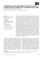
spectrometry and a spin probe 5-doxyl-stearic acid (Fig. 5).
The lower value of the order parameter the higher the
violaxanthin de-epoxidation rate is observed.
DISCUSSION
This paper is the first work where VDE has been isolated
from a monocotyledonous plant. The action and properties
of this enzyme are the same as VDE isolated previously
from dicotyledonous plants [25].
The presented results show that VDE-mediated conver-
sion of violaxanthin via antheraxanthin into zeaxanthin can
occur in PtdCho/monogalactosyldiacylglycerol liposomes.
It is worth noting that the VDE enzyme added to initiate the
reaction was present on the external and not internal side of
the liposome membrane and that similar kinetics and
decline in violaxanthin amount as in the measurements
performed with thylakoids [15] were observed. For this
reason, the PtdCho/monogalactosyldiacylglycerol unilamel-
lar liposome system used in this work is a good model of the
native photosynthetic membrane for studying the VDE
activity.
The presence of monogalactosyldiacylglycerol in Ptd-
Cho-liposomes was found to be indispensable for the
violaxanthin de-epoxidation reaction. As we have demon-
strated, the rate of violaxanthin to antheraxanthin conver-
sion depends on monogalactosyldiacylglycerol/PtdCho
ratio in the liposome membrane even if the absolute amount
of monogalactosyldiacylglycerol in the reaction mixture and
its proportion to violaxanthin and VDE remains constant
(Fig. 2, Table 1). On the basis of these results, we postulate
that VDE binds only to certain membrane domains that are
rich in monogalactosyldiacylglycerol and the de-epoxida-
tion reactions take place in these domains. Violaxanthin
being distributed homogeneously in the lipid bilayer has to
Table 2. Kinetic parameters of a de-epoxidation reaction calculated for the experimental data presented in Figs 3 and 4 by means of the mathematical
model.
Temp.
(°C) VA
0
S
VA
· 10
)3
(min
)1
)
AZ
0
· 10
)3
(min
)1
)S
AZ
Liposomes
4 0.005 0.024 0.065 0.371
12 0.026 0.34 0.150 1.808
25 0.260 19.76 0.810 50.29
Monogalactosyldiacylglycerol system
4 0.004 0.012 0.030 0.0004
12 0.020 0.133 0.100 0.681
25 0.085 21.76 0.400 8.51
Fig. 5. PtdCho/monogalactosyldiacylglycerol liposome membrane flui-
dity and percent of violaxanthin converted after 1 min de-epoxidation
reaction at different temperatures.
Ó FEBS 2002 Violaxanthin de-epoxidation in liposomes (Eur. J. Biochem. 269) 4661
enter the monogalactosyldiacylglycerol-enriched domains
by lateral diffusion to be converted to antheraxanthin. The
higher monogalactosyldiacylglycerol/PtdCho ratio, the
higher the amount of such domains in the liposomal
membrane. This shortens the diffusion path of violaxanthin
molecules to these domains and results in higher rate of
violaxanthin de-epoxidation (see the values of VA
0
in
Table 1). It is well known that nonbilayer prone lipids (e.g.
monogalactosyldiacylglycerol) may form reversed hexa-
gonal phase in model lipid membranes and it has been
reported that such structures exist in biological membranes
[26–28].
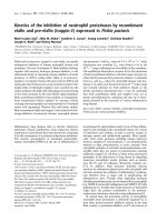
31
P-NMR spectra shown in Fig. 7 clearly demon-
strate the existence of the reversed hexagonal phase domains
in our system of PtdCho/monogalactosyldiacylglycerol
liposomes. The presence of the reversed hexagonal phase
in thylakoid membranes has been also reported in our
earlier papers and by other authors using
31
P-NMR
and freeze-fracturing techniques [23,29–31]. These observa-
tions give a sound basis for our model of violaxanthin
de-epoxidation in liposomes and thylakoid membranes.
According to the presented model, the second reaction of
de-epoxidation, i.e. conversion of antheraxanthin to zea-
xanthin, also occurs in the monogalactosyldiacylglycerol
rich domains, and is greatly facilitated because antheraxan-
thin, formed in such domains, has an immediate access to
the VDE enzyme. Therefore, and in contrast to the
de-epoxidation of violaxanthin to antheraxanthin, the
conversion of antheraxanthin to zeaxanthin seems to be
not limited by diffusion process. The conclusion that the
conversion of violaxanthin to antheraxanthin is more
sensitive to monogalactosyldiacylglycerol concentration
than the conversion of antheraxanthin to zeaxanthin is
supported by relatively high value of the AZ
0
parameter
even in conditions of very low VA
0
(e.g. at 5 mol% of
monogalactosyldiacylglycerol, Table 1).
The model assuming the existence of monogalactosyldi-
acylglycerol reversed hexagonal phase domains in the
membrane to which VDE binds and rather homogeneous
distribution of violaxanthin molecules explains also the
strong correlation between the rate of violaxanthin
de-epoxidation and value of the membrane lipid order
parameter. It seems that decreasing value of the order
parameter permits faster lateral diffusion of violaxanthin in
the membrane and the molecules of this xanthophyll may
reach sooner the monogalactosyldiacylglycerol rich
domains where they are de-epoxidated (Fig. 5, Tables 2
and 3). This model can also explain a clear temperature
effect on the level and the time of antheraxanthin appear-
ance in the liposome system. The conversion of anther-
axanthin to zeaxanthin is less dependent on the changes in
membrane physical properties (Tables 2 and 3) for the
reasons already discussed. Application of the proposed
model to the results obtained shows why the conversion of
violaxanthin to antheraxanthin is much slower and more
sensitive to temperature than transition from the anther-
axanthin to zeaxanthin. On the basis of our results and
literature data [15,22], we postulate that changes in mem-
brane fluidity may play an important role in regulation of
the violaxanthin de-epoxidation rate in membranes.
The higher rate of violaxanthin de-epoxidation to
antheraxanthin and stronger temperature effect on this
process in liposomes than in monogalactosyldiacylglycerol
systems is probably related to the different availability of
violaxanthin for VDE in both systems studied. As revealed
by PCS and electron microscopy, the size of monogalacto-
syldiacylglycerol aggregates differed greatly from that of
PtdCho/monogalactosyldiacylglycerol liposomes. The
monogalactosyldiacylglycerol structures were found as
large, heterogeneous aggregates with a mean diameter of
% 600 nm and a large standard deviation. Thus, the
previous assumption that monogalactosyldiacylglycerol
creates small micelles with only one molecule of violaxan-
thin inside [32] was not confirmed in our study. The average
diameter of liposomes was % 110 nm (as expected) with
narrow standard deviation. Apparently, the availability of
violaxanthin for VDE is higher in liposomes than in the
monogalactosyldiacylglycerol system where access of the
enzyme to its substrate may be impeded by large scale
aggregation of monogalactosyldiacylglycerol structures.
Neither the molecular arrangement of monogalactosyldi-
acylglycerol in such aggregates nor the orientation of
violaxanthin in these structures have been precisely deter-
mined [23].
To convert violaxanthin into zeaxanthin, VDE has to
remove two epoxy groups attached to two rings of the
violaxanthin molecule. In the unilamellar liposome system,
where all the xanthophylls are oriented perpendicularly to
the plane of the membrane and VDE is present only outside
the vesicles, the formed antheraxanthin molecule, to be
converted into zeaxanthin, has to reverse its orientation as a
whole in such a way that the end group containing the ring
with the remaining epoxy group appears on the other side of
membrane. Such a Ôflip-flopÕ of antheraxanthin molecule is a
necessary step assuming that VDE cannot penetrate
through the lipid bilayer and has access to the outer surface
of the liposome only. Table 3 shows the maximal time in
which all molecules of violaxanthin are converted to
antheraxanthin and all molecules of antheraxanthin are
converted to zeaxanthin at a given temperature at saturating
amount of VDE and at the initial reaction rate. While the
time for VfiA conversion is shortened considerably on the
increase of temperature from 4 to 25 °C,thetimeforAfiZ
transition changes is in a much more narrow range. In the
liposome system studied, the time for AfiZ transition takes
3.1 min at the temperature of 25 °C and 2.9 and 2.0 min at
12 and 4 °C, respectively. This means that flip-flop of
antheraxanthin in PtdCho/monogalactosyldiacylglycerol
liposomes at a given temperature has to be shorter than
the times specified in Table 3. Moreover, faster conversion
of antheraxanthin to zeaxanthin (AZ
0
values) than violax-
anthin to antheraxanthin (VA
0
values) was observed at all
temperatures studied (Tables 2 and 3) suggesting that flip-
flop of antheraxanthin is not the limiting step in the
transformation of violaxanthin into zeaxanthin in the
Table 3. The maximal time required for the conversion of all violaxan-
thin molecules into antheraxanthin (T
VA
) and all molecules of anther-
axanthin into zeaxanthin (T
AZ
) at three different temperatures in
PtdCho/monogalactosyldiacylglycerol liposomes.
Temp. (°C) T
VA
(min) T
AZ
(min)
4 217.4 2
12 30 2.9
25 5.6 3.1
4662 D. Latowski et al. (Eur. J. Biochem. 269) Ó FEBS 2002
membrane system investigated. Our results are in agreement
with those of Arvidsson et al. [15] who suggested that in the
isolated thylakoids the flip-flop of antheraxanthin is not the
limiting factor in zeaxanthin formation.
The apparently longer time necessary for antheraxanthin
to zeaxanthin conversion at higher temperatures (Table 3)
can be explained in terms of our model assuming diffusion
controlled rate of violaxanthin to antheraxanthin conver-
sion. Violaxanthin and antheraxanthin compete for the
same active site of the VDE enzyme. At elevated tempera-
ture, when violaxanthin lateral diffusion in the membrane is
faster, more violaxanthin molecules reach the monogalacto-
syldiacylglycerol rich domains in time unit. In such a
situation, violaxanthin competes successfully with anther-
axanthin for the VDE active site; this results in enlargement
of the antheraxanthin pool. As a consequence, a relatively
lower number of antheraxanthin molecules of the pool can
reach the VDE active site and become converted to
zeaxanthin. This conclusion is supported by the data
presented in Fig. 6, which shows that at higher temperatures
a lower proportion of total antheraxanthin pool is conver-
ted into zeaxanthin. It should be also added that because
VDE was present in excess in the reaction mixture, only part
of it could be bound to the membrane, depending on the size
and number of monogalactosyldiacylglycerol-rich domains.
Some conclusions drawn from our results obtained with
model lipid bilayer can be extrapolated to describe the role
of the xanthophyll cycle in the regulation of thylakoid
membrane fluidity. In the darkness, due to zeaxanthin
epoxidase activity, violaxanthin accumulates in thylakoids.
Illumination of plants with strong light causes acidification
of thylakoid lumen, which is a prerequisite for VDE binding
to thylakoid membrane, and also it usually increases leaf
temperature, which results in the increase of the membrane
dynamics. A temperature-induced increase of thylakoid
membranes dynamics facilitates diffusion of violaxanthin
molecules into monogalactosyldiacylglycerol-VDE domains
where it is converted into antheraxanthin and zeaxanthin.
This conclusion is supported by the results of Sarry et al.
[33] who found that illumination of plants at low tempera-
ture results in a lower amount of zeaxanthin formed than at
higher temperature. There are reports that show that
zeaxanthin may act like cholesterol and play important role
in the regulation of thylakoid membrane arrangement.
Gruszecki and Strzalka [34] showed that light induced
accumulation of zeaxanthin affects membrane fluidity.
Tardy and Havaux [35] found that decreased value of the
thylakoid membrane order parameter was proportional to
the amount of zeaxanthin present in the membrane. The
rigidifying effect of this xanthophyll was also found upon
incorporation of exogenous zeaxanthin into isolated thyl-
akoid membranes [36].
Fig. 6. The percentage of total antheraxanthin pool converted into
zeaxanthin during violaxanthin de-epoxidation reaction in PtdCho/
monogalactosyldiacylglycerol liposomes at three different temperatures.
Fig. 7. 31P-NMR spectra of PtdCho liposomes (A) without mono-
galactosyldiacylglycerol and (B) with 30 mol% monogalactosyldiacyl-
glycerol.
Ó FEBS 2002 Violaxanthin de-epoxidation in liposomes (Eur. J. Biochem. 269) 4663
Zeaxanthin formed in the hexagonal phase domains can
probably leave these regions and, due to its membrane
rigidifying properties, it regulates the molecular dynamics of
thylakoid membranes and protects them at elevated
temperatures resulting from intense irradiation.
In conclusion, the PtdCho/monogalactosyldiacylglycerol
liposome system described in this work is more appropriate
than monogalactosyldiacylglycerol aggregates for studying
the mechanism of violaxanthin de-epoxidation catalysed by
VDE in vitro because it approaches the native photosyn-
thetic membranes. The existence of de-epoxidation reactions
in liposomes opens new possibilities in the investigation of
the xanthophyll cycle, which might contribute to a better
understanding of this process.
ACKNOWLEDGEMENTS
This work was supported by a grant no. 6P04A 02819 from Committee
for Scientific Research (KBN) of Poland. We wish to thank Maria
Kozlowska for electron microscopy pictures, Dr Maria Zembala for
PCS measurements and Dr F. Szneler for
31
P- NMR analysis. We are
very grateful to Dr Fabrice Franck from University of Liege, Belgium
for helpful discussion.
REFERENCES
1. Stransky, H. & Hager, A. (1970) The carotenoid pattern and the
occurence of the light induced xanthophyll cycle in various classes
of algae part 6 chemo systematic study. Arch. Microbiol. 73,315–
323.
2. Yamamoto, H.Y., Nakayama, T.O.H. & Chichester, C.O. (1962)
Studies on the light and dark interconversions of leaf xantho-
phylls. Arch. Biochem. Biophys. 97, 168–173.
3. Havir, E.A., Tausta, L.S. & Peterson, R.B. (1997) Purification and
properties of violaxanthin de-epoxidase from spinach. Plant Sci.
123, 57–66.
4. Rockholm, D.C. & Yamamoto, H.Y. (1996) Purification of a
43-kilodalton lumen protein from lettuce by lipid-affinity preci-
pitation with monogalactosyldiacylglyceride. Plant Physiol. 110,
697–703.
5. A
˚
kerlund, H E., Arvidson, P O., Bratt, C.E. & Carlsson, M.
(1995) Partial purification of the violaxanthin de-epoxidase. In
Photosynthesis from Light to Biosphere (P.Mathis,ed.),Vol.IV,
pp. 103–106. Kluwer Academic Publishers, Dortrecht.
6. Bugos, R.C. & Yamamoto, H.Y. (1996) Molecular cloning of
violaxanthin de-epoxidase from romaine lettuce and expression in
Escherichia coli. Proc. Natl Acad. Sci. USA 93, 6320–6325.
7. Hager. A. (1969) Lichtbedingte pH-Erniedringung in einem
Chloroplasten-Kompartiment als Ursache der Enzymatichen
Violaxanthin-zu Zeaxanthin – Umwandlung Beziehungen zur
Photophosphorylierung. Planta 89, 224–243.
8. Bratt, C.E., Arvidsson, P O., Carlsson, M. & A
˚
kerlund, H E.
(1995) Regulation of violaxanthin de-epoxidase activity by pH and
ascorbate concentration. Photosynth. Res. 45, 169–175.
9. Hager, A. & Holocher, K. (1994) Localization of the xanthophyll
cycle enzyme violaxanthin de-epoxidase within the thylakoid
lumen and abolition of its mobility by a (light-dependent) pH
decrease. Planta 192, 581–589.
10. Yamamoto,H.Y.,Chenchin,E.E.&Yamada,D.K.(1974)Effect
of chloroplast lipids on violaxanthin de-epoxidase activity. Pro-
ceedings of the 3rd International Cong. Photosyn, pp. 1999–2006.
Elsevier, Amsterdam.
11. Siefierman-Harms, D., Ninnemann, H. & Yamamoto, H.Y.
(1987) Reassembly of solubilized chlorophyll-protein complexes in
proteolipid particles-comparison of monogalactosyldiacylglycerol
and two phospholipids. Biochim. Biophys. Acta 892, 303–313.
12. Webb, M. & Green, B. (1991) Biochemical and biophysical
properties of thylakoid acyl lipids. Biochim. Biophys. Acta 1060,
133–158.
13. Israelachvili, J.N. & Mitchell, D.J. (1975) A model for the packing
of lipids in bilayer membranes. Biochim. Biophys. Acta. 389,13–
19.
14. Gruszecki, W.I. (1995) Different aspects of protective of the xan-
thophyll cycle under stress conditions. Acta Physiol. Plant. 17,
145–152.
15. Arvidsson, P O., Carlsson, H., Stefansson, H., Albertsson, P A.
&A
˚
kerlund, H E. (1997) Violaxanthin accessibility and tem-
perature dependency for de-epoxidation in spinach thylakoid
membranes. Photosynth. Res. 52, 39–48.
16. Milton, A., Wolff, G., Ourisson, G. & Nakatani, Y. (1986)
Organization of carotenoid-phospholipid bilayer systems
incorporation of zeaxanthin and their C-50 homologues into
dimyristoylphosphatidylcholine vesicles. Helv. Chim. Acta 69,
12–24.
17. Latowski, D., Kostecka, A. & Strzalka, K. (2000) Effect of
monogalactosyldiacylglycerol and other thylakoid lipids on vio-
laxanthin de-epoxidation in liposomes. Bioch. Soc. Trans.,Vol.28
Part. 6, 810–812.
18. Enoch, H.G. & Stritmatter, P. (1979) Formation and properties of
1000 A
˚
-diameter, single-bilayer phospholipidvesicles. Proc. Natl
Acad. Sci. USA 76, 145–149.
19. Davies, B.H. (1976) Carotenoids. In Chemistry and Biochemistry
of Plant Pigments (Goodwin, T.W., ed.), pp. 65–66. Academic
Press, London New York San Francisco.
20. Yamamoto, H.Y. (1995) Xanthophyll cycle. Meth Enzymol. 110,
303–312.
21. Gilmore, A.M. & Yamamoto, H.Y. (1991) Resolution of lutein
and zeaxanthin using a non-endcappted, lightly carbon-loaded C
18
high-performance liquid chromatographic column. J. Chrom. 543,
137–145.
22. Latowski, D., Burda, K. & Strzalka, K. (2000) A mathematical
model describing kinetics of conversion of violaxanthin to
zeaxanthin via intermediate antheraxanthin by the xanthophyll
cycle enzyme violaxanthin de-epoxidase. J. Theor. Biol. 206,507–
514.
23. Sprague, G.S. & Staehelin, L.A. (1984) Effect of Reconstitution
Method on the structural organization of isolated chloroplast
membrane lipids. Biochim. Biophys. Acta 777, 306–322.
24. Phillip, D., Molnar, P., Toth, G. & Young, A.J. (1999) Light-
induced formation of 13-cis violaxanthin in leaves of Hordeum
vulgarum. J. Photochem. Photobiol. B: Biol. 49, 89–95.
25. Hieber, A.D., Bugos, R.C. & Yamamoto, H.Y. (2000) Plant
lipocalins: violaxanthin de-epoxidase and zeaxanthin epoxidase.
Biochim. Biophys. Acta. 1482, 84–91.
26. De Kruijff, B., Verkleij, A.J., Van Echteld, C.J.A., Gerritsen, W.J.,
Mombers, C., Noordam, P.C. & Gier, J. (1979) The occurance of
lipid particles in lipid bilayers as seen by
31
P NMR and freeze-
fracture electron-microscopy. Biochim. Biophys. Acta. 555,200–
209.
27. Walde, P., Giuliani, A.M., Boicelli, C.A. & Luisi, P.L. (1990)
Phospholipid-based reversed micelles. Chem. Phys. Lipids 53,265–
288.
28. Venetie
¨
, R.V. & Verkleij, A.J. (1981) Analysis of the hexagonal
II phase and its relations to lipidic particles and the lamellar
phase. A freeze-fracture study. Biochim. Biophys. Acta. 645,262–
269.
29. Haran
˜
czyk, H., Strza
3
ka,K.,Dietrich,W.&Blicharski,J.S.(1995)
31
P-NMR observation of the temperature and glycerol induced
non-lamellar phase formation in wheat thylakoid membranes.
J. Biol. Phys. 21, 125–139.
30. Quinn, P.J. & Williams, W.P. (1983) The structural role of lipids
in photosynthetic membranes. Biochim. Biophys. Acta. 737,223–
266.
4664 D. Latowski et al. (Eur. J. Biochem. 269) Ó FEBS 2002
31. Gounaris, K., Sen, A., Brain, A.P.R., Quinn, P. & Williams, W.P.
(1983) The formation of non-bilayer structures in total polar lipid
extracts of chloroplast membranes. Biochim. Biophys. Acta. 728,
129–139.
32. Yamamoto, H.Y. & Higashi, R.M. (1978) Violaxanthin de-epo-
xidase. Lipid composition and substrate specifity. Arch. Bioch.
Biophys. 190, 514–522.
33. Sarry, J E., Montillet, J L., Sauvaire, Y. & Havaux, M. (1994)
The protective function of the xanthophyll cycle in photosynthesis.
FEBS Lett. 353, 147–150.
34. Gruszecki, W.I. & Strzalka, K. (1991) Does the xanthophyll cycle
take part in the regulation of fluidity of the thylakoid membrane?
Biochim. Biophys. Acta. 1060, 310–314.
35. Tardy, F. & Havaux, H. (1997) Thylakoid membrane fluidity
and thermostability during the operation of the xanthophyll
cycle in higher-plant chloroplasts. Biochim. Biophys. Acta 1330,
179–193.
36. Strzalka, K. & Gruszecki, W. (1997) Modulation of thylakoid
membrane fluidity by exogenously added carotenoids. J. Biochem.
Mol. Biol. Biophys. 1, 103–108.
Ó FEBS 2002 Violaxanthin de-epoxidation in liposomes (Eur. J. Biochem. 269) 4665