Báo cáo Y học: A catalytically inactive b1,4-N-acetylglucosaminyltransferase III (GnT-III) behaves as a dominant negative GnT-III inhibitor potx
Bạn đang xem bản rút gọn của tài liệu. Xem và tải ngay bản đầy đủ của tài liệu tại đây (376.38 KB, 9 trang )
A catalytically inactive b1,4-
N
-acetylglucosaminyltransferase III
(GnT-III) behaves as a dominant negative GnT-III inhibitor
Hideyuki Ihara, Yoshitaka Ikeda, Souichi Koyota, Takeshi Endo, Koichi Honke and Naoyuki Taniguchi
Department of Biochemistry, Osaka University Medical School, Suita, Osaka, Japan
b1,4 -N-Acetylglucosaminyltransferase III (GnT-III) plays a
regulatory role in the biosynthesis of N-glycans, and it has
been suggested that its product, a bisecting GlcNAc, is
involved in a variety of biological events as well as in regu-
lating the b iosynthesis of t he oligosaccharides. I n t his s tudy,
it was f ound, on the basis of sequence homology, that GnT-
III contains a small region that is signi®cantly homologous
to both snail b1,4GlcNAc transferase and b1,4Gal trans-
ferase-1. Subsequent mutational analysis demonstrated an
absolute requirement for two conserved Asp residues
(Asp321 and Asp323), which are located in the most
homologous region of rat GnT-III, for enzymatic activity.
The overexpression of Asp323-substituted, catalytically
inactive GnT-III in Huh6 cells led to the suppression of the
activity of endogenous GnT-III, but no signi®cant decrease
in its expression, and led to a speci®c inhibition of the f or-
mation of bisected sugar chains, as shown by s tructural
analysis of the total N-glycans f rom t he cells. These ®ndings
indicate that the mutant serves a dominant negative eect on
a speci®c step in N-glycan biosynthesis. This type of Ôdomi-
nant negative glycosyltransferaseÕ, identi®ed has potential
value as a powerful tool for d e®ning the precise biological
roles of the bisecting GlcNAc structure.
Keywords: GnT-III; glycosyltransferase; bisecting G lcNAc;
N-glycan synthesis; dominant negative eect.
b1,4 -N-Acetylglucosaminyltransferase III (GnT-III) cata-
lyzes the transfer of GlcNAc from UDP-GlcNAc, a glycosyl
donor, to a core b-mannose residue in N-linke d oligosac-
charides via a b1 ® 4 linkage, resulting in the formation o f
a bisected sugar chain [1]. The resulting GlcNAc r esidue is
referred to a s a bisecting G lcNAc, and i s known to p lay a
role in regulating the biosyn thesis of N-glycans, as the
addition of this unique structure inhibits the action of other
N-acetylglucosaminyltransferases, such as GnT-IV and
GnT-V, both o f which are i nvolved in t he formation o f
multiantennary sugar chains [2]. Thus, GnT-III can be
regarded as a key glycosyltransferase in N-glycan biosyn-
thetic pathways.
It has been suggested that GnT-III and/or the bisecting
GlcNAc residue are involved in a variety of biological
processes, such as the intracellular s orting of glycopro-
teins [ 3], secretion of apo B100 from the liver [4], cell
adhesion [5] a nd cancer metastasis [6,7], as evidenced by
gene transfection experiments using GnT-III cDNA.
Although studies using GnT-III-de®cient mice have
revealed that a defect in GnT-III suppresses diethylni-
trosamine-induced hepatocarcinogenesis [8,9], they do not
provide any explanations for the changes c aused b y an
overexpression of GnT-III, as mentioned above. The
mechanisms that u nderlie such a v ariety of biological
events, which are c aused by ectopic expression, overex-
pression and defects in GnT-III, remain obscure. In
addition, the issue of whether phenotypes associated with
altered expression of GnT-III are the actual consequences
of N-glycan modi®cation by GnT-III r emains unresolved.
To address these issues, a catalytically inactive GnT-III
and/or dominant negative mutant GnT-III would be
valuable because i t may potentially de®ne whether
bisected N-glycan structures are actually important.
Alternatively, the i ssue of whether G nT-III protein i s
directly involved regardless of formation of bisected sugar
chains could also be examined.
The i denti®cation of catalytic residues, the absence of
which leads to the complete loss of activity, is required to
produce a catalytically inactive GnT-III. The enzymatic
properties of t his enzyme h ave been extensively studied in
terms of s ubstrate speci®city t owards acceptors a nd
donors, and these collective data p rovide an enzymatic
basis for the role of the bisecting GlcNAc in the regulation
of N-glycan biosynthesis [1,10,11]. Nevertheless, because
the catalytic mechanism of GnT-III has not yet been
analyzed in suf®cient detail, the chemical basis of the
enzymatic reaction is not known. The r eaction catalyzed
by GnT-III is accompanied by inversion at the anomeric
center of the transferred monosaccharide and the forma-
tion of a b1 ® 4 linkage, while chemically similar
reactions are a lso catalyzed by other g lycosyltransferases
such as b1,4GlcNAc transferase (b1,4GlcNAcT) from
snail, Lymnaea stagnalis [12], and mammalian b1,4Gal
transferases, (b1,4GalTs), both of w hich ar e mutually
homologous [13]. It s eems more likely that the common
properties of these three enzymes are associated with the
catalytic mechanism, rather t han substrate binding, a s the
Correspondence to N. Taniguchi, Department of Biochemistry, Osaka
University Medical School, 2-2 Yamadaoka, Suita, O saka 565-0871,
Japan. Fax: + 81 6 6879 3429, Tel.: + 81 6 6879 3420,
E-mail:
Abbreviations: GnT-III, b1,4-N-acetylglucosaminyltransferase III;
PNGase, peptide-N-glycosidase; H RP, horseradish peroxidase;
DMEM, Dulbecco's modi®ed Eagle's medium.
Enzyme: b1,4-N-acetylglucosaminyltransferase III (EC 2.4.1.144).
Note: a web site is available at />biochem/index.html
(Received 17 April 2001, revised 15 October 2001, accepted 29 October
2001)
Eur. J. Biochem. 269, 193±201 (2002) Ó FEBS 2002
donor and acceptor substrates are d ivergent. Therefore, a s
the residues that are conserved among these enzymes
would be expected to be involved i n the presumed
common catalytic mechanism, such residues must be
essential for enzyme activity.
In this study, amino-acid residues of GnT-III, which are
conservedinmammalianb1,4GalT-1 a nd snail b1,4GlcN-
AcT were identi®ed by a c omparison of s equences using a
dot matrix analysis, a nd were then e xamined for their
requirement with respect to GnT-III activity. In addition,
the effect of the inactive m utant G nT-III, in which a residue
identi®ed as being essential was replaced, was examined in
the biosynthesis of bisected sugar chains in cells. These
experiments were performed, in order to determine if the
mutant serves as a Ôdominant negative g lycosyltransferaseÕ
toward a speci®c step in the oligosaccharide biosynthesis.
EXPERIMENTAL PROCEDURES
Materials
Restriction endonucleases and DNA-modifying e nzymes
were purchased from Takara (Kyoto, Japan), Toyobo
(Shiga, Japan) a nd New England Biolabs ( UK). UDP-
GlcNAc and GlcNAc w ere obtained f rom Sigma (MO,
USA). O ligonucleotide p rimers wer e synthes ized b y Greiner
Japan (Tokyo, Japan). Antibodies were obtained from
following sources: monoclonal anti-(GnT-III) Ig from
Fujirebio Inc. (Tokyo, Japan); horseradish peroxidase
(HRP)-conjugated anti-(mouse IgG) Ig from Promega
(WI, USA). Peptide-N-glycosidase F (PNGase F) was
obtained from Roche Diagnostics (IN, USA). Standards of
pyridylaminated sugar chains were purchased from Takara
and Seikagaku Corp. (Tokyo, Japan). Other common
chemicals were obtained from Wako pure chemicals
(Osaka, Japan), Nacalai Tesque (Kyoto, Japan) and Sigma.
Construction of expression plasmids
For transient expression in COS-1 cells, a cDNA encoding
rat GnT-III [14] was subcloned into t he Ec o RI sites of an
SV40-based expression vector, pSVK3 (Amersham Phar-
macia Biotech, Buckinghamshire, UK). Wh en the enzyme
was stably expressed in Huh6 cells, the cDNA was
subcloned into the EcoRI sites of another expression vector,
pCXNII, which contains a neo
r
gene [15]. In this vector, the
GnT-III was expressed under the contro l of t he b-a ctin
promoter and the CMV enhancer.
Site-directed mutagenesis
Site-directed mutagenesis experiments were carried out
according to Kunkel [16], as described previously [17]. A
0.6-kb fragment obtained by digestion of rat GnT-III cDNA
with EagIandHindIII was subcloned into pBluescript
KS
+
, and the r esulting plasmid was used for t ransformation
of CJ236 (dut
±
, ung
±
). The uracil-substituted ssDNA was
prepared by infection of the transformed CJ236 with a
helper phage M13K07. This template was then used with
oligonucleotide primers to replace the conserved aspartic
acid residues with alanine. The primers used in this study
were 5¢-TTTATCATCGACGCCGCGGACGAGATCC-
3¢ for replacement of Asp321 (designated D321A),
5¢-ATCATCGACGACGCCGCGGAGATCCC TGCGT-
3¢ for Asp323 (D323A), and 5¢-ATCCCTGCGCGTGCC
GGCGTGCTGTTCCTGAAG-3¢ for Asp329 (D329A).
The resulting mutations were veri®ed by dideoxy sequencing
using a DNA sequencer (model 373 A, Applied Biosystems,
CA, U SA), and t he entire sequences that had been subjected
to mutagenesis were also veri®ed. T he corresponding region
of the wild-type cDNA was replaced by each mutant
sequence. The plasmids for the expression of these mutants
were constructed, as were those for the wild-type enzyme,
and used for transfection.
Cell culture
Huh6 cells, a human hepatoblastoma cell line, and C OS-1
cells were maintained in Dulbecco's modi®ed Eagle's
medium (DMEM) containing 10% fetal bovine serum,
100 U ámL
)1
penicillin, 1 00 lgámL
)1
streptomycin and
5gáL
)1
glucose under a humidi®ed atmosphere of 95% air
and 5% CO
2
.
Protein determination
Protein concentration was determined with BCA Kit
(Pierce, IL, USA) using BSA as a standard.
Electrophoresis and immunoblot analysis
SDS/PAGE was carried out on 8% gels, according to
Laemmli [18]. The separated proteins were transferred onto
a n itrocellulose membrane (PROTORAN, Schleicher &
Schuell Inc., NH, USA). The resulting membrane was
blocked with 5 % skimmed m ilk and 0.5% BSA in NaCl/P
i
containing 0.05% Tween-20, and was then incubated with
an anti-(GnT-III) Ig. After washing with NaCl/P
i
that
contained 0.05% Tween-20, the membrane was reacted with
a HRP-conjugated goat anti-(mouse I gG) Ig. The i mmuno-
reactive protein bands w ere visualized by chemiluminescence
using an ECL system (Amersham Pharmacia). Ponceau
staining of the t ransferred membrane was performed b efore
blocking with skimmed milk and B SA to verify equal
amounts of proteins loaded. For digestion by PNGase F,
the samples were denatured by boiling for 3 min in 20 m
M
phosphate buffer (pH 7.0) containing 0.2% SDS, 1%
2-mercaptoethanol and 0.5% Triton X-100. Deglycosyla-
tion by PNGase F was performed according to the manu-
facturer's instructions.
DNA transfection
Expression plasmids were transfected into cells by electro-
poration [19] using a Gene Pulser (Bio-Rad, CA, USA), as
described p reviously [14]. In a typical experiment, the cells
were washed with Hepes-buffered saline and resuspended i n
the same solution. Plasmids (30 lg), puri®ed by CsCl
gradient ultracentrifugation, were added to the cell suspen-
sion, followed by electri®cation. For t ransient expression in
COS-1 cells, the transfected cells were harvested after an
appropriate growth period. When stable tran sfectants of
Huh6 cells were established, the transfected cells were
subjected to s election by geneticin resistance. The expression
of GnT-III was veri®ed by immunoblot analysis and
enzyme activity assay for GnT-III.
194 H. Ihara et al. (Eur. J. Biochem. 269) Ó FEBS 2002
Enzyme activity assays for glycosyltransferases
GnT-III and GnT-V activities were assayed using a
pyridylaminated b iantennary sugar chain as an acceptor
substrate, as described previously [20,21]. A large-scale
preparation of pyridylaminated biantennary sugar chain
was performed as reporte d previously [22±24]. Standard
assays were performed in a ®nal volume of 15 llof125 m
M
Mes/NaOH buffer (pH 6.25) containing 0.5% Triton
X-100, 200 m
M
GlcNAc. The assay mixture also contained
10 m
M
MnCl
2
for GnT-III assay or 10 m
M
EDTA for
GnT-V. The concentration of pyridylaminated biantennary
sugar chain was 10 l
M
for both enzymes. The concentra-
tions of the donor, UDP-GlcNAc, for GnT-III and GnT-V
were 20 and 40 m
M
, respectively. After incubation for 2±4 h,
the reactions were terminated by rapidly heating to 100 °C.
The reaction mixtures were t hen centrifuged at 10 000 g,for
10 min and the resulting supernatants were applied to an
HPLC equipped with a TSK-gel, ODS-80TM column
(4.6 ´ 150 mm) (Tosoh, Tokyo, Japan) in order to separate
and quantitate the products. Elution was performed
isocratically at 55 °Cusinga20-m
M
acetate buffer
(pH 4 .0) containing 0.3% and 0.15% butanol for GnT-III
and G nT-V assays, respectively. The column eluate was
monitored for ¯uo rescence using a detector (model
RF-10AXL, Shimadzu, Kyoto, Japan) operating at excita-
tion and e mission wavelengths of 320 and 400 nm, respec-
tively. The amounts of products were estimated from the
¯uorescence intensity. b1,4GalT activity was also assayed
using a pyridylaminated biantennary sugar chain as an
acceptor substrate [25]. The assay mixture for b1,4GalT
consisted of 50 m
M
Mops buffer (pH 7.4) containin g 20 m
M
MnCl
2
, 0.5% T riton X-100, 5 m
M
UDP-Gal and 1 0 l
M
pyridylaminated biantennary sugar chain. After incubation
for 2 h, the reaction was stopped, and t he reaction mixture
was a nalyzed b y norm al phas e HPLC. In this assay, a TSK-
gel Amide-80 column (4.6 ´ 250 mm) (Tosoh) was used at
40 °C. The elution buffer was 1.14% acetic acid/triethyl-
amine (pH 7.3) that also contained 62% acetonitrile.
Structural analysis of sugar chains
Huh6 cells and the transfectant cells, whic h were harvested
from 10 10-cm dishes of con¯uent cultures were sonicated
and lyophilized. Each preparation of the whole cells was
then hydrazinolyzed to liberate Asn-linked o ligosaccharides,
as described p reviously [23,24,26]. The free oligosaccharides
were then re-N-acetylated in 10 mL saturated ammonium
bicarbonate with 554 ll acetic anhydride, followed by
desalting on a column of AG 50 W-X12 resin (Bio-Rad).
Fluorescence labeling of the sugar chains was c arried out by
reductive amination involving the use of a ¯uorescent
reagent, 2-aminopyridine and sodium cyanoborohydride
[24]. After pyridylamination, the excess reagents were
removed b y gel ®ltration with H W-40F (Toy opearl, Tosoh).
The pyridylaminated sugar chains were then desialylated,
degalactosylated and defucosylated by sialidase (Arthro-
bacter ureafaciens, N acarai tesque) , b-g a lactosidase (ja ck
bean, Seikagaku Corp.) and a-fucosidase (bovine kidney,
Sigma), respectively. The digested samples were then
analyzed by HPLC using a TSK-gel ODS-80TM column
(4.6 ´ 150 mm), and elution was performed at 55 °Cbya
linear gradient of butanol from 0.1% to 0.25% in 20 m
M
ammonium/acetate buffer (pH 4.0). The eluted sugar chain
peaks w ere i denti®ed by comparison with standard pyr-
idylaminated sugar chains (Takara and Seikagaku Corp.).
RT-PCR
Total RNA from parental Huh6 cells and various transfec-
tants was prepared using TRIZOL (Gibco-BRL, MD,
USA), and the cDNAs were synthesized by reverse
transcriptase with an oligo d T-adaptor p rimer from RNA
LA PCR Kit (Takara). In order to speci®cally detect the
expression of endogenous human GnT-III, PCR was
performed with the selective primers for human GnT-III
in a PCR Thermal Cycler 480 (Takara). The primers used in
this study were designed to detect mRNA for human GnT-
III but not rat GnT-III: 5¢-AAGACCCTGTC CTAT-3¢
(nucleotide position 85±99 in the ORF) for sense, and
5¢-GTTGGCCCCCTCAGG-3¢ (position 415±429) for
antisense. This differential detection was con®rmed by PCR
using plasmid DNA containing human and rat GnT-III
cDNAs as templates. The primers to detect b-actin mRNA
as a control were 5¢-CAAGAGATGGCCACGGCTGCT-
3¢ (nucleotide position 673±693 in the ORF of human
b-a ctin)and5 ¢-TCCTTCTGCATCCTGTCGGCA-3¢(posi-
tion 927±947), f or sense and antisense, respectively. T he
sizes of the products that were yielded by the PCR using
these primers were expected to be 345 bp and 275 bp for
human GnT-III and b-actin, respectively. The absence of
ampli®cation of the genomic DNA was veri®ed by subject-
ing total RNA directly to the PCR without reverse
transcription, because the open reading frame of GnT-III
is encoded by a single exon.
RESULTS AND DISCUSSION
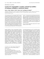
Comparison of the amino-acid sequence of GnT-III
with snail b1,4GlcNAcT and mammalian b1,4GalT-1
In order to identify the essential residues that are important
for GnT-III activity, the candidate residues for examination
were selected on the basis of amino-acid sequence homology
amongratGnT-III,mammalianb1 ,4-galactosylt ransfera se-1
(b1,4GalT-1) and snail b1,4-N-acetylglucosaminyltrans-
ferase (b1,4GlcNAcT). Dot matrix analyses comparing the
GnT-III sequence with that of either of the other enzymes
showed signi®cant similarities in the small region, in which
three aspartic acid residues and several other residues are
perfectly conserved (Fig. 1A,B). Only this homologous
region was detected in all three enzymes. These Asp residues
correspond to Asp321, Asp323 and A sp329 in the rat GnT-
III sequence, as shown by sequence alignment (Fig. 1C).
The sequence of Asp321-Val322-Asp323 appears to corre-
spond to the D -X-D motif, as have been suggested for the
catalytic importance in many other glycosyltransferases
[27±41], and, thus, the sequence comparison suggests that
these Asp residues represent likely candidates for investiga-
tion as having a role in GnT-III activity.
Site-directed mutagenesis of the conserved
aspartic acid residues in GnT-III
To explore the requirement o f the cand idate amino acids,
Asp321, Asp323 and Asp329, for enzyme activity, mutant
Ó FEBS 2002 A dominant negative glycosyltransferase (Eur. J. Biochem. 269) 195
GnT-IIIs in which these Asp residues were replaced by Ala
were prepared using site-directed mutagenesis, and the
respective mutants were designated as D321A, D323A and
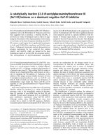
D329A. When these mutants were transiently expressed in
COS-1 cells, t heir protein expression levels were f ound to b e
similar to that of wild-type, as indicated by immunoblot
analysis (Fig. 2A,B). However, in the standard activity
assay, the D321A and D323A mutants had no detectable
catalytic activity, whereas the D329A mutant was fully
active, a s shown by an activity similar to that of the wild-
type enzyme (Table 1). No activity was detected in the
D321A and D323A mutants even under the conditions of
longer incubation time and UDP-GlcNAc concentration as
high as 100 m
M
, ind icating that the replacement of Asp321
and Asp323 lead to a complete loss of activity. These d ata
suggest that Asp321 and Asp323 play a n essential role in t he
activity of enzyme, while Asp329 does not, even though t his
Asp r esidue is conserved. Our previous study showed that
three N-linked glycosylation sites in rat GnT-III are fully
glycosylated when expressed in C OS cells [42], a nd all
mutants used in this study were also found to be fully
glycosylated, as indicated by the digestion with PNGase F
(Fig. 2C). Immuno¯uorescence microscopic a nalysis
showed that the i ntracellular localization of the mutants
are identical to that of the wild-type enzyme, indicating that
the replacements of Asp321 and Asp323 have no effect on
intracellular localization (data not shown). Thus, it seems
unlikely that t hese mutations lead to gross conformational
alterations or misfolding of the protein, but it is more likely
that the loss o f the activity is the result of the deletion of the
active site residues.
The possible role of Asp321 and Asp323
in the catalysis of GnT-III
The absolute requirement for Asp321 and Asp323 and their
conservation in b1,4GalT-1 a nd snail b1,4GlcNAcT sug gest
that the short sequence of Asp321-Val322-Asp323 in GnT-
III plays a role that i s a nalogous to the function of the D -X-D
motif, found in b1,4GalT-1 [13]. Although an essential r ole
for the D-X-D motif has been demonstrated in many
glycosyltransferases [27±35], the function of this motif
seemed divergent. While a large clostridial glucosyltransfer-
ase requires the D-X-D m otif for UDP or UDP-sugar
binding [27], this motif appears not to be critical for
nucleotide binding in GM2 synthase [34] and Fringe [31] in
Fig. 1. Dotplot analyses for rat GnT-III vs. snail b1,4GlcNAcT or bovine b1,4G alT-1. Dotplots for (A) rat G nT-III vs. L. stagnalis b1,4GlcNAcT
and ( B) rat GnT-III vs. bovine b1,4GalT-1 are shown. The am ino-aci d sequences we re compared under the conditions of a window size of 10
residues and 50% of identity using the computer software program,
ALIGN
. The numbers beside the axis indicate the residue number of each
enzyme. Diagonal plots show the homologous regions, which satisfy the ab ove conditions. T he sequences of the most ho mologous region are given
outside the matrices. (C) Multiple alignment of the homologous regions of GnT-III, b1,4GalT-1 and L. stagnalis b1,4GlcNAcT were carried out by
CLUSTALV
. The amino-acid residues which are conserved in all enzymes are highlighted by shaded boxes, and two homologous residues are also
indicated by grey-shaded boxes. The conserved asp artic acid residues that were examined by mutational a nalysis are indicated by arrowheads.
GenBank a ccession numbers for the glycosyltransferases are: G nT-III (human), D13789; GnT-III (rat), NM_01 9239; GnT-III (mouse),
NM_010795; b1,4GlcNA cT (L. stagnalis), X80228; b1,4 GalT-1 (human), X14085; b1,4 GalT-1 (bovine), X14558; b1,4 GalT-1 (mouse), J03880;
b1,4 GalT-2 (mouse), AB019541.
196 H. Ihara et al. (Eur. J. Biochem. 269) Ó FEBS 2002
spite of i ts requirement for their enzyme activities. On the
other h and, crystallographic analyses of b1,4GalT-1 [36] as
well as other glycosyltransferases [37±41] have sugg ested
that this motif serves the coordination of a divalent cation
such as Mn
2+
along with a phosphoryl group of the donor.
The motif would thereby allow t he enzyme to interact with a
nucleotide portion of the d onor nu cleotide sugar and also
may facilitate the reaction via electrostatic catalysis involv-
ing t he divalent cation. Although the function of the D -X-D
motif is not known for GnT-III, it is possible t hat the
D-X-D m otif in GnT-III plays a similar role to that in
b1,4GalT-1 because of the signi®cant sequence homology in
the region containing this motif. In a large clostridial
glucosyltransferase mutant similar to the GnT-III D321A
and D323A mutants, the activity could be recovered in the
presence of extremely high concentrations of Mn
2+
,
supporting the suggestion that the equivalent aspartic acid
residues in the motif are involved in the coordination with
the d ivalent cation [ 27]. Nevertheless, in the case o f GnT-III,
activity was not detected even at concen trations of Mn
2+
as
high as 100 m
M
, suggesting an a bsolute requirement of
Asp321 and Asp323 for the coordination of Mn
2+
during
the reaction of GnT-III (data not shown).
Expression of a catalytically inactive GnT-III
in Huh6 cells, a hepatoblastoma cell line
In order to determine whether a catalytically inactive GnT-
III mutant serves a dominant negative function by prevent-
ing the action of the wild-type endogenou s e nzyme, Huh6
cells, a human hepatoblastoma cell line, were transfected
with the rat GnT-III mutants because a structural pro®le of
the N-glycans in these cells had been previously ch aracter-
ized through a structural analysis of N-linked sugar chains
of a-fetoprotein produced by the cells [43]. H uh6 cells
express relatively high levels of GnT-III and produce
bisected sugar chains, the products of this glycosyltransfer-
ase [ 43]. Following the selection of the transfected cells by
geneticin resistance, clones were grown separately, and the
expression of the rat mutant enz yme was veri®ed by
immunoblot analysis. As a result, we obtained three clones,
which overexpress the D323A GnT-III mutant (Fig. 2D). In
Huh6 cells, a s was in COS-1 cells, the D323A mutant was
expressed a t a similar level to the wild-type, and appeared to
be fully glycosylated (Fig. 2D,F). In the case of the other
mutant, D321A, however, we were not successful in
establishing such clones.
The speci®c suppression of endogenous GnT-III
activity by expression of the catalytically inactive
D323A mutant in Huh6 cells
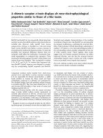
When assays for GnT-III activity were performed in the
transfected cells, it was found that the activity in the
transfected cells were as low as less than 5 % of the activity in
the parental or mock-transfected Huh6 cells, which were
used as controls. On the other hand, the activities of GnT-V,
another GlcNAc transferase which is involved in the
formation of b1,6-branches, and b1,4GalT were not essen-
tially affected in the transfe cted cells, indicating that the
overexpression of the D323A mutant has no effect on these
glycosyltransferase activities (Fig. 3). Therefore, it appears
that the overexpression of the m utant does not impair
Fig. 2. Expression of the wild-type and mutant GnT-III proteins. The
wild-type and mutant enzymes were transiently expressed in COS-1
cells (A±C), and stably expressed in Huh6 c ells (D ±F). (A,D) The cell
homogenates were separated on 8% SDS-gels and were analyz ed by
immunoblot using a nti-(GnT -III) Ig. (B ,E) The amounts of protein
loaded were veri®ed by Ponceau staining, prior to t he immunoblot
analysis. (C,F) The s amples were treated w ith PNGase F, followed b y
SDS/PAGE and immunoblotting. COS-1 an d pSVK3 indicate non-
transfected and vector-transfected (mock) COS-1 cells, respectively.
Huh6, H uh6/D 323A a n d H uh6/WT represent parental nontrans-
fected, D323A-transfected and wild-type-transfected Huh6 cells,
respectively. Details of the conditions are described in Experimental
procedures.
Table 1. Activities of the wild-type and mutant GnT-IIIs transiently
expressed in CO S-1 cells. Gn T-III ac tivity wa s determ ined a s descr ibed
in Experimental procedures. The mock plasmid w as transfected with a
vector, pSVK3. ND, not d etectable .
Plasmid
GnT-III activity
(nmoláh
)1
ámg protein
)1
)
Not transfected ND
Mock ND
Wild-type 0.98
D321A ND
D323A ND
D329A 1.0
Ó FEBS 2002 A dominant negative glycosyltransferase (Eur. J. Biochem. 269) 197
glycosyltransferase a ctivities in a nonspeci®c manner, for
example, via the downregulation of their expression or
damage to the Golgi apparatus. Quite s imilar r esults were
obtained in all three of the obtained clones. These results
suggest that the overexpression of the D323A mutant
speci®cally suppresses the activity of endogenous GnT-III.
To examine whether the mutant GnT-III inhibits the
intrinsic G nT-III in vitro, the extracts from t he parental
cells were mixed with the extract from the D323A-
transfected ce lls, and were then analyze d by the activity
assay. As shown in Fig. 4, no inhibitory effect of the mutant
was observed, and, thus, it seemed unlikely that the in vivo
inhibition by the mutant is due to direct competition for
substrate. Furthermore, RT-PCR using a primer set that is
speci®c to the human GnT-III sequence showed that the
mRNAs for endogenous human GnT-III were not signif-
icantly d ecreased (Fig. 5), suggesting t hat the decreased
GnT-III activity is not due to the downregulation of
expression of the intrinsic human GnT-III gene. Therefore,
it is more likely that t he activity of the i ntrinsic human
enzyme is decreased as the result of control at the
translational or post-translational level including protein±
protein interactions.
Blockage of a speci®c step, namely the formation
of a bisecting GlcNAc residue, in N-glycan biosynthesis
via the expression of the D323A mutant
In order to further examine the issue of whether the
biosynthesis of bisected sugar chains are inhibited by
overexpression of the D323A mutant, total N-linked sugar
chains were prepared from the cells and labeled with
2-aminopyridine, a ¯uorescent reagent. The resulting free
oligosaccharides were digested wi th sialidase, b- galactosi-
dase and a-fucosidase, and then s ubjected to r eversed phase
HPLC, in order to analyze the core structures, i.e . the
addition of a b isecting GlcNAc and t he extent of branching.
As shown in Fig. 6, when parental cells, which express
various sugar chains with bi-, tri- and tetra-antennae were
examined, substantial fractions (20±30%) of these sugar
chains were found to contain t he bisecting GlcNAc. Almost
Fig. 4. Eect of the D323A mutan t on the endogenous GnT-III activity
in vitro. After indicated amounts of the extracts from parental Huh6
cells and the D323A-transfected cells were mixed, GnT-III activity was
assessed. The data are expr essed as the relative value to the activity
determined in the absence of the m utan t extract.
Fig. 3. GnT-III, b1,4GalT and GnT-V activities in the D323A-trans-
fectant. Activities of GnT-III (A), b1,4GalT (B) and GnT-V (C) were
assayed using a pyridylaminated oligosaccharide a cceptor, as desc ribed
in Exp erimental proc ed ures. D ata f or the transfectants are expressed as
mean values from three dierent clones for D323A and 2 clones for the
wild-type GnT-IIIs and mock-transfect ed cells. In the D323A-trans-
fectants, standard deviations are also shown.
198 H. Ihara et al. (Eur. J. Biochem. 269) Ó FEBS 2002
the s ame p ro®le was obtained in the case of the mock-
transfected cells (data not shown). On the other hand, the
D323A-transfected cells were found to expres s n egligible
levels of bisected sugar chains. These results indicate that the
overexpression of the D323A mutant blocks the biosynthe-
sis of bisected sugar chains as the result of a decrease in the
activity of endogenous GnT-III. The results demonstrate
that the c atalytically inactive mutant enzyme acts as a
dominant negative g lycosyltransferase toward the forma-
tion of a bisecting GlcNAc in vivo.
Fig. 5. Detection of m RNA for endogenous h uman GnT-III in the D323A-transfectant by RT-PCR. m RN A expression of endogenous human GnT-
III in parental Huh6 cells and the transfectants for the D323A and wild-type GnT-IIIs were investigated by RT-PCR (upper panel). Reverse
transcription was omitted t o verify the absence o f ampli®cation of genomic DNA in the lanes indicated by RT (±). Human b-actin mRNA
expression was a lso examined as a control (lower pan el). S peci®city t o the endogenous h uman enzyme was con®rme d b y PCR using plasmid D NAs.
Lanes: h uman GnT-III and rat GnT-III indic a te PCR-amp li®cation of plasmids c ontaining cDNAs for hu man and rat e nzym e, resp ec tively. D e tails
regarding the PCR procedures are described in Experimental p rocedures.
Fig. 6. Structural analysis of Asn-linked sugar
chains from parental and transfected Huh6
cells. Elution p ro®les of pyridylaminated sugar
chains in reversed phase HPLC are shown for
parental Huh6 cells ( a), wild-type GnT-III-
transfectant (b) and D 323A-transfectant (c).
Numbers at the top indicate peaks for bisected
sugar chain s: 1, as ialo -agala cto-bisec ted
biantennary sugar chain; 2, asialo-agalacto-
bisected tetraantennary sugar c hain; 3, a sialo-
agalacto-bisected triantennary sugar chain
containing a b1,4-GlcNAc residue on the
Mana1,3 arm. Arro wheads in dicate nonbi-
sected sugar chains: left arrowhead, overlap-
ping peaks of asialo-agalacto biantenna ry and
asialo-agalacto tetraantennary s ugar chains;
right, asialo, agalacto triantennary sugar chain
containing a b1,4-GlcNAc residue on the
Mana1,3 arm. These structures are shown
above the pro®le.
Ó FEBS 2002 A dominant negative glycosyltransferase (Eur. J. Biochem. 269) 199
CONCLUSIONS
Our present study identi®ed Asp321 and Asp323 as being
essential residues in the enzyme activity of GnT-I II, and
provides further support for the v iew that the D-X-D motif
in glycosyltransferases is important and signi®cant, even
though the exact roles of Asp321 and Asp323 in GnT-III
remains to be elucidated. On the other hand , the utility of
the catalytically inactive GnT-III as a dominant negative
glycosyltransferase toward bisecting GlcNAc formation is
clearly demonstrated. The ®ndings contribute to the estab-
lishment of a distinct strategy for in vivo oligosaccharide
manipulation, which permits the speci®c inhibition of a
particular glycosylation step in the biosynthetic pathway in
a manner that is independent of gene targeting. Although a
similarattemptwasmadefora1,3GalT [44], the mechanism
of the inhibition is not known. It is generally thought that
dominant negatively acting molecules involve competition
with the c orresponding endogenous wild-type molecules.
Such competition could also occur in the case of the
dominant negative GnT-III. Possible steps of the c ompeti-
tion could involve: (a) competition for localization in the
Golgi, as o bserved in competition for cell surface b1,4GalT-1
[45]; (b) homophilic interaction involved in the formation of
the active enzyme; (c) association with a presently unknown
molecule that activates the enzym e; a nd (d) activation of the
enzyme by post-translational m odi®cation. Although the
mechanism of a ction of the dominant negative mutant is not
yet known, an understanding of this mechanism could lead
to the discovery of a novel regulatory mechanism for
glycosyltransferase activity.
ACKNOWLEDGEMENTS
We thank Dr Milton S. Feather for correcting t his manuscript. This
research was supported, in part, by a Grant-in-Aid f or Scienti®c
Research on Priority Area no. 10178104 from the Ministry o f
Education, Culture, Sports, Science and Technology of Japan, and
by the Sumitomo Foundation.
REFERENCES
1. Narasimhan, S. (1982) Control of glycoprotein synthesis. UDP-
GlcNAc: glycopeptide beta 4-N-acetylglucosaminyltransferase III,
an enzyme in hen oviduct which adds GlcNAc in beta 1 ® 4
linkage to the beta-linked mannose of the trimannosyl core of
N-glycosyl oligosaccharides. J. Biol. Chem. 257, 10235±10242.
2. Schachter, H. (1986) Biosynthe tic controls that determine the
branching and microhete rogeneity of prote in-bou nd oligosac char-
ides. Biochem. Cell Biol. 64, 163±181.
3. Sultan, A .S., Miyoshi, E., Ihara, Y., Nishikawa, A., T sukada, Y.
& Taniguchi, N. (1997) Bisecting G lcNAc structures act as nega-
tive sorting signals for cell surface glycoproteins in forskolin-
treated rat hepatoma cells. J. Biol. Chem. 272, 2866±2872.
4. Ihara,Y.,Yoshimura,M.,Miyoshi,E.,Nishikawa,A.,Sultan,
A.S., Toyosawa, S., Ohnishi, A., Suzuki, M., Yamamura, K.,
Ijuhin, N . & Taniguch i, N. (1998) E ctopic expression of N-acety-
lglucosaminyltransferase I II in t ransgenic hepatocytes disrupts
apolipoprotein B secretion and induces aberrant cellular mor-
phology with lipid storage. Proc. Natl Acad. Sci. USA 95, 2526±
2530.
5. S heng, Y., Yoshimura, M., Inoue, S., Oritani, K., Nishiura, T.,
Yoshida,H.,Ogawa,M.,Okajima,Y.,Matsuzawa,Y.&
Taniguchi, N. (1997) Remodeling of glycoconjugates on CD44
enhances cell adhesion to hyaluronate, tumor growth and meta-
stasis in B16 melanoma cells expressing b1,4-N-acetylglucosami-
nyltransferase III. Int. J. Cancer 73, 850±858.
6. Yoshimura, M., Ihara, Y., Matsuzawa, Y. & Taniguchi, N. (1996)
Aberrant glycosylation of E-cadherin enhances cell±cell binding to
suppress metastasis. J. Biol. Chem. 271, 13811±13815.
7.Yoshimura,M.,Nishikawa,A.,Ihara,Y.,Taniguchi,S.&
Taniguchi, N. (1995) Suppression of lung metastasis of B16 mouse
melanoma by N-acetylglucosaminyltransferase III gene transfec-
tion. Proc. Natl Acad. Sci. USA 92, 8754±8758.
8. Bhaumik, M., Harris, T., S undaram, S., Johnson, L., Guttenplan,
J., Rogler, C. & Stanley, P. (1998) Progression of hepatic neo-
plasms is severely retarded in mice lacking the bisecting
N-acetylglucosamine on N-glycans: evidence for a glycoprotein
factor that facilitates hepatic tumor p rogression . Cancer Res.
58, 2881±2887.
9. Yang, X., Bhaumik, M., Bhattac haryya, R., G ong, S., Rogler,
C.E. & S tanley, P . (2000) Ne w e vidence for an extra-hepatic role of
N-acetylglucosaminyltransferase III in the progression of diethyl-
nitros amin e-in duce d liver tumors in m ice . Cancer Res. 60, 3313±
3319.
10. Schachter, H ., Narasimhan, S ., Gleeson, P. & Vella, G. (1983)
Control of branching during th e biosynthesis of asparagine-linked
oligosaccharides. Can. J. Biochem. Cell Biol. 61, 1049±1066.
11. I keda, Y., K oyota, S., Ihara, H., Yamaguchi, Y., Ko rekane, H.,
Tsuda, T., Sasai, K. & Taniguchi, N. (2000) Kinetic basis for the
donor nucleotide-sugar speci®city o f b1,4-N-acetylglucosaminyl-
transferase III. J. Biochem. (Tok yo) 12 8 , 609±619.
12. Bakker, H., A gterberg, M., Van Tetering, A., Koeleman, C.A.,
Van den Eijnden, D.H. & Van Die, I. (199 4) A Lymnaea stagnalis
gene, with s equence s imilarity to t hat o f m ammalian b1 ®
4-galactosyltransferases, encodes a novel U DP-GlcNAc: GlcNAc
b-R. b1 ® 4-N-acetylglucosaminyltransferase. J. Biol. Chem. 269 ,
30326±30333.
13. B reton, C., B ettler, E., Joziasse, D.H., Geremia, R. & Imberty, A.
(1998) Sequence±function relationships of prokaryotic a nd eukar-
yotic galactosyltransferases. J. Biochem. (Tokyo) 123, 1000±1009.
14. Nishikawa, A., Ihara, Y ., Hatakeyama, M., Kangawa, K. &
Taniguchi, N. (1992) Puri®cation, c DNA c loning, and expression
of UDP-N-acetylglucosaminyltransferase III from rat kidney.
J. Biol. Chem. 267, 18199±18204.
15. Niwa, H., Yamamura, K. & Miyazaki, J. (1991) Ecient selection
for high-expression transfectants with a n ovel e ukaryotic vector.
Gene 108, 193±199.
16. Kunkel, T.A. (1985) Rapid and ecient site-speci®c mutagenesis
without phenotypic selectio n. Proc.NatlAcad.Sci.USA82, 488±
492.
17. Ikeda, Y., Fujii, J., Taniguchi, N. & Meister, A. (1995) Human
c-glutamyl transpeptidase mutants involving conserved aspartate
residues and the unique cystein e residue of the ligh t subunit. J. Biol.
Chem. 270, 12471±12475.
18. Laemmli, U.K. (1970) Cleavage of structural proteins during the
assembly of the h ead of bacteriophage T4. Nature 227, 680±685.
19. Chu, G., H ayakawa, H. & Berg, P. (1987) Elec troporation for the
ecient transfection of mammalian cells with DNA, Nucleic Acids
Res. 15, 1311±1326.
20. T aniguchi, N., Nishikawa, A., F ujii, S. & Gu, J.G. (1989) Gly-
cosyltransferase assays using p yridylaminated acceptor: N-acetyl-
glucosaminyltransferase III, IV, and V. M ethod s Enzymol. 179,
397±408.
21. Nishikawa, A., Fujii, S., Sugiyama, T. & Taniguchi, N. (1988) A
method for the determin atio n o f N-acetylglucosaminyltransferase
III activity in rat tissues involving HPLC. Anal. Biochem. 170,
349±354.
22. S eko, A., Koketsu, M., N ish izono, M., Enoki, Y ., Ibrahim, H.R.,
Juneja, L.R., Kim, M. & Yamamoto, T. (1997) Occurrence of a
sialylglycopeptide and free sialylglycans in hen's egg yolk.
Biochem. Biophys. Acta 1335, 23±32.
200 H. Ihara et al. (Eur. J. Biochem. 269) Ó FEBS 2002
23. Patel,T.,Bruce,J.,Merry,A.,Bigge,C.,Wormald,M.,Jaques,A.
& Parekh, R. (1993) Use of hydrazine to release in i ntact and
unreduced form both N- and O-linked oligosaccharides from
glycoproteins. Biochemistry 32, 679±693.
24. Hase, S., Ibuki, T. & Ikenaka, T . (1984) Reexamination of the
pyridylamination used for ¯uorescence labeling o f oligosacchar-
ides and its app licatio n to glycoproteins. J. Biochem. (Tokyo) 95,
197±203.
25. Morita, N., Hase, S., Ikeh ara, K., Mikoshiba, K. & Ikenaka, T.
(1988) Pyridylamino sugar chain a s an a cceptor for galactosyl-
transferase. J. Biochem. (Tokyo) 103, 332±335.
26. Nishiura, T., Fujii, S., Kanayama, Y., Nishikawa, A., Tomiyama,
Y., Iida, M., Karasuno, T., Nakano, H., Yonezawa, T., Tanigu-
chi, N. & Tarui, S. (1990) Carbohydrate analysis of immuno-
globulin G myeloma proteins by lectin and high performance
liquid chromatography: role of glycosyltransferase. Cancer Res.
50, 5345±5350.
27. Busc h, C., Hofmann, F., Selzer, J., Munro, S., Jeckel, D. &
Aktories, K. (1998) A common motif of eukaryotic glyco-
syltransferases is essential for the enzyme activity of large clostri-
dial cytotoxins. J. Biol. Chem. 273, 19566±19572.
28. Wiggins, C.A.R. & Munro, S. (1998) Activity of the yeast MNN1
a-1,3-mannosyltransferase requires a motif conserved in many
other families of glyc osyltransf erases. Proc. Natl Acad. Sci. USA
95, 7945±7950.
29. Shibayama, K., Ohsuka, S., Tanaka, T., Arakawa, Y . & Ohta, M.
(1998) Conserved structural regions involved in the catalytic
mechanism of Escherichia coli K-12 WaaO (RfaI). J. Bacteriol.
180, 5313±5318.
30. Hagen, F.K., H azes, B., de Rao, R.Sa, D. & Tabak, L.A. (1999)
Structure-function an alysis of the UDP -N-acetyl-
D
-galactos-
amine: polypeptide N-acetylgalactosaminyltransferase. Essential
residues lie in a predicted active site cleft resembling a lactose
repressor fold. J. Biol. Chem. 274, 6797±6803.
31. Munro, S. & Freeman, M. (2000) The notch signalling regulator
fringe acts in the Golgi apparatus and requires the glycosyltrans-
ferase sig natu re mo tif D XD. Curr. Biol. 10, 813±820.
32. Keusch, J.J., Manzella, S.M., Nyame, K.A., Cummings, R.D. &
Baenziger, J.U. (2000) E xpression cloning of a new member of the
ABO blood g roup g lycosyltransferases, iGb3 s ynthase, t hat directs
the synthesis of isoglobo-glycosphingolipids. J. Biol. Chem. 275,
25308±25314.
33. Keusch, J.J., Manzella, S.M., Nyame, K.A., Cummings, R.D. &
Baenziger, J.U. (2000) Cloning of G b3 syn thase, t he key e nzyme i n
globo-series glycosphingolipid synthesis, predicts a family of
a1,4-glycosyltransferases conserved inplants, insects, and mam-
mals. J. Biol. Chem. 275, 25315±25321.
34. Li, J ., Rancour, D.M., Allende, M.L., Worth, C.A., D arling, D.S.,
Gilbert,J.B.,Menon,A.K.&Young,W.W.Jr(2001)TheDXD
motif is required for GM2 synthase activity but is not critical for
nucleotide binding. Glycobiology 11, 217±229.
35. Maeda, Y., Watanabe, R., Harris, C.L., Hong, Y., Ohishi, K.,
Kinoshita, K. & K inoshita, T. (2001) PIG-M transfers the ®rst
mannose t o glycosylphosphatidylinositol on the lumenal side of
the ER. EMBO J. 20, 250±261.
36. Gastinel, L.N., Cam billau, C. & Bourne, Y. ( 1999) Crystal
structures of t he b ovine b4-galactosyltransferase catalytic domain
and its complex with urid ine diphosp hogalactose. EMB O J. 13,
3546±3557.
37. Charnoc k, S .J. & Davies, G.J. (1999) Structure of the nu cleo tide-
diphospho-sugar transferase, SpsA from Bacillus subtilis,innative
and nucleotide-complexed forms. Biochemistry 38, 6380±6385.
38. Pedersen, L.C., T suchida, K., Kitagawa, H., Sugahara, K.,
Darden, T.A. & Negishi, M. (2000) Heparan/chondroitin sulfate
biosynthesis. Structure and m ech anism of hu man glucuronyl-
transferase I. JBiolChem.275, 34580±34585.
39. Unligil, U.M., Zhou, S., Yuwaraj, S., Sarkar, M., Schachter, H. &
Rini, J.M. (2000) X -ray crystal s tructure of rabbit N-acetylglu-
cosaminyltransferase I: catalytic mechanism and a new protein
superfam ily. EMB O J. 19, 5269±5280.
40. Persson, K., Ly, H.D., Dieckelmann, M., Wakarchuk, W.W.,
Withers, S.G. & Strynadk a, N.C. (2001) Crystal structure of the
retaining galactosyltransferase LgtC from Neisseria meningitidis in
complex with donor an d acceptor sugar analogs. Nat. Struct. Bio l.
8, 166±175.
41. Gastinel, L.N., Bignon, C., Misra, A.K., Hindsgaul, O., Shaper,
J.H. & Joziasse, D.H. (2001) Bovine a1,3-galactosyltransferase
catalytic domain structure and its relatio nship with ABO histo-
blood gro up and glycosphingolipid glycosyltransferases. EMBO J.
20, 638±649.
42. Nagai, K., Ihara, Y., Wada, Y. & Taniguchi, N. (1997) N-Gly-
cosylation is requisite for the enzyme activity and Golgi r eten-
tion of N-acetylglucosaminyltransferase III. Glycobiology 7,769±
776.
43. Ohno, M., Nishikawa, A., Koketsu, M., Taga, H., Endo, Y.,
Hada, T., Higashino, K. & Taniguchi, N. (1992) E nzymatic basis
of sugar structures of a-fetoprotein in hepatoma and hepato-
blastoma cell l ines: correlation with activities of a1,6,fucosyl-
transferase and N-acetylglucosaminyltransferases III and V. Int. J.
Cancer 8, 315±317.
44. Ogawa, H ., Kobayashi, I., Nagasaka, T., N amii, Y ., Hayashi, S.,
Kadomatsu, K., Muramatsu, T. & Takagi, H. (1999) Suppression
of porcine xenoantigen expression by dominant-negative eect of
a-1,3-galactosyltransferase (a-1,3-GT) splicing variants. Tr ans-
plant. Proc. 32, 58.
45. Evans, S.C., Lopez, L.C. & Shur, B.D. ( 1993) Dom inant ne gative
mutation in cell su rfac e b1,4-galactosyltransferase inhibits cell±cell
and cell±matrix interactions. J. Cell Biol. 120, 1045±1057.
Ó FEBS 2002 A dominant negative glycosyltransferase (Eur. J. Biochem. 269) 201