Báo cáo Y học: Molecular cloning, bacterial expression and properties of Rab31 and Rab32 docx
Bạn đang xem bản rút gọn của tài liệu. Xem và tải ngay bản đầy đủ của tài liệu tại đây (778.5 KB, 13 trang )
Molecular cloning, bacterial expression and properties of Rab31
and Rab32
New blood platelet Rab proteins
Xiankun Bao
1
, Andrea E. Faris
2
, Elliott K. Jang
1
and Richard J. Haslam
1,2
Departments of
1
Pathology and Molecular Medicine and
2
Biochemistry, McMaster University, Hamilton, Ontario, Canada
GTP-binding proteins of the Rab family were cloned from
human platelets using RT-PCR. Clones corresponding to
two novel Rab proteins, Rab31 and Rab32, and to Rab11A,
which had not been detected in platelets previously, w ere
isolated. The coding sequence of Rab31 (GenBank acces-
sion no. U59877) corresponded to a 194 amino-acid protein
of 21.6 kDa. The Rab32 sequence was exten ded to 1000
nucleotides including 630 nucleotides of coding sequence
(GenBank accession no. U59878) but t he 5¢ coding
sequence was only completed later by others (GenBank
accession no. U71127). Human Rab32 cDNA encodes a 225
amino-acid protein of 25.0 kDa with the unusual GTP-
binding sequence DIAGQE in place of DTAGQE. North-
ern blots for Rab31 and Rab32 identi®ed 4.4 kb and 1.35 kb
mRNA species, respectively, in some human tissues and in
human erythroleukemia (HEL) cells. Rabbit polyclonal
anti-peptide antibodies to Rab31, Rab32 and Rab11A
detected platelet proteins of 22 kDa, 28 kDa and 26 kDa,
respectively. Human platelets were highly enriche d in
Rab11A ( 0.85 lgámg of platelet protein
)1
) a nd contained
substantial amounts of Rab32 (0.11 lgámg protein
)1
). Little
Rab31 was present (0.005 lgámg protein
)1
). All three Rab
proteins were found in both granule and membrane frac-
tions from platelets. In rat platelets, the 28-kDa R ab32 was
replaced by a 52-kDa immunoreactive p rotein. Rab31 and
Rab32, expressed as glutathione S-transferase (GST)-fusion
proteins, did not bind [a-
32
P]GTP on nitrocellulose blots but
did bind [
35
S]GTP[S] in a Mg
2+
-dependent manner. Bind-
ing of [
35
S]GTP[S] was optimal with 5 l
M
Mg
2+
free
and was
markedly inhibited by higher Mg
2+
concentrations in the
case of GST±Rab31 but not GST±Rab32. Both proteins
displayed low steady-state GTPase activities, w hich were
not inhibited by mutations (Rab31
Q64L
and Rab32
Q85L
)
that abolish the GTPase activities of most low-M
r
GTP-
binding proteins.
Keywords: Rab protein; GTP-binding protein; GTPase;
Mg
2+
; p latelet.
Low-M
r
GTP-binding proteins of the Rab subfamily play
important roles in vesicle and granule targeting [1]. Because
blood platelets secrete the contents of three distinct granule
types, namely dense g ranules, a-granules and lysosomes, i n
response to physiological stimuli [2], the identities and
subcellular locations of platelet Rab proteins are of
considerable interest. Immunoblotting experiments using
antibodies to known Rab proteins have demonstrated the
presence in human platelets of Rab1, Rab3B, Rab4, Rab5,
Rab6 and R ab8 [3,4], a s well as Rab27A [5,6] and Rab27B
[6,7]. Rab6 and R ab8 were detected on a-granules [3],
although evidence has been presented that Rab4 regulates
a-granule secretion [ 4]. Over 50 different Rab proteins have
now been identi®ed, some of which are highly tissue- or cell-
speci®c [1,8]. Consequently, it is not certain that all the
major p latelet Rab proteins have been identi®ed. We
therefore adopted a different ap proach to the identi®cation
of Rab proteins in human platelets and cloned sequences
from platelet mRNA by RT-PCR, using a degenerate
oligonucleotide corresponding to the conserved protein
sequence WDTAGQE, found in members of t he Rab and
Rho families of low-M
r
GTP-binding proteins. Antibodies
to unique C-terminal peptide sequences were then prepared
and used to con®rm the presence of the proteins in platelets.
By these methods, we identi®ed two previously unknown
Rab proteins, Rab31 and Rab32, in human platelets and
also demonstrated the presence in these platelets o f large
amounts of Rab11A. Here, we d escribe the cloning and
tissue distributio n o f R ab31 and Rab32, their bacterial
expression and s ome unusual b iochemical properties of t he
recombinant p roteins. Brief reports of some of our ®ndings
have been published in abstract form [9,10].
EXPERIMENTAL PROCEDURES
Materials
An AmpliFINDER
TM
RACE kit, a Marathon
TM
cDNA
ampli®cation kit and a human multiple tissue Northern blot
were obtained from Clontech Laboratories Inc.
Correspondence to R. J. Haslam, Department of Pathology and
Molecular Medicine, McMaster University, 1200 Main Street West,
Hamilton, Ontario, Canada L8N 3Z5. Fax: + 905 777 7856,
Tel.: + 905 525 9140 Ext. 22475, E-mail:
Abbreviations: ACS, aqueous counting scintillant; ECL, enhanced
chemiluminescence; GAPDH, glyceraldehyde 3-phosphate
dehydrogenase; GTP[S], guanosine 5¢-O-(c-thio)triphosphate; GST,
glutathione S-transferase.
Enzyme: g lutathione S-transferase ( EC 2.5.1.18).
(Received 10 July 2001, revised 16 October 2001, accepted 30 October
2001)
Eur. J. Biochem. 269, 259±271 (2002) Ó FEBS 2002
[a-
32
P]dCTP (3000 Ciámmol
)1
)and[
35
S]guanosine 5¢-O-
(c-thio)triphosphate ([
35
S]GTP[S], 1250 Ciámmol
)1
)were
from NEN and [a-
32
P]GTP (> 3000 Ciámmol
)1
)was
from ICN Pharmaceuticals. Immobilon-P membrane for
blotting proteins, HAWP ®lters (0.45 lm, 25 mm) and
Centricon YM-10 ®lters were from Millipore. RPMI 1640
medium, f oetal bovine serum,
L
-glutamine, T4 D NA ligase,
Taq DNA polymerase, MMLV r everse transcriptase,
Superscript
TM
II reverse transcriptase/Taq mix and
restriction enzymes were all from Life Technologies. QIA-
quick
TM
PCR puri®cation kits and QIAprep
TM
plasmid
DNA puri®cation kits were from Qiagen. pBluescript
SK+ DNA was from Stratagene. pGEX-4T-1 DNA,
glutathione-Sepharose 4B, aqueous counting scintillant
(ACS), secondary antibody for immunoblotting and
enhanced chemiluminescence (ECL) reagents w ere f rom
Amersham Pharmacia Biotech. Avid A L c olumns were
from BioProbe International. UltraLink Iodoacetyl gel for
peptide a f®nity-puri®cation of antibodies was from Pierce.
Darco G60 activated carbon was from Fisher Scienti®c.
Oligonucleotides and peptides were synthesized and DNA
sequenced in the Central Facility of the Institute f or
Molecular B iology and Technology (McMaster University,
Canada). The
PEPTOOL
TM
program used f or sequence
alignment w as obtained from B ioTools Inc. The authors
are very grateful to P. D. Stahl (Washington University
School of Medicine, St Louis, MO, USA) f or providing
Escherichia coli expressing GST±Rab5A [11]. A sample of
recombinant Rab11A protein [12] was generously supplied
by J. R. Goldenring (Institute for M olecular Medicine and
Genetics, Medical College of Georgia, Augusta, GA, USA).
Iloprost was a gift from Schering AG.
Human cell lines
MEG-01 cells (a megakaryoblastic leukemia cell line) were
donated b y K . K. Wu ( University of Texas, H ealth Science
Center, Houston, TX, USA). Cultures of other human cell
lines were obtained from the following sources in the
Department of Pathology a nd Molecular Medicine at
McMaster University, Hamilton, Canada: HEL cells (an
erythroleukemia cell line) from B. J. Clarke, K562 cells (a
multipotential haematopoietic cell line) and Jurkat cells
(a T-cell line) from K. Rosenthal, and KU812 ce lls (a
basophilic leukemia cell line) from J. Marshall. Cell lines
were routinely grown in RPMI 1640 m edium supplemented
with antibiotics and 10% foetal bovine serum (heated at
56 °C for 30 min).
L
-Glutamine (0.03%, w/v) was added
into the medium for MEG-01 cells only.
Isolation of human platelets and their subcellular
fractionation
Platelets were isolated by a modi®cation of the method of
Mustard et al. [13]. Blood (100 mL) w as taken from
healthy human donors and centrifuged for 5 min at 140 g
to separate platelets from o ther blood cells. T o minimize
contamination by other cells, only the top one third of the
platelet-rich plasma was collected when mRNA was
prepared. The platelets were isolated by centrifugation
for 5 min at 1160 g and the pellet was washed three times
in 10 mL of Ca
2+
-free Tyrode's solution containin g 0.35%
BSA, 5 m
M
Pipes, pH 6.5, 90 lgámL
)1
of apyrase,
50 IU ámL
)1
of heparin and 20 n
M
iloprost. Care was
taken to remove any residual red and white cells during
washes. In s ome experiments, platele t cytosol and partic-
ulate fractions enriched in either granules or membranes
were prepared by differential centrifugation of platelet
sonicates [14].
Isolation of mRNA and total RNA
Micro-FastTrack mRNA Isolation Kits (Invitrogen) were
used to extract mRNA from platelets and HEL cells,
whereas TRIzol Reagent (Life Technologies) was used to
isolate total RNA from HEL, K562 and Jurkat cells,
according to the manufacturer's protocol.
Cloning of platelet
Rab
cDNA sequences
cDNA was synthesized from human platelet mRNA using
MMLV reverse transcriptase and an RT primer
(5¢-GGACTAGTGTCGACAAGCTTGAATTCT
17
-3¢,
43-mer) consisting of oligo-dT with four added restriction
sites (SpeI, SalI, HindIII and EcoRI, shown in bold). The
RT reaction mixture was then added to a PCR cocktail. The
sense oligonucleotide used for PCR ampli®cation was a
128-fold degenerate oligonucleotide encoding the amino-
acid sequence, WDTAGQE, with BamHI and XbaI
restriction sites (in bold) at the 5¢ end (5¢-CGGGATCCTCT-
AGATGGGA(T/C)AC(A/G)GC(A/T/C/G)GG(A/T/C/G)
CA(A/G)GAG-3¢, 35-mer). In some r eactions, this primer
was replaced by an oligonucleotide identical except for the
replacement o f t he 3 ¢Gby3¢A. Two separate 5 ¢ primers
were used to avoid degeneracy in any of the three bases at
their 3¢ ends. The antisense oligonucleotide w as a 26-mer
adaptor identical to the 5 ¢ half of the R T primer. PCR w ith
Taq DNA polymerase was carried out by heating the
mixtur e a t 9 5 °C for 2 min, followed by 4 0 cycle s of 1 min
at 9 5 °C and 4 min at 68 °C, and t hen a ®nal 7 min a t
72 °C. The products were then cloned into pBluescript
SK+ using the XbaIandHindIII or EcoRI restriction sites
and inserts larger than 450 bp were sequenced. After
identi®cation o f novel Rab cDNAs, the 5¢ nucleotide
sequences were obtained by 5 ¢-RACE, using antisense
primers based on 5¢ sequences of partial clones of Rab
proteins and either an AmpliFINDER
TM
RACE kit or a
Marathon
TM
cDNA ampli®cation kit. PCR products were
then cloned into pBluescript SK+. After a complete
(Rab31) or nearly complete (Rab32) sequence was assem-
bled, the ORF was reampli®ed from a platelet Marathon
TM
cDNA library using a ppropriate sp eci®c primers, cloned i n
pBluescript SK+ and resequenced in both directions to
verify the assembled sequence.
Northern blotting analysis
Total RNA from HEL, K562 and Jurkat cells was
electrophoresed on a 1% agarose/formaldehyde gel, blotted
on to nylon membrane and cross-linked with UV light.
Probes ( 50 ngámL
)1
and 10
7
c.p.m.ámL
)1
) labelled with
[a-
32
P]dCTP were prepared by PCR ampli®cation of Rab
cDNA sequences encoding amino-acid residues f rom t he
C-terminal halves of the proteins ( and 3¢-untranslated
sequence), as follows: Rab31, nucleotides 366±788; Rab32,
nucleotides 546±868 (see Fig. 1A,B). For a control probe,
260 X. Bao et al. (Eur. J. Biochem. 269) Ó FEBS 2002
nucleotides 252±793 of human glyceraldehyde 3-phosphate
dehydrogenase (GAPDH) cDNA (GenBank accession no.
M33197) were ampli®ed f rom a HEL cell cDNA library
(prepared using the Marathon
TM
cDNA ampli®cation kit).
Hybridization was carried out at 68 °C f or 1 h as described
previously [15]. The same membranes w ere probed succes-
sively for Rab31, Rab32 and GAPDH mRNAs, with
intermediate stripping by heating at 100 °Cin0.5%SDS
for 10 min.
Immunoblotting
Rabbit anti-peptide antibodies were prepared to peptide
sequences in the hypervariable C-terminal regions of
cloned R ab proteins. T he peptides synthesized were
(CH
3
CO)TIKVEKP TMQASRRC for Rab31 (Fig. 1 A),
(C)NEENDVDKIKLDQE(CONH
2
) for Rab32 (Fig. 1B),
and (C)QKQMSDRRENDMS(CONH
2
) f or Rab11A
(amino-acid residues 178±190). These peptides were coupled
to keyhole limpet haemocyanin via their endogenous
(Rab31) or added (Rab32, Rab11A) cysteine residues, using
4-(N-maleimidomethyl) cyclohexane-1-carboxylate. Rabbits
were immunized by intradermal injection of the conjugated
peptides (0.5±1.0 mg) with Freund's complete adjuvent.
Sera with adequate titres were obtained a fter 3±4 boosts
with conjugated peptide i n incomplete adjuvent. Protein for
immunoblotting was analysed by SDS/PAGE, using 13%
acrylamide in the separating gel, and then transferred
electrophoretically to Immobilon-P. Immunoreactive pro-
teins were detected using the rab bit immune sera, immune
IgG puri®ed on Avid AL columns or antibody af®nity-
puri®ed on an UltraLink I odoacetyl column containing
covalently bound peptide. Bound antibody was visualized
using horse-radish peroxidase-conjugated donkey anti-
(rabbit IgG) Ig as t he se condary antibody and ECL reagents.
Bacterial expression of Rab31 and Rab32
To generate an expression construct, Rab31 cDNA was
ampli®ed from a platelet Marathon
TM
cDNA library, using
as PCR primers, 5¢-TAGGATCCGCGATACGGGAGC-
TCAAAG-3¢ (P31-1) and 5 ¢-ATCTCGAGGATGTGGG-
Fig. 1. Nucleotide and deduced amino-acid s equences of Rab31 and Rab32. (A) Rab31. The nu cleotide sequence shown ( Ge nBank a cc ession no.
U59877) was obtained as described under Experimental procedures. An almost identical cDNA cloned at the same time from human melanocytes
[30] diers by two bases and one amino acid in the open reading frame (see box). (B) Rab32. The nucleotide sequence shown is derived from two
clones, o ne obtained as describ ed i n the Experimental proc edures (GenBank accession no. U59878) and a second, which completed the 5¢ end of the
open reading frame, obtained later from GenBank (U71127). Sequence variants found in Rab32 cDNA from HEL cells are boxed. For both Rab31
and Rab32, the conserved amino-acid sequences involved in binding GDP/GTP are shown white on black, the glutamine residues mutated in this
study a re shade d and the pe ptide sequences used for generating a ntib odies are doubly un derlined. The nucleotide se qu ences that were u sed f o r
ampli®cation of cD NAs that were ligated into pG EX -4T-1 are also indicated (P31-1, P31-2, P32-1, P32-2, see E xperim ental procedure s).
Ó FEBS 2002 Rab proteins from platelets (Eur. J. Biochem. 269) 261
CTCTGGCTTCT-3¢ (P31-2), containing BamHI and Xh oI
restriction sites (in bold), respectively (Fig. 1A). The PC R
reaction conditions (with Taq DNA polymerase) were
1 min at 95 °C, 2 min at 60 °C (®rst cycle) or 2 min at 65 °C
(remaining cycles) a nd 2 min at 72 °C, for a tot al o f 21
cycles. T he PCR p roduct was puri®ed using a QIAquick
TM
kit and cloned into the BamHI and XhoIsitesofthe
pGEX-4T-1 expression vector. The sequence of the insert
was veri®ed in both directions and competent E. coli BL21
(DE3) cells transformed. In the expressed GST±Rab31
fusion protein, the initiating methionine of Rab31 was
replaced by GS residues (from the BamHI restriction site).
To verify the coding seq uence of Rab32 and generate an
expression construct, human platelet mRNA was ®rst
isolated using a Micro-FastTrack
TM
kit. Reverse trans crip-
tion of this platelet mRNA was carried out using a
Superscript
TM
II RT/Taq mix (30 min at 55 °Cand2min
at 94 °C) and a primer (P32-2) containing XhoIandSalI
restriction sites (in bold) and the complement of nucleotides
700±724 of Rab32 cDNA (5¢-AAGCTCGAGTCGAC-
TTCTTCAGAGCTGAGGCACACAC-3¢). The resulting
cDNA was a mpli®ed by PCR using 5¢-TGGGATCC-
GGAGGAGCCGGGGACCCCGGCCTG-3¢, containing
a BamHI si te, a s the 5¢ primer (P32-1) and P32-2 as the
3¢ primer (Fig. 1B). The PCR reaction conditions (with Taq
DNA polymerase) were 1 min at 94 °C, 2 min at 60 °Cand
2 min at 72 °C, for 3 5 cycles. The p roduct was c loned into
the BamHI a nd XhoI sites of pGEX-4T-1 a nd sequenced in
both directions. Th e GST±Rab32 fusion protein was
expressed in E. coli BL21(DE3) cells, as for Rab31. In this
fusion pro tein, the ®rst three a mino-acid residues of Rab32
(MAG) were replaced by GS residues.
To express GST-fusion proteins and GST itself, 4 mL of
Luria±Bertani medium (containing 100 lg of ampicillin per
mL) was inoculated with E. coli BL21(DE3) cells contain-
ing the appropriate pGEX-4T-1 construct and grown
overnight at 37 °C. This culture was used to seed a larger
culture (200±500 mL), which was grown at 37 °C until
D 0.5. The fusion protein was then induced with 0.1 m
M
isopropyl t hio-b-
D
-galactoside and the culture grown for a
further 3 h, when the b acteria were isolated by centrifuga-
tionandfrozenat)70 °C until needed. B acterial pellets
(each from 50 m L of culture) were resuspended in 9.8 mL
of NaCl/P
i
(pH 7.4) containing lysozyme (100 lgámL
)1
).
After incubation of the cells for 30 min at 0 °C, 1 m
M
phenylmethanesulfonyl ¯uoride and 5 m
M
dithiothreitol
were ad ded and the cells were sonicated f or 10 min in a
bath sonicator. Bacterial supernatant was then isolated by
centrifugation and mixed with Triton X-100 (®nal concen-
tration 0.1%), MgCl
2
(10 m
M
) and glutathione-Sepharose
4B beads. After s haking the mixture for 60 min at room
temperature, the beads were isolated, washed three times
with NaC l/P
i
containing 0.1% Triton X-100 and 10 m
M
MgCl
2
andthenelutedwith10m
M
reduced glutathione in
50 m
M
Tris/HCl (pH 8.0) containing 10 m
M
MgCl
2
.The
eluted protein was concentrated by centrifugation at 4 °C
in a Centricon ®lter, diluted with Buffer A (100 m
M
KCl,
20 m
M
Hepes, pH 7.5, 1 m
M
EDTA, 1 m
M
dithiothreitol)
containing 10 m
M
MgCl
2
, and reconcentrated. GST-fusion
proteins were stable in this solution for 1±3 weeks at 4 °C.
Protein concentrations were determined by the Lowry
method using Sigma protein standard diluted in Buffer A.
Inclusion of MgCl
2
in the above solutions was essential to
obtain GST±Rab proteins capable o f binding guanine
nucleotides.
Mutagenesis of Rab31 and Rab32
An attempt was made to create constitutively active forms
of these R ab proteins by mutating the g lutamine re sidue in
the DTAGQE GTP-binding motif to a leucine r esidue
(Fig. 1A,B). A dual PCR method [16] was applied to the
Rab31 and Rab32 c onstructs in pGEX-4T-1 t o g ive clones
encoding GST±Rab31
Q64L
and GST±Rab32
Q85L
,respec-
tively. These proteins were expressed and puri®ed, as
described for GST±Rab31 and GST±Rab32.
Binding of [a-
32
P]GTP by GST±Rab proteins
on nitrocellulose blots
After SDS/PAGE, GST±Rab proteins were electroblotted
onto nitrocellulose, renatured and p robed w ith [a-
32
P]GTP
by two different methods [17,18]. I n one, t he binding buffer
contained 2 l
M
MgCl
2
[17] and in t he other, 10 m
M
MgCl
2
[18].
Binding of [
35
S]GTP[S] by GST±Rab proteins
A modi®cation of the method of Kabcenell et al. [19] was
used. Puri®ed protein (10±200 pmol) was incubated at
37 °C in Buffer A containing [
35
S]GTP[S] (usually 5 l
M
at a
speci®c radioactivity of 0.5 Ciámmol
)1
) and any additional
MgCl
2
required to g ive a de®ned concentration of Mg
2+
free
(usually 5 l
M
or 10 m
M
). The amount of MgCl
2
required
was calcu lated u sing a computer version of the programme
described by Fabiato & Fabiato [20] and the binding
constants used by the same authors. At speci®c times,
triplicate 10 lL samples of the incubation mixture were
diluted into 100 lL of wash buffer [100 m
M
KCl, 20 m
M
Hepes (pH 7.5), 0.5 m
M
MgCl
2
] and immediately applied to
HAWP ®lters in a Millipore vacuum ®ltration unit. After
three washes with 2 mL of wash buffer, the ®lters were
placed in vials with 0 .5 mL of water a nd 8 mL of A CS and
counted for
35
S by liquid scintillation. After correction for
the [
35
S]GTP[S] observed on ®lters from control incubations
without protein, t he results w ere e xpressed as p mol of
[
35
S]GTP[S] bound per 100 pmol of protein ( mean SE);
this is equivalent to the percentage of protein containing
bound [
35
S]GTP[S].
GTPase assays
The GTPase activities of GST±Rab proteins were measured
by a modi®cation o f the method of Kabcenell et al.[19].
Puri®ed protein (10±200 pmol) was incubated at 37 °Cin
40±200 lL o f Buffer A containing [c-
32
P]GTP (usually
5 l
M
at a speci®c radioactivity of 0.5 Ciámmol
)1
)and
suf®cient additional MgCl
2
to give the required concentra-
tion of Mg
2+
free
(usually 5 l
M
or 10 m
M
). At appropriate
times (usually 180 min), triplicate 10 lLsampleswere
mixed with 0.75 mL of 50 m
M
NaH
2
PO
4
(at 0 °C)
containing 10 m
M
EDTA and activated carbon (5% w/v)
to remove unhydrolysed [c-
32
P]GTP. After centrifugation,
0.4 m L of each supernatant was diluted in 0 .01% 4-
methylumbelliferone and counted for C
Ï
erenkov radiation.
Nonenzymatic release of [
32
P]P
i
was subtracted. GTPase
262 X. Bao et al. (Eur. J. Biochem. 269) Ó FEBS 2002
activity was expressed as nmol of GTP hydrolysedámg
protein
)1
ámin
)1
.
RESULTS
Cloning of platelet Rab proteins
We used degenerate sense primers corresponding to the
conserved sequence WDTAGQE and 3¢-RACE to amplify
Rab-related sequences from platelet cDNA. Previously, this
consensus se quence had been successfully used to clone the
5¢ ends of Rab sequences from mouse kidney [21]. Although
the deoxynucleotides corresponding to the speci®c trypto-
phan residue were at the 5¢ end of the primers t hat we used,
we found that the s equences ampli®ed were restricted to
members o f t he Rab and Rho families o f l ow-M
r
GTP-
binding proteins. The 3¢ sequences that we obtained were
extended i n the 5¢ direction by 5¢-RACE. In this study, two
novel Rab s equences (Rab31 and Rab32) were cloned from
platelet mRNA (Fig. 1A,B). These sequences were readily
recognized as those of low-M
r
GTP-binding proteins, in
that in addition to the D XXG sequence present in the
cloning primer, they encoded the GXXXXGK(S/T),
NKXD and EXSA amino-acid residues also i nvolved i n
binding GDP or GTP [22] (Figs 1A,B and 2). In addition,
the glycine residue present in the Switch I region and the two
C-terminal cysteine residues found in both Rab31 and
Rab32 are characteristic of Rab proteins [22].
The coding sequence of Rab31 that we obtained (Gen-
Bank accession no. U59877) corresponded to a 194-residue
protein with a nominal molecular mass (ignoring p renyla-
tion) of 21.6 kDa. Of two adjacent potential translation
initiation codons (nucleotides 58±60 and 61±63 in Fig. 1A)
only the second is surrounded by a plausible K ozak
consensus sequence [23]. A 5¢ in-frame stop codon (nucle-
otides 28±30 in Fig. 1A), precludes translation of a larger
protein. The Rab32 sequence obtained by ourselves (Gen-
Bank accession no. U59878) was incomplete but a later
sequence submitted by Seabra and colleagues (GenBank
accession no. U71127) completed a plausible coding
sequence with an additional 15 amino acids at the
N-terminus, though no 5¢ in-frame stop codon was found.
This sequence of Rab32 encodes a 225-residue protein with a
nominal m olecular mass of 25.0 kDa (Fig. 1B). In a Rab32
clone o btained later from HEL cells, we observed two
nucleotide (and amino acid) changes (Fig. 1 B). One unusual
feature was the ®nding that the W DTAGQE s equence
typical of Rab proteins was replaced by WDIAGQE in
Rab32 (Fig. 1B). In addition to Rab31 and Rab32, several
clones encoding human Rab11A were isolated from human
platelets. For unknown reasons, clones corresponding to the
Rab proteins previously detected in platelets by immunob-
lotting [3,4,6] were not obtained.
The Rab protein s equences most closely resembling
Rab31 and Rab32 were identi®ed by
BLAST
searches of the
NCBI nonredundant database [24] and were aligned with
Rab31 and Rab32 by using
PEPTOOL
TM
with some manual
adjustments (Fig. 2). The re sults initially showed that
human Rab31 was most closely related to canine Rab22
with which it shared 71% amino-acid identity. In a
simultaneous study, a protein a lmost identical to our
Rab31 w as cloned from human melanocytes and named
Rab22B [25]. The coding sequence o f the latter differed by
two nucleotides and one amino-acid residue from that
Fig. 2. Alignment of the deduced amino-acid sequences of Rab31 and Rab32 with those of closely related Rab proteins. Multiple sequence alignments
were carried out using the
PEPTOOL
TM
programme; m inor manual adjustments were made t o the alignment of the N- and C- terminal amino-acid
residues. Consensus sequences are shown white on black. T he individual perc ent identities of proteins related to Rab31 and Rab32 are shown on the
right (% ID). Conserved residues that participate in the binding of guanine nucleotide [22] are mar ked with asterisks. The Switch I and Switch II
regions (from Rab3A [54]) are also indicated. (A) The deduced amino-acid sequence of Rab31/22B (Fig. 1A) is aligned with those of human
Rab22A (XM_009454), human Rab5A (U18420) and tobacco Rhn1 (P31583). (B) The deduced amino-acid sequence of Rab32 (Fig. 1B) is aligned
with th ose of related Rab proteins containing an isoleucine su bstitution (.) in the PM3 GTP-binding motif [22]. These proteins are a mouse Rab32-
like protein (NM_026405), human Rab38 (AF235022 [27]), Rab7L1 (D84488) [29] which is the human ortholog of rat Rab29 [30] and Dictyostelium
RabE (AF116859). Hs, Homo sa piens;Mm,Mus m usculus;Np,Nicotiana p lumbaginifolia;Dd,Dictyostelium discoideum.
Ó FEBS 2002 Rab proteins from platelets (Eur. J. Biochem. 269) 263
obtained b y ours elves (Fig. 1A). The next most similar
Rab-related p rotein with 49% identity w as Rhn1 from
tobacco [26], which is related to Rab5A (Fig. 2A). A
comparison of human Rab32 with m ore recently identi®ed
Rab proteins d emonstrated 84% i dentity with a mouse
protein p redicted from a RIKEN cDNA clone, which is
probably a murine form of Rab32 (Fig. 2B). In addition,
66% identity was observed b etween human Rab32 and
human Rab38 [27], which is the human ortholog of an
uncharacterized Rab p rotein previously cloned from rat
alveolar type II cells (GenBank accession no. M94043).
RabE from Dictyostelium [28] and human Rab7L1 [29],
apparently the human ortholog of rat Rab29 [30], w ere also
related t o R ab32 (Fig. 2B). T his g roup of Rab32-related
proteins are c haracterized by the presence of t he
WDIAGQE seque nce, as well as a high overall similarity,
suggesting that they form a discrete subfamily o f Rab
proteins (see Discussion).
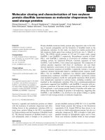
Expression of Rab31 and Rab32
Northern blots demonstrated that human tissues and
cultured cells expressed a 4.4-kb Rab31 mRN A and a
1.35-kb Rab32 mRNA, though the distribution of these two
mRNAs was very different (Fig. 3). Rab31 mRNA was
expressed most strongly in placenta and brain and to a lesser
extent in heart and lung, but no signal was detected from
liver, skeletal muscle, kidney and pancreas. H EL cells, and
to a lesser extent K 562 cells expressed Rab3 1 mRNA,
whereas Jurkat c ells did not. In c ontrast, the 1.35-kb Rab32
mRNA was expressed in most of the human tissues
examined, but particularly in heart, liver and kidney, and
was also found in HEL and K562 cells (Fig. 3). A 2.0-kb
Rab32 mRNA was also detected in some preparations of
RNA from HEL cells.
To demonstrate the presence of Rab31, Rab32 and
Rab11A proteins in platelets, rabbit antibodies were gen-
erated to unique peptides from the variable C -terminal
regions of the proteins ( see Experimental p rocedures and
Fig. 1A,B). These antibodies gave strong signals that were
blocked by the peptides to which they were prepared. As
shown i n Fig. 4, a ll these a ntibodies detected proteins in
platelets with m olecular m asses s imilar t o or slightly higher
than those predicted from their cDNA sequences (Rab31,
22 kDa; Rab32, 28 kDa; Rab11A, 26 kDa; Fig. 4). Pre-
sumably, the higher values re¯ect geranylgeranylation of the
proteins. For unknown reasons, HEL cells did n ot contain
detectable amounts of Rab31 and Rab32, using immuno-
blotting techniques. Rab31 protein was found in both
MEG-01 cells and KU812 cells, whereas Rab32 was not.
Rab11A was found in all cells tested, but appeared to be
present in particularly large amounts in platelets (Fig. 4). To
determine the amounts of these Rab proteins in platelets,
immunoblots of 10±20 lg of platelet protein were compared
with those of s tandard amounts o f the recombinant Rab
proteins (0.5±20 ng), which were subjected to SDS/PAGE
Fig. 3. Expression of Rab31 and Rab32
mRNA in human tissues and cell lines.
A human multiple tissue N orthern blot ( lanes
1±8, 2 lg of p olyA+ RNA per lane) was
obtained from Clontech (lane 1, heart; lane 2,
brain; lane 3, placenta; l ane 4, lung; lane 5,
liver; l ane 6, s keletal muscle; l ane 7, kidney;
lane 8, pancreas). In addition, total RNA
(12±15 lg per lane) was extracted from three
human cell lines, electrophoresed on a 1%
agarose-formaldehyde gel and blo tted onto a
nylon membrane, as described under Experi-
mental procedures (lane 9, HEL cells; lane 10,
K562 ce lls; lane 11, Jurkat cells). These two
membranes were probed successively with
32
P-labelled DNA (300±400 nucleotides) s yn-
thesized by PCR a m pli®cation of sequences
from Rab31, Rab32 and GAPDH (see
Experimental procedures). Autoradiographs
are shown. T he positions of RNA standards
are indicated on t he left.
264 X. Bao et al. (Eur. J. Biochem. 269) Ó FEBS 2002
at the same time. These results showed that human platelets
contained, per mg of total platelet protein, 0.85 0.13 lg
of Rab11A, 0.11 0.02 lg of Rab32 and 0.005 0.001 lg
of Rab31 ( mean valu es SE from f our, s ix and four
determinations, respectively, using platelets from different
donors). The subcellular distributions of these R ab proteins
were studied by differential centrifugation using a simple
method shown to y ield one particulate f raction enriched
in granules and mitochondria and another enriched in
both plasma and intracellular membranes [14]. The
results (Fig. 5) showed that Rab31, Rab32 and Rab11A
were equally enriched in the granule/mitochondrion and
membrane fractions and that no R ab31 or R ab32, and only
a s mall amount of Rab11A, was present in the supernatant
(cytosol) fraction.
Rab31 and Rab11A were detected in rat platelets in
amounts similar to those observed in human platelets, when
the same antibodies were used. Far smaller amounts of
Rab11A were observed in p rotein from rat kidney, liver,
heart, lung and brain, con®rming that platelets have an
exceptionally high Rab11A content (not shown). In contrast,
no 28 kDa immunoreactive species corresponding to human
Rab32 was found in rat platelets or in any rat tissue examined
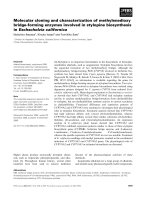
Fig. 4. Comparison of the a mounts of Rab31, Rab32 and Rab11A in
human platelets and related human cell lines. Protein (30 lgperlane)
from human platelets (lane 1), HEL cells (lane 2), MEG-01 cells (lane
3) and KU812 cells (lane 4) was subjected to SDS/PAG E, electro-
blotted onto Immobilon-P and probed for Rab31, Rab32 and
Rab11A, u sing rabbit ser a containing polyclonal antibodies raised
against speci®c Rab peptides (se e Experimental proced ures). Immu-
noreactive proteins were detected by ECL. The positions of prestained
protein standards are s hown on the right.
Fig. 5. Subcellular distributions of Rab proteins in human platelets.
Protein (30 lg) from platelet lysate (lane 1 ), from platelet fractions
enriched in granules (lane 2) or mem branes (la ne 3), a nd f rom the
platelet sup ernata nt f ract ion ( lane 4) was subjected to SDS/PAGE and
electroblotted onto Immobilon-P. Rab31 was detected using a 1 : 100
dilution of rabb it anti-p eptide se rum, Rab32 w ith r abbit a nity-
puri®ed immune I gG and Rab11A with rabbit i mmune I gG puri®ed
on Avid AL anity gel. In each case, b ound antibody was vi sualized
by ECL. T he positions of prestained p rotein standards are s hown on
the right.
Fig. 6. Immunoblot of rat platelet and tissue
proteins usi ng anti-Rab32 I g. Samples
containing 20 lg of p rotein were analys ed
by SDS/PAGE and e lectroblo tted onto
Immobilon-P. Immunoreactive proteins were
detected using anity-puri®ed antibody t o
Rab32 and visualized by ECL. Lane 1, human
platelets; lane 2, rat platelets; lane 3, rat aorta;
lane 4, rat heart; lane 5 , rat kidney.
Ó FEBS 2002 Rab proteins from platelets (Eur. J. Biochem. 269) 265
(Fig. 6). Instead, an equivalent amount of an immunoreac-
tive protein of 52 kDa was observed in rat platelets and a
much smaller amount was detected in rat heart. A very weak
52-kDa signal was also observed in s amples of human
platelet protein (Fig. 6). We conclude that the 52 kDa
protein m ay be a long form of Rab32 (see Discussion).
Bacterial expression of Rab31 and Rab32
GST±Rab31, GST±Rab32 and the potentially GTPase-
de®cient mutants of t hese proteins, GST±Rab31
Q64L
and
GST±R ab32
Q85L
, were cloned and expressed as described in
Experimental procedures. GST±Rab5A was expressed using
bacteria provided by P. Stahl. The puri®ed fusion proteins
(and GST itself) were almost homogeneous (Fig. 7A) and
suitable for experimental studies. To determine whether t he
recombinant Rab31 and Rab32 proteins bound GTP, we
®rst u sed [a-
32
P]GTP t o probe nitrocellulose blots of the
proteins, using two different Mg
2+
concentrations [17,18],
but no binding of [a-
32
P]GTP was detected (e.g. Fig. 7B). To
con®rm t hat the methods were working, samples of p latelet
protein and of GST-Rab5A were included and bound
[a-
32
P]GTP (Fig. 7B). We conclude that GST±Rab31 and
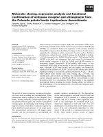
Fig. 7. Puri®cation and properties o f wild-type and mutant Rab31 and
Rab32 expressed a s GST±fusion proteins. Samples of platelet particu-
late fraction protein and of puri®ed GST and GST±Rab proteins were
subjected to SDS/PAGE as follows: lane 1, platelet protein; lane 2,
GST; lane 3, GST±Rab31; lane 4, GST±Rab31
Q64L
;lane5,GST±
Rab32; lane 6, GST±Rab32
Q85L
; lane 7 , GST±Rab5A. Gels were
processed as follows: (A) a Coomassie Blue-stained gel showing 10 lg
of platelet protein and 0.5 lg of GST and GST±Rab p roteins; (B) an
[a-
32
P]GTP overlay [17] of a nitrocellulose blot of a gel containing
40 lg of platelet protein, 0.5 lgofGSTand0.5lgofGST±Rab
proteins, except for GST±Rab5A (0.1 lg); (C) an immunoblot of a gel
containing 20 lgofplateletproteinand0.1lg of GST and GST±Rab
proteins, using a 1 : 1000 dilution of anti-Rab31 antiserum; (D) a
similar immunoblot using a 1 : 1000 dilution of anti-Rab32 antiserum.
The positions of prestained protein standards are shown on the right.
Fig. 8. Kinetics of GTP[S] binding by GST±Rab31 and GST±Rab32 at
low and high Mg
2+
free
concentrations. Puri®ed GST±Rab31 (A) or
GST±Rab32 (B) (in each case 200 pm ol of prote in in 0.2 m L of Bu er
A) was i ncub ated at 37 °Cwith5l
M
[
35
S]GTP[S] in the presence of
5 l
M
Mg
2+
free
(d)or10m
M
Mg
2+
free
(j). [
35
S]GTP[S]-binding by the
proteins was measured at the indicated tim es, as described under
Experimental procedures. Values are means SE from three d eter-
minations.
266 X. Bao et al. (Eur. J. Biochem. 269) Ó FEBS 2002
GST±Rab32 (and t he mutant proteins) were unable to
renature after binding to nitrocellulose.
The speci®city of our antibodies was studied in experi-
ments with t he GST-fusion proteins . Antibody to Rab31
detected only Rab31 and not GST, GST±Rab32 or
GST±Rab5A (Fig. 7C). Similarly, antibody to Rab32
detected only Rab32 (and minor proteolytic fragments)
(Fig. 7D).
[
35
S]GTP[S] binding by GST±Rab31 and GST±Rab32
Several studies have shown that Mg
2+
can have a critical
in¯uence o n GTP or GTP[S] binding by decreasing the o ff-
rates for both GDP and GTP/GTP[S] [31,32]. Figure 8
shows that the time-course of binding of [
35
S]GTP[S] to
GST±Rab31 and GST±Rab32 depended on the Mg
2+
concentration in t he medium. W ith both GST±Rab31
(Fig. 8 A) and GST±Rab32 (Fig. 8B), binding of
[
35
S]GTP[S] (5 l
M
) reached a maximum within 30 min at
37 °Cwhen5l
M
Mg
2+
free
was present, whereas with
10 m
M
Mg
2+
free
the binding of [
35
S]GTP[S] did not reach
this maximum in the case of GST±Rab31 and required 1 ±
2 h incubation with GST±Rab32. In control experiments,
GST did not bind [
35
S]GTP[S] at either Mg
2+
concentra-
tion. Studies on the e ffects o f d ifferent buffered Mg
2+
concentrations on binding of [
35
S]GTP[S] in 120 min
incubations also showed this major difference b etween the
two proteins (Fig. 9). Binding of [
35
S]GTP[S] to GST±
Rab31 reached a sharp maximum with 5 l
M
Mg
2+
and
then declined as the Mg
2+
free
increased, reaching the low
level s een in Fig. 8A with 10 m
M
Mg
2+
free
. GST±Rab32
was much less sensitive than GST±Rab31 to the ability of
Mg
2+
concentrations above 5 l
M
to inhibit [
35
S]GTP[S]
binding. These effects of Mg
2+
ions on [
35
S]GTP[S] binding,
as seen in several different experiments, are summarized in
Table 1, w hich shows that t he ratio o f [
35
S]GTP[S] binding
with 10 m
M
and 5 l
M
Mg
2+
free
after 3 h of incubation was
0.19 with GST±Rab31 a nd 0.98 with GST±Rab32, a highly
signi®cant difference (2P < 0.001). Table 1 also shows the
effects of the Rab31
Q64L
and Rab32
Q85L
mutations on
[
35
S]GTP[S] binding by the f usion p roteins. Less binding
was seen w ith 5 l
M
Mg
2+
free
in both cases and with 1 0 m
M
Mg
2+
free
in the case of GST±Rab32
Q85L
. This re¯ects a
progressive loss of stability of these proteins under
incubation conditions in which GDP could dissociate
relatively rapidly. Because optimal equilibrium binding of
[
35
S]GTP[S] to GST±Rab31 was only observed with 5 l
M
Mg
2+
free
,theK
d
values for [
35
S]GTP[S] dissociation from
both GST±Rab31 and GST±Rab32 were determined at this
Mg
2+
concentration and gave v alues o f 0.82 0.10 l
M
and 1.7 0.3 l
M
, r espectively (means range from two
determinations).
Fig. 9. Dependence of GTP[S] binding by GST±Rab31 and GST±
Rab32 on t he concentration o f Mg
2+
free
. Pur i®ed GST±Rab31 (A) o r
GST±Rab32(B)(ineachcase50pmolin0.1mLofBuerA)was
incubatedfor120minat37°Cwith5l
M
[
35
S]GTP[S] and t he indi-
cated concentrations of Mg
2+
free
(buered by 1 m
M
EDTA).
[
35
S]GTP[S]-binding by the proteins was then measured as described
under Experimental procedures. Values are means SE from three
determinations.
Table 1. [
35
S]GTP[S]-binding by puri®ed GST±Rab proteins and mutants. GST±Rabproteinswereexpressedandisolatedasdescribedunder
Experimental procedures. Binding of [
35
S]GTP[S] was determined in 180-min incubations at 37 °C in the p resen ce of 5 l
M
[
35
S]GTP[S] and the
indicated concentrations of Mg
2+
free
, and is expressed as mol of [
35
S]GTP[S] bound per 100 m ol of protein. Mean values SE from the numbers
of separate protein preparations indicated in parentheses are shown. The ratio of [
35
S]GTP[S] binding at 10 m
M
Mg
2+
free
to that at 5 l
M
Mg
2+
free
was calculated f or each protein preparation f or which both values w ere obtained; me an ratios SE are given.
Protein
[
35
S]GTP[S] binding
(mol [
35
S]GTP[S] per 100 mol of protein)
Ratio of [
35
S]GTP[S] binding
(10 m
M
Mg
2+
free
/5 l
M
Mg
2+
free
)
5 l
M
Mg
2+
free
10 m
M
Mg
2+
free
GST-Rab31 35.3 2.8 (8) 7.0 0.7 (6) 0.19 0.01 (6)
GST-Rab31
Q64L
18.3 2.6 (4) 9.7 1.8 (4) 0.55 0.12 (4)
GST-Rab32 26.5 2.7 (9) 25.4 1.7 (8) 0.98 0.10 (8)
GST-Rab32
Q85L
11.2 4.2 (4) 5.2 1.1 (4) 0.69 0.26 (4)
Ó FEBS 2002 Rab proteins from platelets (Eur. J. Biochem. 269) 267
GTPase activities of GST±Rab31, GST±Rab32 and mutants
As expected, the steady-state GTPase activities of these Rab
proteins were low, but as they were linear with time for up to 4
hat37 °Cwith5 l
M
[c-
32
P]GTP as substrate, whether 5 l
M
Mg
2+
free
or 10 m
M
Mg
2+
free
was used, valid measurements
could be obtained (Table 2). Under both of these conditions,
the GTPase activity of GST±Rab32 was signi®cantly higher
than that of GST±Rab31 (2P < 0.05, unpaired t-test)
(Table 2). Control e xperiments with recombinant GST
prepared similarly showed negligible contaminating bacterial
GTPase activity. V
max
and a pparent K
m
values f or the
GTPase activity of GST±Rab31 obtained i n the presence of
10 m
M
Mg
2+
free
were 0.964 0.272 nmolámg pro-
tein
)1
ámin
)1
and 14.8 2.1 l
M
, respectively ( means SE,
four preparations), wher eas those for GST±Rab32 were
1.80 0.35 n molámg protein
)1
ámi n
)1
and 6.7 0.9 l
M
,
respectively (means SE, three p reparations). These re-
sults suggest k
cat
values for the GTPase activities of GST±
Rab31 and GST±Rab32 of 0.046 min
)1
and 0.092 min
)1
,
respectively, though it is unlikely that all the recombinant
Rab prot ein w as c atalytica lly a ctive. A ssays w ith 10 m
M
Mg
2+
free
and 5 l
M
[c-
32
P]GTP (Table 2) showed that the
GST±R ab31
Q64L
and GST±Rab32
Q85L
mutants did no t
exhibit the expected decreases in GTPase activities, though
diminished activities were often observed after prolonged
incubation, with 5 l
M
Mg
2+
free
. Reproducible kinetic con-
stants were not obtained, probably because of the instability
of these mutan t proteins i n the longer incubations required
to measure GTPase activities at high GTP concentrations.
DISCUSSION
Relationships of Rab31 and of Rab32 to other Rab
proteins
After the initial cloning of Rab31 in 1996 [9], a
BLAST
search
showed that the most closely related Rab protein, with 71%
identity, was canine Rab22 [33]. Because the accepted
guideline for the u se of the next available R ab number w as,
at that time, an identity less than 85% [34], we named the
new p rotein Rab31 r ather than Rab22B. However, a
protein c loned from human melanocytes that is almost
identical to Rab31 has been named Rab22B [25]. There have
been at least two attempts to de®ne criteria that permit
classi®cation of Rab proteins into subfamilies on the basis of
their primary amino -acid sequences [8,35]. In addition to
®ve short s equences considered characteristic of the R ab
family as a whole (RabF1-F5), which include one within the
Switch I region (RabF1) and two within the Switch II region
(RabF3 and RabF4), three [35] or four [8] sequences have
been identi®ed that a re highly conserved only in the
members of putative subfamilies of Rab proteins
(RabSF1±SF4). It has been proposed that such sequences
may c onvey effector speci®city, whereas the Switch I and II
domains primarily convey sensitivity t o the binding of GTP
[8]. There is support f or this view from the crystal structure
of the complex of Rab3A/GTP/Mg
2+
with the effector
domain of r abphilin-3A, which shows that elements o f
RabSF1, RabSF3 and RabSF4 form a complementarity-
determining region t hat binds a structural element of
rabphilin-3A [36]. Particular importance has been attached
to RabSF4 (in the C-terminal hypervariable region) in
identi®cation o f subfamilies of Rab proteins [8]. However,
the RabSF4 r egions of Rab31 and Rab22A (amino-acid
residues 168±180) contain only one residue out of 13 in
common (8%), compared with 58% in Rab subfamilies as a
whole [ 8]. Moreover, based on t he RabSF1-SF4 criteria for
de®ning Rab protein subfamilies [8], Rab32 and Rab38
resemble each other more closely than do Rab31 and
Rab22A. F inally, Rab37 is 74% i dentical to Rab26 [37].
Thus, it is certainly possible that Rab31 and Rab22A act
through distinct e ffectors and we are unable to support t he
suggestion [8] that Rab proteins with > 70% identity
should be assigned the same number.
The amino-acid sequence of human Rab32 was most
similar to that predicted for a recently cloned mouse Rab
protein (Fig. 2B). However, the percent identity o f these
proteins (84%) was less than usual for orthologous Rab
proteins. Thus, human and mouse Rab11A are 100%
identical and human and mouse R ab5A 97% identical. The
main sequence differences between human Rab32 and the
related mouse p rotein are in the N- and C-terminal regions
and it is unlikely that our antibody to the human protein
would recognize this mouse protein. This raises the possi-
bility that a mouse protein that is more closely related to
human Rab32 remains to be identi®ed.
Rab32 contains amino-acid sequences that are shared
with only a small number of other Rab proteins. Most
conspicuously, the threonine in the WDTAGQE sequence
found in almost all Rab proteins was replaced by isoleucine.
This WDIAGQE sequence is also found in the above mouse
protein, Rab38 [27] and Rab7L1/29 [29], amongst mam-
malianRabproteinsidenti®edtodate,anditisalsopresent
Table 2. GTPase activities of puri®ed GST±Rab proteins and mutants. GST-Rab proteins were expressed and isolated as described in Experimental
procedures. GTPase activities were determined in 180±240 min incubations at 37 °C in the presence of 5 l
M
[c-
32
P]GTP and the indicated
concentrations of Mg
2+
free
, and are expressed as nmol of GTP hydrolysedámg protein
)1
ámin
)1
. Mean values SE from the numbers of separate
protein preparations indicated in p arentheses are sho wn. The r atio of the GTPase activity at 10 m
M
Mg
2+
free
to that at 5 l
M
Mg
2+
free
was
calculated for each protein preparation f or which b oth values w ere obtained; mean ratios S E are g iven.
Protein
GTPase activity
(nmolámg protein
)1
ámin
)1
)
Ratio of GTPase activities
(10 m
M
Mg
2+
free
/5 l
M
Mg
2+
free
)
5 l
M
Mg
2+
free
10 m
M
Mg
2+
free
GST-Rab31 0.205 0.057 (8) 0.195 0.044 (9) 1.15 0.13 (8)
GST-Rab31
Q64L
± 0.262 0.078 (6) ±
GST-Rab32 0.335 0.071 (8) 0.498 0.088 (8) 1.72 0.29 (8)
GST-Rab32
Q85L
± 0.515 0.151 (6) ±
268 X. Bao et al. (Eur. J. Biochem. 269) Ó FEBS 2002
in RabE from the slime mold, Dictyostelium discoideum [28].
Rab28, which is more distantly related t o other Rab
proteins, contains isoleucine in the sequence, WDIGGQT
[38]. Two other u nusual structural features ar e shared by
Rab32, the related mouse protein, Rab38 and Rab7L1/29,
namely replacement of the glycine residue that usually
precedes the guanine nucleotide-binding NKXD motif by
alanine and replacement of a conserved phenylalanine 10
residues C-terminal to the EXS[AV] motif by alanine,
threonine, serine or methionine (compare Figs 2A,B). These
structural features of the Rab32 group of proteins all lie
within or close to the guanine nucleotide-binding motifs of
these proteins, suggesting that they could affect guanine
nucleotide b inding. T his p ossibility i s s upported b y t he
demonstration that in one conformation of Sec4p, the
threonine residue that is replaced by isoleucine in Rab32
makes contacts with other residues and water molecules
involved in Mg
2+
and nucleotide binding [39].
Expression of Rab31, Rab32 and Rab11A in platelets
Use of a ntibodies directed against C-terminal a mino-acid
sequences speci®c to each Rab protein studied demonstrat-
ed the presence in human platelets of proteins with
molecular masses corresponding closely to those predicted
from the corresponding cDNA sequences. I n the case of
Rab31, an in-frame stop codon 5¢ to the initiating methi-
onine precludes translation of a larger protein but this is not
thecasewithRab32. The recently published human genomic
DNA sequence shows that the Rab32 gene is on fragment
Al133539 of chromosome 6 and that the ORF encoding the
®rst 83 amino acids of the p rotein ( nucleotides 41277±
41526) extends in a 5¢ directi on to a nother translation
initiation codon at nucleotide 40473, which is preceded by
two in-frame stop c odons (at nucleotides 40458 and 40461).
Both potential translation initiation codons are embedded
in weak but acceptable Kozak consensus sequences [23].
This 5¢ sequence could encode an additional 268 amino
acids to give a long form of Rab32 (Rab32L) with a
molecular mass o f 52.7 kDa. The additional sequence
contains a proline-rich region detected by
PROSITE
and a
perfect 16 amino-acid repeat. Evidence for transcription of
much of this 5¢ extension is provided by dbEST sequences,
the longest of which covers nucleotides 40750±41196
(dbEST Id: 3 133919). N evertheless, it is clear from both
our Northern and Western blots that t he principal form of
Rab32 expressed in human cells is the short form shown in
Fig. 1B. A predicted transcription start s ite and T ATA box
are present in the 5¢ untranslated sequence of the 28 kDa
Rab32 and can account for the 1.35 kb Rab32 mRNA,
which is t oo short t o e ncode Rab32L. However, three lines
of evidence suggest that small amounts of Rab32L may be
expressed in human cells; ®rst, a 2-kb mRNA was
sometimes observed in HEL cells; second, small amounts
of an immunoreactive 52-kDa protein were detected on
some Western blots of human platelets; third, the above
expressed sequence tag (EST) includes the predicted tran-
scription start site and TATA box of the 28-kDa Rab32. In
rat platelets and heart, the 28-kDa Rab32 appeared to be
completely replaced by a protein corresponding to Rab32L.
Although we cannot yet exclude the possibility that the
antibody to Rab32 detects an u nrelated 52-kDa protein, an
N-terminal extension of a Rab protein is not without
precedent. T hus, Rab36 has 110 more amino acids a t t he
N-terminus than other Rab proteins [40].
Our demonstration that p latelets contain Rab31, Rab32
and Rab11A, a dds three to the eight Rab proteins already
known to b e present (Rab1, Rab3B, Rab4, R ab5, Rab6,
Rab8, R ab27A, Rab27B [3±7]). O nly in one case, Rab4,
has evidence for a speci®c function, n amely a role in the
exocytosis of a-granule constituents, been obtained [4].
Signi®cant differences in the subcellular distributions of
these Rab proteins in platelets have been observed.
Whereas Rab3B was found largely in the cytosol fraction,
the other Rab proteins were predominantly particulate
[3,4]. Rab6 and Rab8 were associated with a-granules [3]
and other Rab proteins (Rab4, Rab5, Rab27A and
Rab27B) have been found in fractions containing
a-granules, a s well a s in m embrane fractions [4,7]. No
Rab p roteins h ave yet been c learly a ssociated with the
platelet dense granules. In the present study, Rab31,
Rab32 and Rab11A were also found in both granule and
membrane fractions, and only R ab11A was also present in
the cytosol. T his observation may be explained b y the
®nding that Rab11A is a preferential target for GDP
dissociation inhibitor [41]. The very high content of
Rab11A in platelets ( 0.85 lg per mg of platelet protein)
is consistent with a major role for this protein. Although
studies in a variety of cells have indicated that Rab11A is
closely associated with pericentriolar recycling endosomes
[42,43], other studies have found eviden ce of roles in
the constitutive and r egulated pathways of exocytosis
[41,44,45]. Rab11A is f ound in the trans-Golgi network
and in vesicles and granules derived therefrom [41,44].
Moreover, it a ppears to b e required f or transport o f
vesicles to the plasma m embrane [41]. Finally, in h eart
muscle, R ab11A is found together with Rab4 in the
insulin-mobilized vesicles containing the glucose trans-
porter (GLUT4) and could therefore play a role in the
fusion of these vesicles with the plasma membrane [45].
Binding of guanine nucleotides and hydrolysis of GTP
by GST±Rab31 and GST±Rab32
Many studies have shown that bacterially expressed R ab
proteins, devoid of post-translational modi®cations, contain
bound GDP and retain their native GTPase activities after
exchange of G DP for GTP [11,19,31,32,46±49]. T hese
include GST±Rab proteins, which have been shown to bind
[a-
32
P]GTP after blotting and renaturation in the cases of
Rab5A [11] and Rab4 [48], and to possess GTPase activity
in the case of R ab4 [48]. M oreover, a GST±Rab3 and a
chimeric Rab3 cleaved from this fusion protein with
thrombin acted similarly to in hibit secretion after microin-
jection into Aplysia neurons [49]. Finally, GST±Rab5A
binds Rab5A effectors in a GTP- or GTP[S]-dependent
manner [50]. Because o f these ®ndings, we used the Rab31
and Rab32 GST-fusion proteins to characterize the inter-
actions of these Rab proteins with guanine nucleotides.
Contrary to expectation, neither GST±Rab31 nor GST±
Rab32 b ound [a-
32
P]GTP a fter blotting to a nitrocellulose
membrane, though GST±Rab5A did so [11]. However, low-
M
r
GTP-binding proteins differ markedly in their ability to
renature on nitrocellulose, a process that depends critically
on C-terminal amino-acid sequences [51]. Binding of
[
35
S]GTP[S] by the native proteins provides a more critical
Ó FEBS 2002 Rab proteins from platelets (Eur. J. Biochem. 269) 269
test and was found to be highly dependent on the Mg
2+
concentration. Not only was Mg
2+
required for binding of
[
35
S]GTP[S] by both GST±Rab31 and GST±Rab32 but
Mg
2+
free
concentrations above 5 l
M
markedly inhibited the
rate and ®nal extent of [
35
S]GTP[S] binding by GST±Rab31.
With GST±Rab32, h igh Mg
2+
concentrations were less
inhibitory. T hese observations can m ost easily be explained
by an ability of the higher Mg
2+
concentrations to inhibit
the dissociation of GDP bound to the Rab proteins, as
previously found for R ab3A [31] and Rab5A [32]. Thus,
with 10 m
M
Mg
2+
free
,thek
o
for GDP release from GST±
Rab31 may be much lower than the k
o
for GDP release
from GST±Rab32.
The GTPase activities of Rab proteins are very low in the
absence o f a GTPase-activating protein (GAP) [19,31,47,
52,53], t hough variations up to 20-fold, as between Rab5 and
Rab7, have b een reported [53]. Both the steady-state
GTPase activities of Rab proteins incubated with
[c-
32
P]GTP [19,31] and the intrinsic GTPase activities o f
Rabproteinsloadedwith[a-
32
P]GTP [47,52,53] h ave been
measured. In the former case, exchange of G TP for bound
GDP may be rate-limiting f or GTPase activity, as f ound
for Rab3A at high Mg
2+
concentrations [31]. With GST±
Rab31,thesameGTPaseactivitieswereseenwith5l
M
and 10 m
M
Mg
2+
free
. This could be coincidental, with GTP
hydrolysis rate-limiting at 5 l
M
Mg
2+
free
and GDP-GTP
exchange rate-limiting a t 10 m
M
Mg
2+
free
. In t he present
study, GST±Rab32 consistently exhib ited a higher GTPase
activity than GST±Rab31 and GTP hydrolysis could be rate-
limiting under all co nditions, b ecause Mg
2+
was much less
effective in i nhibiting GDP release f rom this p rotein.
Studies on some Rab proteins, including Rab3A [ 46] and
Rab5A [11,47] have shown that mutation of glutamine to
leucine in the PM3 G TP-binding motif (DTAGQE) [22]
greatly decreases the GTPase activities of the proteins, as
predicted from t he corresponding mutation in Ras (Q61L).
This is consistent with evidence that this glutamine residue
may participate in GTP h ydrolysis by stabilizing t he
catalytic transition state [36]. Moreove r, the Q79L mutant
of Rab5A has constitutive activity in prom oting endosome
fusion [47]. Nevertheless, there is evidence that this gluta-
mine residue is not essential for the GTPase activities of a ll
Rab proteins. Thus, the corresponding mutation in Rab11A
(Q70L) did not d ecrease GTP hydrolysis in t he absence or
presence of cytosol containing GAP activity [43]. Moreover,
Rab25 has considerable GTPase activity [43], although the
corresponding glutamine residue is naturally replaced by
leucine. Our results showed no decreases i n the GTPase
activities of GST± Rab31
Q64L
and GST±Rab32
Q85L
relative
to the unmutated proteins, suggesting that Rab31 and
Rab32 fall into the same category as Rab11A and m ay not
be constitutively activated by this mutation. These obser-
vations emphasize the importance of establishing the
biochemical properties of m utant Rab GTPases before t hey
are used to study the functions of individual Rab proteins in
more co mpl ex sy stems .
ACKNOWLEDGEMENTS
We thank Dr Brian Golding of Biology Department, McMaster
University, f or assisting with a nalysis o f the human Rab32 gene. T his
work was supported by a grant from the Canadian Institutes of Health
Research (MOP-5626).
REFERENCES
1. Zerial, M. & McBride, H. (2 001) Rab proteins as m embrane o r-
ganizers. Nat. Rev. 2, 107±117.
2. Siess, W. (1989) Molecular m echanisms of platelet a ctivation.
Physiol. Rev. 69, 58±178.
3. Karniguian, A., Zahraoui, A. & Tavitian, A. (1993) Identi®cation
of small GTP-binding rab proteins in human platelets: thrombin-
induced phosphorylation of rab3B, rab6, and rab8 proteins. Proc.
Natl Acad. Sci. USA 90 , 7647±7651.
4. Shirakawa, R., Yoshioka, A., Horiuchi, H., Nishioka, H.,
Tabuchi, A . & Kita, T. (2000) Small G TPase Rab4 r egulates
Ca
2+
-induced a-granule secretion in platelets. J. Biol. C hem. 275,
33844±33849.
5. Nagata, K., Okano, Y., Suzuki, T. & Nozawa, Y. (1993) Evidence
for the presence of a low M
r
GTP-binding protein, ram p25, in
human platelet me mbrane s. Platelets 4, 268±274.
6. Chen,D.,Guo,J.,Miki,T.,Tachibana,M.&Gahl,W.A.(1997)
Molecular cloning and c haracterization o f Rab27a a nd Rab27b,
novel human Rab p rote ins sh ared by me lanoc ytes an d p latelets.
Biochem. Mo l. Med. 60 , 27±37.
7. Nagata, K. & Nozawa, Y. (1993) Subcellular localization of a low
M
r
GTP-binding protein, c25KG, in resting and stimulated
human platelets. Biochem. Biophys. Re s. Commun. 195, 108 1±
1088.
8. Pereira-Leal, J.B. & Seabra, M.C. (2000) The mammalian Rab
family of small GTPases: de®nition of family and subfamily
sequence motifs suggests a mechanism for functional speci®city in
the Ras superfamily. J. M o l. Biol. 301, 1077±1087.
9. Bao, X., Jang, E.K. & Haslam, R.J. (1996) Mole cular cloning of
two novel Rab proteins f rom human platelets. FASE B J. 10,
A1011.
10. Bao, X., Faris, A.E. & Haslam, R.J. (1997) Cloning, tissue dis-
tribution and bacterial expression of human Rab31 and Rab32.
Mol. Biol. Cell 8, 199a.
11. Li, G. & Stahl, P.D. (1993) Structure±function relationship of the
small G TPase rab5. J. Biol. Chem. 268, 24475±24480.
12. Goldenring, J.R., Smith, J., Vaughan, H.D., Cameron, P.,
Hawkins, W. & Navarre, J. (1996) Rab11 is an apically located
small GTP-binding protein in epithelial tissues. Am.J.Physiol.
270, G515±G525.
13.Mustard,J.F.,Perry,D.W.,Ardlie,N.G.&Packham,M.A.
(1972) Preparation of suspensions of washed plate lets from
humans. Br. J. Haematol. 22 , 193±204.
14. Fox, J.E.B., Say, A.K. & Haslam, R.J. (1979) Subcellular
distribution of the dierent platelet proteins phosphorylated on
exposure of intact platelets to ionophore A23187 or to prosta-
glandin E
1
. P ossible role of a membrane ph osphop olypeptide
in the regulation o f calcium-ion transport. Biochem. J. 184 ,651±
661.
15. Yang, T T. & Kain, S.R. (1 995) Fast hy bridization solution
for the detection of immobilized nucleic acids. Biotec hnique s 18,
498±503.
16. Landt, O., Grunert, H P. & H ahn, U. (1990) A general method
for rapid site-directed mutagenesis using t h e polymerase chain
reaction. Gene 96, 125±128.
17. Bhullar, R.P. & Haslam, R.J. (1987) De tection of 23±27 kDa
GTP-binding proteins in platelets and other cells. Biochem. J. 245,
617±620.
18. Schmitt, H.D., Wagner, P., Pfa, E. & Gallwitz, D. (1986) The
ras-related YPT1 gene product in yeast: a GTP-bin ding protein
that might be involved in microtubule org anization. Cell 47,401±
412.
19. Kabcenell,A.K.,Goud,B.,Northup,J.K.&Novick,P.J.(1990)
Binding a nd h ydro lysis of guanine nucleotides by Sec4p, a y east
protein i nvo lved in the regulation o f vesicular t rac. J. B io l.
Chem. 265, 9366±9372.
270 X. Bao et al. (Eur. J. Biochem. 269) Ó FEBS 2002
20. Fabiato, A. & Fabiato, F. (1979) Calculator programs fo r com-
puting the composition of the solutions containing multiple metals
and ligands used for experiments in skinned muscle cells. J. Physiol.
(Paris) 75, 463±505.
21. Chavrier, P., Simons, K. & Zerial, M. (1992) The complexity of the
Rab and Rho GTP-binding protein subfamilies revealed by a PCR
cloning a pproach. Gene 112, 261±264.
22. Valencia, A., Chardin, P., Wittinghofer, A. & Sander, C. (1991)
The ras protein fam ily: evolutionary tree and role of conserved
amino acids. Biochemistry 30 , 4637±4648.
23. Kozak, M . (1991) Structural features in eukaryotic mRNAs that
modulate the initiation o f translation. J. Bio l. Chem. 26 6, 19867±
19870.
24. Altschul, S.F., Madden, T.L., Scha
È
er, A.A., Zhang, J., Zhang,
Z., Miller, W. & Lipman, D.J. (1997) Gapped BLAST a nd PSI-
BLAST: a new ge neration of protein database search programs.
Nucleic Acids Res. 25 , 3389±3402.
25. Chen, D., Guo, J., Miki, T., Tachibana, M. & Gahl, W.A. (1996)
Molecular cloning of two novel rab genes from human melano-
cytes. Gene 174, 129±134.
26. Terryn, N., Anuntalabhochai, S., Van Montagu, M. & Inze
Â
,D.
(1992) Analys is of a Nicotiana plumbaginifolia cDNA encoding a
novel s mall GTP-binding protein. FEBS Lett. 299, 287±290.
27. Ja
È
ger, D., S tock ert, E., J a
È
ger, E., Gu
È
re, A .O., Scanlan, M.J.,
Knuth, A., Old, L.J. & Chen, Y T. (2000) Serological cloning of a
melanocyte rab guanosine 5 ¢-triphosphate-binding protein and a
chromosome condensation protein from a melanoma comple-
mentary D NA library. Cancer Res. 60, 3584±3591.
28. Norian, L., Dragoi, I.A. & O'Halloran, T. (1999) Molecular char-
acterization of rabE, a developmentally regulated Dictyostelium
homolog of mammalian rab GTPases. DN A Cell Biol. 18, 59±64.
29. Shimizu, F., Katagiri, T., Suzuki, M., Watanabe, T.K., Okuno, S.,
Kuga,Y.,Nagata,M.,Fujiwara,T.,Nakamura,Y.&
Takahashi, E . (1997) Cloning and chromosome assignment to
1q32 of a h uman cDNA (RA B7L1) encoding a s mall GTP-
binding protein, a member of the RAS superfamily. Cytogenet.
Cell Genet. 77 , 261±263.
30. Massmann, S., Schu
È
rmann, A. & Joost, H G. (1997) Cloning of
two splicing variants of t he novel R as-related GTPase Rab29
which is predominately expressed in kidney. Biochim. Biophys.
Acta 1352, 48±55.
31. Burstein, E.S. & Macara, I.G. (1992) Interactions of the ras-like
protein p25
rab3A
with Mg
2+
and guanine nucleotides. Biochem. J.
282, 387±392.
32. Pan, J.Y., Sanford, J.C. & Wessling-Resnick, M. (1996) In¯uence
of Mg
2+
on the structure and function of Rab5. J. Biol. C he m.
271, 1322±1328.
33. Olkkonen, V.M., Dupree, P., Killisch, I., Lu
È
tcke,A.,Zerial,M.&
Simons, K . (1993) Molecular cloning and subcellular localization
of three GTP-binding proteins of the rab subfamily. J. Cell Sci.
106, 1249±1261.
34. Kahn,R.A.,Der,C.J.&Bokoch,G.M.(1992)Therassuper-
family of GTP-binding proteins: guidelines on nomenclature.
FASEB J. 6, 2512±2513.
35. Moore, I., Schell, J. & Palme, K. (1995) Subclass-speci®c sequence
motifs identi®ed in Rab GTPases. Trends Bioche m. Sci. 20, 10±12.
36. Ostermeier, C. & Brunger, A.T. (1999) Structural basis of Rab
eector speci®city: crystal structure of the small G protein Rab3A
complexed with the eector domain of rabphilin-3A. Cell 96, 363±
374.
37. Masuda, E.S., Luo, Y., Young, C., Shen, M., Rossi, A.B., Huang,
B.C.B.,Yu,S.,Bennett,M.K.,Payan,D.G.&Scheller,R.H.
(2000) Rab37 is a novel mast cell speci®c GTPase localized to
secretory granules. FEBS Lett. 470, 61±64.
38. Brauers, A., Schu
È
rmann, A., Massmann, S., Mu
È
hl-Zu
È
rbes, P.,
Becker, W., Kainulainen, H., Lie, C. & Joost, H G. (1996)
Alternative mRNA s plicing of t he novel GT Pase Rab 28 gene rates
isoforms with dierent C-termini. Eur. J. Bioche m. 237, 833±840.
39. Stroupe, C. & Brunger, A.T. (2000) Crystal structures of a Rab
protein in its inactive and active conformations. J. Mol. Biol. 304,
585±598.
40. Mori, T., Fukuda, Y., Kuroda, H ., Matsumura, T ., Ota, S.,
Sugimoto, T., Nakamura, Y. & Inazawa, J. (1999) Cloning and
characterization of a novel Rab-family gen e, Rab36,within
the region a t 22q11.2 that is homozygously deleted i n m alignant
rhabdoid tumors. Biochem. Biophys. Res. Commun. 254, 594±600.
41. Chen, W., Feng, Y., Chen, D. & Wandinger-Ness, A. (1998)
Rab11 is r equired f or trans-Golgi network-to-plasma m embrane
transport and a preferential target for GDP dissociation inhibitor.
Mol. Biol. Cell 9, 3241±3257.
42. Ullrich, O., Reinsch, S., Urbe
Â
,S.,Zerial,M.&Parton,R.G.(1996)
Rab11 regulates recycling through the p ericentriolar r ecycling
endosome. J. Cell B iol. 135, 913±924.
43. Casanova, J.E., Wang, X., Kumar, R., Bhartur, S.G., Navarre, J.,
Woodrum, J.E., Altschuler, Y., Ray, G.S. & Goldenring, J.R.
(1999) Association of Rab25 and Rab11a with the apical recycling
system of polarized Madi n-Darby canine kidney cells. Mol. Biol.
Cell 10, 47±61.
44. Urbe
Â
,S.,Huber,L.A.,Zerial,M.,Tooze,S.A.&Parton,R.G.
(1993) Rab11, a small GTPase associated with both c onstitutive
and r egulated secretory pathways i n PC12 c ells. FEBS Lett. 334,
175±182.
45. Kessler, A ., Tomas, E., I mmler, D., Meyer, H .E., Zorzano, A. &
Eckel, J. (2000) Rab11 is associated with GLUT4-containing
vesicles and redistributes in response to insulin. Diabetologia 43,
1518±1527.
46. Brondyk, W.H., McKiernan, C.J., Burstein, E.S. & Macara, I.G.
(1993) Mutants of Rab3A a nalogous to oncogenic Ras mutants;
sensitivity to Rab3A-GTPase activating p rotein and Rab3A-
guanine nucleotide r eleasing factor. J. Biol. C h em. 268, 9410±
9415.
47. Stenmark, H., Parton, R.G., S teele-Mortimer, O., Lu
È
tcke, A.,
Gruenberg, J. & Zerial, M. (1994) Inhibition of rab5 GTPase
activity stimulates membrane fusion in endocytosis. EMBO J. 13,
1287±1296.
48.Cormont,M.,Bortoluzzi,M N.,Gautier,N.,Mari,M.,Van
Obberghen, E . & Le Marc hand-Brustel, Y. (1996) Po tential role of
Rab4 in t he regulation o f subcellular localization of Glut4 in
adipocytes. Mol. Cell. Biol. 16, 6879±6886.
49. Johannes, L., Doussau, F., Clabecq, A., Henry, J P., Darchen , F.
& Poulain, B. (1996) Evidence for a functional link between Rab3
and the SNARE complex. J. Cell Sci. 109, 2875±2884.
50. Christoforidis, S., McBride, H.M., Burgoyne, R.D. & Zerial, M .
(1999) The Rab5 eector EEA1 is a core component of endosome
docking. N ature (London) 397, 621±625.
51. Klinz, F J . (1994) GT P-blot analysis o f small GTP -binding pro-
teins. The C -termin us is involved in renaturatio n of blotted pro-
teins. Eur. J. Biochem. 225, 99±105.
52. Shapiro, A.D., Riederer, M.A. & Pfeer, S.R. (1993) Biochemical
analysis of rab9, a ras-like GTPase i nvolved in protein transpor t
from late endosomes to the trans Golgi network. J. Biol. Chem.
268, 6925±6931.
53. Simon,I.,Zerial,M.&Goody,R.S.(1996)Kineticsofinteraction
of Rab5 and Rab7 with nucleotides and magnesium i ons. J. Biol.
Chem. 271, 20470±20478.
54. Dumas, J.J., Zhu, Z., Connolly, J.L. & Lambright, D.G. (1999)
Structural basis of activation and GTP hydrolysis in Rab proteins.
Structure 7, 413±423.
Ó FEBS 2002 Rab proteins from platelets (Eur. J. Biochem. 269) 271