Báo cáo Y học: Solution structure of the HIV gp120 C5 Domain pptx
Bạn đang xem bản rút gọn của tài liệu. Xem và tải ngay bản đầy đủ của tài liệu tại đây (280.38 KB, 8 trang )
Solution structure of the HIV gp120 C5 Domain
Laure Guilhaudis, Amy Jacobs and Michael Caffrey
Department of Biochemistry and Molecular Biology, University of Illinois at Chicago, IL, USA
In HIV the viral envelope protein is processed by a host cell
protease to form gp120 and gp41. The C1 and C5 domains of
gp120 are thought to directly interact with gp41 but are
largely missing from the available X-ray structure. Bio-
physical studies of the HIV gp120 C5 domain (residues 489–
511 of HIV-1 strain HXB2), which corresponds to the
carboxy terminal region of gp120, have been undertaken.
CD studies of the C5 domain suggest that it is unstructured
in aqueous solutions but partially helical in trifluoroethanol/
aqueous and hexafluoroisopropanol/aqueous buffers. The
solution structure of the C5 peptide in 40% trifluoroethanol/
aqueous buffer was determined by NMR spectroscopy. The
resulting structure is a turn helix structural motif, consistent
with the CD results. Fluorescence titration experiments
suggest that HIV C5 forms a 1 : 1 complex with the HIV
gp41 ectodomain in the presence of cosolvent with an
apparent K
d
of 1.0 l
M
. The absence of complex forma-
tion in the absence of cosolvent indicates that formation of
the turn-helix structural motif of C5 is necessary for complex
formation. Examination of the C5 structure provides insight
into the interaction between gp120 and gp41 and provides a
possible target site for future drug therapies designed to
disrupt the gp120/gp41 complex. In addition, the C5 struc-
ture lends insight into the site of HIV envelope protein
maturation by the host enzymes furin and PC7, which
provides other possible targets for drug therapies.
Keywords:AIDS;gp41;gp120;HIV;NMR.
Envelope proteins are known to play roles in directing viral
particles to the appropriate target cell and initiating the
fusion of the viral and cellular membranes in diverse viruses
including retroviridae, herpesviridae, filoviridaie and para-
myxoviridae [1]. In the retrovirus HIV, which is the
causative agent of AIDS, the envelope proteins exist as a
noncovalent complex of a surface subunit (gp120) and a
transmembrane subunit (gp41), which are proteolytic
products of the env gene. HIV infection, as well as SIV
(simian immunodeficiency virus) infection, is thought to
occur in three discreet steps [2]. During the initial step, the
gp41/gp120 complex associates with the CD4 receptor. In
the second step, gp120 associates with a chemokine receptor
that is specific to the cell type and dissociates from gp41. In
the final step, gp41 mediates fusion of the viral and the
cellular membranes by a poorly understood mechanism,
which is thought to involve the N-terminal hydrophobic
region of gp41, termed the fusion peptide. gp120 is known
to be well exposed on the HIV surface and play an initial
and critical role in HIV infection. Thus, gp120 is an
attractive target for vaccines and structure-based drugs
designed for the prevention and treatment of AIDS. The
search for new AIDS therapies is of critical importance in
light of recent reports that current highly active antiretro-
viral treatments are ineffective in 20–50% of treated patients
[3]. Moreover, therapies designed to disrupt gp120 function
are especially attractive because of their potential prophy-
lactic properties, as well as being alternative and comple-
mentary therapies to the protease and reverse transcriptase
inhibitors currently utilized.
In HIV, the precursor envelope protein, gp160, is proteo-
lytically cleaved to form gp120 and gp41 by the host specific
proteases furin and PC7 [4]. Cleavage of the precursor viral
envelope protein at the carboxyl side of the amino acid
sequence REKR is critical to HIV function, as shown by
mutagenesis studies [5]. Moreover, inhibitors of furin and
PC7 have been shown to block gp160 processing and HIV
infection [6]. The mature gp120 consists of 5 conserved
regions (C1-C5) and five variable regions (V1–V5). Recently,
the structure of HIV gp120 in complex with regions of the
CD4 receptor and a monoclonal antibody that blocks gp120
binding to the chemokine receptor has been determined by
X-ray crystallography [7]. However, for technical reasons,
large regions of the conserved and variable domains of gp120
are missing from the X-ray structure. Mutagenesis and
deletion studies have suggested that the C1 and C5 domains
of gp120, which are largely missing in the crystal structure
but would be expected to be proximal to one another, are
involved in the interaction with gp41 [8,9]. Furthermore, it
has recently been shown that the introduction of non-native
intermolecular disulfide bonds between the gp41 loop region
and the C1 or C5 domains of gp120 stabilizes the gp41/gp120
complex and mimics the antigenic properties of the native
envelope complex [10]. Taken together, the mutagenesis
studies suggest that there is a direct interaction between the
gp120 C1 and C5 domains and the gp41 loop region. In an
effort to further characterize the gp41/gp120 interaction, the
solution structure of the C5 domain of HIV gp120 has been
determined by NMR spectroscopy.
EXPERIMENTAL PROCEDURES
The HIV gp120 C5 domain was prepared by solid phase
peptide synthesis (Biological Resource Center, University of
Illinois at Chicago). The peptide was modified by the
Correspondence to M. Caffrey, Department of Biochemistry and
Molecular Biology, University of Illinois at Chicago, 1819 W Polk St.,
Chicago, IL 60612, USA.
Fax: + 1 312 413 0364, Tel.: + 1 312 996 4959.
E-mail: caff
(Received 12 June 2002, revised 9 August 2002,
accepted 19 August 2002)
Eur. J. Biochem. 269, 4860–4867 (2002) Ó FEBS 2002 doi:10.1046/j.1432-1033.2002.03187.x
additions of acetyl and amido groups to the amino and
carboxy termini, respectively. The identity of the peptide
was verified by mass spectrometry (mass ¼ 2700.9). The
wild-type HIV gp41 ectodomain was prepared as previously
described [11]. CD spectra were recorded on a Jasco J-710
spectropolarimeter with a 0.1-cm path length at 23 °C.
Fluorescence titrations were performed with a PTI Fluo-
rescence System using a 0.1-cm path length at 25 °C. Before
performing the titrations, the HIV gp41 ectodomain and
gp120 C5 were equilibrated in 50 m
M
sodium formate
(pH 3.0), 40% trifluoroethanol (v/v). Data were analyzed
with
KALEIDAGRAPH
3.08.
For the NMR experiments, the conditions were 1 m
M
C5, 50 m
M
PO
4
(pH 6.0), 40% trifluoroethanol-d
3
(Cam-
bridge Isotopes), 5% D
2
O. NMR experiments were per-
formed on a Bruker DRX 600 equipped with a triple
resonance probe. TOCSY and NOESY experiments were
acquired at 285, 290, 295, 300 and 305 K. For the TOCSY
experiments the isotropic mixing time was 72 ms; for the
NOESY experiments, the mixing times were 75, 150, and
250 ms.
13
C-edited HSQC were acquired at 300 and 305 K.
The HIV gp120 C5 structure was calculated with the
program
CNS
[12]. The protocol consisted of four steps: (a)
high temperature torsion angle molecular dynamics start-
ing from an extended conformation; (b) slow-cooling
torsion angle molecular dynamics; (c) slow-cooling Carte-
sian dynamics; and (d) conjugate gradient minimization.
For the first step, the temperature was set to 50 000 K for
2000 steps of 15 fs. In this step, the force constants for NOE,
dihedral angles, and van der Waals forces were 150 kcalÆ
mol
)1
ÆA
˚
)2
, 100 kcalÆmol
)1
Ærad
)2
and 0.1 kcalÆmol
)1
ÆA
˚
)4
,
respectively. For the second step, the temperature was set
to 50 000 K and cooled to 0 K over 1000 steps of 15 fs. In
this step, the force constants for NOE, dihedral angles, and
van der Waals forces were 150 kcalÆmol
)1
ÆA
˚
)2
, 200 kcalÆ
mol
)1
Ærad
)2
and 0.1–1.0 kcalÆmol
)1
ÆA
˚
)4
, respectively. For
the third step, the starting temperature was set to 2000 K
and cooled to 0 K in 3000 steps of 5 fs. In this step, the force
constants for NOE, dihedral angles, and van der Waals
forces were 150 kcalÆmol
)1
ÆA
˚
)2
, 200 kcalÆmol
)1
Ærad
)2
and
1.0–4.0 kcalÆmol
)1
ÆA
˚
)4
, respectively. In the final step, 200
steps of conjugate gradient minimization were performed.
In this step, the force constants for NOE, dihedral angles,
and van der Waals forces were 75 kcalÆmol
)1
ÆA
˚
)2
, 400 kcalÆ
mol
)1
Ærad
)2
and 1.0 kcalÆmol
)1
ÆA
˚
)4
, respectively. In all
steps, an energy term for a conformational database was
included with a force constant of 1.0 [13]. The / and u
dihedral angles were set to the values derived from
1
Hand
13
C chemical shifts using the program
TALOS
[14]. For the
structure calculation, errors of ± 30° were used for the
dihedral restraints. NOEs were classified as strong
(<2.7 A
˚
), medium (<3.3 A
˚
or <3.5 A
˚
for amide protons),
weak (<5.0 A
˚
)andveryweak(<6.0A
˚
). A correction
of 0.5 A
˚
was added to proton distances involving
methyl groups. The electrostatic map and structural
figures were generated by the molecular graphics program
MOLMOL
[15].
RESULTS
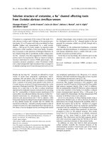
To characterize the HIV gp120 C5 domain, a 23 residue
peptide was synthesized, corresponding to residues 489–511
of HIV-1 strain HXB2 (Fig. 1A). The synthetic C5 peptide
was found to be soluble under all experimental conditions
that were examined. As shown in Fig. 1B, the CD spectra of
C5 in aqueous solution suggest the absence of regular
secondary structure. The absence of secondary structure
was found to be independent of temperature, pH and salt
concentration (data not shown). In contrast, the CD spectra
of C5 in trifluoroethanol/H
2
O or hexafluoroisopropanol/
H
2
O mixtures suggest the presence of helix. In trifluoro-
ethanol/H
2
O mixtures, the maximal amount of helical
structure is observed in 40% trifluoroethanol (Fig. 1B). A
similar amount of helical structure is observed in 60%
hexafluoroisopropanol (v/v, data not shown). In the
presence of 40% trifluoroethanol, the molar ellipticity at
222 nm suggested that the helical content of C5 is 40%
(i.e. 10 residues). Importantly, the use of trifluoroethanol
or hexafluoroisopropanol poses no technical difficulties for
high-resolution NMR studies due to the availability of
deuterated compounds.
We found that the NMR spectra of HIV C5 in 40%
trifluoroethanol were superior to those of C5 in 60%
hexafluoroisopropanol. Consequently, we chose to further
characterize the 40% trifluoroethanol sample. The
1
H
resonances of C5 in 40% trifluoroethanol were assigned by
a series of TOCSY and NOESY experiments at different
temperatures (285–305 K). The
13
C
a
and
13
C
b
were assigned
by
13
C-edited HSQC experiments on a natural abundance
sample. Due to the small degree of spectral dispersion, it
was critical to make assignments at various temperatures.
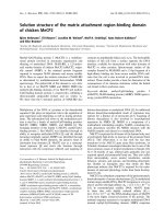
The NOESY spectra of C5 at lower temperatures are
superior to those at higher temperatures, due to an increased
correlation time (and thus an increased NOE). An example
of the NOESY spectrum at 285 K is given in Fig. 2A. The
Fig. 1. (A) Amino acid sequence and (B) CD spectra of the HIV gp120
C5 peptide. (A) Numbering corresponds to that of HIV-1 strain HXB2
[16]. (B) Dotted lines represent the spectrum in aqueous solution
(25 l
M
C5, 100 m
M
Tris/HCl/pH 8.0); solid lines represent the spec-
trum in the presence of an organic solvent (25 l
M
C5, 40% trifluoro-
ethanol, 100 m
M
Tris/HCl/pH 8.0).
Ó FEBS 2002 HIV Gp120 C5 solution structure (Eur. J. Biochem. 269) 4861
presence of sequential H
N
-H
N
crosspeaks is characteristic of
helical regions. Indeed, the crosspeak pattern shown in
Fig. 2A suggests the presence of a continuous helix between
residues 499 and 509. The secondary structure was further
characterized by
1
H
a
and
13
C
a
chemical shifts. As shown by
Fig. 2B, the
1
H
a
and
13
C
a
chemical shifts of residues 499–
509 suggest the presence of a helix. The prolines at positions
493 and 498 are apparently in the trans conformation, based
on the presence of strong a/d NOEs. Moreover, the absence
of Ôpeak splittingÕ in the TOCSY spectra of C5 indicates that
the trans isomer is the major isomer. Interestingly, the H
N
and H
a
line-widths of the region encompassing the prolines
and preceding the helix (residues 489–498) are similar to
those of the helix. Accordingly, the relaxation properties of
this region indicate that it is not appreciably more mobile
than the helix.
The tertiary structure of HIV gp120 C5 was determined
by an iterative method of torsion angle molecular dynam-
ics followed by simulated annealing [18]. A summary of
theNOEsusedinthecalculationispresentedinFig.3.
From Fig. 3, it is apparent that there are numerous long-
range contacts between residues in the N- and C-terminal
regions of C5 (e.g. V489 to V505 and I491 to V506). An
ensemble of 20 low energy structures is presented in
Fig. 4A and the structural statistics of the ensemble are
summarized in Table 1. The overall backbone RMSD of
the ensemble to the mean is 0.53 A
˚
and the overall heavy
atom RMSD (i.e. nonhydrogen) of the ensemble to the
mean is 1.25 A
˚
. In Fig. 4B, the backbone RMSD of the
C5 ensemble is plotted as a function of residue number
to illustrate the relative uncertainties of the backbone.
Figure 5A shows the ribbon diagram of the energy
minimized mean structure of HIV gp120 C5. The
structural motif of C5 is that of a turn-helix with the
amino and carboxy termini in close proximity. As shown
by Fig. 5A, the amino terminal end of the C5 domain
would be attached to remaining domains of gp120 in the
native structure. The carboxy terminal end of the C5
domain would be attached to the amino terminus of gp41
in the unprocessed form (termed gp160). Note that the
last four residues of the C5 domain form the furin/PC7
recognition site (amino acid sequence ¼ REKR) and
processing at this site by furin liberates the gp41 amino
terminus, which contains the fusion peptide. In Fig. 5B,
the electrostatic map of C5 is presented. Pertinent
features include a relatively uncharged hydrophobic
ÔelbowÕ in the turn (residues 493–499, amino acid
sequence ¼ PLGVAPT), the presence of two negative
charges (E492 and E509), and the presence of numerous
positive charges along the exterior of the helix
(e.g. K490, K500, R504, R508 and R511). Interestingly,
the hydrophobic ÔelbowÕ is absolutely conserved among
Fig. 2. (A) NOESY spectrum and (B) secondary chemical shifts of
1
H
a
and
13
C
a
of HIV gp120 C5 in 40% trifluoroethanol. (A) Experimental
conditions were 1 m
M
C5, 40% trifluoroethanol, 50 m
M
PO
4
(pH 6.0), 5% D
2
O at 285 K with a mixing time of 150 ms. (B) Secondary chemical
shifts of
1
H
a
(a) and
13
C
a
(b) of the HIV gp120 C5 in 40% trifluoroethanol. Experimental conditions were 1 m
M
C5, 40% trifluoroethanol, 50 m
M
PO
4
(pH 6.0) at 300 K. Random coil values of a protein in trifluoroethanol were taken from Merutka et al.[17].
Fig. 3. (A) Summary of the number and type of
NOEs observed for each residue and (B) NOE
contact map for HIV gp120 C5 in 40% tri-
fluoroethanol. (A) The NOE classification is
given in Table 1.
4862 L. Guilhaudis et al. (Eur. J. Biochem. 269) Ó FEBS 2002
HIV species [16]. Moreover, the charged residues at
positions 490, 500, 504, 508, 509, 510 and 511 are highly
conserved among HIV species [16].
The next logical step is to assay the ability of HIV C5 to
bind to HIV gp41. As discussed in the introduction, the loop
region of gp41 is the most likely binding site for C5. The
gp41 loop region contains four highly conserved tryptophan
residues (W85, W99, W103, and W112), which could
possibly serve as fluorescent reporters of a gp41/C5
interaction. Importantly, the C5 domain employed in the
present study does not possess any tryptophan residues.
Therefore, we performed fluorescence titrations of the HIV
gp41 ectodomain in the presence of trifluoroethanol as
cosolvent. As shown by Fig. 6A, the tryptophan fluores-
cence of HIV gp41 increases as HIV C5 is added. The
increased tryptophan fluorescence at 340 nm suggests that
at least some of the gp41 tryptophan sidechains are being
shielded from solvent by the addition of HIV C5 (Fig. 6B).
Based on the fluorescence changes shown by Fig. 6A, HIV
C5 forms a 1 : 1 complex with the HIV gp41 ectodomain
with an apparent K
d
of 1.0 l
M
. Interestingly, titrations
performed in the absence of cosolvent did not exhibit
measurable changes in tryptophan fluorescence, which
suggests that the turn-helix structural motif present of C5
is critical to complex formation.
DISCUSSION
The CD results suggest that HIV gp120 C5 is in a random
coil conformation in aqueous solutions, which is in agree-
ment with previous studies of similar C5 peptides [20,21]. In
contrast the presence of helix is clearly indicated by CD
Fig. 4. (A) Ensemble of 20 low-energy struc-
tures of HIV gp120 C5 in 40% trifluoroethanol
and (B) plot of the backbone RMSD of the final
C5 ensemble as a function of residue number.
(A) For clarity the amino and carboxy termini
have been denoted by their residue number.
(B) For this calculation, the backbone was
defined as the N, C
a
and C atoms.
Table 1. Structural statistics for the final 20 simulated annealing structures (ÆSAæ) and the minimized mean structure (ÆSAæ
r
). None of the structures
exhibited distance violations greater than 0.5 A
˚
or dihedral violations greater than 6°.
ÆSAæÆSAæ
r
RMS deviations from distance restraints all (190) 0.062 ± 0.003 0.075
intraresidue (16) 0.042 ± 0.012 0.032
sequential (| i-j | ¼ 1) (93) 0.081 ± 0.004 0.095
short range (1 < | i- j| < 5) (63) 0.030 ± 0.004 0.054
longrange (|i–j | > 5) (18) 0.030 ± 0.015 0.001
RMS deviations from dihedral restraints (°) (47)
a
0.99 ± 0.21 1.46
Deviations from idealized covalent geometry bonds (A
˚
) 0.006 ± 0.001 0.004
angles (°) 0.61 ± 0.02 2.52
impropers (°) 0.48 ± 0.04 2.74
Measures of structure quality
b
% residues in most favorable regions 84.2 ± 4.1 88.9
% residues in additionally allowed regions 15.8 ± 4.1 11.1
% residues in generously allowed regions 0.0 ± 0.0 0.0
% residues in disallowed regions 0.0 ± 0.0 0.0
number of bad contacts/100 residues 2.2 ± 3.3 8.7
Coordinate precision
c
backbone (A
˚
) 0.53 ± 0.20
heavy (A
˚
) 1.25 ± 0.72
helix backbone (A
˚
) 0.39 ± 0.06
helix heavy (A
˚
) 1.22 ± 0.65
a
Torsion angle restraints consisted of 21 /,21w,3v
1
, and 2 v
2
.
b
The overall quality of the structure was assessed by the program
PROCHECK
3.5 [19].
c
Defined as average RMS difference between the final 20 simulated annealing structures and the mean coordinates.
Ó FEBS 2002 HIV Gp120 C5 solution structure (Eur. J. Biochem. 269) 4863
when C5 is in the presence of the cosolvents trifluoroethanol
or hexafluoroisopropanol. The high-resolution solution
structure of HIV gp120 C5 in 40% trifluoroethanol was
determined by NMR spectroscopy. The presence of a
carboxy terminal helix (residues 499–510) was consistent
with the CD experiments, which predicted a helical content
of 10 residues. We recognize that from a physiological
standpoint the structural and dynamic characterization of
C5 in the presence of a cosolvent is less desirable than under
strictly aqueous conditions. However, it is increasingly
apparent that trifluoroethanol only stabilizes helical struc-
tures in protein domains with inherent propensity for helix
formation [22–24]. Moreover, trifluoroethanol/aqueous
mixtures are thought to mimic the hydrophobic environ-
ment of protein interiors [22,25–28]. In the present case, C5
in isolation is missing the other domains of gp120 as well as
its putative hydrophobic interaction site on gp41 [10,18,29].
Consequently, the trifluoroethanol/aqueous mixture may
very well mimic the hydrophobic environment of the gp120
C5 domain in vivo. Interestingly, the presence of helix at
residues 500–511 was predicted for HIV gp120 by various
methods of secondary structure prediction [30] and thus the
C5 propensity for helical structure is not surprising.
Importantly, we have shown by fluorescence titration that
HIV C5 directly interacts with the HIV gp41 ectodomain in
the presence of cosolvent, suggesting that the C5 structure
presented herein is physiologically relevant.
The HIV gp120 C5 structure provides insight into the
functional properties of the C5 domain. First, the C5
structure encompasses the site of envelope protein matur-
ation. Specifically, the furin/PC7 recognition site REKR is
present at the C terminal helix of the present C5 construct
(Fig. 5). Mutations at this cleavage site inhibit fusion and
hence HIV infection [5] and thus this site is a valid target for
drug therapies against HIV. However, it is important to
note that the presence of helix in this site is unknown in the
precursor envelope protein and further study is clearly
warranted. Second, the C5 structure provides insight into
the structure and action of a peptide that displays antiviral
activity against HIV-1 and HIV-2 [31]. This peptide, which
is called CLIV, corresponds to four linked copies of residues
499–511. The peptide has been shown to disrupt the fusion
process after binding of CD4, which indicates the CLIV
peptide does not function by disrupting gp120 maturation
[32]. Third, the C5 structure can be used to interpret the
results of mutagenesis studies of the gp120–gp41 interaction.
For example substitutions of C5 residues inhibit association
of gp120 with gp41 [8]. Moreover, substitution of I491,
P493, G495, A497, P498, T499 or A501 by cysteines resulted
in intermolecular crosslinks to non-native cysteines intro-
duced into the gp41 loop region [10]. Together the
mutagenesis, structural studies, and fluorescence titration
experiments [7,18,29,33] (and the present work) provide
compelling evidence that the hydrophobic loop of gp41
interacts with the hydrophobic turn region of C5.
The HIV gp120 C5 structure can be used to model the
gp41/gp120 complex. In Fig. 7, we present a model of the
gp41 and gp120 components, which were determined by
Fig. 5. (A) Ribbon diagram of the minimized mean structure of HIV
gp120 C5 in 40% trifluoroethanol and (B) electrostatic map of the
minimized mean structure of HIV gp120 C5. (A) The location of the
furin/PC7 site is shown. The locations of the missing gp120 domains
and gp41 are denoted. (B) In this orientation, the conserved Ôhydro-
phobic elbowÕ of C5 is located at the upper left corner of the figure.
Fig. 6. (A) Fluorescence titration of HIV gp41 ectodomain by HIV gp120 C5 and (B) location of tryptophan residues in HIV gp41 ectodomain
(coordinates taken from reference [33]). (A) Experimental conditions were 20 l
M
HIV gp41 ectodomain in 50 m
M
sodium formate (pH 3.0), 40%
trifluoroethanol at 25 °C. Titrations of a buffer blank by HIV C5 exhibited no change in the 340 nm fluorescence. The solid line represents the least
squaresfitofthedatawithK
d
¼ 1.0 ± 0.8 l
M
and n ¼ 0.98 ± 0.13. (B) The exposed tryptophans found in the gp41 loop are labeled. The
unlabeled tryptophans, which include W60, W117 and W120, are found in the protein interior and not exposed to solvent [18,33].
4864 L. Guilhaudis et al. (Eur. J. Biochem. 269) Ó FEBS 2002
NMR spectroscopy and X-ray crystallography [7,18,33]
and the present work. As discussed above, knowledge
of the gp41/gp120 structure is limited by the absence of
structural information for the C1 and C5 domains of
gp120, which are indicated to interact directly with gp41.
Accordingly, we have modeled the complex by first
placing C5 in the context of the gp120 core structure
determined by X-ray crystallography [7]. The first three
residues of the C5 domain used in the present study
(amino acids VKI) are observed in the gp120 X-ray
structure; thus, the C5 domain can be overlaid with the C
terminus of the gp120 X-ray structure (the RMSD of the
NMR VKI backbone to the X-ray VKI backbone is
0.4 A
˚
). The extended conformation of these residues in
solution is consistent with being part of a b sheet observed
in the gp120 X-ray structure (Fig. 7). The location of C5
with respect to the gp120 core is also consistent with
immunological studies that suggest that the C5 domain
interacts directly with the C1 and C2 domains [34]. In the
second step, we have used mutagenesis studies of gp41
and gp120 to reveal residues that are in close contact in
the gp41/gp120 complex. As discussed above, a number of
non-native cysteines placed within the gp41 loop form
intermolecular crosslinks to non-native cysteines placed in
the C5 domain [10]. Interestingly, the most reactive non-
native cysteines occur at position 94 of gp41 (denoted by
the blue sphere in Fig. 7) and at position 501 of gp120
(denoted by the red sphere in Fig. 7), which can be placed
in close proximity in the present model. The C5/gp41
interactiondepictedinFig.7wouldbeexpectedto
partially shield W85, W99 and W103 of gp41 from
solvent (cf. Figure 6B), which is consistent with the
fluorescence titration experiments discussed above. Finally,
the placement of the viral and target cell membranes is
facilitated by the structures of gp41 and CD4 receptor.
Specifically, gp41 contains a transmembrane domain that
is C-terminal to the ectodomain and thus located at the
ÔleftÕ of Fig. 7. The CD4 receptor contains a C-terminal
membrane anchor and thus is located at the ÔrightÕ of
Fig. 7. In addition, the location of the target cell
chemokine receptor can be inferred by the binding site
of the anitbody 17b, which blocks binding of gp120 to the
chemokine receptor [7]. Note that the model is consistent
with immunological studies that indicate that residues
501–511 are well exposed and immunodominant in the
native complex [35], consistent with its being charged and
thus hydrophilic (Fig. 5B). Furthermore, antibody acces-
sibility studies have suggested that the amino terminal
region of the gp120 C5 domain (residues 489–500) is not
exposed in the gp120/gp41 complex but exposed in the
monomeric form when gp120 dissociates from gp41 [36],
consistent with the notion that the hydrophobic ÔelbowÕ of
C5 (Fig. 5B) interacts with the hydrophobic loop region
of gp41 [18,33]. In total, the resulting model is consistent
with a large number of biochemical and structural studies.
It is important to note that the gp41 helical regions are
generally thought to undergo large-scale structural chan-
ges during the binding of gp120 to CD4 and the
subsequent fusion event (cf. [37] but see [18,38] for an
alternative viewpoint). Accordingly, the known structures
for gp41 [18,33] may not represent the gp41 conformation
bound to gp120. However, we are unaware of any
evidence for structural changes in the gp41 loop connect-
ing the helical regions.
In conclusion, we feel that the HIV gp120 C5 structure is
relevanttobiomedicalresearchonAIDSingeneral
and vaccine development in particular. Firstly, a full
understanding of gp120 function requires high-resolution
structural and dynamic information for all regions of gp120.
The present work presents the first high-resolution struc-
tural information for C5, albeit in 40% trifluoroethanol.
Secondly, based on the immunological studies [35,36] and
the model of the gp41/gp120 complex presented in Fig. 7,
the C5 domain is expected to be partially accessible to
solvent and thus accessible to the immune system. Conse-
quently, the gp120 C5 domain, which is highly conserved
among HIV and SIV isolates, may be an attractive target for
vaccine development. Finally, as discussed above, the C5
domain represents the most likely site for gp41 binding.
Disruption of this binding is expected to inhibit HIV
infection and thus the C5 domain is an attractive target for
structure-based drug therapies.
Fig. 7. Model for the gp41–gp120 complex
upon association with the target cell receptors.
The HIV gp41 ectodomain is shown in blue
and is a taken from the model structure [33],
which is based on the NMR structure of the
SIV gp41 ectodomain [18]. The gp120 C5
structureisshowninredandistakenfromthe
present work. The structure of the core gp120-
CD4 receptor-antibody 17b complex is taken
from Kwong et al. [7]. The gp120 core is
shown in green; the CD4 receptor is shown in
cyan, and the antibody 17b is shown in violet.
The sites of the most reactive non-native
cysteines in gp41 (at position 94) and gp120
(at position 501) are shown as spheres and
are taken from Binley et al.[10].
Ó FEBS 2002 HIV Gp120 C5 solution structure (Eur. J. Biochem. 269) 4865
ACKNOWLEDGEMENTS
The coordinates of the minimized mean structure of the HIV gp120 C5
have been deposited in the Protein Data Bank (ID 1MEQ). This work
was supported by RO1 grant AI47674.
REFERENCES
1. White, J.M. (1992) Membrane fusion. Science 258, 917–924.
2. Dimitrov, D.S. (1997) How do viruses enter cells? The HIV
coreceptors teach us a lesson in complexity. Cell 91, 721–730.
3. Major, E., Rausch, D., Marrak, C. & Clifford, D. (2000) HIV-
associated dementia. Science 288, 440–441.
4. Hallenberger, S., Moulard, M., Klenk, H. & Garten, W. (1997)
The role of eukaryotic subtilisin-like endoproteases for the acti-
vation of human immunodeficiency virus glycoproteins in natural
host cells. J. Virol. 71, 1036–1045.
5. Bosh, V. & Pawlita, M. (1990) Mutational analysis of the human
immunodeficiency virus type 1 env gene product proteolytic
cleavage site. J. Virol. 64, 2337–2344.
6. Hallenberger, S., Bosch, V., Angliker, H., Shaw, H., Klenk, H. &
Garten, W. (1992) Inhibition of furin-mediated cleavage activation
of HIV-1 glycoprotein-gp160. Nature 360, 358–361.
7. Kwong,P.D.,Wyatt,R.,Robinson,T.,Sweet,R.W.,Sodroski,J.
& Hendrickson, W. (1998) Structure of an HIV gp120 envelope
glycoprotein in complex with the CD4 receptor and a neutralizing
antibody. Nature 393, 648–659.
8. Helseth, E., Olshevsky, U., Gabuzda, D., Ardman, B., Haseltine,
W. & Sodroski, J. (1990) Changes in the transmembrane region of
the human immunodeficiency virus type -1 gp41 envelope glyco-
protein affect membrane fusion. J. Virol. 64, 6314–6318.
9. Ivey-Hoyle, M., Clark, R.K. & Rosenberg, M. (1991) The NH2-
terminal 31 amino acids of human immunodeficiency virus type-1
envelope protein gp120 contain a potential gp41 contact site.
J. Virol. 65, 2682–2685.
10. Binley,J.,Sanders,R.,Clas,B.,Schuelke,N.,Master,A.,Guo,
Y., Kajumo, T., Anselma, D., Maddon, P., Olson, W. & Moore, J.
(2000) A recombinant human immunodeficiency virus type 1
envelope glycoprotein complex stabilized by an intermolecular
disulfide bond between the gp120 and gp41 subunits is an anti-
genic mimic of the trimeric virion-associated structure. J. Virol. 74,
627–643.
11. Wingfield, P., Stahl, S., Kaufman, J., Zlotnick, A., Hyde, C.,
Gronenborn, A. & Clore, G. (1997) The extracellular domain of
immunodeficiency virus gp41 protein: expression in Escherichia
coli, purification, and crystallization. Prot. Sci. 6, 1653–1660.
12. Brunger, A., Adams, P., Clore, G., DeLano, W., Gros, P.,
Grosse-Kunstleve, R., Jiang, J S., Kuszewski, J., Nilges, M.,
Pannu, N., Read, R., Rice, L., Simonson, T. & Warren, G.
(1998) Crystallography and NMR System: a new software suite
for macromolecular structure determination. Acta Cryst. D54,
905–921.
13. Kuszewski, J., Gronenborn, A. & Clore, G. (1997) Improvements
and extensions in the conformational database potential of NMR
and x-ray structures of proteins and nucleic acids. J. Magn. Reson.
125, 171–177.
14. Cornilescu, G., Delaglio, F. & Bax, A. (1999) Protein backbone
angle restraints from searching a database for chemical shift and
sequence homology. J. Biomol. NMR 13, 289–302.
15. Koradi,R.,Billeter,M.&Wuthrich,K.(1996)MOLMOL:a
program for display and analysis of macromolecular structures.
J. Mol. Graphics 14, 52–55.
16. Douglas, N.W., Munro, G.H. & Daniels, R.S. (1997) HIV/SIV
glycoproteins: structure-function relationships. J. Mol. Biol. 273,
122–149.
17. Merutka, G., Dyson, H. & Wright, P. (1995) Random coil H-1
chemical-shifts obtained as a function of temperature and
trifluoroethanol concentration for the peptide series GGXGG.
J. Biomolec. NMR 5, 14–24.
18. Caffrey, M., Cai, M., Kaufman, J., Stahl, S.J., Wingfield, P.T.,
Gronenborn, A.M. & Clore, G.M. (1998) Solution
structure of the 44 kDa ectodomain of SIV gp41. EMBO J. 17,
4572–4584.
19. Laskowski,R.,MacArthur,M.,Moss,D.&Thorton,J.(1993)
PROCHECK – a program to check the stereochemical quality of
protein structures. J. Appl. Cryst. 26, 283–291.
20. Moulard, M., Challoin, L., Canarelli, S., Mabroluk, K. &
Darbon, H. (1998) Retroviral envelope glycoprotein processing:
Structural investigation of the cleavage site. Biochemistry 37,
4510–4517.
21. Ferrer, M., Sullivan, B., Godbout, K., Burke, E., Stump, H.,
Godoy, J., Golden, A., Profy, A. & van Schravendijk, M. (1999)
Structural and functional characterization of an epitope in the
conserved C-terminal region of HIV-1 gp120. J. Peptide Res. 54,
32–42.
22. Segawa, S., Fukuno, T., Fujiwara, K. & Noda, Y. (1991) Local
structures in unfolded lysozyme and correlation with secondary
structures in the native conformation – helix-forming or helix-
breaking propensity of peptide segments. Biopolymers 31,497–
509.
23. Sonnichsen, F., Van Eyk, J., Hodges, R. & Sykes, B. (1992) Effect
of trifluoroethanol on protein secondary structure – an NMR and
CD study using a synthetic actin peptide. Biochemistry 31, 8790–
8798.
24. Hamada,D.,Kuroda,Y.,Tanaka,T.&Goto,Y.(1995)High
helical propensity of the peptide-fragments derived from beta-
lactoglobulin, a predominantly beta-sheet protein. J. Mol. Biol.
254, 737–746.
25. Dyson, H., Sayre, J., Merutka, G., Shin, H., Lerner, R. & Wright,
P. (1992) Folding of peptide-fragments comprising the complete
sequence of proteins – models for initiation of protein folding. 2.
Plastocyanin. J. Mol. Biol. 226, 819–835.
26. Jasanoff, A. & Fersht, A. (1994) Quantitative-determination
of helical propensities from trifluoroethanol titration curves.
Biochemistry 33, 2129–2135.
27. Waterhous, D. & Johnson, W. (1994) Importance of environment
in determining secondary structure in proteins. Biochemistry 33,
2121–2128.
28. Hirota, N., Mizuno, K. & Goto, Y. (1998) Group additive con-
tributions to the alcohol-induced alpha-helix formation of
melittin: Implication for the mechanism of the alcohol effects on
proteins. J. Mol. Biol. 275, 365–378.
29. Caffrey, M., Cai, M., Kaufman, J., Stahl, S.J., Wingfield, P.T.,
Gronenborn, A.M. & Clore, G.M. (1997) Determination of the
secondary structure and global topology of the 44 kDa ecto-
domain of gp41 of the simian immunodeficiency virus by multi-
dimensional nuclear magnetic resonance spectroscopy. J. Mol.
Biol. 271, 819–826.
30. Hansen, J., Lund, O., Nielsen, J., Brunak, S. & Hansen, J. (1996)
Prediction of the secondary structure of HIV-1 gp120. Proteins 25,
1–11.
31. Sabatier, J., Moulard, M., Rochat, H., Van Rietschoten, J. &
Fenouillet, E. (1996) Anti-HIV activity of multibranched peptide
constructs derived either from the cleavage sequence or from the
transmembrane domain (gp41) of the human immunodeficiency
virus type 1 envelope. Virology 223, 406–408.
32. Barbouche, R., Decroly, E., Kieny, M. & Fenouillet, E. (2000) An
anti-human immunodeficiency virus multiple antigen peptide
encompassing the cleavage region of the env precursor interferes
with membrane fusion at a post-CD4 binding step. Virology 273,
169–177.
33. Caffrey, M. (2001) Model for the structure of the HIV gp41
ectodomain: insight into the intermolecular interactions of the
gp41 loop. Biochim. Biophys. Acta 1536, 116–122.
4866 L. Guilhaudis et al. (Eur. J. Biochem. 269) Ó FEBS 2002
34. Moore, J., Willey, R., Lewis, G., Robinson, J. & Sodroski, J.
(1994) Immunological evidence for interactions between the first,
second and fifth conserved domains of the gp120 surface glyco-
protein of human immunodeficiency virus type 1. J. Virol. 68,
6836–6847.
35. Palker,T.,Matthews,T.,Clark,M.,Cianciolo,G.,Randall,R.,
Langlois, A., White, G., Safai, B., Snyderman, R., Bolognesi, D. &
Haynes, B. (1987) A conserved region at the cooh terminus of
human-immunodeficiency-virus gp120 envelope protein contains
an immunodominant epitope. Proc. Natl. Acad. Sci. USA 84,
2479–2483.
36. Moore, J., Sattentau, Q., Wyatt, R. & Sodroski, J. (1994) Probing
the structure of the human immunodeficiency virus surface gly-
coprotein gp120 with a panel of monoclonal antibodies. J. Virol.
68, 469–484.
37. Chan, D. & Kim, P. (1998) HIV entry and its inhibition. Cell 93,
681–684.
38. Caffrey, M., Kaufman, J., Stahl, S., Wingfield, P., Gronenborn, A.
& Clore, G. (1999) Monomer-trimer equilibrium of the ecto-
domain of SIV gp41: insight into the mechanism of peptide
inhibition of HIV infection. Prot. Sci. 8, 1904–1907.
Ó FEBS 2002 HIV Gp120 C5 solution structure (Eur. J. Biochem. 269) 4867