Báo cáo Y học: The refolding of type II shikimate kinase from Erwinia chrysanthemi after denaturation in urea pot
Bạn đang xem bản rút gọn của tài liệu. Xem và tải ngay bản đầy đủ của tài liệu tại đây (594.71 KB, 9 trang )
The refolding of type II shikimate kinase from
Erwinia chrysanthemi
after denaturation in urea
Eleonora Cerasoli
1
, Sharon M. Kelly
1
, John R. Coggins
1
, Deborah J. Boam
1
, David T. Clarke
2
and Nicholas C. Price
1
1
Division of Biochemistry and Molecular Biology, Institute of Biomedical and Life Sciences, Joseph Black Building,
University of Glasgow, Scotland, UK;
2
Synchrotron Radiation Department, CLRC Daresbury Laboratory, Warrington UK
Shikimate kinase was chosen as a convenient representative
example of the subclass of a/b proteins with which to
examine the mechanism of protein folding. In this paper we
report on the refolding of the enzyme after denaturation in
urea. As shown by the changes in secondary and tertiary
structure monitored by far UV circular dichroism (CD) and
fluorescence, respectively, the enzyme was fully unfolded in
4
M
urea. From an analysis of the unfolding curve in terms of
the two-state model, the stability of the folded state could be
estimated as 17 kJÆmol
)1
. Approximately 95% of the
enzyme activity could be recovered on dilution of the urea
from 4 to 0.36
M
. The results of spectroscopic studies indi-
cated that refolding occurred in at least four kinetic phases,
the slowest of which (k ¼ 0.009 s
)1
) corresponded with the
regain of shikimate binding and of enzyme activity. The two
most rapid phases were associated with a substantial increase
in the binding of 8-anilino-1-naphthalenesulfonic acid with
only modest changes in the far UV CD, indicating that a
collapsed intermediate with only partial native secondary
structure was formed rapidly. The relevance of the results to
the folding of other a/b domain proteins is discussed.
Keywords: shikimate kinase; protein folding; protein
unfolding; circular dichroism; fluorescence.
Despite considerable experimental and theoretical efforts
over the past 30 years, the mechanism by which proteins
achieve their functional three-dimensional structure repre-
sents a major area of uncertainty [1,2]. The importance of
an understanding of protein folding is illustrated by both
biotechnological applications (for example, in the recovery
of properly folded expressed proteins [3]) and by clinical
consequences (where disease states are caused by protein
misfolding [4]). In addition, an understanding of the
principles governing protein folding would help to allow
the huge amount of information from genome sequencing
projects to feed through to accurate predictions of three-
dimensional structure of the encoded proteins. Because of
the difficulties in applying structural techniques to the
acquisition of structure accompanying or following trans-
lation in vivo, the usual experimental approach has been to
study the refolding of denatured proteins when conditions
have been changed to promote folding. Several lines of
evidence indicate that this approach can give valid insights
into the process of protein folding in vivo [5]. Detailed
studies have allowed the pathways of folding of a number of
small proteins, such as barnase [6], dihydrofolate reductase
[7], chymotrypsin inhibitor 2 [8], lysozyme [9] and CheY
[10] to be mapped out, but a key requirement is to examine
the behaviour of protein fold families in a systematic
manner.
The most structurally diverse of the classes of proteins,
introduced by Chothia and colleagues [11], is the a/b class,
which contains nearly 100 different kinds of protein folds.
One of these subclasses is the P-loop-containing nucleotide
triphosphate hydrolases, the core of which forms a classical
mononucleotide-binding fold found in a number of struc-
turally diverse proteins such as myosin, elongation factor
EF-Tu, p21
ras
, the NDB domain of the ABC transporters,
Rec A and adenylate kinase. The structural conservation of
the core within this group of proteins is illustrated by the
fact that superimposition of the P-loops results in root mean
square deviations in alpha C atoms of only 0.3–0.4 A
˚
[12].
The isoenzyme II of shikimate kinase (SK, EC 2.7.1.71), an
enzyme which catalyses the specific phosphorylation of the
3-hydroxyl group of shikimate using ATP as the phosphoryl
donor [13,14], is a member of this subclass. This step is the
fifth in the seven-step pathway leading to the synthesis of
chorismate, the precursor of aromatic compounds. From
the X-ray structure of SK [15], it is clear that the ordering of
the strands 23145 in the parallel b sheet places the enzyme in
thesamestructuralfamilyastheNMPkinases(adenylate
kinase, guanylate kinase, uridylate kinase and thymidine
kinase).
SK has a number of experimental advantages in estab-
lishing the mechanism of protein folding. It is a monomeric
enzyme without disulphide bonds and, with a molecular
mass of 19 kDa, it is amongst the smallest kinases so far
reported. SK has a single Trp residue (Trp54) that is located
in the region near the shikimate binding site [15]. Binding of
shikimate leads to quenching of Trp fluorescence [16],
thereby providing a convenient probe for the integrity of the
shikimate binding site. An additional feature of SK is that
the side chains of Arg11, Arg58 and Arg139 provide a
Correspondence to N. C. Price, Synchrotron Radiation Department,
CLRC Daresbury Laboratory, Warrington WA4 4AD, UK.
Fax: + 44 141 330 6447; Tel.: + 44 141 330 2889;
E-mail:
Abbreviations: SK, shikimate kinase; ANS, 8-anilino-1-naphthalene-
sulfonic acid; PK, pyruvate kinase; LDH, lactate dehydrogenase;
GdmCl, guanidinium chloride; SRS, synchrotron radiation source.
(Received 14 November 2001, revised 6 February 2002, accepted
1 March 2002)
Eur. J. Biochem. 269, 2124–2132 (2002) Ó FEBS 2002 doi:10.1046/j.1432-1033.2002.ejb.02862.x
highly positively charged environment around the Trp side
chain and the shikimate binding site [15]. The use of the
iodide ion as a quencher of protein fluorescence provides an
additional means of investigating the integrity of this region
of the protein.
In the present paper, we have undertaken a study of the
unfolding and refolding of the type II SK from E. chry-
santhemi, using studies of CD, fluorescence, activity and
ANS fluorescence, and employing both manual mixing and
rapid reaction techniques. From these studies, we have been
able to formulate an outline pathway for the folding process
in which at least three intermediates are involved. The
results extend the less complete data available for the
refolding of adenylate kinase [17] indicating that the
pathway described for SK should act as a model for many
other members of this subclass of a/b proteins.
MATERIALS AND METHODS
Enzyme purification
The purification protocol was based on those used for the
purification of SK II from Escherichia coli [18] and for the
previous purification of the enzyme from E. chrysanthemi
[19]. The latter method was adapted by reducing the salt
concentration so as to prevent protein precipitation. After
cell breakage, all steps were performed at 4 °C.
E. coli BL21(DE3)pLysS cells (10 g) were resuspended in
10 mL of buffer (20 m
M
Tris/HCl, pH 7.5 containing
0.4 m
M
dithiothreitol plus one tablet of ÔComplete
TM
Õ
(Boehringer) to inhibit protease action. Cells were broken
by passing them through a French pressure cell twice at
6.9 MPa and the resulting mixture was centrifuged at
100 000 g for 1 h.
The supernatant was dialysed for 4 h against buffer A
(20 m
M
Tris/HCl, pH 7.5 containing 0.4 m
M
dithiothreitol
and 1 m
M
MgCl
2
) and loaded on to a pre-equilibrated
DEAE-Sephacel anion exchange column (30 cm · 2.6 cm
diameter, flow rate 50 mLÆh
)1
). The column was then
washed with buffer A until A
280
< 0.1. Elution of shiki-
mate kinase was achieved using a linear gradient of
0–300 m
M
KCl in 600 mL buffer A with a flow rate of
50 mLÆh
)1
and a fraction volume of 14 mL.
Pooled fractions were dialysed against buffer A. Before
adding the solution to a phenyl–Sepharose CL-4B column
(4 · 2 cm), solid (NH
4
)
2
SO
4
was added to 30% saturation
(164 gÆL
)1
). The solution was stirred for 20 min and then
centrifuged at 20 000 g for 15 min. The supernatant was
loaded onto the column pre-equilibrated in buffer B
[100 m
M
Tris/HCl, pH 7.5 containing 0.4 m
M
dithiothreitol
and 1.2
M
(NH
4
)
2
SO
4
]. The column was washed overnight
with buffer B at low flow rate (5 mLÆh
)1
)and10mL
fractions were collected. The enzyme was eluted using a
linear gradient of 400 mL 1.2–0.0
M
(NH
4
)
2
SO
4
in buffer B
with a flow rate of 20 mLÆh
)1
and a fraction volume of
10 mL. At the end of the gradient the column was washed
with 250 mL of 100 m
M
Tris/HCl, pH 7.5 containing
0.4 m
M
dithiothreitol until residual shikimate kinase had
been eluted. Active fractions were dialysed overnight against
buffer A containing 10% (v/v) glycerol to concentrate the
enzyme sample.
After this step, the sample was loaded on to the pre-
equilibrated Sephacryl S200 (superfine grade) column
(120 · 2.5 cm) and eluted at a flow rate of 10 mLÆh
)1
in
buffer C (50 m
M
Tris/HCl, pH 7.5 containing 0.4 m
M
dithiothreitol, 5 m
M
MgCl
2
and 500 m
M
KCl) with a
fraction volume of 4 mL. Active fractions were pooled and
dialysed overnight against 50 m
M
Tris/HCl, pH 7.5 con-
taining 0.4 m
M
dithiothreitol, 5 m
M
MgCl
2
and 50% (v/v)
glycerol. The purified SK was stored at )20 °C.
Before use, SK was dialysed against buffer D (35 m
M
Tris/HCl, pH 7.6 containing 5 m
M
KCl, 2.5 m
M
MgCl
2
and 0.4 m
M
dithiothreitol) and used within a 2-day period.
Enzyme activity and CD measurements showed that the
protein is stable if stored overnight at )20 °Cinthis
buffer.
The concentration of SK was determined spectrophoto-
metrically using a value of 0.62 for the A
280
of a 1 mgÆmL
)1
solution in a cuvette of 1-cm pathlength. This value was
calculated from the amino-acid composition of the enzyme
[20], using the observed ratio (1.09) of absorbances in buffer
andin6
M
GdmCl. This value was within 10% of that
obtained using the dye-binding method [21]. The ratio A
280
/
A
260
was greater than 1.8, confirming the absence of
significant contaminant by nucleotide.
Assay of enzyme activity
The activity of the shikimate kinase was determined by a
double coupled assay involving pyruvate kinase (PK) and
lactate dehydrogenase (LDH). The production of ADP in
the shikimate kinase-catalysed reaction leads to the conver-
sion of NADH to NAD
+
, which is monitored by the
decrease in A
340
.
The assay was carried out at 25 °C in a buffer consisting
of 50 m
M
triethanolamine hydrochloride containing 50 m
M
KCl and 5 m
M
MgCl
2
,titratedtopH7.2withKOH.
Concentrations of the assay components were 1.6 m
M
shikimate, 5 m
M
ATP, 1 m
M
phosphoenolpyruvate,
0.2 m
M
NADH, 1 U of each of PK and LDH. Stock
solutions of the substrates were stored at )20 °Cafter
neutralization with KOH. Under these conditions, the
specific activity of the enzyme was 350 lmol min
)1
Æmg
)1
.
In order to measure the activity of SK in the presence
of urea it was necessary to use a quenched assay because
of the effects of this agent on the coupling enzymes [22].
The SK-catalysed reaction was carried out in an assay
solution containing 5 m
M
ATP, 1.6 m
M
shikimate and
the appropriate concentration of urea in the assay buffer.
At chosen times after the start of the reaction aliquots of
this solution were diluted 30-fold into a quench mixture
containing the appropriate concentrations of PEP,
NADH, PK and LDH. From the decrease in A
340
,the
concentration of ADP produced in the SK-catalysed
reaction at the chosen times can be determined, and
hence the rate of this reaction calculated. The errors in
assays of enzyme activity were less than 5% of the quoted
values.
Spectroscopic measurements
Except where indicated, all spectroscopic measurements
were made on enzyme samples in buffer D.
Most CD measurements were made using a Jasco J-600
spectropolarimeter, using cells of pathlength 0.2 or 0.5 mm
and protein concentrations in the range 0.1–0.5 mgÆmL
)1
.
Ó FEBS 2002 Refolding of shikimate kinase (Eur. J. Biochem. 269) 2125
Some CD data were obtained on experimental station 3.1 of
the CLRC Daresbury Laboratory’s Synchrotron Radiation
Source (SRS). This facility comprises a vacuum-UV 1 m
Seya-Namioka monochromator, which provides a high flux
of linearly polarized light in the wavelength range 120–
300 nm, which is converted to circularly polarized light
using a photoelastic modulator [23]. The SRS CD facility
was particularly useful when spectra were recorded in the
presence of high concentrations of NaCl or urea which
absorb strongly in the far UV. Spectra were recorded using
cells of pathlength 0.1 or 0.01 mm and protein concentra-
tions in the range 1–2 mgÆmL
)1
. Fluorescence data were
obtained using a PerkinElmer LS50 spectrofluorimeter.
The fluorescence of ANS was measured using excitation
and emission wavelengths of 380 nm and 480 nm, respec-
tively. The concentrations of solutions of ANS were
checked spectrophotometrically using a value of 6.0 for
the A
350
of a 1-m
M
solution in a cuvette of 1-cm pathlength
[24].
The quenching of protein fluorescence by sodium iodide
(over the range of quencher concentrations from 0 to 0.2
M
)
was analysed by Stern–Volmer plots as described previously
[25].
Stopped flow measurements were made using an Applied
Photophysics SX-17
M
Vapparatususinga10:1mixing
ratio. The dead times for the fluorescence and CD modes
have been determined as 1.7 and 8 ms, respectively [26]. As
recommended by the manufacturer, the time filter applied
was less than 10% of the half time of the process being
studied, in order to avoid distortion of the kinetic analysis.
This analysis was undertaken using the
PRO/K
software
supplied with the instrument. The data reported represent
the averages of three runs each of 10 shots. Unless otherwise
stated, the errors in the amplitudes and rate constants
derived were less than 10% of the stated values. The
concentration of enzyme during refolding was in the range
60–110 lgÆmL
)1
in different experiments, with no signifi-
cant variation in rate constants observed over this range.
Light scattering was measured using the PerkinElmer
LS50 spectrofluorimeter with excitation and emission
wavelengths of 320 nm.
Unfolding and refolding studies
Stock solutions of Ultrapure grade urea (10
M
)weremade
up by weight in buffer D; the actual concentrations were
checked using refractive index data [27].
Unfolding and refolding of SK was performed essentially
as described in our previous studies on type II dehydroqu-
inase [28]. To study the extent of unfolding of SK, the
enzyme was routinely incubated in buffer D in the stated
concentration of denaturant for 1 h at 20 °C, before the
CD, fluorescence and activity data were recorded. Refolding
was routinely initiated after unfolding for 1 h in the
presence of 4
M
urea, by dilution with 10 vol. of buffer D,
to give a residual concentration of denaturant of 0.36
M
.In
preliminary experiments, it was shown that unfolding in 4
M
urea for periods ranging from 5 min to 3 h had no effect on
either the spectroscopic properties of the unfolded enzyme,
or the kinetics of refolding as monitored by changes in
protein fluorescence. Where indicated ANS was included in
the unfolding and refolding mixtures at a concentration of
40 l
M
.
RESULTS
Unfolding of enzyme
Stability of the enzyme. The loss of secondary and tertiary
structure during unfolding of SK by urea were monitored
by changes in far UV CD and fluorescence, respectively.
On incubation of the enzyme in 4
M
urea, there was
essentially a complete loss of secondary structure with the
ellipticity at 225 nm reduced to less than 10% of the value
characteristic of native enzyme. The degree of unfolding was
monitored by changes in the ellipticity at 225 nm.
When excited at 290 nm, the fluorescence emission
maximum of SK is 346 nm, indicating that the single Trp
(Trp54) is significantly exposed to the solvent, a conclusion
consistent with the high value of the Stern–Volmer constant
for quenching of the fluorescence by succinimide [16]. When
incubated in 4
M
urea, the emission maximum shifts to
356 nm, indicating that the Trp has become completely
exposed to solvent. The degree of unfolding was monitored
by changes in the emission intensity at 346 nm.
The unfolding data for SK (Fig. 1) could be analysed
satisfactorily in terms of a two-state model [27], suggesting
that no intermediate species were populated to a significant
extent. From the plot of free energy change against
denaturant concentration the stability of native enzyme in
the absence of denaturant could be estimated as
17 ± 1kJÆmol
)1
with no significant difference in stability
observed using the two measures of structural changes
employed. The value of the stability is towards the lower
end of those observed for a range of globular proteins [29]
and is similar to the value estimated for the structurally
similar enzyme adenylate kinase (19.6 kJÆmol
)1
)from
studies of the unfolding by urea [17]. However, given the
difficulties in estimating the contributions of the various
non–covalent interactions to the overall stability of
globular proteins [29], it is not profitable to analyse this
degree of similarity in greater detail.
Changes in activity in the presence of urea. Incubation
with urea leads to losses in activity which run roughly in
parallel with the structural changes, with 85 and 40%
activity retained in the presence of 1 and 2
M
urea,
respectively. In the presence of 4
M
urea, shikimate kinase
retains no detectable activity (< 0.1% of the control value).
Refolding of enzyme
All experiments on the refolding of shikimate kinase
involved unfolding in 4
M
urea for unfolding and 11-fold
dilution (to 0.36
M
urea) to initiate refolding. During this
process, there was no significant increase in light scattering
at 320 nm during refolding showing that aggregation
occurred to a negligible effect.
Regain of activity. The first time point at which activity
can be accurately assessed was estimated to be about 80 s
after the start of refolding, taking into account the time
taken for appropriate dilution into the assay solution and
for the double coupled assay system to achieve a constant
rate. By this time 35% of the activity of the control sample
(in the presence of 0.36
M
urea) had been regained. Over the
next 15 min, a further 60% activity was regained in a first
2126 E. Cerasoli et al. (Eur. J. Biochem. 269) Ó FEBS 2002
order process with a rate constant 0.007 s
)1
. Thus overall
95% of the activity of the control was regained (Fig. 2).
Extrapolation of the curve shows that after 15 s, the regain
of activity is 10% or less. If dithiothreitol was omitted from
the unfolding and refolding buffers, the extent of regain of
activity was reduced to 60%, showing that some damage
had occurred to either or both of the two Cys side chains
(Cys13 and Cys162) in the enzyme during the unfolding/
refolding procedure.
Regain of secondary structure on refolding. When the
enzyme was unfolded in 4
M
urea and subsequently refolded
by an 11-fold dilution using manual mixing, 75% of the
recovery of ellipticity at 225 nm was complete within the
dead time (20 s) of the start of recording the ellipticity. A
further 15% of the signal was regained over the subsequent
500 s with a rate constant of 0.009 s
)1
.Attheendofthis
period the far UV CD spectrum of the refolded enzyme was
very similar to that of native enzyme (data not shown).
Using stopped flow mixing to initiate refolding it was
shown that the regain of ellipticity at 225 nm occurred in a
number of phases. From data obtained over the first 20 s of
refolding, it was shown that, within 20 ms, 15% of the total
signal corresponding to the folded enzyme (i.e. the differ-
ence between denatured and folded enzyme) had been
regained. A further 20% of the signal was regained in a first
order process with a rate constant of 8 s
)1
; in the third phase
a further 40% was regained with a rate constant 0.08 s
)1
.
Finally from data over the time range 20–200 s, a fourth
phase was observed accounting for an additional 10%
change with a rate constant 0.008 s
)1
. Taken together, the
four phases account for a regain of 85% of the native
secondary structure (Fig. 3).
Regain of tertiary structure. The regain of tertiary struc-
ture was monitored by changes in protein fluorescence at
350 nm after dilution of the denaturant from 4
M
to 0.36
M
.
In the manual mixing mode, the first time point at which
reliable data could be obtained was 20 s after refolding had
been initiated. Within this dead time, 35% of the fluores-
cenceofnativeenzyme(inthepresenceof0.36
M
urea) had
been regained. Over the course of 20 min, a further 55% of
the fluorescence was regained in a first order process with a
rate constant of 0.009 s
)1
(data not shown). Thus overall
90% of the signal of native SK was regained. Using stopped
flow mixing techniques, it was found that less than 5% of the
total change occurred within 5 ms and that the subsequent
changes in fluorescence occurred in two phases with
amplitudes 42 and 45% of the total change with first order
rate constants of 0.08 and 0.009 s
)1
, respectively (Fig. 4A).
The rate of the slower process corresponds to that observed
using manual mixing techniques.
Refolding in the presence of shikimate. Refolding of
shikimate kinase in the presence of shikimate was carried
out in order to assess the stage in the process at which the
shikimate binding site is formed, using the quenching of the
protein fluorescence by the ligand as the index of binding.
For these experiments it was necessary to monitor the
refolding by fluorescence at 330 nm, rather than 350 nm. At
the latter wavelength, the quenching caused by the binding
of shikimate to folded enzyme was nearly equal to the
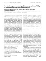
Fig. 1. The unfolding of SK in the presence of urea. (A) Structural
changes monitored by changes in ellipticity at 225 nm (triangles) and
protein fluorescence at 350 nm (squares) as described in the text. The
concentration of protein in each sample was 0.2 mgÆmL
)1
.Thedata
shown combine the results of three separate sets of experiments for
each technique, with the results of replicate determinations within 5%.
(B) Data analysed according to the two-state model [27], with the
regression line shown.
Fig. 2. The kinetics of regain of activity of SK after denaturation in 4
M
urea. Activity values are expressed relative to a control sample incu-
bated in the presence of the final concentration of urea, i.e. 0.36
M
.The
dashed line shows a fit to a first order process with a rate constant of
0.007 s
)1
.
Ó FEBS 2002 Refolding of shikimate kinase (Eur. J. Biochem. 269) 2127
enhancement of protein fluorescence which occurred on
refolding, leading to a very small overall change. In a
separate experiment (data not shown) the binding of
shikimate to the enzyme in the presence of 0.36
M
urea
was shown to be very rapid. When 2 m
M
shikimate was
added to the enzyme (0.09 mgÆmL
)1
), over 95% of the
fluorescence change occurred within the dead time of the
stopped-flow instrument (1.8 ms), implying a rate constant
for the association reaction > 7 · 10
5
M
)1
Æs
)1
.
The refolding of enzyme in the absence of shikimate led
to a biphasic increase in fluorescence at 330 nm (Fig. 4B);
the kinetics of this process were essentially indistinguishable
from those observed at 350 nm (Fig. 4A). When the
refolding was carried out in the presence of 2 m
M
shikimate,
however, a markedly different kinetic pattern was observed
(Fig. 4B). After a rapid increase in fluorescence, essentially
complete within 15 s, there was a slow small decrease over
the next 185 s. The rate constant for this decline (0.025 s
)1
)
was rather higher than that of the slow increase in the
absence of shikimate (0.009 s
)1
), which could indicate that
the presence of ligand has a nucleating effect on folding of
this area of the enzyme [5]. The folding of the protein (which
wouldbeexpectedtoleadtoanincreaseinprotein
fluorescence) leads to the formation of a Ônative-typeÕ
shikimate binding site and the consequent quenching results
in the overall decrease in fluorescence in this phase of the
process. The simplest interpretation of these results is that
the formation of this Ônative-typeÕ site is only associated with
the slowest phase of the folding process.
ANS as a probe during refolding. ANS has been used
extensively as a probe for the existence of Ômolten globuleÕ or
Ôcompact intermediateÕ states of proteins and their forma-
tion during folding [30,31]. However, there have been
concerns raised that the presence of ANS may in fact
perturb the folding process [32].
InthecaseofSK,thepresenceof40l
M
ANScausedan
18% decrease in the activity of enzyme when assayed under
the standard conditions. The presence of ANS caused less
than 10% change in the K
d
for shikimate using the
fluorescence quenching titration. When unfolding and
refolding were performed in the presence of 40 l
M
ANS,
the regain of activity was 95% that of the control (with
ANS); this activity was regained in a first order process with
a rate constant 0.008 s
)1
.Fromthesedata,itisclearthat
ANS has only relatively minor effects on the catalytic site of
the enzyme and its ability to refold after denaturation.
During the refolding process, a characteristic pattern of
changes in ANS fluorescence during refolding was observed.
When refolding was initiated by manual mixing techniques,
Fig. 3. The kinetics of changes in ellipticity at 225 nm during refolding
of SK after denaturation in 4
M
urea. The refolding was initiated by
stopped flow mixing; the inset shows data in the first second of the
reaction. Curves a, b and c refer to enzyme in the presence of 4
M
urea,
enzyme in the presence of 0.36
M
urea, and enzyme during refolding,
respectively. The pattern of residuals to the curve fitting is shown.
Fig. 4. The kinetics of changes in protein fluorescence at during refold-
ing of SK after denaturation in 4
M
urea. Refolding was initiated by
stopped flow mixing and the fluorescence signals have been corrected
for the buffer signal. (A) Refolding in the absence of shikimate. Curves
a, b and c refer to enzyme in the presence of 4
M
urea, enzyme in the
presence of 0.36
M
urea, and enzyme during refolding, respectively. (B)
Comparison of refolding in the absence and presence of 2 m
M
shiki-
mate. In (A), fluorescence was monitored at 350 nm; in (B) fluores-
cence was monitored at 330 nm. The pattern of residuals to the curve
fitting in (A) is shown.
2128 E. Cerasoli et al. (Eur. J. Biochem. 269) Ó FEBS 2002
there was a rapid increase in fluorescence within the dead
time of observation (20 s) corresponding to 10 times the
fluorescence of the starting solution (enzyme in 4
M
urea)
and 2.5 times the value of the end solution (enzyme in
0.36
M
urea). This increase was followed by a decrease over
the subsequent 600 s to reach a value similar to that
observed for the enzyme in the final concentration of urea
(0.36
M
); the rate constant for this decrease was 0.009 s
)1
(data not shown). Using stopped flow mixing techniques
(Fig. 5A,B) to initiate refolding, the initial rapid increase in
ANS fluorescence was found to be at least 50% complete
within 5 ms. Further analysis of the changes in fluorescence
over the first 200 ms after mixing suggested that the increase
occurred in two phases of approximately equal amplitude,
one very fast (k >100s
)1
) and the other with a rate
constant 11 s
)1
. (It should be noted that the magnitude of
the faster rate constant could not be estimated accurately;
the value quoted is based on the half time being less than or
equal to 5 ms). The subsequent decrease in ANS fluores-
cence (to reach a value similar to that of the enzyme in the
presence of the final concentration of urea) occurred in two
first order processes with rate constants 0.08 and 0.012 s
)1
;
the amplitude of the faster of these two phases corresponded
to 25% of the total decrease observed (Fig. 5A).
Refolding in the presence of sodium iodide. The I
–
ion is a
very effective quencher of SK fluorescence as shown by the
high Stern–Volmer constant (K
sv
)of19.8
M
)1
, compared
with 10.1
M
)1
for the model compound, N-acetyltryptophan
amide. It is likely that the high degree of quenching of SK is
due to the positively charged environment provided by the
three Arg side chains (Arg11, Arg58 and Arg139) in
the neighbourhood of Trp54 [15]. This is confirmed by the
observation that in the presence of 4
M
urea the K
sv
values
for SK is reduced dramatically to 4.0
M
)1
; by contrast the
addition of 4
M
urea has only a very small effect on the K
sv
of the model compound (9.5
M
)1
). In the presence of 0.36
M
urea the K
sv
for SK (19.0
M
)1
) is very similar to that of
native enzyme.
We have studied the changes in protein fluorescence
during refolding of SK in the presence of 0.1
M
NaI to assess
at what stage in the folding process the positively charged
environment of Trp54 in native enzyme is formed. From the
data obtained when manual mixing was used to initiate
refolding, it was found that after 600 s, the degree of
quenching caused by 0.1
M
NaI, corresponded to a K
sv
of
17.8
M
)1
(i.e. very similar to that of enzyme in the presence
of 0.36
M
urea). After 20 s, the degree of quenching
corresponded to a K
sv
of 12.0
M
)1
. The changes in
fluorescence over the period from 20 to 600 s could be
fitted to a first order process with a rate constant 0.008 s
)1
,
which is very similar to that observed in the absence of NaI
(0.009 s
)1
) (data not shown). Using stopped-flow mixing to
initiate refolding (Fig. 6), the degree of quenching after 20 s
was found to correspond to a K
sv
of 12.0
M
)1
, identical to
that observed by manual mixing. After 2 s, the quenching
corresponded to a K
sv
of 6.4
M
)1
, which is similar to the
value for denatured enzyme. From these results, it is clear
that the high degree of quenching and hence the positively
charged environment of the Trp is formed progressively
during the two (relatively slow) processes during which the
changes in the Trp fluorescence itself occur.
Model of folding pathway and properties of intermedi-
ates. Detailed studies of the refolding of a number of
proteins after denaturation have led to the development of
the Ônucleation-condensationÕ model; this seeks to draw
together ideas from earlier proposals which focussed
attention on aspects such as formation of secondary
structure or hydrophobic collapse [33]. In energy terms,
the transition from denatured to native state is viewed in
terms of a Ôfolding landscapeÕ in which kinetic flow can
occur through a series of states of progressively lower energy
in a Ôfolding funnelÕ [1,34,35].
Although there are some differences in detail between the
results of the various techniques employed in the present
work to monitor the refolding of SK after denaturation in
urea, when taken together the data indicate that there are
probably four kinetic phases contributing to the folding
process. The average rate constants for these phases are
Fig. 5. The kinetics of changes in ANS fluorescence at 480 nm during
refolding of SK after denaturation in 4
M
urea. Refolding was initiated
by stopped flow mixing and changes were monitored over the time
ranges (A) 0–220 s and (B) 0–220 ms. In each panel, curves a, b and c
refer to enzyme in the presence of 4
M
urea, enzyme in the presence of
0.36
M
urea, and enzyme during refolding, respectively. The pattern of
residuals to the curve fitting is shown.
Ó FEBS 2002 Refolding of shikimate kinase (Eur. J. Biochem. 269) 2129
> 100 s
)1
(half-life < 7 ms), 10 s
)1
(half-life 70 ms), 0.08 s
)1
(half-life 9 s) and 0.009 s
)1
(half-life 80 s). A simple outline
model could thus be proposed which involves three interme-
diates (I
1
,I
2
and I
3
) between the unfolded state (U) and the
native state (N); these are linked in a sequential fashion:
U
!
> 100 s
À1
I
1
!
10 s
À1
I
2
!
0:08 s
À1
I
3
!
0:009 s
À1
N
The properties of these are indicated in Table 1, in which
the various properties of the unfolded and final states have
been normalized to 0 and 100, respectively, in order to
facilitate comparison.
The increase in ANS fluorescence occurs very rapidly
implying that the formation of a collapsed intermediate
precedes substantial regain of secondary structure. This type
of result is analogous to that previously observed for the
refolding of the 89 amino-acid protein barstar [35]. It might
be informative to explore the nature of the early formed
intermediate(s) by using CD over at shorter wavelengths in
the far UV than can be accessed using current commercially
available stopped flow CD instruments. The generation of
the shikimate binding site and the regain of most, if not all,
of the activity occurs during the final slow phase. This phase
is associated with the completion of regain of native
fluorescence and its quenching by I
–
, the further extrusion
of ANS, together with small changes in secondary structure.
Comparison with studies on related proteins
The refolding of SK to generate active enzyme occurs
considerably more slowly than for many proteins of a
similar size [5,36,37]. It has been suggested that the low rate
might be a feature of a number of a/b domain proteins,
where the formation of the central b sheet core is expected to
be a slow process requiring the formation of a large number
of specific long-range contacts in the proper orientation
[38,39]. In contrast, the formation of a helices is much more
rapid, as short-range interactions are involved. The final
steps in formation of the native structure of a/b domain
proteins can involve slow rearrangement of domains, as
observed in the case of the p21
ras
protein [40].
In the refolding of a number of proteins, the cis/trans
isomerization of Xaa–Pro imide bonds appears to account
for some or all of the slow steps involved [41,42]. Upon
unfolding of the protein, a slow isomerization (with a time
constant of the order of 100–1000 s [41]) of the Xaa–Pro
imide bonds occurs to give a mixture containing typically
10–20% cis species at equilibrium. Upon refolding, proteins
in which the Xaa–Pro bonds are in their native state can
refold rapidly. Slow refolding species represents proteins in
which a Xaa–Pro imide bond is trapped in the non-native
conformation; productive folding can only occur after
isomerization has occurred. In many such cases, the slow
step(s) can be accelerated by addition of peptidyl prolyl
isomerase. While it is possible that the slowest phase of the
folding of shikimate kinase could reflect Xaa–Pro isomeri-
zation, there is evidence that this is not the case. Firstly, none
of the seven proline residues in the native enzyme contain a
cis imide bond [15]. Secondly, as indicated in Materials and
methods, we have found no difference in the rates or
amplitudes of the slow phases of the refolding process using
unfolding times ranging from 5 min to 3 h. Thirdly, the
amplitudes of those slow phases which require cis/trans
isomerization are typically 10–20%, reflecting the propor-
tion of cis Xaa–Pro imide bonds at equilibrium in the
unfolded state. In the case of shikimate kinase the slowest
phase in the refolding has an amplitude of 55% of the total
fluorescence change, and greater than 55% of the total
changes in ANS desorption, shikimate binding and catalytic
activity (Table 1). Further detailed studies of the refolding of
mutants of SK in which the proline residues had been
systematically substituted and of the refolding after very
short periods of unfolding (the Ôdouble jumpÕ technique [42])
would help to establish the role, if any, played by isomeri-
zation of Xaa–Pro bonds in the refolding of the enzyme.
Fig. 6. The kinetics of changes in quenching of protein fluorescence at
350 nm by iodide during refolding of SK after denaturation in 4
M
urea.
Refolding was initiated by stopped flow mixing. The concentration of
NaI present during refolding was 0.1
M
,andtheK
sv
value at any given
time was calculated by comparing the fluorescence intensities in the
absence (F
0
) and presence (F) of iodide, using the equation:
K
sv
¼
F
0
=F À 1
0:1
m
À1
Curves a, b and c refer to enzyme in the presence of 4
M
urea, enzyme
in the presence of 0.36
M
urea, and enzyme during refolding, respect-
ively. The pattern of residuals to the curve fitting is shown.
Table 1. Properties of intermediates in the refolding of shikimate kinase
after denaturation in 4
M
urea. In the table, U and N represent the
unfolded and refolded states of the enzyme and I
1
,I
2and
I
3
the inter-
mediates inferred from the kinetic analysis of changes in activity and
spectroscopic parameters during refolding. In order to facilitate com-
parisons, the values of U and N have been normalized to 0 and 100,
respectively, and the properties of intermediates scaled accordingly. In
each case, more than 85% of the property of native enzyme was
regained after refolding.
Property U I
1
I
2
I
3
N
ANS fluorescence (480 nm) 0 150 300 250 100
CD at 225 nm 0 20 45 90 100
Protein fluorescence (350 nm) 0 0 5 50 100
Fluorescence quenching (I
–
) 0 0 15 55 100
Shikimate binding 0 0 0 0 100
Activity 0 0 0 < 10 100
2130 E. Cerasoli et al. (Eur. J. Biochem. 269) Ó FEBS 2002
The results we have obtained can be compared with the
less complete data reported by Zhang et al.[17]onthe
refolding of the structurally similar adenylate kinase after
unfolding in urea. Because Zhang et al.[17]usedonly
manual mixing techniques to initiate refolding, the early
steps in the refolding pathway were not examined. Zhang
et al. [17] observed that most of the ellipticity at 225 nm of
adenylate kinase was regained within the dead time of
manual mixing and estimated the rate constant for the
regain of secondary structure as > 0.16 s
)1
at 25 °C. Our
data on shikimate kinase show that 75–80% regain of
ellipticity at 225 nm occurs within 20 s, but that this occurs
in three stages. The last stage, during which most of the
remaining ellipticity is regained, occurs with a rate constant
of % 0.009 s
)1
at 20 °C. The rate constant for the regain of
activity of adenylate kinase reported by Zhang et al. [17]
was 0.025 s
)1
at 25 °C, which is of a comparable magnitude
to the value obtained for SK (0.009 s
)1
at 20 °C) in the
present work.
Zhang et al. [17] reported that in the case of adenylate
kinase there was a rapid increase in ANS fluorescence upon
initiation of the refolding process, followed by a decline as
the probe was released from the protein. The desorption
step in the case of adenylate kinase occurred with a single
rate constant (0.004 s
)1
), which is of a similar magnitude to
that of the slowest step we observed, associated with regain
of the activity of shikimate kinase.
It is clear that our results extend the results provided by
Zhang et al. [17] and indicate that the model we have
proposed for refolding, which emphasizes the rapid hydro-
phobic collapse and the somewhat slower rate of secondary
structure formation, would be more generally applicable to
this subclass of a/b proteins. A more complete understand-
ing of the folding mechanism will be derived from further
experimental investigations using a range of selected
mutants in conjunction with theoretical studies such as
those reported by Kumar et al.[43].Theyusedmolecular
dynamics calculations to demonstrate the importance of the
N-terminal 36-residue block of adenylate kinase from
Saccharomyces cerevisiae in directing the folding of the
protein. Indirect evidence suggesting that this may also be
the case for the folding of SK comes from our observations
that the native structure of the shikimate-binding domain is
only formed at the later stages of the process (Table 1).
ACKNOWLEDGEMENTS
We wish to thank the Biotechnology and Biological Sciences Research
Council of the UK for financial support, the Universities of Stirling and
Glasgow for studentship support to E. C., and John Greene for
assistance with enzyme purification.
REFERENCES
1. Brockwell, D.J., Smith, D.A. & Radford, S.E. (2000) Protein
folding mechanisms: new methods and emerging ideas. Curr.
Opin. Struct. Biol. 10, 16–25.
2. Radford, S.E. (2000) Protein folding – progress made and promise
ahead. Trends Biochem. Sci. 25, 611–618.
3. Thatcher, D.R. & Hitchcock, A. (1994) Protein folding in bio-
technology. In Mechanisms of Protein Folding (Pain, R.H., ed.),
IRL Press, Oxford, pp. 229–261.
4. Cohen, F.E. (1999) Protein misfolding and prion diseases. J. Mol.
Biol. 293, 313–320.
5. Jaenicke, R. (1987) Folding and association of proteins. Prog.
Biophys. Mol. Biol. 49, 117–237.
6. Fersht, A.R. (1993) Protein folding and stability: the pathway of
folding of barnase. FEBS Lett. 325, 5–16.
7. Gegg, C.V., Bowers, K.E. & Matthews, C.R. (1997) Probing
minimal independent folding units in dihydrofolate reductase by
molecular dissection. Protein Sci. 6, 1885–1892.
8. Itzhaki, L.S., Otzen, D.E. & Fersht, A.R. (1995) The structure of
the transition state for folding of chymotrypsin inhibitor 2 ana-
lysed by protein engineering methods: evidence for a nucleation-
condensation mechanism for protein folding. J. Mol. Biol. 254,
260–288.
9. Radford, S.E., Dobson, C.M. & Evans, P.A. (1992) The folding of
hen lysozyme involves partially structured intermediates and
mutliple pathways. Nature 358, 302–307.
10. Lopez-Hernandez, E. & Serrano, L. (1996) Structure of the tran-
sition state for folding of the 129 amino acid protein CheY
resembles that of a smaller protein CI-2. Fold. Des. 1, 43–55.
11. Murzin, A.G., Brenner, S.E., Hubbard, T. & Chothia, C. (1995)
SCOP, a structural classification of proteins database for the
investigation of sequence and structure. J. Mol. Biol. 247, 536–540.
12. Krell, T., Maclean, J., Boam, D.J., Cooper, A., Resmini, M.,
Brocklehurst, K., Kelly, S.M., Price, N.C., Lapthorn, A.J. &
Coggins, J.R. (2001) Biochemical and X-ray crystallographic
studies on shikimate kinase: the important structural role of the
P-loop lysine. Protein Sci. 10, 1137–1149.
13. Whipp, M.J. & Pittard, A.J. (1995) A re-assessment of the
relationship between aroK- and aroL-encoded shikimate kinase
enzymes of Escherichia coli. J. Bacteriol. 177, 1627–1629.
14. De Feyter, R.C. & Pittard, J. (1986) Purification and properties of
shikimate kinase II from Escherichia coli K-12. J. Bacteriol. 165,
331–333.
15. Krell, T., Coggins, J.R. & Lapthorn, A.J. (1998) The three-
dimensional structure of shikimate kinase. J. Mol. Biol. 278,
983–997.
16. Idziak, C., Price, N.C., Kelly, S.M., Krell, T., Boam, D.J.,
Lapthorn, A.J. & Coggins, J.R. (1997) The interaction of shiki-
mate kinase from Erwinia chrysanthemi with substrates. Biochem.
Soc. Trans. 25, S627.
17. Zhang, H J., Sheng, X R., Pan, X M. & Zhou, J M. (1998)
Refolding of urea-denatured adenylate kinase. Biochem. J. 333,
401–405.
18. Millar, G., Hunter, M., Lewendon, A. & Coggins, J.R. (1986) The
cloning and expression of the aroL gene from Escherichia coli
K-12. Biochem. J. 237, 427–437.
19. Krell, T., Coyle, J.E., Horsburgh, M.J., Coggins, J.R. &
Lapthorn, A.J. (1997) Crystallisation and preliminary X-ray
crystallographic analysis of shikimate kinase from Erwinia
chrysanthemi. Acta Crystallogr. D53, 612–614.
20. Gill, S.C. & von Hippel, P.H. (1989) Calculation of protein
extinction coefficients from amino-acid sequence data. Anal. Bio-
chem. 182, 319–326.
21. Bradford, M.M. (1976) A rapid and sensitive method for the
quantitation of microgram quantities of protein utilizing the
principle of protein-dye binding. Anal. Biochem. 72, 248–254.
22. Johnson, C.M. & Price, N.C. (1987) Denaturation and renatura-
tion of the monomeric phosphoglycerate mutase from Schizo-
saccharomyces pombe. Biochem. J. 245, 525–530.
23. Clarke, D.T. & Jones, G.R. (1999) Extended circular dichroism
measurements using synchrotron radiation show that the assembly
of clathrin coats requires no change in secondary structure. Bio-
chemistry 38, 10457–10462.
24. Dodd, G.H. & Radda, G.K. (1968) 1-Anilinonaphthalene-8-sul-
phonate, a fluorescent conformational probe for glutamate
dehydrogenase. Biochem. J. 114, 407–417.
25. Moore, J.D., Hawkins, A.R., Charles, I.G., Deka, R., Coggins,
J.R., Cooper, A., Kelly, S.M. & Price, N.C. (1993) Characterisa-
Ó FEBS 2002 Refolding of shikimate kinase (Eur. J. Biochem. 269) 2131
tion of the type I dehydroquinase from Salmonella typhi. Biochem.
J. 295, 277–285.
26. McClelland, D.A. & Price, N.C. (1998) Stopped flow analysis of
the refolding of hen egg white riboflavin binding protein in its
native and dephosphorylated forms. Biochim. Biophys. Acta 1382,
157–166.
27. Pace, C.N. (1986) Determination of urea and guanidine hydro-
chloride denaturation curves. Methods Enzymol. 131, 266–280.
28. Price, N.C., Boam, D.J., Kelly, S.M., Duncan, D., Krell, T.,
Gourley, D.G., Coggins, J.R., Virden, R. & Hawkins, A.R. (1999)
The folding and assembly of the dodecameric type II dehy-
droquinases. Biochem. J. 338, 195–202.
29. Pace, C.N. (1990) Conformational stability of globular proteins.
Trends Biochem. Sci. 15, 14–17.
30. Christensen, H. & Pain, R.H. (1991) Molten globule intermediates
and protein folding. Eur. J. Biophys. 19, 221–229.
31. Ptitsyn, O.B. (1995) Molten globule and protein folding. Adv.
Protein Chem. 47, 83–229.
32. Engelhard, M. & Evans, P.A. (1995) Kinetics of interaction of
partially folded proteins with a hydrophobic dye – evidence that
molten globule character is maximal in early folding intermediates.
Protein Sci. 4, 1553–1562.
33. Fersht, A.R. (1999) Structure and Mechanism in Protein Science: a
Guide to Enzyme Catalysis and Protein Folding.Freeman,New
York, pp. 540–614.
34. Dill, K.A. & Chan, H.S. (1997) From Levinthal to pathways to
funnels. Nat. Struct. Biol. 4, 10–19.
35. Agashe, V.R., Shastry, M.C.R. & Udgaonkar, J.B. (1995) Initial
hydrophobic collapse in the folding of barstar. Nature 377,
754–757.
36. Grantcharova, V., Alm, E., Baker, D. & Horwich, A.L. (2001)
Mechanisms of protein folding. Curr. Opin. Struct. Biol. 11,
70–82.
37.Gunasekaran,K.,Eyles,S.J.,Hagler,A.T.&Gierasch,L.M.
(2001) Keeping it in the family: folding studies of related proteins.
Curr. Opin. Struct. Biol. 11, 83–93.
38. Houry, W.A., Frishman, D., Eckerskorn, C., Lottspeich, F. &
Hartl, F.U. (1999) Identification of in vivo substrates of the cha-
peronin GroEL. Nature 402, 147–154.
39. Plaxco, K.W., Simons, K.T. & Baker, D. (1998) Contact order,
transition state placement and the refolding rates of single domain
proteins. J. Mol. Biol. 277, 985–994.
40. Zhang, J. & Matthews, C.R. (1998) The role of ligand binding in
the kinetic folding mechanism of human p21
H–ras
protein. Bio-
chemistry 37, 14891–14899.
41. Schmid, F.X., Mayr, L.M., Mu
¨
cke, M. & Scho
¨
nbrunner, E.R.
(1993) Prolyl isomerases: role in protein folding. Adv. Prot. Chem.
44, 25–66.
42. Nall, B.T. (1994) Proline isomerization as a rate-limiting step. In
Mechanisms of Protein Folding (Pain, R.H., ed.), IRL Press,
Oxford, pp. 80–103.
43. Kumar,S.,Sham,Y.Y.,Tsai,C J.&Nussinov,R.(2001)Protein
folding and function: the N-terminal fragment in adenylate kinase.
Biophys. J. 80, 2439–2454.
2132 E. Cerasoli et al. (Eur. J. Biochem. 269) Ó FEBS 2002