Báo cáo Y học: High affinity binding between laminin and laminin binding protein of Leishmania is stimulated by zinc and may involve laminin zinc-finger like sequences doc
Bạn đang xem bản rút gọn của tài liệu. Xem và tải ngay bản đầy đủ của tài liệu tại đây (224.78 KB, 8 trang )
High affinity binding between laminin and laminin binding protein
of
Leishmania
is stimulated by zinc and may involve laminin
zinc-finger like sequences
Keya Bandyopadhyay, Sudipan Karmakar, Abhijit Ghosh and Pijush K. Das
Molecular Cell Biology Laboratory, Indian Institute of Chemical Biology, Jadavpur, Calcutta, India
In the course of trying to understand the pathogenesis of
leishmaniasis in relation to extracellular matrix (ECM)
elements, laminin, a major ECM protein, has been found to
bind saturably and with high affinity to a 67-kDa cell surface
protein of Leishmania donovani. This interaction involves a
single class of b inding sites, which are ionic in nature,
conformation-dependent and possibly involves sulfhydryls.
Binding a ctivity was significantly enhanced by Zn
2+
,an
effect possibly mediated through Cys-rich zinc finger-like
sequences on laminin. Inhibition studies with monoclonals
against polypeptide chains and specific peptides with adhe-
sive properties revealed t hat the binding site was localized in
one of the nested zinc finger consensus sequences of B1 chain
containing the specific pentapeptide sequence, YIGSR.
Furthermore, incubation of L. donovani promastigotes with
C(YIGSR)
3
-NH
2
peptide amide or antibody directed
against the 67-kDa laminin-binding protein (LBP) induced
tyrosine phosphorylation of proteins with a molecular mass
ranging from 115 to 130 kDa. These stud ies suggest a role
for LBP in the interaction of parasites w ith ECM elements,
which may mediate one or more downstream signalling
events nece ssary for establishment of infection.
Keywords: Leishmania donovani; l aminin; laminin-binding
protein; zinc finger sequence; cell adhesion.
Protozoan parasites of the genus Leishmania cause a diverse
group of diseases collectively called leishmaniases, which
range in severity from spontaneously healing cutaneous
ulcers to potentially fatal visceral disease. These parasites
have a digenetic life cycle, passing from the i nfected sand fly
vector to the mammalian host as the vector takes a blood
meal. The flagellated promastigote invades mammalian
cells, primarily the resident macrophages, where in succes-
sive steps they adhere, penetrate, transform into amastigotes
and replicate. In this process the host macrophage is lysed,
parasites move in search o f fresh target cells and thus
infection is spread to the neighbouring cells. In order to
migrate from b lood vessels, where they circulate, to the
interior of the cell lysosome, where they differentiate, these
parasites have to surpass the formidable barrier of the
extracellular matrix (ECM) and basement membrane (BM).
The ability to adhere to ECM components may rep resent a
mechanism by which pathogens avoid entrapment within
the ECM, thus playing an important role in pathogenesis.
Pathogens like trichomonads, Paracoccidioides brasiliensis
and Candida albicans possess cell surface molecule s c apable
of interacting with ECM [1–3]. Trypomastigotes of
Trypanosoma c ruzi express a set of surface glycopr oteins
known collectively as Tc-85, at least one member of which
has adhesive property to laminin [ 4]. We h ave recently
reported the presence of a 67-kDa transmembrane glyco-
protein on the surface o f Leishmania donovani that binds to
laminin, the major glycoprotein of ECM and BM [5].
Detailed characterization has revealed that it may a ct as an
adhesin [6]. However, neither the mode of binding nor the
possible factors cooperating in binding protein are under-
stood in any d etail. Laminin is a glycoprotein consisting of
three chains (A, B1 and B 2), which are joined b y disulfide
bonds into a cruciform structure w ith three N -terminal
short arms a nd one C-ter minal long arm. Many of the
functional sites exist o n individual chains of laminin, w hile
others seem to be formed by folding o f all three chains. It is
also possible that some sites are cryptic in native trimeric
protein and become exposed under certain conditions [7].
Although various functional sites of laminin have been
identified using proteolytic fragments and synthetic pep-
tides, little i s known about the p hysical natu re of t hese
binding sites or t he regulatory factors that govern these
interactions.
A recent study focussing on BM assembly showed the
involvement of zinc and implicated lam inin zinc finger-like
sequences [8]. The assembly of BM is believed to involve the
independent polymerization of collagen type IV and
laminin, as well as high affinity interactions between
laminin, enactin/nidogen, perlecan and collagen t ype IV.
Zn
2+
was found to be most effective i n enhancing laminin–
enactin and laminin–collagen type I V binding. Previously,
the enactin binding site was mapped to one of the zinc-finger
containing repeats on t he laminin A chain [9]. More
recently, high affinity binding between laminin and Alzhei-
mer’s a myloid precursor protein, serum a myloid A, was
Correspondence to P. K. Das, Molecular Cell Biology Laboratory,
Indian Institute of Chemical Biology, 4 Raja S.C. Mullick Road,
Jadavpur, Calcutta 700 032, India.
Fax: + 91 33 473 5197, Tel.: + 91 33 473 6793,
E-mail:
Abbreviations: ECM, extracellular matrix; BM, basement membrane;
LBP, laminin binding protein.
(Received 2 6 October 2001, revised 10 January 2002, accepted 17
January 2002)
Eur. J. Biochem. 269, 1622–1629 (2002) Ó FEBS 2002
attributed to be mediated through Cys-rich zinc finger-like
sequences on laminin [10].
Attempts have been made in the present study to reveal
the physicochemical nature of the binding between laminin
and laminin-binding protein (LBP) of Leishmania, believed
to be important for the homing o f the parasites. We
investigated the influence of pH and various essential ions
on laminin–LBP interactions. Of all the essential ions tested,
zinc was the most effective at enhancing laminin–LBP
interactions. The zinc effect was saturable and the binding
site was l ocalized in one of the nested zinc finger consensus
sequences of B1 chain containing the specific pentapeptide
sequence, YIGSR. It is now beginning to be believed that
cell–matrix i nteractions do not merely provide structural
anchors, but, at least in some cases, transmit signals that
trigger downstream biochemical events [11,12]. We here
provide evidence that YIGSR, the binding motif of laminin,
as well as polyclonal anti-LBP Ig induce protein tyrosine
phosphorylation.
MATERIALS AND METHODS
Parasites
L. donovani AG83 (MHOM/IN/1983/AG83) was isolated
from an Indian patient with visceral leishmaniasis [13].
Parasites were m aintained in BALB/c m ice by intravenous
passage every 6 wee ks. For experiments involving promas-
tigotes, parasites were used a t or near t he stationary phase
of growth from passages 2–5 after in vitro transformation
from liver and spleen-derived amastigotes. Promastigotes
were cultured at 22 °C in medium 199 with Hanks salts
(Gibco laboratories, Grand Island, NY, USA) containing
Hepes (12 m
M
),
L
-glutamine (20 m
M
), 10% fetal bovine
serum, 50 UÆmL
)1
penicillin and 50 lgÆmL
)1
streptomycin.
L. donovani promastigotes were surface-labelled w ith
125
I
by using lactoperoxidase-glucose oxidase as described pre-
viously [14] and metabolically labelled with [
35
S]methionine
according to [15].
Purification of LBP
Membrane proteins were isolated by biotinylation and
streptavidin–agarose extraction. L. donovani promastigotes
(2 · 10
8
) were incubated at 2 2 °C for 10 min with 100 lg
of sulfo-NHS biotin (Pierce Chemical Co., Rockford, IL,
USA). Cells were then washed and l ysed in 1 mL lysis
buffer [5 m
M
Tris/HCl (pH 7.5), 0.5% Triton X-100,
25 m
M
KCl, 5 m
M
MgCl
2
,0.5lgÆmL
)1
leupeptin,
1 lgÆmL
)1
aprotinin, 50 lgÆmL
)1
soybean trypsin inhib-
itor, 10 lgÆmL
)1
phenylmethanesulfonyl fluoride. Cells
were then centrifuged at 12 000 g for 30 min at 4 °C,
supernatant absorbed on to a streptavidin–agarose column
(1 mL, Pierce Chemical Co.) and membrane proteins
eluted with 25 m
M
Tris/HCl (pH 7.5) containing 5 m
M
MgCl
2
/30 m
M
b-octylglucoside.
Membrane proteins were first passed through a DEAE-
cellulose column (1 · 10 cm) previously equilibrated with
buffer I [50 m
M
Tris/HCl (pH 7.4), 1 m
M
EDTA, 0 .5 m
M
phenylmethanesulfonyl fluoride, 25 UÆmL
)1
aprotinin].
Bound proteins were eluted with 100 mL of a linear
gradient of 0–400 m
M
NaCl in buffer I. The eluate was
then passed through a Con A–Sepharose column previously
equilibrated with buffer II [10 m
M
Tris/HCl (pH 7 .4), 0.2
M
NaCl, 0.1% Nonidet P40) and eluted with buffer II
containing 1
M
a-met hyl-
D
-mannopyranoside. The purifi ed
LBP was obtained by mixing the eluate with an equal
volume of laminin–Sepharose [prepared by co upling Engel-
breth-Holm-Swarm laminin (25 lg, Sigma Chemical Co., St
Louis, MO, USA) with 100 lL of cyanogen bromide-
activated Sepharose CL-4B] and incubated for 16 h at 4 °C.
The bound protein was eluted with 2
M
glycine, dialyzed
against 10 m
M
Tris/HCl (pH 7.4) and stored at )70 °C.
Authenticity of the purified protein w as checked by
autoradiography of immunoprecipitated p rotein from
metabolically ([
35
S]methionine) labelled parasites as well as
direct and indirect immunoblotting as described p reviously
[6]. Direct immunoblotting denotes treatment of nitrocellu-
lose paper containing proteins with anti-LBP Ig followed by
alkaline phosphatase conjugated secondary antibody
whereas indirect immunoblotting denotes sequential treat-
ment with laminin, anti-laminin Ig and secondary antibody.
Anti-LBP Ig
Polyclonal a ntibody to the LBP was raised by intraperito-
neal injection of 20 lg LBP emulsified in complete Freund’s
adjuvant into male New Zealand rabbit. Three booster
doses were administered at intervals o f 2 weeks by injecting
LBP emulsified in incomplete F reund’s adjuvant. After
10 days from the fourth injection blood was collected from
rabbit ear and the anti-LBP Ig separated a ccording to Hall
et al .[16].
Peptides and antibodies
The synthetic peptides RNIAEIIKDI, GPRPPERHQS,
SIKVAV, LRYESK, YIGSR, HEIPA, RGD, LGTIPG,
RYVVLPR, C(YIGSR)
3
NH
2
and CYKNVRSKIGSTE
NIKHQPGGGKV were synthesized on a 430-A peptide
synthesizer ( Applied B iosystems) and further purified by
HPLC. Before use, the peptides were dissolved in 10 m
M
HCl and immediately added to indicated buffer. Anti-
laminin and anti-(P-Tyr ) Ig were from Sigma Chemical C o.
Monoclonal antibodies against human laminin A, B1 and
B2 chains were from Life Technologies Inc.
Zinc analysis
Laminin zinc c ontent was assayed b y atomic absorption
spectroscopy using elemental zinc standards (0–2 p .p.m.).
Laminin was assayed either directly or after loading with
ZnCl
2
, which involved sequential dialysis first against NaCl/
Tris [20 m
M
Tris/HCl (pH 7.4), 150 m
M
NaCl] c ontaining
50 l
M
ZnCl
2,
then against NaCl/Tris containing 0.1 m
M
EDTA and finally against NaCl/Tris to remove unbound
Zn
2+
. Samples at 0.5 mgÆmL
)1
protein were dissolved in
2% nitric acid prior to analysis.
Assay of laminin binding to LBP
Laminin binding to p ure LBP was assayed according to
Malinoff & Wicha [17]. Nitrocellulose discs (6 m m dia-
meter) were spotted with 200 ng of protein each in a total
volume of 10 lL and blocked by 5 % BSA in NaCl/P
i
at 37 °C for 1 h. The discs were incubated in presence of
Ó FEBS 2002 Zinc-finger sequence in laminin binding (Eur. J. Biochem. 269) 1623
125
I-labelled laminin in a final volume of 50 lL and incuba-
tedfor30minat20°C. The discs were then washed thrice
with 5% BSA and measured for radioactivity retained in
them. Laminin was iodinated with 1 mCi of
125
I (carrier-
free, Amersham, Arlington Heights, IL, USA) by the
chloramine-T method [18] to a specific activity of (3–5) ·
10
6
c.p.m.lg
)1
. The binding of
125
I-labelled laminin to
L. donovani was quantified as described previously [5].
Solid phase adhesion assay
Microtiter wells were coated with 50 lL of laminin
(100 lgÆmL
)1
) and blocked with BSA. To the wells,
125
I-labelled parasites (5 · 10
5
parasitesÆmL
)1
) were added
and allowed to incubate for 60 min at 22 °C. The wells were
then washed extensively with NaCl/P
i
containing 0.l%
Tween 20 and the radioactivity measured. All readings were
corrected f or background values, which represented radio-
activity recovered in wells coated with BSA alone.
Tyrosine phosphorylation
L. donovani promastigotes (2 · 10
8
) a t l og phase culture
were first washed twice with medium M199 devoid o f fetal
bovine s erum and then suspended in 1 mL of the same
medium. Then, 100 lgÆmL
)1
of either C(YIGSR)
3
-NH
2
or
an unrelated peptide as negative control was added. The
cells were incubated at 22 °C for various time periods,
washed twice with ice cold NaCl/P
i
and immediately f rozen
in liquid nitrogen. Cells were lysed in 100 lLofSDS/PAGE
sample buffe r by boiling for 5 min, p roteins were resolved
by means of 7.5% S DS/PAGE a nd analysed by immuno-
blotting with monoclonal a nti-(P-Tyr) antibody followed by
alkaline phosphatase conjugated goat anti-(rabbit IgG) Ig
as secondary antibody. Protein bands were developed with
Nitro B lue tetrazolium and 5-b romo-4-chloro-indolyl-
3-phosphate in 50 m
M
Tris/HCl (pH 9.5), 150 m
M
NaCl,
5m
M
MgCl
2
[19]. For selective adhesion to coated
polystyrene latex beads, these (0.05 mL) were first suspen -
dedin0.45mLNaCl/P
i
containing 100 lgofC(YIGSR)
3
-
NH
2
peptide amide or 100 lg of anti-LBP Ig followed by
incubation for 30 min at room temperature, centrifugation
at 2000 g for 10 min and r esuspending in 0.5 mL NaCl/P
i
.
Serum-starved L. donovani promastigotes (0.2 mL, 5 · 10
7
cells) were mixed with 0.1 mL (2.1 · 10
8
) l atex beads coated
with C(YIGSR)
3
-NH
2
peptide amide or anti-LBP Ig,
incubated at room temperature for 30 min and harvested
by centrifugation for 10 min at 2000 g. Cells were solubi-
lized by boiling in SDS sample buffer for 5 min and the
extracted proteins were resolved by means of 7.5% SDS/
PAGE followed by immunoblotting with anti-(P-Tyr) Ig.
RESULTS
Isolation of LBP
To isolate the laminin-binding component, L. donovani
promastigote membrane proteins obtained by b iotinylation
and streptavidin–agarose extraction were subjected to a
three-step purification procedure involving DEAE-cellulose,
Con A –Sepharose and a laminin–Sepharose affinity chro-
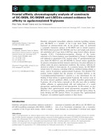
matography. Silver staining of the purified protein showed a
single band of molecular mass of 67 kDa (Fig. 1, lane 1).
Indirect immunoblotting revealed a 67-kDa protein band
using laminin as the primary probe followed b y treatment
with anti-laminin Ig and alkaline phosphatase-conjugated
secondary Ig (lane 2). The control nitrocellulose strip
(lane 3), which was devoid of laminin t reatment, failed to
reveal any band t hereby suggesting the s pecifi city of t he
reaction. Blotting with avidin probes also did not reveal any
band (lane 4). Direct immunoblotting using anti-LBP Ig
and secondary antibody also resulted in a 67-kDa band
(lane 5) confirming the a uthenticity of the protein. Finally,
the parasitic origin of the protein was demonstrated by
immunoprecipitating LBP from metabolically labelled
L. donovani using a nti-LBP Ig and protein A–Sepharose
beads. When these immune complexes were dissociated and
run on SDS/PAGE and autoradiographed, we observed a
single band at 67 kDa (lane 6).
Requirements for optimal laminin-LBP binding
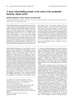
Denaturation by heat had similar effects on both laminin
and LBP (Fig. 2A). The binding activities of both laminin
or LBP wer e completely destroyed b y heat denaturation
(100 °C, 5 min) indicating that the conformation of both
the receptor an d ligand are essential for binding. Changes in
pH of the binding buffer also had mar ked effect on binding
constant with a change of as little as 0.5 pH units from
pH 7.5 being enough to lower specific binding activity
Fig. 1. Isolation and identification o f LBP. L. donovani me mbrane
proteins isolated by biotinylation and streptavidin–agarose extraction
and passed through DEAE-cellulose, Con A– Sepharose and laminin–
Sepharose were analysed by 7.5% SDS/PAGE under reducing
conditions. The gel was silver stained (lane 1). The molecular masses are
indicated to the left of th e panel. A ffinit y purified prot ein from lamin in–
Sepharose was transferred to nitrocellulose membrane and subjected to
indirect immunoblot analysis using l aminin as the p rimary probe fol-
lowed by rabbit anti-laminin IgG, goat anti-(rabbit IgG) Ig, Nitro Blue
tetrazolium and 5-bromo-4-chloro-indolyl-3-phosphate; (lane 2). Lane
3 was incubated with BSA instead of laminin. Lane 4 represents
immunoblot analysis using avidin as the primary probe and anti-
(rabbit avidin) IgG as the secondary antibody. Affinity purified protein
was subjected to direct immunoblot analysis using rabbit anti-LBP
antiserum as primary probe (lane 5). Promastigotes were metabolically
labelled with [
35
S]methionine, lysed and the LBP w as immunoprecipi-
tated by anti-LBP Ig and autoradiographed (lane 6).
1624 K. Bandyopadhyay et al. (Eur. J. Biochem. 269) Ó FEBS 2002
(Fig. 2 B). Both affinity and binding maxima were optimum
at pH 7.5. Nonspecific binding to BSA was not changed
over the pH range (data not shown). Involvement of surface
charge in the binding may be one of the reasons for pH
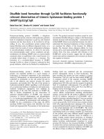
dependence. A number of c ompounds were also found to
affect laminin–LBP interaction (Fig. 3A). The protein
denaturant urea at 2
M
prevented binding, i ndicating again
that the in teraction is conformation-dependent. Increasing
the NaCl concentration to 0.3
M
also significantly reduced
binding suggesting the ionic nature of the binding sites. Free
sulfhydryl groups were also implicated as alkylation of
laminin with N-ethylmaleimide without reduction of disul-
fide bonds also reduced th e b inding significantly. No such
reduction in binding was observed when LBP was treated
with N-ethylmaleimide (data not shown ). The inhibition of
laminin binding activity with EDTA suggested the involve-
ment of divalent metal ions and a series of common trace
elements were tested at their respective plasma concentra-
tions (Fig. 3B). Zn
+2
was found to be the most effective of
all metal ions tested at enh ancing the laminin-binding
activity (K
d
¼ 1.92 ± 0.42 n
M
and B
max
¼ 10.20 ±
0.90 ng). Mn
2+
and Cu
2+
are the other two metals,
which promoted binding to a small extent whereas Ca
2+
and Mg
2+
showed inhibitory effect compared with EDTA.
The zinc effect on laminin b inding was saturable with
optimal binding occurring at physiological Zn
2+
concen-
tration (15 l
M
), above which the amount of nonspecific
binding increased. Preincubation of LBP with either Zn
2+
or EDTA (Fig. 3C) did not alter the binding activity
suggesting thereby that the cofactor requirement of Zn
2+
is
for laminin only. Treatment of l aminin with diethyl
pyrocarbonate, a histidine modifying agent, did not change
the binding parameters (Fig. 3 A) suggesting thereby that
Zn
2+
binding did not occur via the His-Xaa-His sites, which
are known to bind certain metals with high affinity [20].
Significant reduction in binding after alkylation with
N-ethylmaleimide on the other hand may suggest the
involvement o f cysteine sulfhydryl groups in Zn
2+
binding.
Laminin (0.5 mgÆmL
)1
) dialyzed agains t an excess o f ZnCl
2
(50 l
M
), followed by extensive dialysis against N aCl/Tris to
remove free metal, was found to contain 9.84 ± 1.51 nmol
of Zn
2+
per mol of laminin. A s mall amount of Zn
2+
,
1.21 ± 0.32 nmolÆmol
)1
of laminin was also detected in
control laminin preparation not dialyzed against ZnCl
2
.
Incidentally, laminin has 4 2 Cys-rich repeats found on the
amino terminal e nds of its three subunits (A, B1 and B2),
of which 12 contained nested zinc-finger consensus
sequences known to be involved in several protein–protein
interactions [21].
Fig. 2. Laminin binding activity for LBP (A) after heat denaturation and
(B) at different pH. (A) Bindi ng expe rimen ts we re carrie d ou t a fter
heating laminin in 20 m
M
Tris/HCl (pH 7.4), 150 m
M
NaCl and LBP
in 20 m
M
Na
2
CO
3
,NaHCO
3
(pH 9.6), 4
M
urea at 10 0 °Cfor5min.
Binding of untreated laminin to BSA is also included. (B) Laminin-
LBP binding was carried out at d ifferen t pH l evels: pH 6.5 and 7.0
(20 m
M
phosphate), pH 7.5 and 8.0 (20 m
M
Tris/HCl) and pH 9.0
(20 m
M
glycine/NaOH) w ith usual amount of NaCl (150 m
M
). Dis-
sociation constants and binding maxima (where applicable) are shown
for each curve on graph. All binding was carried in presence of 15 l
M
ZnCl
2
and are represented as mean of three separate experiments.
Fig. 3. Effect o f various agents on laminin-LBP binding. (A) LBP was
coated onto nitrocellulose discs and incubated with increasing con-
centrations of laminin under different conditions ( shown on the right
of the graph). (B) The influence of different divalent metal ions on
binding was evaluated at their respective plasma concentrations (2 m
M
CaCl
2
,15l
M
CuCl
2
,1m
M
MgCl
2
,1m
M
MnCl
2
and 1 5 l
M
ZnCl
2
).
(C) Binding was carried out after pretreating either laminin or LBP
with Zn
2+
and EDTA. Data rep resent mean of three separate
experiments.
Ó FEBS 2002 Zinc-finger sequence in laminin binding (Eur. J. Biochem. 269) 1625
Localization of the binding region of laminin
The binding of radiolabelled laminin was almost completely
inhibited by excess nonradioactive laminin, but not by
excess heparin or chondroitin sulfate or hyaluronic acid or
vitronectin (Table 1). B inding of radiolabelled laminin was
also inhibited by purified LBP in a concentration-dependent
manner (Table 1 ). Consistent with this finding is the
observation that polyclonal a nti-laminin serum resulted in
abolishing the parasite adherence to laminin-coated wells
(Fig. 4 A). In order to determine which polypeptide chain of
laminin harbour the LBP binding site, monoclonal anti-
bodies against various laminin chains were tested for their
potential of competitive inhibitions of leishmanial adher-
ence to laminin-coated substrata (Fig. 4A). Of the v arious
monoclonals tested, only that against B1 chain could
abrogate parasite adherence to laminin-coated wells. To
further localize the domain of laminin responsible for LBP
binding, we took advantage of the fact that a number of
peptides responsible for the attachment activity for a variety
of cell types have been derived from laminin. The first
peptide, YIGSR, a component of the B1 chain of laminin, is
included in the major c ell b inding and cell migration site of
laminin [22,23]. The second one, RNIAEIIKDI, a compo-
nent of B2 chain of laminin, i s associated with the
promotion of neurite outgrowth and cell binding [24]. The
hexapeptide, SIKVAV, a component of the A chain o f
laminin has been described as an angiogenic factor in vivo
[25]. Control peptides of t he same length, but with different
structures were also included for all the sequences. Of all
these peptides tested in adherence inhibition studies only
YIGSR and C(YIGSR)
3
-NH
2
were found to inhibit
laminin binding significantly (59% and 65%, respectively)
(Fig. 4 B). In order to ascertain whether YIGSR in a protein
environment would be more active, YIGSR fused to protein
A was also tested. The inhibitory effect was similar to that of
the pentapeptide (Fig. 4B). Other signature sequences of B1
chain with adhesion property such a s RYVVLPR (21),
LGTIPG [26] and RGD [27] did not show any inhibitory
activity (data n ot shown). A ll these molecules with adher-
ence inhibitory activity could effectively block laminin
binding to LBP (Table 2).
Tyrosine phosphorylation through LBP
Results suggest that the zinc finger motif of B1 chain of
laminin containing YIGSR sequence may provide the
Table 1. Inhibition of radiolabelled laminin binding to L. donovani
promastigotes. Data represent mean ± SD of triplicate determinations.
Values include the significance (*P < 0.001) of the difference between
inhibition in the p resence a nd absence o f inhibitors a s determined by
analysis of variance.
Bound c.p.m.
Bound
laminin (ng)
b
(A) By soluble glycosaminoglycans
Competitor
a
None 20 987 ± 2868 7.20 ± 0.98
Laminin 2846 ± 845 0.98 ± 0.29*
Heparin 18 467 ± 2255 6.34 ± 0.77
Chondroitin sulfate 16 870 ± 2032 5.79 ± 0.70
Hyaluronic acid 17 121 ± 1983 5.87 ± 0.68
(B) By purified LBP
LBP (lgÆmL
)1
)
0.25 13 897 ± 1835 4.77 ± 0.63
0.50 8658 ± 1246 2.97 ± 0.43*
0.75 5396 ± 887 1.85 ± 0.30*
1.00 2124 ± 636 0.73 ± 0.22*
a
Unlabelled competitors were used at a final concentration of
1mgÆmL
)1
.
b
The amount of
125
I-labelled laminin per 10
7
pro-
mastigotes.
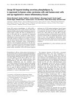
Fig. 4. Inhibition of attachment of L. donovani promastigotes to lami-
nin-coated micro titer wells b y (A) various antibodies and (B) synthetic
peptide s. (A) Laminin-coated surfaces (5 lg per well) were overlaid
with 5 · 10
5
cells of a suspension of
125
I-labelled parasites and incu-
bated for the indicated periods of time i n presenc e of (s) none (d)
anti-laminin Ig (n)anti-B1chainIg(h)anti-B2chainIgand(m)anti-
A chain Ig. All antibodies were at 1 : 10 dilution. After extensive
washing of the unbound parasites with NaCl/P
i
, the adherence of
parasites was determined by counting the wells in a gamma counter.
(B) Parasites (1 · 10
6
) were surface labelled with
125
I and incubated for
1 h at 22 °C with l aminin-coated m icro titer w ells in the presence of
0.1 m gÆmL
)1
of various synthetic peptides. Data are mean ± SD from
incubations p erformed in triplicate. The amount of attached cells is
given as a percent of the number of cells that were attached to the wells
in the absence of peptides. For the decapeptide RNIAEIIKDI related
to the cell binding site from th e B2 c hain of laminin, the decapeptide
GPRPPERHQS was used as control. For the hexapeptide SIKVAV
related to the A chain, LRYESK was used as control whereas for the
pentapeptide YIGSR related to the B1 chain, HEIPA was used as
control.
Table 2. The effect of various agents on laminin-LBP binding. Means
of three determinations ± S D. Values in clude the s ignificance
(* P < 0.001) of the difference b etween inhibition in the p resence and
absence of inhibitors as determined by analysis o f variance.
Agents applied % Inhibition
None 0 ± 3
Laminin B1 81 ± 6*
YIGSR 66 ± 5*
HEIPA 8 ± 2
C(YIGSR)
3
-NH
2
76 ± 6*
YIGSR grafted protein A 53 ± 5*
1626 K. Bandyopadhyay et al. (Eur. J. Biochem. 269) Ó FEBS 2002
physiological scaffolding required f or LBP binding. It is
likely that b inding o f laminin t o cell surface LBP through
YIGSR sequence may involve s pecific downstream signal-
ling events, one of which may be phosphorylation of
tyrosine residues of some intracellular proteins. We there-
fore analysed th e response of L. donovani promastigotes to
the presenc e of C(YIGSR)
3
-NH
2
as compared to an
unrelated peptide. Exposure of 2 · 10
8
promastigotes to
100 lgÆmL
)1
of C(YIGSR)
3
-NH
2
peptide induced tyrosine
phosphorylation of several proteins with a molecular mass
of 115–130 kDa (Fig. 5A). The induction of tyrosine
phosphorylation was rapid a nd transient, reaching a
maximum level within 1 min. In contrast, when cells
were exposed to an unrelated polypeptide
(CYKNVRSKIGSTENIKHQPGGGKV) of similar
length, and the same molar concentration, tyrosine phos-
phorylation of these proteins was hardly detected (Fig. 5A,
lanes 4 and 5). It seems therefore that at least some high
molecular mass proteins of 115–130 kDa underwent phos-
phorylation on tyrosine residues following binding of
YIGSR repeat to the cell surface 67-kDa LBP. In order
to further ascertain that the induction of tyrosine phos-
phorylation is not due to any growth factors, serum-starved
parasites were allowed to adhere in suspension to polysty-
rene late x beads coated with C(YIGSR)
3
-NH
2
for 1 min a t
22 °C. As shown in Fig. 5B (lane 2), the same high
molecular mass proteins of 115–130 kDa underwent phos-
phorylation on tyrosine residues. Phosphorylation w as not
detected in the presence of uncoated beads (lane 1). In order
to know whether clustering of LBP by anti-LBP Ig also
could induce tyrosine phosphorylation, serum-starved cells
were allowed to adhere in suspension to polystyrene latex
beads coated with a nti-LBP Ig and incubated for 1 min at
22 °C. Figure 5B (lane 3) s hows that clustering of LBP by
the corresponding antibod y resulted in phosphorylating the
same group of proteins that were phosphorylated in
response to C(YIGSR)
3
-NH
2
coated beads.
DISCUSSION
Adhesion of pathogen to host tissue is a prerequisite for
many types of infections. Diseases such as leishmaniases are
is generally initiated when sand fly, the vector, regurgitates
promastigote form of the parasite at the time of taking a
blood meal from human body. This developmental form
migrates through the blood stream into various definite
organs like liver and s pleen and u ltimately takes refuge
within the resident macrophages where it transforms into
the amastigote f orm a nd multiplies in number. Eventually
parasites are released into the interstitial tissue by macro-
phage lysis, invade fresh cells and the cycle i s repeated. This
way the entire reticuloendothelial system b ecomes progres-
sively infected. Evidently during transit in the interstitial
tissue, t hese intracellular parasites must be in contact with
the extracellular matrix and the basement membrane. We
have identified and characterized a laminin binding protein
(LBP) from the surface of L. donovani that may mediate cell
adhesion by helping the parasite to home in their physio-
logical address [5,6]. Laminin is a multidomain molecule
[24], and it is known t hat there are several specific binding
domains on laminin for each of the laminin binding
proteins. Studies with proteolytic fragments, domain-speci-
fic antibodies, and synthetic peptides have identified differ-
ent regions of laminin w ith biolo gical a ctivity [ 21]. T his
paper is mainly concerned with the identification of a
specific domain of laminin mediating the binding of
leishmanial LBP.
The purified 67-kDa LBP isolated from the membrane
fraction behaved as one would expect of a laminin receptor
and laminin binding to LBP was found to be dose-
dependent, specific and saturable. Laminin–LBP interaction
also involved a single class of binding sites, which appeared
to be conformation-dependent, ionic in nature, and signi-
ficantly enhanced by Zn
2+
. Detailed binding studies at
various pH indicated the presence of His and Cys at the
binding site. However, t he un altered binding parameters
after diethyl pyrocarbonate treatment preclude the possi-
bility of the pre sence of His at the b inding site. It may be
mentioned that the ionization state of amino-acid residues is
influenced by their unique microenvironment; therefore,
predicting the impact of the residues based solely on
theoretical pK
a
of their individual side c hains is speculative.
The positive e ffect of zinc o n laminin binding activity
suggests that it could be a potential metal cofactor for
L. donovani interaction with ECM and BM. Both Zn
2+
and
free sulfhydryls may be required for LBP binding site on
laminin a s e videnced by the stimulatory and inhibitory
effects of ZnCl
2
and N-ethylmaleimide, respectively. Prein-
cubating LBP with ZnCl
2
did not enhance laminin-binding
activity, indicating that zinc was affecting laminin only.
Moreover, treating LBP with EDTA had little effect on its
binding with laminin, consistent with the indication of the
role of zinc as laminin-specific cofactor. L aminin is known
Fig. 5. Tyrosine phosphorylation via LBP. (A) L. donovani promasti-
gotes ( 2 · 10
8
cells) were washed twice with medium M199 a nd
incubated with 100 lgÆmL
)1
of either C(YIGSR)
3
-NH
2
for 1 min (lane
1), 5 min (lane 2) or 15 min (lane 3) or with 100 lgÆmL
)1
of un relate d
peptide for 1 m in (lane 4) and 5 m in (lane 5). Cells were washed with
ice-cold NaCl/P
i
, lysed, subjected to 7.5% SDS/PAGE and transferred
to nitrocellulose membrane. The blotted membranes were incubated
with anti-(P-T yr) monoclonal antibodies followed by alk aline phos-
phatase c onjugated secondary antibody and developed by Nitro Blue
tetrazolium and 5-bromo-4-ch loro-indolyl-3-phosph ate. (B) Serum-
starved promastigotes (5 · 10
7
cells) were incubated with uncoated
latex b eads (lane 1), latex beads coated with C(YIGSR)
3
-NH
2
(lane 2)
or with antibodies directed against the 67 kDa LBP (lane 3). Following
incubation, cells were collected, lysed, subjected to SDS/PAGE and
blotted with anti-(P-Tyr) m ono clonal antibodies.
Ó FEBS 2002 Zinc-finger sequence in laminin binding (Eur. J. Biochem. 269) 1627
to contain 42 Cys-rich repeats of which 12 represent the
consensus sequence for Cys-rich Zn
2+
fingers. Taken
together, the data therefore suggest that Zn
2+
finger like
sequence may represent the actual LBP binding site or at
least contribute to i t significantly. Laminin bound zinc
detected by flame atomic absorption spectroscopy was
about 10 molÆmol
)1
. The amount is consistent with the
predicted number of zinc finger sequences. It is now well
known that metal-binding domains, particularly Zn
2+
finger motifs, play central roles in mediating interactions
between proteins and man y d ifferent macromolecules [ 28].
This may b e due to the formation of bumps and ridges that
extend from the s urfaces of proteins t hat are well suited for
interactions with other m acromolecules. Laminin zinc
fingers are known to participate in binding to Alzheimer’s
amyloid precursor protein and collagen IV [8,29]. The
enactin binding site was recently mapped to Cys-rich repeats
on the laminin B2 chain which happens to contain Zn
2+
finger like sequence [9]. Although the present study was
carried out with mouse laminin, t he putative z inc-finger
motifs are known to be highly conserved between human
[30–32], mouse [33,34] and Drosophila [35–37]. Inhibition
studies with Fab fragments of monoclonal antibodies
against various chains of laminin are indicative of the
presence of LBP binding site on the B1 chain of laminin.
Moreover, a number of small peptide r ecognition sequences
have been reported to d ate i n l aminin, w hich ar e a ttributed
to various biological activities of laminin [38]. YIGSR, a
short sequence of the B1 chain of l aminin, was reported to
be a potential binding site for specific laminin b inding
proteins, particularly 67-kDa laminin receptor present on
normal and cancer cell surface [39, 40]. This sequence is no t
present in the A and B2 chains. C ompetitive inhibition o f
laminin-LBP binding by YIGSR indicates that interaction
of LBP with this peptide is specific. However, YIGSR
grafted in protein A could not enhance the inhibitory effect
over that of the peptide alone. All these studies suggest that
zinc finger motif of B 1 chains containing YIGSR sequence,
may provide the physiological scaffolding required for LBP
binding.
Cell–matrix interactions have recently been shown to
trigger many signalling processes [11,12]. For example,
tyrosine phosphorylation is i nvolved in collagen s ignalling
in amoebas, which m ight play a role i n the invasiveness
capability of this parasite [41]. In the present studies one
class of proteins was found to be phosphorylated in
respon se to the interaction of C(YIGS R)
3
-NH
2
with the
67-kDa LBP. T hese proteins h ad a molecular mass of
115–130 kDa, but their identity remains to be determined.
It is possible t hat the above proteins may undergo
autophosphorylation on a tyrosine residue, which generally
implies that it encodes a phosphotyrosine kinase, as a result
of activation by cell adhesion to YIGSR sequence.
Alternatively, the proteins may be phosphorylated by
another unknown phosphotyrosine kinase. As an antibody
directed against the 67-kDa LBP can induce tyrosine
phosphorylation of these proteins, it is likely that dimeri-
zation or oligomerization of LBP is required f or activating
an associated tyrosine kinase.
The ability of L. donovani LBP to bind a major ECM
protein like laminin probably plays a role in pathogenesis of
the disease process this species exhibits in mammalian host.
The ECM protein binding ability of the leishmanial LBP
could allow the parasite to persist within the host and thus
contribute to virulence. For example, binding of ECM
protein to the surface of the parasite via LBP could block or
reduce host’s immune response to the parasite by sterically
masking immunogenic epitope. The ability to bind ECM
proteins might also facilitate adhesion of the pathogen to
host cells such as macrophages via laminin receptors present
on the cell surface. The elucidation of the binding region o f
laminin may therefore help i n better understanding the
pathogenesis as well as developing effective therapeutic
strategies.
ACKNOWLEDGEMENTS
We are indebted to the Council for Scientific and Industrial Research
and the Department of Biotechnology, Government of India for
financial help.
REFERENCES
1. Silva Filho, F.C., de Souza, W. & Lopes, L.D. (1988) Presence of
laminin-binding proteins in trichomonads and their role i n adhe-
sion. Proc. Natl Acad. Sci. US A 85, 8042–8046.
2. Vicentini, A.P., G esztesi, J., Franco, M.F., d eSouza, W ., d eMoraes,
J.Z., Travassos, L.R. & Lopes, J.D. (1994) Binding of Para-
coccidioides brasiliensis to laminin through surface glycoprotein gp
43 leads to enhancement of fungal pathogenesis. Infect. Immun. 62,
1464–1469.
3. Lopez-Ribot, J.L., Casanova, M., Monteagudo, C., Sepulveda, P.
& Martinez, J.P. (1994) Evidence for the presence of a high affinity
laminin receptor-like molecule on the surface of Candida albicans
yeast cells. Infect. Immun. 62, 742–746.
4. Giordano, R., Chammas, R., Veiga, S.S., Colli, W. & Alves,
M.J.M. (1994) An acidic component o f the heterogene ous Tc-85
protein fam ily from the surface of Trypanosoma cruzi is a laminin
binding glycoprotein. Mol. Biochem. Parasitol. 65, 85–94.
5. Ghosh, A., Kole, L., Bandyopadhyay, K., Sarkar, K. & Das, P.K.
(1996) Evide nce of a laminin binding protein on the s urface
of Leishmania donovani. Biochem. Biophys. Res. Commun. 226,
101–106.
6. Ghosh, A., Bandyopadhyay, K., Kole, L. & Das, P.K. (1999)
Isolation of a laminin-binding protein from the protozoan parasite
Leishmania donovani that may m ed iate cell adhesion. Biochem. J.
337, 551–558.
7. Aumailley, M., Gerl, M., Sonnenberg, A., Deutzmann, R. &
Timpl, R. (1990) Identification of the Arg-Gly-Asp sequence in
laminin A chain as a latent cell-binding site being exposed in
fragment P1. FEBS Lett. 262, 82–86.
8. Ancsin, J.B. & Kisilevsky, R. (1996) Laminin interactions
important for basement membrane assembly are promoted by zinc
and implicate l aminin zinc finger-like sequences. J. Biol. Chem.
271, 6845–6851.
9. Mayer, U ., N ischt, R., Poschl, E., Mann, K ., Fukuda, K., G erl,
M., Yamada, Y. & Timpl, R . ( 1993) A single EGF-like motif of
laminin is responsible for high affinity nidogen binding. EMBO J.
12, 1879–1885.
10. Ancsin, J.B. & Kisilevsky, R. (1997) Characterization of high
affinity binding between laminin and the acute-phase protein,
serum amyloid A. J. Biol. Chem. 272, 406–413.
11. Hynes, R.O. (1992) Integrins: versatility, modulation, and s igna-
ling in cell adhesion. Cell 69 , 11–25.
12. Schwartz, M.A. & Ingber, D.E. (1994) Integr ating with integrins.
Mol. Biol. C ell 5, 389–393.
13. Sarkar, K. & Das, P.K. (1997) Protective effect of neoglycoprotein
conjugated muramyl dipeptide against Leishmania donovani
infec ti on . J. Immunol. 158, 5357–5365.
1628 K. Bandyopadhyay et al. (Eur. J. Biochem. 269) Ó FEBS 2002
14. Chakraborty, P. & Das, P.K. (1988) Role of mannose/N-ace-
tylglucosamine receptors in blood cle arance and cellular a ttac h-
ment of Leishmania donovani. Mol. Biochem. Parasitol. 28 , 55–62.
15. Kahl, L.P. & McMahon-Pratt, D.J. (1987) Structural and anti-
genic characterization of a species- and promastigote-specific
Leishmania mexicana amazonensis m embrane protein. J. Immunol.
138, 1587–1595.
16. Hall, D.E., Frazer, K.A., Hann, B.C. & Reichardt, L.F. (1988)
Isolation and characterization of a laminin-binding protein from
rat and chick muscle. J. Ce ll Biol. 107, 687–697.
17. Malinoff, H.L. & Wicha, M.S. (1983) Isolation of a cell surface
receptor protein for laminin from murine fibrosarco ma cells.
J. Cell Biol . 96, 1475–1479.
18. Greenwood,F.,Hunter,W.&Glover,J.(1963)Thepreparation
of
131
I-labeled human growth hormone of high specific radio-
activity. Biochem. J. 89, 114–123.
19. Leary, J.J., Brigati, D.J. & Ward, D.C. (1983) Rapid and sensitive
colorimetric method for visua lizing biotin-labeled DNA probes
hybridised to DNA o r R NA immobilized on nitrocellulose: bio-
blots. Proc. Natl Acad. Sci. USA 80, 4045–4049.
20. K urz, L.L., Klink, H., Jakob, I., Kuchenbecker, M., Benz, S.,
Lehmann-Horn, F. & Rudel, R. (1999) Identification of three
cysteines as targets for the Zn
2+
blockade of the human sk eletal
muscle chloride channel. J. Biol. C hem. 274, 11687–11692.
21. Yamada, K.M. (1991) Adhesive recognition sequences. J. Biol.
Chem. 266, 12809–12812.
22. Iwamoto,Y.,Robey,F.A.,Graf,J.,Sasaki,M.,Kleinman,H.K.,
Yamada,Y.&Martin,G.R.(1987)YIGSR,asyntheticlaminin
pentapeptide, inhibits experimental metastasis formation. Science
238, 1132–1134.
23. Iwamoto, Y., Graf, J., Sasaki, M., Kleinman, H.K., Greatorex,
D.R., Martin, G.R., Robey, F.A. & Yamada, Y. (1988) Synthetic
pentapeptide from the B1 chain of laminin promotes B16F10
melanoma cell migration. J. Cell. Physiol. 134, 287–291.
24. Engel, J. (1992) Laminins and other strange proteins. Biochemistry
31, 10643–10651.
25. Grant, D.S., Kinsella, J.L., Fridman, R., Auerbach, R., Piasecki,
B.A., Yamada, Y., Zain, M. & Kleinman, H.K. (1992) Interaction
of endothelial cells with a laminin A chain peptide (SIKVAV)
in vitro and induction of angiogenic behavio ur in vivo. J. Cell.
Physiol. 153, 614–625.
26. Hinek, A., Wrenn, D.S., Mecham, R.P. & Borondes, S.H. (1988)
The elastin rec eptor: a galac toside-bin ding protein. Science 239,
1539–1541.
27. Mecham, R.P., Hinek, A., Griffin, G.L., S enior, R.M. & Liotta,
L.A. (1989) The elastin recep tor shows structural a nd functional
similarities to the 67-kDa tumor cell laminin receptor. J. Biol.
Chem. 264, 16652–16657.
28. B erg, J.M. (1990) Zinc fingers and other m etal-binding domains.
Elements for interactions between macromolecules. J. Biol. C hem.
265, 6513–6516.
29. Narindrasorasak, S., Lowery, D .E., Altman, R.A., Gonzalez-
DeWhitt, P.A., Greenberg, B.D. & Kisilevsky, R. (1992) Char-
acterization of high affinity binding be tween laminin and Alzhei-
mer’s disease amyloid precursor proteins. La boratory Invest. 67,
643–652.
30. N issinen, M., Vuolteenaho, R., Boot-Handford , R., Kallunki, T .
& Tryggvason, K. (1991) Primary structure of the human laminin
A chain. Limited expression in human tissues. Biochem. J. 276,
369–379.
31. Pikkarainen, T., Eddy, R., Fukushima, Y., Byers, M., Shows, T.,
Pihlajaniemi, J., Saraste, M. & Tryggvason, K. (1988) Human
laminin B1 chain. A multidomain p rotein with gene (LAMB1)
locus in the q22 region of chromosome 7. J. Biol. Chem. 262,
10454–10462.
32. Pikkarainen, T., Kallunki, T. & Tryggvason, K . (1988) Human
laminin B2 chain. Comparison of the complete amino acid
sequence with the B1 chain reveals variability in sequence
homology betwee n differen t structural d omains. J. Biol. Chem.
263, 6751–6758.
33. Sasaki, M. & Yamada, Y. (1987) The laminin B2 chain has a
multidomain structure homologous to the B1 chain. J. Biol. Chem.
262, 17111–17117.
34. Sasaki, M., Kleinman, H.K., Huber, H., Deutzmann, R. &
Yamada, Y. (1988) Laminin, a multidomain protein. Th e A chain
has a unique globular domain and h omology with the baseme nt
membrane proteoglycan and the laminin B chains. J. Biol. Chem.
263, 16536–16544.
35. K usche-Gullber g, M., Garrison, K., M acKrell, A.J., Fessler, L.I.
& F essler, J.H. ( 1992) Laminin A chain: e xpression during
Drosophila development and genomic sequence. EMBO J. 11,
4519–4527.
36. Montell, D.J. & Go odman, C.S. ( 1988) Drosophila substrate
adhesion molecule: seq uence of laminin B1 chain reveals dom ains
of homology with mouse. Cell 53, 463–473.
37. Ch i, H .C. & Hui, C.F. (1989) Primary s tructure of the Drosophila
laminin B2 c hain and comparison with h uman, m ouse, a nd
Drosophila laminin B1 a nd B2 chains. J. Biol. Chem. 264,
1543–1550.
38. Mecham, R.P. (1991) Receptors for laminin on mammalian cells.
FASEB J. 5, 2538–2546.
39.Graf,J.,Iwamoto,M.,Sasaki,M.,Martin,G.R.,Kleinman,
H.K., Robey, F.A. & Yamada, Y. (1987) Identification of an
amino acid sequence in laminin mediating cell attachment,
chemotaxis, and receptor binding. Cell 48, 989– 996.
40. Clement, B., Segui-Real, B., Savagner, P., Kleinman, H.K. &
Yamada, Y. (1990) Hepatocyte attachment to laminin is mediated
through multiple receptors. J. Cell Biol. 110, 185–192.
41. Perez, E., Munoz, M.L. & Ortega, A. (1996) Entamoeba histo-
lytica: Involvement of pp125
FAK
in collagen-induced signal
transduction. Exp. Parasitol. 82 , 164–170.
Ó FEBS 2002 Zinc-finger sequence in laminin binding (Eur. J. Biochem. 269) 1629