Outcomes Associated With Isolated Agenesis of the Corpus Callosum A Meta analysis REVIEW ARTICLEPEDIATRICS
Bạn đang xem bản rút gọn của tài liệu. Xem và tải ngay bản đầy đủ của tài liệu tại đây (1 MB, 15 trang )
Outcomes Associated With
Isolated Agenesis of the Corpus
Callosum: A Meta-analysis
Francesco D’Antonio, MD, PhD,a Giorgio Pagani, MD,b Alessandra Familiari, MD,c Asma Khalil, MD,d TallyLerman Sagies, MD, PhD,e,f Gustavo Malinger, MD,e,g Zvi Leibovitz, MD,e,h Catherine Garel, MD, PhD,i Marie
Laure Moutard, MD, PhD, j Gianluigi Pilu, MD, PhD,k Amar Bhide, MD,d Ganesh Acharya, MD, PhD,a Martina
Leombroni, MD,l Lamberto Manzoli, MD, PhD,m,n Aris Papageorghiou, MD,d Federico Prefumo, MD, PhDo
CONTEXT: Antenatal counseling in cases of agenesis of the corpus callosum (ACC) is
abstract
challenging.
OBJECTIVES: To ascertain the outcome in fetuses with isolated complete ACC and partial ACC.
DATA SOURCES: Medline, Embase, CINAHL, and Cochrane databases.
STUDY SELECTION: Studies reporting a prenatal diagnosis of ACC. The outcomes observed
were: chromosomal abnormalities at standard karyotype and chromosomal microarray
(CMA) analysis, additional anomalies detected only at prenatal MRI and at postnatal
imaging or clinical evaluation, concordance between prenatal and postnatal diagnosis and
neurodevelopmental outcome.
DATA EXTRACTION: Meta-analyses of proportions were used to combine data.
RESULTS: Twenty-seven studies were included. In cACC, chromosomal anomalies occurred
in 4.81% (95% confidence interval [CI], 2.2–8.4) of the cases. Gross and fine motor control
were abnormal in 4.40% (95% CI, 0.6–11.3) and 10.98% (95% CI, 4.1–20.6) of the cases,
respectively, whereas 6.80% (95% CI, 1.7–14.9) presented with epilepsy. Abnormal
cognitive status occurred in 15.16% (95% CI, 6.9–25.9) of cases. In partial ACC, the rate of
chromosomal anomalies was 7.45% (95% CI, 2.0–15.9). Fine motor control was affected
in 11.74% (95% CI, 0.9–32.1) of the cases, and 16.11% (95% CI, 2.5–38.2) presented with
epilepsy. Cognitive status was affected in 17.25% (95% CI, 3.0–39.7) of cases.
LIMITATIONS: Different neurodevelopmental tools and time of follow-up of the included studies.
CONCLUSIONS: Children wih a prenatal diagnosis of isolated ACC show several degrees of
impairment in motor control, coordination, language, and cognitive status. However, in view
of the large heterogeneity in outcomes measures, time at follow-up, and neurodevelopmental
tools used, large prospective studies are needed to ascertain the actual occurrence of
neuropsychological morbidity of children with isolated ACC.
aDepartment
of Clinical Medicine, Faculty of Health Sciences, UiT - The Artic University of Norway, Tromsø, Norway; bDepartment of Obstetrics and Gynecology, Fondazione Poliambulanza,
Brescia, Italy; cDepartment of Maternal-Fetal Medicine, Catholic University of the Sacred Heart, Rome, Italy; dFetal Medicine Unit, Division of Developmental Sciences, St. George’s University
of London, London, United Kingdom; eSackler School of Medicine, Tel Aviv University, Tel Aviv, Israel; fFetal Neurology Clinic and Paediatric Neurology Unit, Wolfson Medical Centre, Holon,
Israel; gGYN Ultrasound Division, Tel Aviv Medical Center, Tel Aviv, Israel; hFetal Neurology Clinic and Institute of Medical Genetics, Wolfson Medical Center, Holon, Israel; iService de
Radiologie, Hôpital d'Enfants Armand-Trousseau, Paris, France; jService de Neuropédiatrie, Hôpital Trousseau, Hôpitaux Universitaires de l'Est Parisien, Université Pierre et Marie Curie,
Paris, France; kDepartment of Obstetrics and Gynaecology, Sant'Orsola-Malpighi Hospital, University of Bologna, Bologna, Italy; lDepartment of Obstetrics and Gynecology, University of
To cite: D’Antonio F, Pagani G, Familiari A, et al. Outcomes Associated With Isolated Agenesis of the Corpus Callosum: A Meta-analysis. Pediatrics. 2016;138(3):e20160445
Downloaded from www.aappublications.org/news at Viet Nam:AAP Sponsored on July 6, 2021
PEDIATRICS Volume 138, number 3, September 2016:e20160445
REVIEW ARTICLE
Agenesis of the corpus callosum
(ACC) is one of the most common
congenital brain anomalies, with
an estimated prevalence ranging
from 1.8 per 10 000 in the general
population to 230–600 per 10 000 in
children with neurodevelopmental
disabilities.1–3
Neurodevelopmental outcome
for individuals with callosal
abnormalities is extremely variable
even between children sharing
similar neuroanatomic profiles, and
there is often significant overlapping
in the neuropsychological
performance between patients with
complete ACC (cACC) and those with
partial ACC (pACC).4 Delay in motor
and cognitive functions, epilepsy, and
social and language deficits are the
most common symptoms reported in
individuals with ACC; furthermore,
ACC has been linked with the
occurrence of autism, schizophrenia,
and attention-deficit disorders.5–9
However, pediatric series are biased
by the fact that only symptomatic
cases are reported.
Advances in prenatal imaging
techniques have led to an increase
the detection rate of ACC; however,
antenatal counseling when a fetus is
diagnosed with this anomaly is still
challenging.5
Chromosomal abnormalities
are common in ACC, especially
when associated anomalies are
present, and prenatal invasive
tests are usually performed in
pregnancy to rule out aneuploidies.
Chromosomal microarray (CMA)
allows the detection of small genomic
deletions and duplications that
are not routinely seen on standard
cytogenetic analysis (copy number
variations [CNVs]). Fetuses with
central nervous system (CNS)
anomalies and normal karyotype
have been shown to have a
significantly higher risk of genetic
anomalies at CMA analysis; however,
the risk of clinically significant CNVs
in fetuses with isolated callosal
anomalies has not been completely
ascertained yet.10,11
Antenatal MRI is usually performed
to rule out associated anomalies,
which are major determinants of
outcome in cases of ACC; however,
the actual diagnostic accuracy of fetal
MRI in isolated ACC is still debated.12
Neurodevelopmental outcome
in fetuses with isolated ACC has
been reported to be normal in a
large majority of cases, especially
in complete agenesis. However, a
precise categorization of the burden
of neuropsychological disabilities is
required to counsel parents more
appropriately.13
The first aim of this systematic
review was to ascertain the rate
of associated genetic or anatomic
abnormalities in those patients with
an initial ultrasound examination
showing isolated ACC; the
secondary aim was to explore the
neurodevelopmental status of these
children.
METHODS
Protocol, Eligibility Criteria,
Information Sources, and Search
This review was performed according
to an a priori designed protocol
and recommended for systematic
reviews and meta-analysis.14,15
Medline, Embase, CINAHL, and
Cochrane databases were searched
electronically on February 15, 2014
using combinations of the relevant
medical subject heading terms,
key words, and word variants for
“agenesis of the corpus callosum”
and “outcome”; the search was then
updated on November 26, 2015
(Supplemental Table 5). The search
and selection criteria were restricted
to English. Reference lists of relevant
articles and reviews were hand
searched for additional reports.
PRISMA guidelines were followed.16
Study Selection, Data Collection, and
Data Items
Studies were assessed according to
the following criteria: population,
type of callosal agenesis (cACC and
pACC) outcome, type of imaging
assessment, and outcome (Table 1).
Two authors (F.D. and G.P.) reviewed
all abstracts independently.
Agreement regarding potential
relevance was reached by consensus;
full-text copies of those papers were
obtained and the same 2 reviewers
independently extracted relevant
data regarding study characteristics
and pregnancy outcome.
Inconsistencies were discussed by
the reviewers and consensus reached
with a third author. If >1 study was
published for the same cohort with
identical end points, the report
containing the most comprehensive
information on the population
was included to avoid overlapping
populations. For those articles in
which information was not reported
but the methodology was such that
this information would have been
recorded initially, the authors were
contacted.
Quality assessment of the included
studies was performed using the
Newcastle-Ottawa Scale (NOS) for
cohort studies (Table 2). According
to NOS, each study is judged on 3
broad perspectives: the selection of
the study groups, the comparability
of the groups, and the ascertainment
outcome of interest.44 Assessment of
the selection of a study includes the
evaluation of the representativeness
of the exposed cohort, selection of the
nonexposed cohort, ascertainment of
exposure, and the demonstrating that
outcome of interest was not present
at the start of the study. Assessment
of the comparability of the study
includes the evaluation of the
comparability of cohorts on the basis
of the design or analysis. Finally,
the ascertainment of the outcome of
interest includes the evaluation of the
type of assessment of the outcome
of interest, length, and adequacy of
Downloaded from www.aappublications.org/news at Viet Nam:AAP Sponsored on July 6, 2021
2
D’ANTONIO et al
Downloaded from www.aappublications.org/news at Viet Nam:AAP Sponsored on July 6, 2021
PEDIATRICS Volume 138, number 3, September 2016
3
Year
2015
2015
2015
2015
2015
2014
2013
2013
2013
2013
2012
2012
2012
2012
2011
2010
2010
2009
2009
Source
Cesaretti (17)a
Ruland (18)a
Papoulidis (19)
Shen (20)a
Pashaj (21)
Özyüncü (22)a
Lachmann (23)
Kasprian (24)a
Yinon (25)a
Vestergaard (26)a
Moutard (27)a
Wapner (28)a
Yamasaki (29)a
Shaffer (30)a
Mangione (31)
Ghi (32)
Cignini (33)
Tang (34)a
Goetzinger (35)
United States
United States
Italy
Italy
France
United States
Japan
United States
France
Israel
Austria
United Kingdom
Turkey
Albania-Germany
France
Greece
Germany
Italy
Country
TABLE 1 General Characteristics of the Included Studies
Prospective case
series
Retrospective case
series
Retrospective case
series
Prospective case
control study
Retrospective case
series
Prospective case
series
Retrospective case
series
Retrospective case
series
Retrospective case
series
Retrospective case
series
Retrospective case
series
Retrospective case
series
Retrospective case
series
Retrospective case
series
Retrospective case
series
Prospective case
series
Retrospective case
series
Retrospective case
series
Prospective case
series
Study Design
Complete
Complete
Complete
Partial
Complete, partial
Complete, partial
Complete
Complete, partial
Complete, partial
Complete, partial
Complete, partial
Complete, partial
Complete
Complete, partial
Complete, partial
Partial
Complete, partial
Complete, partial
Complete
Type of ACC
US
US, MRI
US
US, MRI
US, MRI
US
US
US
US, MRI
US
US, MRI
US, MRI
US, MRI
US, MRI
US
US, MRI
US
US, MRI
US, MRI
Prenatal
Imaging
9
10
17
14
112
69
10
15
17
4
4
20
15
33
33
77
4
127
62
Fetuses (n)
3
4
15
10
112
45
8
3
17
2
4
12
7
16
6
35
2
39
62
Isolated ACC (n)
NA
CDI (Ireto's Child
Developmental Inventory)
Standard neurologic
examination
Binet-Simon Scale revided
from Stanford
Not performed
Standard neurologic
examination
NA
Wechsler Intelligence
Scale for Children (III),
Dellatolas Protocol,
Pegboard Test, ReyOsterrieth Complex Figure
Test
Not performed
NA
NA
NA
NA
NA
NA
NA
NA
NA
NA
Dedicated
Neurodevelopmental Tool
NA
2–23 mo
4y
2–10 y
4 y (30–74 mo)
NA
Not specified
NA
10 y
NA
NA
NA
NA
NA
3–6 mo
NA
NA
Not reported
NA
Length of
Follow-up
Not specified
Not specified
Not specified
1–6 y
2–10 y
2–16 y
3 y (1–5 y)
The incidence of the following
outcomes was analyzed in fetuses
with a prenatal diagnosis of cACC and
pACC separately:
1. Chromosomal abnormalities
detected with standard karyotype
analysis.
4
5
4
9
3
7
37
13
US, MRI
Complete, partial
14
US, MRI
Complete, partial
8
US, MRI
Complete, partial
4
US, MRI
Partial
19
3. Rate of additional CNS anomalies
detected only at prenatal MRI but
missed at the initial scan.
4. Additional CNS and extra-CNS
anomalies detected only at
postnatal imaging or clinical
evaluation but missed at prenatal
imaging.
5. Concordance between prenatal
and postnatal diagnosis.
6. Neurodevelopmental outcome.
NA, not assessed; US, ultrasound.
a Additional information provided by the authors.
2001
Goodyear (43)
United Kingdom
2002
Malinger (42)a
Israel
2003
Blaicher (41)
Austria
Retrospective case
series
Retrospective case
series
Retrospective case
series
Retrospective case
series
2006
Volpe (40)
Italy
3
US
Complete
Retrospective case
series
2006
Ramelli (39)
Italy
2006
Pisani (38)
Switzerland
9
US, MRI
Complete, partial
117
US, MRI
2007
Fratelli (37)a
United Kingdom
Retrospective case
series
Prospective case
series
Complete, partial
13
Complete, partial
Retrospective case
series
2008
Chadie (36)
France
Type of ACC
Study Design
Country
Year
Source
TABLE 1 Continued
Risk of Bias, Summary Measures,
and Synthesis of the Results
2. Pathogenic CNVs at CMA.
Prenatal
Imaging
US, MRI
Fetuses (n)
Isolated ACC (n)
Dedicated
Neurodevelopmental Tool
Brunet-Lenzine test revised
for children, Wechsler
Preschooland Primary
Scale of Intelligence,
Wechsler Intelligence
Scale for Children-III,
Terman-Merril Scale
Standard neurologic
examination
Griffiths Scales of Mental
Development , Welchler
primary, preschool and
children scales
Wechsler intelligence Scale
for Children-revised,
Griffiths Scales of Mental
Development
Standard neurologic
examination
Standard neurologic
examination
Standard neurologic
examination
Standard neurologic
examination
Length of
Follow-up
3–16 y
follow-up. According to NOS, a study
can be awarded a maximum of 1 star
for each numbered item within the
Selection and Outcome categories. A
maximum of 2 stars can be given for
the Comparability category.44
Only fetuses with a prenatal
diagnosis of ACC either by
transabdominal or transvaginal
ultrasound were included. cACC
was defined as the total absence of
all the anatomically defined regions
of the corpus callosum, whereas
pACC was defined as the presence
of at least 1 region of the corpus
callosum. For the assessment of the
incidence of abnormal karyotype,
only cases of isolated ACC defined
as having no additional CNS and
extra-CNS anomalies detected at
the ultrasound scan were included
in the analysis. Only cases who had
their full karyotype tested either
prenatally or postnatally were
included. For the occurrence of
genetic abnormalities detected only
at CMA only fetuses with isolated
ACC and normal standard karyotype
were considered suitable for the
analysis. The presence of additional
Downloaded from www.aappublications.org/news at Viet Nam:AAP Sponsored on July 6, 2021
4
D’ANTONIO et al
anomalies detected only at prenatal
and postnatal MRI were assessed
only in fetuses with no additional
anomalies and normal karyotype.
For the purpose of this study, mild
to moderate ventriculomegaly
(defined as a lateral ventricle width
≤15 mm) was not included as an
associated cerebral malformation
because its development is related to
brain re-organization due to callosal
agenesis.
The neurodevelopmental outcome
of infants with ACC was ascertained
exclusively in cases of isolated ACC
with normal full standard karyotype
and no other SNC and extra-CNS
anomalies confirmed postnatally.
Cases with isolated ACC confirmed
at postnatal imaging but showing
extracerebral anomalies at clinical
examination were not included in the
analysis. Furthermore, because the
large majority of the studies showing
the contribution of CMA in fetuses
with isolated ACC did not report the
neurodevelopmental outcome, it was
not possible to perform a subanalysis
to ascertain the neurologic profile
of those cases with normal standard
karyotype and no clinically
significant CNVs found at CMA.
Neurodevelopmental outcome was
divided into 3 different categories
(normal, borderline/moderate, and
severe) as defined by the original
study. Furthermore, to provide a
more objective estimation of the
neurologic performance of these
children, we also assessed the
neurodevelopmental outcome in
terms of: (1) gross motor control,
(2) fine motor control, (3) cognitive
status, (4) epilepsy, (5) visual control,
(6) sensory status, (7) language, and
(8) coordination. All of these figures
were ascertained for fetuses with
cACC and pACC separately.
Only studies reporting a prenatal
diagnosis of ACC were considered
suitable for inclusion in the current
systematic review; postnatal
studies or studies from which cases
diagnosed prenatally could not be
TABLE 2 Quality Assessment of the Included Studies
Author
Year
Selection
Comparability
Outcome
Cesaretti (17)
Ruland (18)
Papoulidis (19)
Shen (20)
Pashaj (21)
Özyüncü (22)
Lachmann (23)
Kasprian (24)
Yinon (25)
Vestergaard (26)
Moutard (27)
Wapner (28)
Yamasaki (29)
Shaffer (30)
Mangione (31)
Ghi (32)
Cignini (33)
Tang (34)
Goetzinger (35)
Chadie (36)
Fratelli (37)
Pisani (38)
Ramelli (39)
Volpe (40)
Blaicher (41)
Malinger (42)
Goodyear (43)
2015
2015
2015
2015
2014
2014
2013
2013
2013
2012
2012
2011
2010
2010
2009
2009
2008
2007
2006
2006
2006
2003
2002
2001
2003
2002
2001
★★★
★★
★★★
★★
★★
★★
★★
★★
★★
★★★
★★
★★★
★★★
★★
★★★
★★
★★★
★★
★★★
★★
★★★
★★
★★★
★
★★
★★
★
★★
★
★★
★
★
★
★
★
★
★★
★
★★
★★
★
★
★
★★
★
★
★
★★
★
★★
★
★
★
★
★★
★
★★★
★
★★
★★
★★
★
★★
★★★
★
★★★
★★★
★★
★★
★
★★
★★
★★
★★
★★★
★
★★★
★
★
★
★★
According to NOS a study can be awarded a maximum of one star for each numbered item within the Selection and
Outcome categories. A maximum of two stars can be given for Comparability.44
extracted were excluded. Cases with
dysgenesis and/or hypoplasia of the
corpus callosum and those with lack
of a clear definition of the anomaly
were not considered suitable for
inclusion. Autopsy-based studies
were excluded on the basis that
fetuses undergoing termination of
pregnancy are more likely to show
associated major structural and
chromosomal anomalies. Studies
reporting the concordance between
prenatal and postnatal diagnosis
of ACC were excluded unless they
provided information about whether
the anomaly was isolated or not.
Studies of nonisolated cases of ACC
were excluded as were studies
published before 2000, because we
felt that advances in prenatal imaging
techniques and improvements in
the diagnosis and definition of CNS
anomalies make these studies less
relevant. Finally, studies that did
not provide a clear classification
of the anomaly and those that did
not differentiate between cACC
and pACC were not considered
suitable for inclusion in the current
review. However, because it was not
possible to extrapolate the figures
for the occurrence of pathogenic
CNVs in fetuses with cACC and
pACC separately, this outcome was
ascertained in the overall population
of fetuses with callosal agenesis.
Only full-text articles were
considered eligible for inclusion;
case reports, conference abstracts,
and case series with <3 cases of
ACC, irrespective of whether the
anomalies were isolated or not, were
also excluded to avoid publication
bias.
We used meta-analyses of
proportions to combine data.45
Funnel plots (Supplemental Figs 10,
11, 12, 13, and 14) displaying the
outcome rate from individual
studies versus their precision
(1 per SE) were carried out with an
exploratory aim. Tests for funnel plot
asymmetry were not used when the
Downloaded from www.aappublications.org/news at Viet Nam:AAP Sponsored on July 6, 2021
PEDIATRICS Volume 138, number 3, September 2016
5
total number of publications included
for each outcome was <10. In this
case, the power of the tests is too
low to distinguish chance from real
asymmetry.45,46
relatively short period of follow-up
after birth did not allow a precise
estimation of the overall rate of
additional anomalies detected only
after birth and missed prenatally.
Between-study heterogeneity
was explored using the I2 statistic,
which represents the percentage of
between-study variation that is due
to heterogeneity rather than chance.
A value of 0% indicates no observed
heterogeneity, whereas I2 values
≥50% indicate a substantial level of
heterogeneity. fixed effects model
was used if substantial statistical
heterogeneity was not present. In
contrast, if there was evidence of
significant heterogeneity between
studies included, a random effect
model was used.47
Synthesis of the Results
All proportion meta-analyses were
carried out by using StatsDirect
version 2.7.9 (StatsDirect, Ltd,
Altrincham, Cheshire, United
Kingdom).
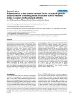
RESULTS
Study Selection and Characteristics
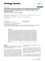
A total of 2296 articles were
identified, 153 were assessed
with respect to their eligibility for
inclusion (Supplemental Table 6),
and 27 studies were included
in the systematic review (Fig 1)
(Table 1).17–43 These 27 studies
included 484 fetuses with isolated
ACC and no other associated CNS
and/or extra-CNS anomalies at
first prenatal assessment.
Quality assessment of the included
studies was performed by using NOS
for cohort studies.44 Some of the
included studies showed an overall
good rate as regard for the selection
and comparability of the study
groups and for the ascertainment of
the outcome of interest. The main
weaknesses of these studies were
represented by their retrospective
design, small sample size , and
lack of a standardized postnatal
confirmation. Furthermore, the
cACC
Twenty studies including 261 fetuses
with isolated cACC were included in
this systematic review.
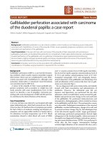
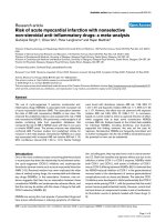
The rate of chromosomal anomalies
was 4.81% (95% confidence
interval [CI], 2.2–8.4) (Fig 2, Table 3).
The figures for the different
chromosomal anomalies found in
fetuses with isolated cACC are shown
in Supplemental Table 7.
It was not possible to extrapolate
data for the rate of clinically
significant CNVs in fetuses with
isolated cACC and normal karyotype,
thus the occurrence of clinically
significant CNVs was assessed in
fetuses with either cACC or pACC.
Overall, the rate of significant CNVs
in fetuses with isolated ACC (either
cACC or pACC) and normal karyotype
was 5.74% (95% CI, 1.3–13.1) (Fig 2).
In 2.99% (95% CI, 0.9–6.1) of the
cases, prenatal diagnosis failed in
correctly identifying cACC, with some
of the cases of pACC misdiagnosed as
having cACC (Supplemental Fig 5).
Additional anomalies not detected at
prenatal ultrasound were diagnosed
at fetal MRI in 7.83% (95% CI,
1.2–19.6) of the cases, whereas
the rate of additional structural
anomalies diagnosed only after birth
and missed at prenatal evaluation
was 5.49% (95% CI, 2.4–9.7)
(Table 3, Supplemental Figs 6 and
7). Individual case descriptions
of the anomalies detected only at
fetal MRI and postnatal imaging/
clinical investigation are shown in
Supplemental Tables 8 and 9.
In view of the high heterogeneity
in study design, age at and
type of assessment, and time at
follow-up, the rates for abnormal
neurodevelopmental outcomes
might not reflect the actual
neuropsychological performance
of these children and should
be interpreted with caution.
Furthermore, it was not possible to
ascertain the neurodevelopmental
performance of children with either
normal standard full karyotype
and no CNVs on CMA because only
one study reported this outcome.
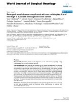
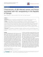
Neurodevelopmental outcome
was reported to be normal in
76.04% (95% CI, 64.3–86.1) of
children with a prenatal diagnosis
of isolated cACC confirmed at
birth (Fig 3, Table 4). The rates of
borderline/moderate and severe
neurodevelopmental outcome in
these children was 16.04% (95%
CI, 7.6–26.8,) and 8.15% (95% CI,
2.5–16.8) respectively. Table 3 shows
the detailed figures for the abnormal
neurodevelopmental performance
in children with isolated cACC.
Gross and fine motor control were
affected in 4.40% (95% CI0.6–11.3)
and 10.98% (95% CI 4.1–20.6) of
the cases, whereas 6.80% (95% CI,
1.7–14.9) of these children presented
with epilepsy. Cognitive status was
affected in 15.16% (95% CI, 6.9–
25.9) of the cases, whereas language
impairment was affected in 8.02%
(95% CI, 2.1–17.3). Finally, abnormal
ocular control and coordination
occurred in 15.84% (95% CI,
4.3–32.9) and 9.50% (95% CI,
3.2–18.7) of the cases, respectively
(Supplemental Fig 8).
Individual outcome descriptions of
children with isolated cACC showing
abnormal neurodevelopmental
profiles are shown in Supplemental
Table 10.
pACC
Fifteen studies including 225 fetuses
with pACC were included in this
review.
The rate of chromosomal anomalies
in fetuses with pACC and no other
structural anomalies visible at
prenatal imaging was 7.45% (95%
Downloaded from www.aappublications.org/news at Viet Nam:AAP Sponsored on July 6, 2021
6
D’ANTONIO et al
FIGURE 1
Systematic review flowchart.
CI, 2.0–15.9) (Fig 2, Table 4). The
figures for the different chromosomal
anomalies found in fetuses with
isolated pACC are shown in
Supplemental Table 11.
Additional anomalies not detected
at prenatal ultrasound were
diagnosed at fetal MRI in 11.86%
(95% CI, 3.2–24.9) of the cases,
whereas the rate of additional
structural anomalies diagnosed
only after birth and missed at
prenatal evaluation was 14.46%
(95% CI, 6.7–24.6) (Table 4,
Supplemental Figs 6 and 7).
Individual case descriptions of
the anomalies detected only at
fetal MRI and postnatal imaging/
clinical investigation are shown in
Supplemental Tables 12 and 13.
A discrepancy between prenatal and
postnatal diagnosis of pACC occurred
in 7.99% (95% CI, 2.5–16.3) of the
cases, mainly consisting in cases of
hypoplastic or dysgenetic corpus
callosum misdiagnosed as pACC
(Supplemental Fig 5).
Assessment of neurodevelopmental
outcome in children with isolated
pACC was even more problematic
in view of the smaller sample
size analyzed compared with
cACC.
Downloaded from www.aappublications.org/news at Viet Nam:AAP Sponsored on July 6, 2021
PEDIATRICS Volume 138, number 3, September 2016
7
FIGURE 2
Pooled proportions for the occurrence of chromosomal anomalies and pathogenic CNVs in fetuses with cACC and pACC.
FIGURE 3
Pooled proportions for the occurrence of abnormal neurodevelopmental outcome in fetuses with cACC.
Neurodevelopmental outcome was
reported to be normal in 71.42%
(95% CI, 53.1–86.7) of children with
a prenatal diagnosis of isolated pACC
confirmed at birth (Table 4). The
rates of borderline/moderate and
severe neurodevelopmental outcomes
in these children was 14.92% (95%
CI, 4.2–30.7) and 12.52% (95% CI,
2.9–27.5), respectively (Fig 4).
Fine motor control was affected
in 11.74 (95% CI, 0.9–32.1) of the
cases, and 16.11% (95% CI, 2.5–
38.2) of these children presented
with epilepsy. Cognitive status
TABLE 3 Pooled Proportions for the Outcomes Explored in This Systematic Review in Fetuses With cACC
Outcome
Pregnancy Outcome
Chromosomal anomalies (standard karyotype)
Chromosomal microarray (CNVs)a
Additional anomalies detected only at prenatal MRI
Additional anomalies detected only post-natally
Discrepancy between pre and post–natal diagnosis
Neurodevelopmental outcome
Normal
Borderline/Moderate
Severe
Detailed neurodevelopmental outcome
Gross motor
Fine motor
Cognitive
Epilepsy
Sensory
Visual
Coordination
Language
a
No. of Studies (n)
Fetuses (n/N)
I2 (%)
Raw % (95% CI)
Pooled Proportion (95% CI)
17
5
8
12
15
5/174
2/56
5/99
9/144
3/156
0
0
59.5
45.9
0
2.87 (0.9-6.6)
3.57 (0.4–12.3)
5.05 (1.7–11.4)
6.25 (2.9–11.5)
1.92 (0.4–5.5)
4.81 (2.2–8.4)
5.74 (1.3–13.1)
7.83 (1.2-19.6)
5.49 (2.4–9.7)
2.99 (0.9–6.1)
9
8
8
41/53
7/51
3/51
29.2
0
0
77.36 (63.8–87.7)
13.73 (5.7–26.3)
5.88 (1.2–16.2)
76.04 (64.3–86.1)
16.04 (7.6–26.8)
8.15 (2.5–16.8)
8
7
7
8
7
7
7
6
1/51
5/50
7/50
1/51
0/50
5/50
5/50
4/45
0
10.5
5
0
0
52.8
47
48.3
2.0 (0.1–10.6)
10.0 (3.3–21.8)
14.0 (5.8–26.7)
2.0 (0.1–10.6)
0 (0–7.1)
10.0 (3.3–21.8)
10.0 (3.3–21.8)
8.89 (2.5–21.2)
4.40 (0.6–11.3)
10.98 (4.1–20.6)
15.16 (6.9–25.9)
6.80 (1.7–14.9)
0 (0–9.2)
15.84 (4.3–32.9)
9.50 (3.2–18.7)
8.02 (2.1–17.3)
The analysis included cases with either isolated cACC and pACC.
Downloaded from www.aappublications.org/news at Viet Nam:AAP Sponsored on July 6, 2021
8
D’ANTONIO et al
TABLE 4 Pooled Proportions for the Outcomes Explored in This Systematic Review in Fetuses With pACC
Outcome
No. of Studies (n)
Fetuses (n/N)
I2 (%)
Raw % (95% CI)
Pooled Proportion (95% CI)
12
5
8
10
9
2/48
2/56
3/29
7/53
3/53
0
0
38.7
1.3
0
4.17 (0.5–14.3)
3.57 (0.4–12.3)
10.34 (2.2–27.4)
13.21 (5.5–25.3)
5.66 (1.2–15.7)
7.45 (2.0–15.9)
5.74 (1.3–13.1)
11.86 (3.2–24.9)
14.46 (6.7–24.6)
7.99 (2.5–16.3)
7
7
7
17/23
3/23
2/23
0
0
0
7.39 (5.2–9.0)
13.04 (2.8–33.6)
8.70 (1.1–28.0)
71.42 (53.1–86.7)
14.92 (4.2–30.7)
12.52 (2.9–27.5)
4
4
4
4
4
4
4
4
0/13
1/13
2/13
2/13
0/13
0/13
1/13
2/13
0
0
42.2
19.4
0
0
0
42.2
0 (0–24.7)
7.70 (0.2–3.6)
15.38 (1.9–45.4)
15.38 (1.9–45.4)
0 (0–24.7)
0 (0–24.7)
7.70 (0.2–3.6)
15.38 (1.9–45.4)
0 (0–23.0)
11.74 (0.9–32.1)
17.25 (3.0–39.7)
16.11 (2.53.2)
0 (0–23.0)
0 (0–23.0)
11.74 (0.9–32.1)
17.25 (3.0–39.7)
Pregnancy outcome
Chromosomal anomalies (standard karyotype)
Chromosomal microarray (CNVs)a
Additional anomalies detected only at prenatal MRI
Additional anomalies detected only postnatally
Discrepancy between prenatal and postnatal diagnosis
Neurodevelopmental outcome
Normal
Borderline/moderate
Severe
Detailed neurodevelopmental outcome
Gross motor
Fine motor
Cognitive
Epilepsy
Sensory
Visual
Coordination
Language
a
The analysis included cases with either isolated completed and partial ACC.
FIGURE 4
Pooled proportions for the occurrence of abnormal neurodevelopmental outcome in fetuses with pACC.
was affected in 17.25% (95% CI,
3.0–39.7) of the cases, whereas
language impairment was noticed
in 17.25% (95% CI, 3.0–39.7) of the
cases. Finally, abnormal coordination
occurred in 11.74% (95% CI, 0.9–
32.1) of the cases (Supplemental
Fig 9).
Individual outcome descriptions of
children with isolated pACC showing
abnormal neurodevelopmental
profile are shown in Supplemental
Table 14.
DISCUSSION
Summary of Evidence
The findings from this systematic
review showed that fetuses with
isolated callosal agenesis (either
cACC or pACC) are at high risk of
chromosomal anomalies. Even
when standard karyotyping is
normal, there is still a significant
risk of genetic anomalies detected
only at CMA analysis. In cases of a
prenatal diagnosis of isolated ACC,
the risk of associated anomalies
detected only at fetal MRI is about
8% and 12% in fetuses with cACC
and pACC, respectively, whereas
associated anomalies detected only
after birth can occur in about 5%
of fetuses with cACC and in 14% of
those with pACC. Short periods of
follow-up, heterogeneity in imaging
protocols, neurodevelopmental
tools used, discrepancies in the
definition of abnormal outcome, and
the small number of included cases
did not allow us to draw any robust
conclusions regarding the occurrence
of abnormal neurodevelopmental
outcome in children with a prenatal
diagnosis of isolated callosal
agenesis. The findings from this
systematic review suggested that
about two-thirds of children showed
a normal neurodevelopmental
outcome, although fine and gross
motor control, coordination,
language, and cognitive status can be
impaired in a significant proportion
of these children. However, these
figures might not reflect the actual
burden of neuropsychological
morbidity in children with isolated
ACC; additional large prospective
Downloaded from www.aappublications.org/news at Viet Nam:AAP Sponsored on July 6, 2021
PEDIATRICS Volume 138, number 3, September 2016
9
studies are needed to confirm these
findings.
Strengths and Limitations
The strengths of this study are its
robust methodology to identify all
possible studies, assess data quality,
and synthesize all suitable data.
For several meta-analyses, the
number of included studies was
small and some studies included
small numbers. The assessment of
the potential publication bias was
also problematic, either because
of the outcome nature (rates with
the left side limited to the value
0), which limits the reliability of
funnel plots, or because of the scarce
number of individual studies, which
strongly limits the reliability of
formal tests. Furthermore, all the
studies included were retrospective,
and thus liable to a considerable
risk of selection bias. In addition,
several outcomes and associations
were not adequately reported in
many studies. Finally, because of the
relatively short postnatal follow-up
period, the overall rate of additional
anomalies detected only after birth
and missed prenatally may have been
underestimated.
The assessment of
neurodevelopmental outcome in
children with a prenatal diagnosis of
isolated ACC was also problematic;
differences in age at follow-up and
neurodevelopmental tools used did
not allow a meaningful stratification
of the different outcomes measures;
therefore, the figures for the
developmental disabilities provided
in the current review might
not reflect the actual burden of
neuropsychological comorbidities
associated with isolated ACC and
should be interpreted with caution.
Furthermore, it was not possible
to stratify the analysis including
only fetuses with normal standard
full karyotype and no pathogenic
CNVs detected at CMA in view
of the lack of data regarding the
neurodevelopmental outcome in
these studies. In this scenario, it
might be entirely possible that cases
with isolated ACC, normal standard
karyotype, and pathogenic CNVs
were included in the analysis, thus
biasing the results. Finally, the
majority of the included studies did
not report a detailed description
of the neurologic performance of
fetuses with isolated ACC and merely
stratified the analysis in 3 different
categories (normal, borderline/
moderate, and severe), for which
inclusion criteria differed among
the studies. In view of all these
limitations, the resulting summary
measures need to be treated with
some caution.
Despite all of these limitations,
our review represents the most
up-to-date overall assessment of
the neurodevelopmental outcome
in callosal agenesis diagnosed
prenatally; this is important because
counseling for parents based
on single, small studies that are
subject to publication bias may be
inadequate.
Implication for Clinical Practice and
Future Perspectives
Advances in prenatal imaging
techniques have led to an increase in
the diagnostic accuracy of ultrasound
in detecting callosal anomalies.
However, prenatal counseling when
a fetus is diagnosed with ACC is
challenging.
The findings from this systematic
review showed that chromosomal
anomalies can occur in a significant
proportion of fetuses with isolated
ACC; furthermore, the risk of
genetic anomalies not detected by
conventional karyotyping is also not
negligible. CMA has recently been
shown to provide useful information
in patients with learning disabilities
and congenital anomalies for which
conventional cytogenetic tests have
proven negative. The findings from
this review support the use of CMA
when ACC is diagnosed prenatally.48
Fetal MRI is usually performed in
cases of prenatal diagnosis of ACC.
In the current review, associated
anomalies not detected at ultrasound
were diagnosed in 7.83% (95%
CI, 1.2–19.6) and in 11.86% (95%
CI, 3.2–24.9) in cACC and pACC,
respectively. However, even in cases
of a prenatal diagnosis of isolated
anomaly, the risk of ACC being not
truly isolated is relatively high, with
additional anomalies detected only
at postnatal imaging and/or clinical
examination, but missed prenatally,
occurring in 5.49% (95% confidence
interval [CI], 2.4–9.7) and 14.46%
(95% confidence interval [CI], 6.7–
24.6) of fetuses with pACC and cACC,
respectively.
Quantifying the real contribution
of fetal MRI in brain anomalies
is challenging. Several factors,
such as operator’s experience,
imaging protocol, time and type
of assessment, interval between
ultrasound and MRI, and type of
anomaly, may play a role in this
scenario and explain the wide
heterogeneity and the conflicting
results reported in previously
published studies. Despite all these
controversies, MRI is routinely
used in clinical practice to confirm
diagnosis and to look for associated
anomalies. The large majority of
additional anomalies detected only
at fetal MRI involved neuronal
migration disorders (Supplemental
Tables 8 and 12), which can be
detected preferentially from the
third trimester of pregnancy. On this
basis, when MRI is performed at the
time of the anomaly scan to confirm
diagnosis, it might be reasonable to
arrange a follow-up scan in the third
trimester to ascertain whether ACC is
truly isolated. These suggestions are
based on the authors’ experience and
further studies looking at the optimal
timing of fetal MRI are needed to
confirm these findings.
Furthermore, even when prenatal
diagnosis rules out associated
anomalies, there is still a significant
Downloaded from www.aappublications.org/news at Viet Nam:AAP Sponsored on July 6, 2021
10
D’ANTONIO et al
risk (5.5% and 14.5% in fetuses
with cACC and pACC, respectively)
to detect additional anomalies after
birth (Supplemental Tables 8 and
12). This should be stressed during
antenatal counseling, underlying
the fact that prenatal imaging is not
always able to differentiate between
complex and isolated cases, and that
postnatal imaging and a thorough
clinical examination are necessary to
confirm that ACC is truly isolated.
Assessing the neurodevelopmental
profile in children with ACC
is challenging. The term
neurodevelopmental outcome can be
misleading and inappropriate when
dealing with brain anomalies because
it encompasses a wide spectrum
of signs with different underlying
disorders and pathologic processes
that are not always easily measured
and that represent a continuous
interaction between pathologic,
environmental, and adaptive factors.
Intellectual abilities in individuals
with ACC have been reported to
be in the lower range of normal;
furthermore, difficulties in pragmatic
language skills and mathematics,
expressive and receptive language,
visual and spatial reasoning, and
attentional skills are impaired
or compromised in a significant
proportion of children.5 However,
postnatal studies are biased by the
fact that only symptomatic patients
are included, thus potentially
overestimating the burden of
disabilities observed in these
anomalies.
The findings from this systematic
review confirmed these results
and showed that children with
ACC may present different degrees
of impairment in neurologic and
neuropsychological domains.
Although a direct comparison
of the neurodevelopmental and
psychological performance of
children with cACC compared
with those with pACC was not
performed in view of the design of
most of the included studies, which
did not allow such a comparison,
the findings of this review do not
suggest a huge difference between
the 2 different entities of callosal
agenesis. The results from this
meta-analysis are surprising and
disagree with what is observed after
birth, where pACC is less likely to be
diagnosed as an isolated finding and
is usually affected by higher rates
of neurodevelopmental disabilities
compared with cACC. In the collective
authors’ opinion, the relatively high
rate of favorable outcome observed
in pACC might be due to the fact that
many of the cases labeled as pACC
prenatally are diagnosed after birth
as having hypoplasia of the corpus
callosum.
CONCLUSIONS
Fetuses with isolated callosal agenesis
are at high risk of chromosomal
anomalies even when a standard
karyotype is negative. Prenatal
imaging is not able to completely
rule out associated anomalies usually
coexisting with this condition, and the
risk of ACC of being not truly isolated
after birth is significant.
In isolated callosal agenesis,
anomalies in fine and gross motor
control, coordination, language,
cognitive status, and intelligence can
occur in a significant proportion of
children. However, in view of the
small number of included cases,
short period of follow-up, and
heterogeneity of neurodevelopmental
tools adopted, these results should be
interpreted with caution, and future
large prospective studies aiming at
assessing the neurodevelopmental
and psychological performance
of children with isolated callosal
agenesis using standardized tools
of neurodevelopmental assessment
at appropriate time intervals are
needed to ascertain the actual
neuropsychological performance and
intellectual impairment of children
with isolated ACC.
ACKNOWLEDGMENTS
We thank Prof O. Glenn, Prof J.
Barkovic, Prof L. Chitty, Prof G.
Kasprian, Prof D. Prayer, Prof Berg,
Prof V. D’Addario, Prof P. Volpe, Prof
G. Rizzo, Prof M. Kilby, Dr A. Rueland,
Prof TA Huisman, Dr O. Shen,
Prof W. Brown, Dr A Yazıcıoğlu, Prof
PH Tang, Dr F. Hadzagic-Catibusic,
Dr. H Slater, Dr M. Yamasaki,
Dr F. Scott, Dr Y. Yinon, Dr M. Nanni,
Dr A. Knafel, Dr L. Kleeman,
Dr S. Pashaj, Dr F. Scott, Dr E.M.
Vestergaard, Dr G. Srebniak,
Dr S. Toru, and Dr I Papoulidis for
their contribution to this systematic
review in terms of additional data
supplied and support.
ABBREVIATIONS
ACC: agenesis of the corpus
callosum
cACC: complete agenesis of the
corpus callosum
CI: confidence interval
CMA: chromosomal microarray
CNS: central nervous system
CNV: copy number variation
NOS: Newcastle-Ottawa Scale
pACC: partial agenesis of the
corpus callosum
Chieti-Pescara, Chieti, Italy; mDepartment of Medicine and Aging Sciences, University of Chieti-Pescara, Chieti, Italy; nEMISAC, Ce.S.I. Biotech, Chieti, Italy; and oDepartment of Obstetrics
and Gynaecology, University of Brescia, Brescia, Italy
Dr D’Antonio designed the study, extracted the data, carried out the initial analyses, drafted the initial manuscript, and reviewed and revised the manuscript;
Dr Pagani designed the study, extracted the data, carried out the initial analyses, and critically reviewed the manuscript; Dr Familiari designed the data collection
instruments, coordinated and supervised data collection at 2 of the 4 sites, and critically reviewed the manuscript; Dr Khalil wrote and critically reviewed the
manuscript; Profs Sagies, Malinger, Garel, Moutard, and Pilu and Drs Leibovitz and Bhide conceptualized and designed the study, designed the data collection,
Downloaded from www.aappublications.org/news at Viet Nam:AAP Sponsored on July 6, 2021
PEDIATRICS Volume 138, number 3, September 2016
11
helped to interpret the results, and wrote the manuscript; Profs Acharya and Papageorghiou conceptualized and designed the study and drafted the initial
manuscript; Dr Leombroni and Prof Manzoli performed the statistical analysis and critically reviewed the manuscript; Dr Prefumo designed the study and the
data collection instruments, coordinated and supervised data collection at 2 of the 4 sites, and critically reviewed the manuscript; and all authors approved the
final manuscript as submitted.
DOI: 10.1542/peds.2016-0445
Accepted for publication Jun 16, 2016
Address correspondence to Francesco D’Antonio, MD, PhD, Department of Clinical Medicine, Faculty of Health Sciences, UiT - The Arctic University of Norway,
Hansine Hansens veg 18, 9019 Tromsø, Norway. E-mail:
PEDIATRICS (ISSN Numbers: Print, 0031-4005; Online, 1098-4275).
Copyright © 2016 by the American Academy of Pediatrics
FINANCIAL DISCLOSURE: The authors have indicated they have no financial relationships relevant to this article to disclose.
FUNDING: No external funding.
POTENTIAL CONFLICTS OF INTEREST: The authors have indicated they have no potential conflicts of interest to disclose.
REFERENCES
1. Paul LK, Brown WS, Adolphs R, et al.
Agenesis of the corpus callosum:
genetic, developmental and functional
aspects of connectivity. Nat Rev
Neurosci. 2007;8(4):287–299
2. Glass HC, Shaw GM, Ma C, Sherr EH.
Agenesis of the corpus callosum in
California 1983-2003: a populationbased study. Am J Med Genet A.
2008;146A(19):2495–2500
3. Jeret JS, Serur D, Wisniewski K, Fisch
C. Frequency of agenesis of the corpus
callosum in the developmentally
disabled population as determined
by computerized tomography. Pediatr
Neurosci. 1985–1986;12(2):101–103
4. Palmer EE, Mowat D. Agenesis of the
corpus callosum: a clinical approach
to diagnosis. Am J Med Genet C Semin
Med Genet. 2014;166C(2):184–197
5. Siffredi V, Anderson V, Leventer
RJ, Spencer-Smith MM.
Neuropsychological profile of
agenesis of the corpus callosum: a
systematic review. Dev Neuropsychol.
2013;38(1):36–57
6. Schaefer GB, Bodensteiner JB.
Evaluation of the child with idiopathic
mental retardation. Pediatr Clin North
Am. 1992;39(4):929–943
7. Hardan AY, Pabalan M, Gupta N,
et al. Corpus callosum volume in
children with autism. Psychiatry Res.
2009;174(1):57–61
8. Tibbo P, Nopoulos P, Arndt S, Andreasen
NC. Corpus callosum shape and size in
male patients with schizophrenia. Biol
Psychiatry. 1998;44(6):405–412
9. Hynd GW, Semrud-Clikeman M, Lorys
AR, Novey ES, Eliopulos D, Lyytinen
H. Corpus callosum morphology in
attention deficit-hyperactivity disorder:
morphometric analysis of MRI. J Learn
Disabil. 1991;24(3):141–146
10. de Wit MC, Srebniak MI, Govaerts
LC, Van Opstal D, Galjaard RJ, Go AT.
Additional value of prenatal genomic
array testing in fetuses with isolated
structural ultrasound abnormalities
and a normal karyotype: a systematic
review of the literature. Ultrasound
Obstet Gynecol. 2014;43(2):139–146
11. Kearney HM, Thorland EC, Brown KK,
Quintero-Rivera F, South ST; Working
Group of the American College of
Medical Genetics Laboratory Quality
Assurance Committee. American
College of Medical Genetics standards
and guidelines for interpretation and
reporting of postnatal constitutional
copy number variants. Genet Med.
2011;13(7):680–685
12. Raybaud C. The corpus callosum, the
other great forebrain commissures,
and the septum pellucidum: anatomy,
development, and malformation.
Neuroradiology. 2010;52(6):447–477
13. Sotiriadis A, Makrydimas G.
Neurodevelopment after prenatal
diagnosis of isolated agenesis of the
corpus callosum: an integrative review.
Am J Obstet Gynecol. 2012;206(4):337.
e1–337.e5
14. Henderson LK, Craig JC, Willis
NS, Tovey D, Webster AC. How
to write a Cochrane systematic
review. Nephrology (Carlton).
2010;15(6):617–624
15. NHS Centre for Reviews and
Dissemination. Systematic reviews:
CRD’s guidance for undertaking
reviews in health care. York, United
Kingdom: University of York; 2009
16. PRISMA statement. Available at: www.
prisma-statement.org/. Accessed
November 12, 2015
17. Cesaretti C, Nanni M, Ghi T, et al
Variability of forebrain commissures
in callosal agenesis: a prenatal MR
imaging study. AJNR Am J Neuroradiol.
2016:37(3):521–527
18. Rüland AM, Berg C, Gembruch U, Geipel
A. Prenatal diagnosis of anomalies of
the corpus callosum over a 13-year
period. Ultraschall Med. 2015. doi:
10.1055/s-0034-1399699
19. Papoulidis I, Sotiriadis A, Siomou
E, et al. Routine use of array
comparative genomic hybridization
(aCGH) as standard approach for
prenatal diagnosis of chromosomal
abnormalities. Clinical experience of
1763 prenatal cases. Prenat Diagn.
2015;35(13):1269–1277
20. Shen O, Gelot AB, Moutard ML,
Jouannic JM, Sela HY, Garel C.
Abnormal shape of the cavum
septi pellucidi: an indirect sign
of partial agenesis of the corpus
callosum. Ultrasound Obstet Gynecol.
2015;46(5):595–599
21. Pashaj S, Merz E. Detection of fetal
corpus callosum abnormalities by
means of 3D ultrasound. Ultraschall
Med. 2016:37(2)185–194
22. Ozyüncü O, Yazıcıoğlu A, Turğal M.
Antenatal diagnosis and outcome
Downloaded from www.aappublications.org/news at Viet Nam:AAP Sponsored on July 6, 2021
12
D’ANTONIO et al
of agenesis of corpus callosum:
A retrospective review of 33
cases. J Turk Ger Gynecol Assoc.
2014;15(1):18–21
23. Lachmann R, Sodre D, Barmpas M,
Akolekar R, Nicolaides KH. Midbrain
and falx in fetuses with absent corpus
callosum at 11-13 weeks. Fetal Diagn
Ther. 2013;33(1):41–46
24. Kasprian G, Brugger PC, Schöpf V,
et al. Assessing prenatal white matter
connectivity in commissural agenesis.
Brain. 2013;136(Pt 1):168–179
25. Yinon Y, Katorza E, Nassie DI, et al.
Late diagnosis of fetal central nervous
system anomalies following a normal
second trimester anatomy scan.
Prenat Diagn. 2013;33(10):929–934
26. Vestergaard EM, Christensen
R, Petersen O, Vogel I. Prenatal
diagnosis: array comparative genomic
hybridization in fetuses with abnormal
sonographic findings. Acta Obstetricia
et Gynecologica Scandinavica .
2013;92(7):762–768
27. Moutard ML, Kieffer V, Feingold
J, et al. Isolated corpus callosum
agenesis: a ten-year follow-up after
prenatal diagnosis (how are the
children without corpus callosum
at 10 years of age?). Prenat Diagn.
2012;32(3):277–283
28. Wapner RJ, Martin CL, Levy B, et al.
Chromosomal microarray versus
karyotyping for prenatal diagnosis.
N Engl J Med. 2012;367(23):2175–2184
29. Yamasaki M, Nonaka M, Bamba Y,
Teramoto C, Ban C, Pooh RK. Diagnosis,
treatment, and long-term outcomes
of fetal hydrocephalus. Semin Fetal
Neonatal Med. 2012;17(6):330–335
30. Shaffer LG, Rosenfeld JA, Dabell MP,
et al. Detection rates of clinically
significant genomic alterations by
microarray analysis for specific
anomalies detected by ultrasound.
Prenat Diagn. 2012;32(10):986–995
31. Mangione R, Fries N, Godard P,
et al. Neurodevelopmental outcome
following prenatal diagnosis of
an isolated anomaly of the corpus
callosum. Ultrasound Obstet Gynecol.
2011;37(3):290–295
32. Ghi T, Carletti A, Contro E, et al.
Prenatal diagnosis and outcome of
partial agenesis and hypoplasia of the
corpus callosum. Ultrasound Obstet
Gynecol. 2010;35(1):35–41
33. Cignini P, D’Emidio L, Padula F,
et al. The role of ultrasonography
in the diagnosis of fetal isolated
complete agenesis of the corpus
callosum: a long-term prospective
study. J Matern Fetal Neonatal Med.
2010;23(12):1504–1509
34. Tang PH, Bartha AI, Norton ME,
Barkovich AJ, Sherr EH, Glenn OA.
Agenesis of the corpus callosum: an
MR imaging analysis of associated
abnormalities in the fetus. AJNR Am J
Neuroradiol. 2009;30(2):257–263
35. Goetzinger KR, Stamilio DM, Dicke JM,
Macones GA, Odibo AO. Evaluating the
incidence and likelihood ratios for
chromosomal abnormalities in fetuses
with common central nervous system
malformations. Am J Obstet Gynecol.
2008;199(3):285.e1–285.e6
36. Chadie A, Radi S, Trestard L, et al;
Haute-Normandie Perinatal Network.
Neurodevelopmental outcome in
prenatally diagnosed isolated agenesis
of the corpus callosum. Acta Paediatr.
2008;97(4):420–424
37. Fratelli N, Papageorghiou AT, Prefumo
F, Bakalis S, Homfray T, Thilaganathan
B. Outcome of prenatally diagnosed
agenesis of the corpus callosum.
Prenat Diagn. 2007;27(6):512–517
38. Francesco P, Maria-Edgarda B,
Giovanni P, Dandolo G, Giulio B.
Prenatal diagnosis of agenesis
of corpus callosum: what is the
neurodevelopmental outcome? Pediatr
Int. 2006;48(3):298–304
39. Ramelli G, Zanda N, Wyttenbach M,
Bronz L, Schnider A. The prognosis of
agenesis of the corpus callosum might
mostly be favourable. Swiss Med Wkly.
2006;136(25-26):404–405
40. Volpe P, Paladini D, Resta M, et al.
Characteristics, associations
and outcome of partial agenesis
of the corpus callosum in the
fetus. Ultrasound Obstet Gynecol.
2006;27(5):509–516
41. Blaicher W, Prayer D, Mittermayer C,
et al. Magnetic resonance imaging
in foetuses with bilateral moderate
ventriculomegaly and suspected
anomaly of the corpus callosum on
ultrasound scan. Ultraschall Med.
2003;24(4):255–260
42. Malinger G, Lerman-Sagie T,
Watemberg N, Rotmensch S, Lev
D, Glezerman M. A normal secondtrimester ultrasound does not
exclude intracranial structural
pathology. Ultrasound Obstet Gynecol.
2002;20(1):51–56
43. Goodyear PW, Bannister CM, Russell
S, Rimmer S. Outcome in prenatally
diagnosed fetal agenesis of the
corpus callosum. Fetal Diagn Ther.
2001;16(3):139–145
44. Wells GA, Shea B, O’Connell D, et al. The
Newcastle-Ottawa Scale for assessing
the quality of nonrandomised
studies in meta-analyses. Available
at: www.ohri.ca/programs/clinical_
epidemiology/oxford.asp
45. Hunter JP, Saratzis A, Sutton AJ,
Boucher RH, Sayers RD, Bown MJ. In
meta-analyses of proportion studies,
funnel plots were found to be an
inaccurate method of assessing
publication bias. J Clin Epidemiol.
2014;67(8):897–903
46. Egger M, Davey Smith G, Schneider
M, Minder C. Bias in meta-analysis
detected by a simple, graphical test.
BMJ. 1997;315(7109):629–634
47. Higgins JPT, Green S, eds. Cochrane
Handbook for Systematic Reviews
of Interventions. Version 5.0.2.
The Cochrane Collaboration, 2011.
Available at: www.cochrane-handbook.
org. Accessed December 10, 2015
48. Sagoo GS, Butterworth AS, Sanderson
S, Shaw-Smith C, Higgins JP, Burton
H. Array CGH in patients with learning
disability (mental retardation) and
congenital anomalies: updated
systematic review and meta-analysis of
19 studies and 13,926 subjects. Genet
Med. 2009;11(3):139–146
Downloaded from www.aappublications.org/news at Viet Nam:AAP Sponsored on July 6, 2021
PEDIATRICS Volume 138, number 3, September 2016
13
Outcomes Associated With Isolated Agenesis of the Corpus Callosum: A
Meta-analysis
Francesco D'Antonio, Giorgio Pagani, Alessandra Familiari, Asma Khalil,
Tally-Lerman Sagies, Gustavo Malinger, Zvi Leibovitz, Catherine Garel, Marie Laure
Moutard, Gianluigi Pilu, Amar Bhide, Ganesh Acharya, Martina Leombroni,
Lamberto Manzoli, Aris Papageorghiou and Federico Prefumo
Pediatrics 2016;138;
DOI: 10.1542/peds.2016-0445 originally published online August 31, 2016;
Updated Information &
Services
including high resolution figures, can be found at:
/>
References
This article cites 43 articles, 3 of which you can access for free at:
/>
Subspecialty Collections
This article, along with others on similar topics, appears in the
following collection(s):
Developmental/Behavioral Pediatrics
/>al_issues_sub
Cognition/Language/Learning Disorders
/>rning_disorders_sub
Fetus/Newborn Infant
/>sub
Birth Defects
/>
Permissions & Licensing
Information about reproducing this article in parts (figures, tables) or
in its entirety can be found online at:
/>
Reprints
Information about ordering reprints can be found online:
/>
Downloaded from www.aappublications.org/news at Viet Nam:AAP Sponsored on July 6, 2021
Outcomes Associated With Isolated Agenesis of the Corpus Callosum: A
Meta-analysis
Francesco D'Antonio, Giorgio Pagani, Alessandra Familiari, Asma Khalil,
Tally-Lerman Sagies, Gustavo Malinger, Zvi Leibovitz, Catherine Garel, Marie Laure
Moutard, Gianluigi Pilu, Amar Bhide, Ganesh Acharya, Martina Leombroni,
Lamberto Manzoli, Aris Papageorghiou and Federico Prefumo
Pediatrics 2016;138;
DOI: 10.1542/peds.2016-0445 originally published online August 31, 2016;
The online version of this article, along with updated information and services, is
located on the World Wide Web at:
/>
Data Supplement at:
/>
Pediatrics is the official journal of the American Academy of Pediatrics. A monthly publication, it
has been published continuously since 1948. Pediatrics is owned, published, and trademarked by
the American Academy of Pediatrics, 345 Park Avenue, Itasca, Illinois, 60143. Copyright © 2016
by the American Academy of Pediatrics. All rights reserved. Print ISSN: 1073-0397.
Downloaded from www.aappublications.org/news at Viet Nam:AAP Sponsored on July 6, 2021