Poor performance of right brain damaged patients on ravens coloured matrices derangement of general intelligence or of specific abilities
Bạn đang xem bản rút gọn của tài liệu. Xem và tải ngay bản đầy đủ của tài liệu tại đây (448.69 KB, 6 trang )
Neuropsychologia,1977, Vol. 15, pp. 675 to 680. PergamonPress.Printedin England
NOTE
POOR PERFORMANCE
OF RIGHT BRAIN-DAMAGED
PATIENTS
ON RAVEN’S COLOURED
MATRICES:
DERANGEMENT
OF
GENERAL
INTELLIGENCE
OR OF SPECIFIC ABILITIES?
G. GAINOTTI, C. CALTAGIRONEand
G.
MICELI
Clinic for Nervous and Mental Diseases, The Catholic University,
Largo A. Gemelli, I-Roma, Italy
(Received
16 December
1976)
Abstract-Two
groups of 170 left and 173 right brain-damaged patients were given the
Raven’s Coloured Matrices, in order to study the influence of the hemispheric side of the
lesion on this test of visual-spatial intelligence. A significant difference was found between the
two hemispheric groups, the right brain-damaged patients scoring worse than the left hemisphere-damaged subjects. Furthermore, patients with right hemispheric lesions showed a
striking tendency to neglect the responses lying on the left side of the page, whereas subjects
with left hemispheric damage showed only a mild tendency to neglect the alternatives lying on
the right half of the sheet. The worse performances obtained on the Coloured Matrices by
right brain-damaged patients seemed due for the most part to unilateral spatial neglect. These
findings suggest that the bad performances of patients affected by retro-rolandic right hemispheric lesions are not due to a general intellectual impairment, but rather to the detrimental
effect of unilateral spatial neglect and of a general disorder in visual-spatial analysis.
RESULTS obtained on Raven’s Progressive Matrices by patients affected by localized cerebral lesions can
be due to three main factors:
-the intellectual status of the patient [l-3];
-his ability in visual-spatial analysis [4-61;
-the presence and severity of unilateral spatial neglect [7-81.
The complexity of the factors underlying the patient’s performance can account, in part at least, for the
conflicting results reported in the literature. ARRIGONI and DE RENZI 191, on the one hand, found significantly lower scores in patients with unilateral left hemisphere lesions. PIERCYand SMYTH [4], on the other
hand, reported much lower scores on the Raven test in patients with right hemisphere damage. Still other
authors [5,6, 10,l l] have reported no significant predominance of either the left or the right brain-damaged
patients.
In a very well controlled study, the problem has again been taken up by BASSOet nl. [12], who have shown
that among the patients affected by localized cerebral lesions two groups are significantly impaired on the
Raven’s Coloured Matrices:
(a) Right brain-damaged patients with visual field defect; and (b) left brain-damaged patients with aphasia.
According to BASSOet al. [12] these findings support the notion that two cerebral areas are critical for
intelligence: (a) the retro-rolandic areas of the right hemisphere; and (b) a region of the left hemisphere
overlapping the language areas. These conclusions are of great interest for the students of brain behaviour
relationships. They raise the question, however, whether the poor performance of the right brain-damaged
patients is really due to general intellectual deterioration rather than to more specific defects, such as spatial
heminattention or impairment of visual perception. BASSOet al. [12] have themselves discarded this hypothesis because the interiority of the right brain-damaged patients with visual field defects “persisted even
when the Raven scores were adjusted for the scores obtained on a test assumed to assess the severity of the
above deficits”.
None the less, the conclusions of BASSO et al. seemed to us at variance both with other data not only in
our own experience but also in the earlier literature. For example, ZANGWILL and co-workers 113-151 have
shown in a series of classical papers, that patients with posterior lesions of the right hemisphere may present
signs of visual spatial agnosia at every level of psychological response, in absence of defects of general
675
676 zyxwvutsrqponmlkjihgfedcbaZYXWVUTSRQPONMLKJIHGFEDCBA
NOTE
intelligence. In more recent years, GAINOTTIand TIACCI 1161have shown that patients with unilateral spatial
neglect perform worse than other right brain-damaged subjects on tasks of visual spatial analysis; and
OXBURY et al. 1171 have demonstrated
that this difference cannot be attributed to a general intellectual
deterioration. The present study was, therefore, undertaken to investigate further the relationship between
poor performance on the Raven Coloured Matrices of patients with right cerebral damage and unilateral
spatial neglect. In particular, we have focused attention on the following points:
(1) Is it possible to demonstrate a significant difference between the performance of right and left braindamaged patients on the Coloured Matrices?
(2) Does the tendency to neglect the designs lying on the side of the page contralateral to the locus of
of lesion differ significantly as between right and left hemisphere-damaged patients?
(3) Are the poor scores of right brain-damaged patients due at least in part to unilateral spatial neglect?
MATERIALS
AND METHODS
Subjects
The present research was carried out on two groups of 170 left and 173 right brain-damaged patients. The
diagnosis of hemispheric damage was made on the ground of the clinical symptoms, supplemented by the
available EEG, brain scan and neuroradiological findings. No selection was made in forming the groups,
and only the following patients were excluded from the study:
(a) Those who considered themselves (or were reported by their relatives) to be left-handed;
(b) those who showed impaired consciousness or were absolutely unable to maintain the set necessary
to carry out the task;
(c) those whose case history or clinical picture pointed to diffuse or clearly bilateral cerebral damage.
No significant difference could be found between right and left brain-damaged patients with respect to
age, educational level and aetiology of illness.
Testing procedures
The Raven’s Coloured
Progressive Matrices were given to all patients without time limits. For each test
problem the subjects were required to point to the item they thought to be the correct choice and the examiner noted it on the record sheet. A failure to respond on any problem was counted as an error. The maximum
score was 36.
In order to assess the possible significance of unilateral spatial neglect in reducing the scores of right and
left brain-damaged patients, it was decided to measure position preference according to a method devised
by COSTAet al. (8) and to compare position preference scores in the two hemispheric groups. The position
preference scores were determined by computing for each patient the difference between the number of
alternatives chosen on the side of the page ipsilateral to the damaged hemisphere and the number chosen
on the contralateral side of the sheet (ipsilateral-minus-contralateral
score). With an error-free performance,
this difference would be zero, because correct responses are equally distributed among the right, the center
and the left positions.
RESULTS
Performance ofright and left brain-damaged patients on the Raven Colorrred Matrices
The mean scores obtained by the right and left brain-damaged patients are shown in Table 1:
Table 1. Scores obtained by right and left brain-damaged
Right brain-damaged
patient (N = 173)
Mean scores
S.D.
x = 16, 375
6,456
patients on Raven’s Coloured Matrices.
Left brain-damaged
patients (N = 170)
y = 18, 608
6, 686
t
3, 134
P
s; oXlO
A significant difference was found to exist between the two hemispheric groups, the right brain-damaged
patients scoring significantly lower than those with left-sided brain damage.
Relationship between hemispheric
to the damaged hemisphere
locus of lesion and tendemy
to neglect the test items on
thepage
zyxwvutsrqponmlkjihgfe
contralateral
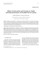
Figure 1 shows the distribution of the position preference scores in our two groups:
It is apparent from inspection of this that right brain-damaged patients show a much more marked position
preference than the left brain-damaged patients. In order to control the statistical value of the prevalence
of position preference scores among the right hemi$pheric patients, we took as cut off point between normal
and pathological performances the position preference of 7 which, according to COSTA et al. [S], has a
677
NOTE
Median value of the
pathological
position
preferenie
scores
+I
I
t36
0 Right
B Left
FJG. 1. Distribution
brain-damaged
brain-damaged
potients
patients
of the position preference scores in right and left brain-damaged
patients.
probability of occurrence of less than 0.01 in normals. The number of patients having an abnormal score
in the expected (positive) direction was 96 (55 %) in the right group and 37 (22%) in the left group. This
difference was found to be highly significant (x2 = 41, 08; P < 0X1001). A further analysis was undertaken
to ascertain whether not only the incidence but zyxwvutsrqponmlkjihgfedcbaZYXWVUTSRQPONMLK
also the strength of positionpreference was higher in fhe right
hemispheric group. The median value of the pathological position preference scores (19.35) was taken as
cut-off point between mild (p.p. +) and severe (p.p. + +-) forms of position preference.
Fifty-two right and only 8 left brain-damaged patients showed the more severe form of position preference
(p.p. + +), whereas 44 right and 29 left hemisphere-damaged patients showed only a mild tendency (p.p. +)
to select the alternatives lying on tilt half of the page ipsilateral to the damaged hemisphere. This difference
was also found to be highly significant (x2 = 11,41; P < 0.001).
Relationships between position preference
Acores and performance
obtained on the Coloured M atrices
In order to ascertain whether the poorer performance on Raven’s Matrices by right hemispheric patients
was due, in part at least, to the tendency to neglect the items lying on the left half of the sheet, both right
and left brain-damaged patients were divided into three subgroups, according to both presence and strength
of the pcsition preference. Table 2 shows the scores obtained on the Coloured Matrices by right and left
brain-damaged patients respectively, with a positionipreference (p.p.) score lower than +7 (p.p. -), a p.p,
score ranging between + 8 and + 19 (pp. i-) and a p.p. score higher than 19 (p.p. + +).
These data show that there is no significant difference between right and left brain-damagedpatcents when
subjects belonging co the same position prejtirence group are compared. On the other hand. a highly significant
di&rence
is found, within each hemispheric group, between patients without position preference
(p.p. -),
subjects with mild position preference (p.p. +) and patients with marked tendency to neglect the alternatives
ly ing on the halfsheet contralateral to the damaged hemisphere (p.p. + +).
Thesedata suggest that the tendency to select the responseslying on the halfsheet ipsilateral to the damaged
hemisphere, paying little or no attention to the contralateral half of the page, is an important factor in
reducing the performance not only of right, but also of the left brain-damaged patients. However, since
both the incidence and strength of position preference were significantly higher in the right hemispheric
groups, these results also suggest that the poorer performance of theright brain-damaged patients was due
for the most part to unilateral spatial neglect.
DISCUSSION
The main results of the present study can be summarized as follows:
(a) If the Coloured Matrices are administered to large groups of unselected patients
with unilateral
678
NATE
2. Scores obtained on the coloured matrices by right and left brain-damaged patients without (p.p. -),
Table zyxwvutsrqponmlkjihgfedcbaZYXWVUTSRQPONMLKJIHGFEDCBA
with slight (D.D. +) and with marked (p.0.
_ _ + +) position preference
Right brain-damaged patients
(N = 173)
X = 20,434
P.P. (N = 77)
Left brain-damaged patients
(N = 170)
x1 = 19, 969
P.P. (N = 133)
p.p. -t
y1 = 14, 827
(N = 29)
t = 7,249 (P < 0.001)
P.P. TV+
z = 10,460
0, 525
n.s.
I, 128
ll.S.
I, 339
“ .S.
t = 3, 205 (P < 0.005)
p.p. + f z 1= 8,875
(N = 8)
(N = 521
P
t = 4, 133 (P < 0X01)
t = 4, 167 (P < 0.001)
p.p. -t
y = 16, 071
(N = 44)
t
brain-damage, right hemispnere-damaged
patients obtain significantly lower scores than do patients with
damage restricted to the left hemisphere.
(b) The tendency to neglect information on the half sheet contralateral to the damaged hemisphere is
dramatically more in evidence in cases in which the lesion is restricted to the right hemisphere.
(c) Patients who pay little or no attention to information lying on the half page contralateral to the
damaged hemisphere obtain significantly lower scores on the Coloured Matrices than patients who show no
clear position preference.
These findings would appear to cast some doubt on the assumption of BASSOet al. [12] that “there is an
area in the right hemisphere, probably adjacent to that serving discrimination of visual information, which is
critically involved in the eduction of the logical relationships existing among different patterns”. Our data
seem, indeed, to suggest that the deficit shown on the Coloured Matrices by right brain-damaged patients with
visualfield defects is not intellectual in nature, since their poor results appear to be chiefly to unilateral spatial
neglect, On the other hand, it should be acknowledged that the association between the presence of uni-
lateral spatial neglect (position preference) and the impairment shown on the Coloured Matrices does not
necessarily mean that poor performance on this test was due only to the effect of position preference. The
alternative hypothesis can also be advanced that it was due in part to the detrimental effect of spatialneglect
and in part to damage to a region ofthe brain subserving a specialized (visual-spatial) form of intelligence.
This area, when damaged, must also be regarded as responsible for the production of unilateral neglect.
This second hypothesis is not definitely at variance with the results of the present research and is in part
supported by some data obtained by ZAIDEL and SPERRY[t8] in patients with cerebral commissurotomy. We
have not attempted to test this hypothesis owing to the difficulty of finding a test of visual-spatial intelligence that does not require lateral spatial exploration. However, since the problem seemed to us interesting
enough, we have constructed a new form of the Coloured Matrices that, without changing the essential
features of the original task, is nevertheless much less sensitive to the presence of unilateral spatial neglect.
This new form of the Progressive Matrices is actually in use in our laboratory and we hope that it will offer
us the opportunity of testing more adequately the hypothesis that there is an area of the right cerebral
hemisphere which is involved in the intellectual processing of visual-spatial data.
REFERENCES
1.
2.
3.
4.
SPEARMAN,0. Theory of a general factor. Br. J. Psychol. 36,117-131,
1946.
RAVEN, J. C. Standard Progressive Matrices. H. K. Lewis, London, 1938.
RAVEN,J. C. Guide to Using the Coloured Progressive Matrices. H. K. Lewis, London, 1973.
COSTA,L. D. and VAUGHAN,H. G. Performance of patients with lateralized cerebral lesions-l.
and perceptual tests. .I. nerv. ment. Dis. 134, 162-168, 1962.
Verbal
5. PIERCY,M. and SMYTH,V. 0. G. Right hemisphere dominance for certain non-verbal intellectual skills.
Brain 85, 775-790,
1962.
6. GAINOTTI,G. and TIACCI, C. Sui rapporti fra aprassia costruttiva, agnosia visuo-spaziale e negligenza
spaziale unilaterale. Riv. Patol. ner. ment. 93, 103-l 16, 1972.
7. GAINOTTI,G. Les manifestations de negligence et d’inattention pour I’hemispace. Cortex 4, 64-91, 1968.
8. COSTA,L. D., VAUGHAN,H. G., JR., HORWITZ, M. and RITTER,W. Pattern of behavioral deficit associated with visual spatial neglect. Cortex 5, 242-263, 1969.
9. ARRXOONX,
G. and DE RENZI, E. Constructional apraxia and hemispheric locus of lesion. Cortex 1,
170-197,1964.
NOTE
679
10. zyxwvutsrqponmlkjihgfedcbaZYXWVUTSRQPONMLKJIHGFEDCBA
DE RENZI, E. and FAGLIONI,P. The comparative efficiency of intelligence and vigilance tests in detecting
hemispheric cerebral damage. Cortex 1,41&433, 1965.
11. COL~NNA, A. and FAGLIONI, P. The performance of hemisphere-damaged
patients on spatial intelligence tests. Cortex 2,293-307, 1966.
12. BASSO,A., DE RENZI, E., FAGLIONI,P., SCO~I, G. and SPINNLER,H. Neuropsychological evidence for
the existence of cerebral areas critical to the performances of intelligence tasks. Bruin 96,715-728,1973.
13. PATERSON,A. and ZANGWILL, 0. L. Disorders of visual space perception associated with lesions of the
right cerebral hemisphere. Bruin 67, 331-358, 1944.
14. MCFIE, J., PIERCY,M. and ZANGWILL, 0. L. Visual spatial agnosia associated with lesions of the right
hemisphere. Brain 73, 167-190, 1950.
15. ETTLINGER,G., WARRINGTON,E. and ZANGWILL, 0. L. A further study of visual-spatial agnosia. Bruin
80, 335-361, 1957.
16. GAINOTTI, G. and TIACCI, C. The relationships between disorders of visual perception and unilateral
spatial neglect. Neuropsychologiu 9,451-455, 1971.
17. OXBURY, J. M., CAMPBELL,D. C. and OXBURY,S. M. Unilateral spatial neglect and impairments of
spatial analysis and visual perception. Bruin 97, 551-564, 1974.
18. ZAIDEL, D. and SPERRY,R. W. Performance on Raven’s colored progressive matrices test by subjects
with cerebral commissurotomy. Cortex 9, 34-39, 1973.
680
NOTE
Zwei Gruppen van 170 links- und 173 rechtshirnig geschsdigten Patienten wurden mit dem RAVEN-Test gepriift(mit &n
RAVENS Coloured Matrices), urnfestzustellen, welchen Einflu6 die Hemisphlre als Verletzungssitz in diesem Test
zur optisch-rrumlichen IRtelligenz hat. Ys wurde eitisignifikanter Unterschied zwischen den beiden HemisphlrenGruppen gefunde?, wobei die rechtshirnig geschldigten
Patien-tenschlechter abschnitten als die 1inkshemisphBrisch
gesch%digter..Dariiberhinaus zeigteq die Patienten mit rechtshemisph4rischen Verletzungen eine ausgepragte Neigung, die
auf der linken Seite des Blattes gelegenen Partien ZU vernachlzissigrl,w4hrend.die Versuchspersonen mit linkshemisph6rischer Sch&di&u??gnur wenig ZUT Vernachl~ssiQxI~ der entsprechenden Partien in der rechten Hrilftedes Blattes neigten. Die schlech&ren Leistungen, die bei der Untersuchung
rechtshirnig Geschadigter mit den RAVEN-Test erzielt wurden
schienen zum grsfltenTeil auf den unilateralen Neglect zuriickzufiihren
zu sein. Diese Befunde sprechen dafiir,daR
schlechte Leistungen bei Patienten mit retrorolandischen
TechtShemiSphariSchen Sct#ligungen nicht auf einer eenerellen
intellektuellen MinderunE;bzsieren, sondern au‘ der nachteiligen Wirkung eines unilateraleriNeglects und eixr
wnerellen
SMrun,rrder FYhiSkeit der optisch-r&xnlichen
,x
Analvse.