breast cancer
Bạn đang xem bản rút gọn của tài liệu. Xem và tải ngay bản đầy đủ của tài liệu tại đây (1.83 MB, 35 trang )
Introduction
Breast cancer is the second leading cause of cancer death in women.
About one in eight women (12.5%) will develop invasive breast cancer during their
lifetime.
The importance of the radiologist’s role has increased over the years.
Radiologic information that may alter stage, prognosis, or treatment includes tumor
size; number of tumor lesions; total span of disease; regional nodal status (axillary
levels I–III, internal mammary, supraclavicular); locoregional invasion (involvement of
the pectoralis muscle, skin, nipple, or chest wall); and distant metastases to bone,
lung, brain, and liver, among other anatomic structures.
The staging information will help in choosing between breast conservation and
mastectomy, preoperative and postoperative chemotherapy or hormonal therapy,
sentinel lymph node biopsy (SLNB) and axillary lymph node dissection (ALND), and
radiation therapy
Risk factors
increasing age
reproductive lifestyle factors increasing unopposed estrogen load: early menarche,
late menopause, nulliparity, infertility, or, if parous, few children with late age at first
delivery, lack of breastfeeding, unopposed estrogen hormone replacement therapy
personal history of breast cancer or a high risk breast lesion.
genetic mutations: BRCA1 or BRCA2 mutation
pathological classification: WHO
Invasive carcinoma of no special type.
Ductal carcinoma in situ
Invasive lobular carcinoma.
Immunophenotype
estrogen receptor (ER)
progesterone receptor (PR)
human epidermal growth factor receptor 2 (HER2; protooncogene Neu; receptor
tyrosine-protein kinase erbB-2)
Ki-67
ACR BI-RADS NHŨ ẢNH 2013
Lymp nodes
diffuse cortical thickening: > 3mm
eccentric cortical thickening
rounded hypoechoic node
complete or partial effacement of the fat hilum.
complete or partial replacement of the node with an ill-defined or irregular mass)
Irregular node, node matting, perinodal extension
microcalcifications in the node
Mammography: mass
Mammography: Calcifications
Carl J. D’Orsi, MD et al. ACR BI-RADS® Atlas Breast Imaging Reporting and Data System. The 5th
Edition. 2013.
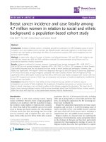
Ultrasound- elastography
Ultrasoundelastography
Ultrasound- elastography
A 53-year-old patient with Rt. breast mass proved to be invasive ductal carcinoma “grade II” on histopathological examination. Left, B-mode ultrasound
showed ill-defined hypoechoic mass with irregular margins and few speculations was seen at 11 o’clock location, which is considered to be BIRADS 5
category. Right, strain elastography, the entire hypoechoic lesion was shaded in blue color with elasticity score (4), strain ratio = 5.72
MRI
The American Joint Committee on Cancer (AJCC)
staging system (7th edition)- TNM
/>
Table 1 illustrates overall preoperative staging as performed with the AJCC’s TNM staging system for breast cancer
Tumor
Tumor
The primary tumor is usually measured in three orthogonal dimensions, with the
largest dimension used for staging purposes.
Tis category includes ductal carcinoma in situ (DCIS), lobular carcinoma in situ, and
Paget disease of the nipple not associated with invasive carcinoma or carcinoma in
situ in the underlying breast parenchyma
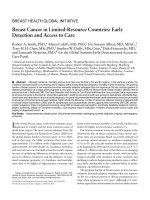
Figure 1. Stage 0, cTisN0M0 tumor. (a, b) Mediolateral oblique (a) and craniocaudal (b) mammographic views of the left
breast demonstrate a cluster of suspicious fine pleomorphic calcifications (arrow). (c) Magnification view more clearly
depicts the calcifications. Stereotactic-guided biopsy revealed DCIS