craniopharygioma U sọ hầu
Bạn đang xem bản rút gọn của tài liệu. Xem và tải ngay bản đầy đủ của tài liệu tại đây (3.58 MB, 35 trang )
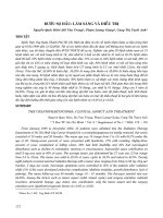
Bệnh nhân nam 21 tuổi, tiền sử khỏe mạnh
Lý do vào viện : đau đầu, nhìn mờ
Bệnh sử: cách 10 tháng bệnh nhân đau đầu âm ỉ từng đợt, chưa đi khám và điều trị
gì, 2 tuần nay bệnh nhân đau đầu tăng, nhìn mờ, khơng yếu liệt chi => vào viện
Các xét nghiệm CTM, sinh hóa, hormon trong giới hạn bình thường
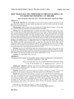
The tumors sellar /suprasellar region with cystic degeneration and solid :
-
Pituitary macroadenoma
-
Hypothalamic chiasmatic glioma
-
Intracranial teratoma
Craniopharyngioma
Pituitary macroadenoma
- All ages
-
Patients typically present with symptoms of local mass effect on adjacent structures
Some may present due to hormonal imbalance, with symptoms of hypopituitarism (from
compression) or secretion
Overall signal characteristics can signifcantly vary depending on tumor components such as hemorrhage, cystic
transformation, or necrosis.
•
•
•
•
T1
–
–
typically isointense to grey matter
larger lesions are often heterogeneous and vary in signal due to areas of cystic
change/necrosis/hemorrhage
T1 C+ (Gd)
–
solid components demonstrate moderate to bright enhancement
T2
–
–
typically isointense to grey matter
larger lesions are often heterogeneous and vary in signal due to areas of cystic
change/necrosis/hemorrhage
GRE/SWI
–
–
most sensitive for detecting any hemorrhagic components, which appear as areas of signal loss
calcifcation is rare but should be excluded by reviewing CT scans
Snowman sign
Hypothalamic chiasmatic glioma:
-
Optic pathway gliomas typically present in children
M~F
10-15% of supratentorial tumors in this age group
10-63% NF1.
The tumors are often low-grade and indolent.
Histologically the majority are pilocytic astrocytomas.
In adults, optic nerve gliomas do occur but are very rare and usually aggressive
tumors
Intracranial teratoma
-
Childhood or early adulthood
Arise in the pineal or suprasellar regions
Sometimes may be associated with elevated levels of: AFP, CEA
T1
– hyperintense components due to fat and proteinaceous/lipid-rich fluid
– intermediate components of soft tissue
– hypointense components due to calcifcation and blood products
T1 C+ (Gd): solid soft tissue components show enhancement
T2: again mixed signal from differing components
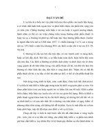
Craniopharyngioma
-
Adamantinomatous craniopharyngiomas :
+ Adamantinomatous craniopharyngiomas are far more common than
papillary
+ Predominantly in children with a second
-
Papillary craniopharyngiomas : identifed most commonly between the ages of
40 and 60 years
MRI
•
cysts
–
–
•
T1: iso- to hyperintense to grey matter
T2: variable but ~80% are mostly or partly T2 hyperintense
solid component
–
–
T1 C+ (Gd): vivid enhancement
T2: variable or mixed