Cell Structure and Function Functi
Bạn đang xem bản rút gọn của tài liệu. Xem và tải ngay bản đầy đủ của tài liệu tại đây (5.15 MB, 80 trang )
Cell Structure and
Cell Structure and
Function
Function
Chapter Outline
Chapter Outline
Cell theory
Cell theory
Properties common to all cells
Properties common to all cells
Cell size and shape –
Cell size and shape –
why are cells so small?
why are cells so small?
Prokaryotic cells
Prokaryotic cells
Eukaryotic cells
Eukaryotic cells
Organelles and structure in all eukaryotic cell
Organelles and structure in all eukaryotic cell
Organelles in plant cells but not animal
Organelles in plant cells but not animal
Cell junctions
Cell junctions
History of Cell Theory
History of Cell Theory
mid 1600s – Anton van Leeuwenhoek
mid 1600s – Anton van Leeuwenhoek
Improved microscope, observed many living cells
Improved microscope, observed many living cells
mid 1600s – Robert Hooke
mid 1600s – Robert Hooke
Observed many cells including cork cells
Observed many cells including cork cells
1850 – Rudolf Virchow
1850 – Rudolf Virchow
Proposed that all cells come from existing cells
Proposed that all cells come from existing cells
Cell Theory
Cell Theory
1.
1.
All organisms consist of 1 or more
All organisms consist of 1 or more
cells.
cells.
2.
2.
Cell is the smallest unit of life.
Cell is the smallest unit of life.
3.
3.
All cells come from pre-existing
All cells come from pre-existing
cells.
cells.
Observing Cells
Observing Cells
(4.1)
(4.1)
Light microscope
Light microscope
Can observe living cells in true color
Can observe living cells in true color
Magnification of up to ~1000x
Magnification of up to ~1000x
Resolution ~ 0.2 microns – 0.5 microns
Resolution ~ 0.2 microns – 0.5 microns
Observing Cells
Observing Cells
(4.1)
(4.1)
Electron Microscopes
Electron Microscopes
Preparation needed kills the cells
Preparation needed kills the cells
Images are black and white – may be colorized
Images are black and white – may be colorized
Magnifcation up to ~100,000
Magnifcation up to ~100,000
•
Transmission electron microscope (TEM)
Transmission electron microscope (TEM)
2-D image
2-D image
•
Scanning electron microscope (SEM)
Scanning electron microscope (SEM)
3-D image
3-D image
SEM
TEM
Cell Structure
Cell Structure
All Cells have:
All Cells have:
an outermost plasma membrane
an outermost plasma membrane
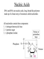
genetic material in the form of DNA
genetic material in the form of DNA
cytoplasm with ribosomes
cytoplasm with ribosomes
1. Plasma Membrane
1. Plasma Membrane
•
All membranes are phospholipid
All membranes are phospholipid
bilayers with embedded proteins
bilayers with embedded proteins
•
The outer plasma membrane
The outer plasma membrane
isolates cell contents
isolates cell contents
controls what gets in and out of the cell
controls what gets in and out of the cell
receives signals
receives signals
2. Genetic material in the
2. Genetic material in the
form of DNA
form of DNA
Prokaryotes – no membrane
Prokaryotes – no membrane
around the DNA
around the DNA
Eukaryotes – DNA is within a
Eukaryotes – DNA is within a
membrane
membrane
3. Cytoplasm with ribosomes
3. Cytoplasm with ribosomes
Cytoplasm – fluid area inside outer
Cytoplasm – fluid area inside outer
plasma membrane and outside
plasma membrane and outside
DNA region
DNA region
Ribosomes – make proteins
Ribosomes – make proteins
Cell Structure
Cell Structure
All Cells have:
All Cells have:
an outermost plasma membrane
an outermost plasma membrane
genetic material in the form of DNA
genetic material in the form of DNA
cytoplasm with ribosomes
cytoplasm with ribosomes
Why Are Cells So Small?
Why Are Cells So Small?
(4.2)
(4.2)
Cells need sufficient surface area to allow adequate transport of nutrients in and wastes out.
Cells need sufficient surface area to allow adequate transport of nutrients in and wastes out.
As cell volume increases, so does the need for the transporting of nutrients and wastes.
As cell volume increases, so does the need for the transporting of nutrients and wastes.
Why Are Cells So Small?
Why Are Cells So Small?
However, as cell volume increases the surface area of the cell does not expand as quickly.
However, as cell volume increases the surface area of the cell does not expand as quickly.
If the cell’s volume gets too large it cannot transport enough
If the cell’s volume gets too large it cannot transport enough
wastes out or nutrients in.
wastes out or nutrients in.
Thus, surface area limits cell volume/size.
Thus, surface area limits cell volume/size.
Why Are Cells So Small?
Why Are Cells So Small?
Strategies for increasing surface
Strategies for increasing surface
area, so cell can be larger:
area, so cell can be larger:
“
“
Frilly” edged…….
Frilly” edged…….
Long and narrow…
Long and narrow…
Round cells will always be small.
Round cells will always be small.
Prokaryotic Cell Structure
Prokaryotic Cell Structure
Prokaryotic Cells are smaller and
Prokaryotic Cells are smaller and
simpler in structure than eukaryotic
simpler in structure than eukaryotic
cells.
cells.
Typical prokaryotic cell is __________
Typical prokaryotic cell is __________
Prokaryotic cells do NOT have:
Prokaryotic cells do NOT have:
•
Nucleus
Nucleus
•
Membrane bound organelles
Membrane bound organelles
Prokaryotic Cell Structure
Prokaryotic Cell Structure
Structures
Structures
Plasma membrane
Plasma membrane
Cell wall
Cell wall
Cytoplasm with ribosomes
Cytoplasm with ribosomes
Nucleoid
Nucleoid
Capsule*
Capsule*
Flagella* and pili*
Flagella* and pili*
*present in some, but not all prokaryotic cells
*present in some, but not all prokaryotic cells
Prokaryotic Cell
Prokaryotic Cell
TEM Prokaryotic Cell
Eukaryotic Cells
Eukaryotic Cells
Structures in all eukaryotic cells
Structures in all eukaryotic cells
Nucleus
Nucleus
Ribosomes
Ribosomes
Endomembrane System
Endomembrane System
•
Endoplasmic reticulum – smooth and rough
Endoplasmic reticulum – smooth and rough
•
Golgi apparatus
Golgi apparatus
•
Vesicles
Vesicles
Mitochondria
Mitochondria
Cytoskeleton
Cytoskeleton
CYTOSKELETON
MITOCHONDRION
CENTRIOLES
LYSOSOME
GOLGI BODY
SMOOTH ER
ROUGH ER
RIBOSOMES
NUCLEUS
PLASMA
MEMBRANE
Fig. 4-15b, p.59
VESICLE
CYTOPLASM
Nucleus
Nucleus
(4.5)
(4.5)
Function
Function
– isolates the cell’s genetic material, DNA
– isolates the cell’s genetic material, DNA
DNA directs/controls the activities of the cell
DNA directs/controls the activities of the cell
•
DNA determines which types of RNA are made
DNA determines which types of RNA are made
•
The RNA leaves the nucleus and directs the synthesis of
The RNA leaves the nucleus and directs the synthesis of
proteins in the cytoplasm at a ______________
proteins in the cytoplasm at a ______________
Nucleus
Nucleus
Structure
Structure
Nuclear envelope
Nuclear envelope
•
Two Phospholipid bilayers with protein
Two Phospholipid bilayers with protein
lined pores
lined pores
Each pore is a ring of 8 proteins with an
Each pore is a ring of 8 proteins with an
opening in the center of the ring
opening in the center of the ring
Nucleoplasm – fluid of the nucleus
Nucleoplasm – fluid of the nucleus
Nuclear pore bilayer facing cytoplasm Nuclear envelope
bilayer facing
nucleoplasm
Fig. 4-17, p.61