Primary end point (six months) results of the ranibizumab for edema of the macula in diabetes (READ 2) study
Bạn đang xem bản rút gọn của tài liệu. Xem và tải ngay bản đầy đủ của tài liệu tại đây (265.67 KB, 8 trang )
Primary End Point (Six Months) Results of
the Ranibizumab for Edema of the mAcula
in Diabetes (READ-2) Study
Quan Dong Nguyen, MD, MSc,1 Syed Mahmood Shah, MBBS,1,2 Jeffery S. Heier, MD,3 Diana V. Do, MD,1
Jennifer Lim, MD,4 David Boyer, MD,5 Prema Abraham, MD,6 Peter A. Campochiaro, MD,1 for the READ-2
Study Group*
Objectives: To compare ranibizumab with focal/grid laser or a combination of both in diabetic macular
edema (DME).
Design: Prospective, randomized, interventional, multicenter clinical trial.
Participants: A total of 126 patients with DME.
Methods: Subjects were randomized 1:1:1 to receive 0.5 mg of ranibizumab at baseline and months 1, 3, and
5 (group 1, 42 patients), focal/grid laser photocoagulation at baseline and month 3 if needed (group 2, 42 patients),
or a combination of 0.5 mg of ranibizumab and focal/grid laser at baseline and month 3 (group 3, 42 patients).
Main Outcome Measures: The primary end point was the change from baseline in best-corrected visual
acuity (BCVA) at month 6.
Results: At month 6, the mean gain in BCVA was significantly greater in group 1 (ϩ7.24 letters, P ϭ 0.01,
analysis of variance) compared with group 2 (Ϫ0.43 letters), and group 3 (ϩ3.80 letters) was not statistically
different from groups 1 or 2. For patients with data available at 6 months, improvement of 3 lines or more
occurred in 8 of 37 (22%) in group 1 compared with 0 of 38 (0%) in group 2 (P ϭ 0.002, Fisher exact test) and
3 of 40 (8%) in group 3. Excess foveal thickness was reduced by 50%, 33%, and 45% in groups 1, 2, and 3,
respectively.
Conclusions: During a span of 6 months, ranibizumab injections by the current protocol had a significantly
better visual outcome than focal/grid laser treatment in patients with DME.
Financial Disclosure(s): Proprietary or commercial disclosure may be found after the references.
Ophthalmology 2009;116:2175–2181 © 2009 by the American Academy of Ophthalmology.
*READ-2 Investigators and Team Members appear in Appendix 1 (available at ).
Diabetic retinopathy is the most prevalent cause of vision
loss in working-age individuals in developed countries.
Severe vision loss occurs because of tractional retinal detachments that complicate retinal neovascularization, but
the most common cause of moderate vision loss is diabetic
macular edema (DME). The pathogenesis of DME is not
completely understood, but hypoxia is a contributing
factor.1 Vascular endothelial growth factor (VEGF) is a
hypoxia-regulated gene, and VEGF levels are increased in
hypoxic or ischemic retina. Hyperglycemia also causes elevation of VEGF, and even before there is evidence of
ischemia, VEGF is elevated in diabetic retinas.2 Injection of
VEGF into mouse eyes causes breakdown of the inner
blood–retinal barrier,3 and sustained release of VEGF in the
eyes of monkeys causes macular edema.4 This combination
of observations in patients and animal models led to the
hypothesis that VEGF plays an important role in the pathogenesis of DME.
An orally active nonselective blocker of VEGF receptors
was found to significantly reduce DME, which recurred
when the drug was stopped, providing the first suggestion
© 2009 by the American Academy of Ophthalmology
Published by Elsevier Inc.
that VEGF antagonists might provide benefit in patients
with DME.5 The development of selective antagonists of
VEGF allowed for more definitive testing of the hypothesis.
Ranibizumab is a Fab fragment of a humanized monoclonal
antibody that binds all isoforms of VEGF-A with high
affinity. In a small open-label study in patients with DME,
it was found that 4 intraocular injections of 0.5 mg of
ranibizumab over the span of 7 months resulted in a mean
reduction in excess foveal thickening of 85% and an average improvement in visual acuity of greater than 2 lines.6
This strongly implicated VEGF in the development of DME
and provided preliminary evidence that ranibizumab could
provide benefit, suggesting that larger controlled clinical
trials should be performed.
The Early Treatment of Diabetic Retinopathy Study
(ETDRS) has shown that focal/grid laser photocoagulation
can reduce the risk for moderate visual loss in eyes with
DME.7 Focal/grid laser therapy is currently standard care
and the gold standard with which new treatments are compared. We now report the results of a multicenter randomized trial in which a regimen of intraocular injections of
ISSN 0161-6420/09/$–see front matter
doi:10.1016/j.ophtha.2009.04.023
2175
Ophthalmology
Volume 116, Number 11, November 2009
ranibizumab was compared with focal/grid laser photocoagulation over the course of 6 months. A potential impediment to the use of ranibizumab is the chronic nature of
DME that could require long-term injections. A potential
problem with focal/grid laser is that severe edema may
make treatment more technically difficult and less effective,
because the edematous retina is less transparent so that
some laser energy is absorbed by the inner retina, which is
undesirable. Also, the lack of transparency makes it difficult
to assess when an appropriate end point is achieved. Injecting ranibizumab 1 week before focal/grid laser could reduce
the amount of thickening and improve the transparency
of the retina, thereby facilitating the laser treatment. Perhaps
the more precise focal/grid laser could lead to long-term
stability and eliminate the need for continued injections of
ranibizumab. Therefore, combination treatment with ranibizumab and focal/grid laser was also tested.
Materials and Methods
an intraocular injection of ranibizumab followed by focal/grid
laser treatment 1 week later. Month 6 was the primary end point of
the study. After month 6, patients were eligible to receive intraocular injections of ranibizumab no more than every 2 months or
focal/grid laser treatment no more than every 3 months if the
retreatment criterion of center subfield thickness of Ն250 m was
met. Safety evaluations, measurement of BCVA, eye examinations, and OCT scans were done at all study visits. Fluorescein
angiography was performed at baseline and 3 and 6 months.
Measurements of glycosylated hemoglobin were done at baseline
and 3 and 6 months. Hematology and blood chemistry tests were
performed at baseline and 6 months.
Administration of Study Drug
A lid speculum was inserted, and after topical anesthesia the
injection site was cleaned with 5% povidone iodine. Additional
topical anesthesia or subconjunctival injection of 2% lidocaine was
given, and 0.5 mg of ranibizumab was injected through the pars
plana into the vitreous cavity. The fundus was examined to ensure
retinal perfusion after the injection, and patients were observed for
1 hour or until intraocular pressure returned to normal. Patients
were called the day after each injection and were asked if they had
decreased vision, eye pain, unusual redness, or any new symptoms.
This is a phase II, randomized clinical trial conducted at 14 sites in
the United States through an investigator-initiated Investigational
New Drug granted by the Food and Drug Administration. The
study adhered to the guidelines of the Declaration of Helsinki, and
the protocol and consent form were approved by a local investigational review board for some sites and by the Western Institutional Review Board for others. Each subject provided written
informed consent. The study was monitored by an independent
data and safety monitoring committee. The study is registered at
www.clinicaltrials.gov under the identifier NCT00407381.
The ETDRS protocol8 with some modifications (50-m light gray
spots) was used for focal/grid laser treatment. Focal treatment was
administered to each leaking microaneurysm, and grid treatment
was placed in areas of thickened retina and areas of nonperfusion
between 500 and 3000 m from the center of the fovea.
Patient Eligibility and Exclusion Criteria
Data Collection and Management
Patients (aged Ն18 years) with type 1 or 2 diabetes and DME were
eligible if they had reduction in visual acuity between 20/40 and
20/320 and met the following criteria: (1) center subfield thickness
measured by optical coherence tomography (OCT) Ն250 m, (2)
glycosylated hemoglobin Ն6% within 12 months before randomization, (3) no potential contributing causes to reduced visual
acuity other than DME, (4) reasonable expectation that scatter
laser photocoagulation would not be required for the next 6
months. Patients were excluded if they had received focal/grid
laser treatment within 3 months, intraocular injection of steroid
within 3 months, or intraocular injection of a VEGF antagonist
within 2 months. If both eyes were eligible, the eye with the
greater center subfield thickness was entered.
The Retinal Imaging Research and Reading Center (RIRRC) at the
Wilmer Eye Institute served as the coordinating, data management,
and reading center. Personnel from the participating sites were
certified by RIRRC to perform digital fluorescein angiography and
OCT based on standardized protocols developed by the RIRRC.
Visual acuity examiners were required to be certified by EMMES
Corporation or by a multicenter Phase II/III clinical trial. Data
were collected online using a customized version of StudyTrax
(ScienceTRAX Inc., Jacksonville, FL), and training was provided
to each site for use of the online system. Files for fluorescein
angiography and OCT were uploaded to the RIRRC web site.
Coordinators at the RIRRC monitored the database weekly and
alerted sites of missed visits or failure to upload files and followed
up until these tasks were completed.
Study Protocol
Consenting patients were screened for the study with a medical
history, physical examination, measurement of best-corrected visual acuity (BCVA) by an experienced examiner using the ETDRS
protocol, a slit-lamp examination, measurement of intraocular
pressure, dilated funduscopic examination, an OCT evaluation, a
fluorescein angiogram, and laboratory tests on blood and urine.
Eligible patients were randomized 1:1:1 to injections of 0.5 mg of
ranibizumab alone (group 1), focal/grid laser alone (group 2), or
combination treatment consisting of injection of 0.5 mg of ranibizumab and focal/grid laser (group 3). Patients in group 1 received
an injection of ranibizumab at baseline and months 1, 3, and 5.
Patients in group 2 received focal/grid laser photocoagulation at
baseline and again at month 3 if center subfield thickness was
Ն250 m. At baseline and month 3, patients in group 3 received
2176
Focal/Grid Laser Photocoagulation
Optical Coherence Tomography
At each clinical site, OCT scans were performed by a certified
technician with a StratusOCT (Carl Zeiss Meditec, Dublin, CA)
using the fast macular scan protocol. This protocol consists of 6
line scans that are 6.0 mm long centered on fixation and spaced 30
degrees apart around the circumference of a circle. Each line
consists of 128 A-scan measurements. With each A-scan, the OCT
software measures the distance between the inner surface of the
retina and the anterior border of retinal pigmented epitheliumchoriocapillaris. The center subfield thickness, the average of 21
measurements along the central 1 mm of each of the 6 scans (total
of 126 measurements), was used as a measure of foveal thickness.
Readers at the RIRRC examined the images for each OCT file to
be sure that there were no artifacts such as misidentification of inner
Nguyen et al ⅐ READ-2 Six-Month Results
or outer surface of the retina. When artifacts were present, corrected
measurements were obtained using RetinaTomographer software
(version 1.1, RIRRC, Baltimore, MD). Macular volume throughout the entire 6-mm zone is calculated using extrapolated values
between the line scans. Excess foveal thickness was calculated by
subtracting the measured foveal thickness value from 212 m, the
upper limit of the normal range of center subfield thickness determined from measurements on a large population of subjects.9
Excess macular volume was determined by subtracting the upper
limit of the normal range of 6.47Ϯ0.37 mm3 from the measured
value.
Data Safety and Monitoring Committee
An independent Data Safety and Monitoring Committee made up
of 2 retina specialists with expertise in clinical trials monitored
adverse events and data at regular intervals.
Statistical Analyses
The primary outcome measure was the change in BCVA between
baseline and month 6. Secondary vision-related outcome measures
were the change in BCVA between baseline and month 3 and the
percentage of patients with 3 or more lines or 2 or more lines
improvement at month 6. Secondary anatomic outcomes were the
change in foveal thickness between baseline and month 6 and the
percentage of patients with elimination of 90% or 50% excess
foveal thickness.
Change from baseline in ETDRS visual acuity and change from
baseline in excess foveal thickness were compared across the 3
groups at months 3 and 6 using 1-way analysis of variance with
Bonferroni post hoc analysis. Outcome comparison between different time points within a group was done using a single-sample
t test. Secondary anatomic and functional outcomes were compared using a 2-sided Fisher exact test.
Results
Baseline Characteristics of the Study Groups
The baseline characteristics of the 126 patients who were randomized in the study are listed in Table 1. The 3 groups were balanced
with respect to mean BCVA, excess foveal thickness, and glycosylated hemoglobin. At baseline, 78 of the 126 patients had hyTable 1. Baseline Characteristics of Randomized Patients
Group 1: Group 2: Group 3:
Ranibizumab Laser Ranibizumab
(n ؍42) (n ؍42) (n ؍42)
Gender (% women)
Race (% Caucasian)
Age (mean yrs)
Mean BCVA (ETDRS letters
read)
Mean BCVA (Snellen
equivalent)
Mean excess foveal thickness
(m)
Hemoglobin A1C (mean mg/dl)
69
76
62
24.85
55
64
62
28.35
52
69
62
24.87
20/80
20/80ϩ3
20/80
198.75
227.67
262.52
7.39
7.77
7.59
BCVA ϭ best-corrected visual acuity; ETDRS ϭ Early Treatment Diabetic Retinopathy Study.
percholesterolemia that required treatment, and this was balanced
among the groups: 26 patients in group 1, 29 patients in group 2,
and 23 patients in group 3. There were no significant differences in
any baseline characteristics among the 3 groups.
Missing Values and Early Terminations
Data were available for 115 of 126 patients at the 6-month primary
end point. Nine patients exited the study before the primary end
point for the reasons listed in Table 2. The treatment randomization, time point of the patients’ last visit, and BCVA at baseline
and last visit are also shown. One patient in group 3 died of a
cerebral vascular accident 6 weeks after study entry and injection
of ranibizumab. Three patients were lost to follow-up, and 3
patients withdrew consent. One patient received scatter laser photocoagulation by an ophthalmologist not participating in the study
and by protocol had to exit the study. One patient in group 2 had
a substantial decrease in BCVA at the 3-month visit, which was
reported to be due to a combination of marked worsening of DME
and vitreous hemorrhage. The treating physician thought it was in
the patient’s best interest to exit the study and receive alternative
treatment. Four patients (1 patient in group 1, 2 patients in group
2, and 1 patient in group 3) withdrew from the study before
receiving any treatment. Two patients (both in group 1) missed the
window for the 6-month visit. All patients who did not have
6-month visit data, but had any posttreatment data at a time point
before month 6, had the last observation carried forward for
analysis of the primary outcome.
Primary Outcome Measure: Mean Change from
Baseline in Best-Corrected Visual Acuity
In group 2, the laser only group, 36 patients had a center subfield
thickness Ͼ250 m at 3 months; 32 patients received additional
focal/grid laser and 4 patients were judged to have maximum laser
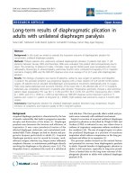
in the macula and were not treated. At month 6, the mean gain in
BCVA was 7.24 letters in group 1, which was significantly better
than the outcome in group 2, in which there was a mean loss of
0.43 letters (Fig 1; P ϭ 0.0001, analysis of variance with Bonferroni post hoc analysis). There was no statistically significant difference between group 1 and group 3, in which there was a mean
gain of 3.8 letters (P ϭ 0.08). Thus, with regard to the primary
outcome of mean change in BCVA at 6 months, the current
regimen of injecting 0.5 mg of ranibizumab for 2 months and then
2 additional injections 2 months apart was superior to focal/grid
laser treatment.
Secondary Vision-Related Outcome Measures
The mean change in BCVA between baseline and 3 months in
group 1 was 3.98 letters, significantly better than the mean loss of
1.48 letters in group 2 (P ϭ 0.01, analysis of variance with
Bonferroni post hoc analysis), but not significantly different from
the 1.93 letters gain (P ϭ 0.22) in group 3 (Fig 1). Of the 37
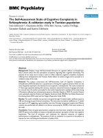
patents in group 1 for whom data were available at 6 months, 8
(22%) had an improvement of 3 or more lines of BCVA and 17
(46%) had an improvement of 2 or more lines (Fig 2A). None of
the 38 patients with only focal/grid laser therapy in group 3
improved by 3 or more lines and 2 patients (5%) improved by 2
more lines, both significantly less than in the ranibizumab group.
Three of 40 patients (8%) in group 2 who had combined treatment
improved by 3 or more lines, and 12 patients (30%) improved by
2 or more lines.
2177
Volume 116, Number 11, November 2009
Ophthalmology
Table 2. Reasons for Withdrawals from Study or Missed Visit at Primary End Point
Discontinued and Excluded from Analysis (n ؍5)
Group
Group
Group
Group
Group
Group
1
2
2
3
3
Last Visit
Reason for Discontinuation
Baseline
Baseline
Baseline
Day 7
Day 7
Lost to follow-up with multiple failed attempts to contact patient.
Lost to follow-up with multiple failed attempts to contact patient.
Patient refused laser treatment and withdrew consent.
Died of stroke 6 wks after enrollment and treatment.
Patient received scatter photocoagulation and by protocol had to be exited from the study.
Discontinued and Analyzed with Last Visit Carried Forward (n ؍4)
Group
Group
Group
Group
Group
1
1
2
2
Last Visit
Month
Month
Month
Month
1
2
3
3
⌬ VA from BL
Reason for Discontinuation
ϩ3
ϩ1
ϩ3
Ϫ16
Anxiety/panic attack during intraocular injection and refused additional injections.
Moved out of state and refused to return.
Lost to follow-up with multiple failed attempts to contact patient.
Patient had vitreous hemorrhage and worsening of DME and was discontinued from the study to
receive alternate treatment.
Missed Primary End Point with Last Visit Carried Forward (n ؍2)
Group
Last Visit
⌬ VA from BL
Reason for Missed Visit
Group 1
Group 1
Month 5
Month 5
ϩ5
ϩ6
Patient was hospitalized during the visit for an elective procedure for a preexisting condition.
Patient missed the visit because of scheduling error.
⌬ VA from BL ϭ change in visual acuity from baseline; DME ϭ diabetic macular edema.
Secondary Anatomic Outcome Measures
Patients in group 1 showed a reduction in mean excess foveal
thickness from 210.0 m at baseline to 103.7 m, approximately
a 50% reduction in macular edema (Fig 3). The improvement in
foveal thickness correlated well with improvement in mean visual
acuity. Patients in group 2 had a mean excess foveal thickness of
227.6 m at baseline that improved to 144.8 m at 6 months,
approximately a 36% reduction, which as noted above was not
accompanied by improvement in mean visual acuity. Patients in
group 3 had a mean excess foveal thickness of 262.5 m at
baseline that improved to 145.3 m at 6 months, a 45% reduction
in macular edema. The mean excess foveal thickness at 6 months
was 103.7 m in group 1, 144.8 m in group 2, and 145.3 m in
group 3. For those patients for whom data were available at 6
months, the percentage of patients who had elimination of 90% or
greater of excess foveal thickness was 24% in group 1, which was
greater than the 8% for groups 2 and 3 (Fig 4A). The percentage
of patients who had elimination of 50% or greater of excess foveal
thickness was not significantly different among the groups: 54%,
48%, and 32% for groups 1, 2, and 3, respectively (Fig 4B).
Adverse Events
Figure 1. Changes in visual acuity from baseline in patients with diabetic
macular edema treated with ranibizumab, focal/grid laser, or a combination
of both (RBZϩlaser). The mean (Ϯstandard error of the mean) change
from baseline in number of letters read at 4 m at 6 and 3 months was
significantly greater for ranibizumab alone versus focal/grid laser alone.
The combination group was not significantly different from the other 2
groups at either time point. *P ϭ 0.01; †P ϭ 0.0003 by 1-way analysis of
variance and Bonferroni post hoc analysis. ETDRS ϭ Early Treatment
Diabetic Retinopathy Study; RBZ ϭ ranibizumab.
2178
There was 1 serious adverse event; a patient in group 3 died of a
cerebral vascular accident 6 weeks after his first injection of
ranibizumab. The patient was at high risk for cerebral vascular
accident because of preexistent cardiovascular disease, and the
event was judged to be unrelated to ranibizumab because of the
long period between its occurrence and the prior injection. Mean
systolic/diastolic readings at baseline, month 3, and month 6 were
134.19/77.64, 131.56/74.15, and 134.75/75.31, respectively, in
group 1 compared with 138.33/80.69, 135.95/79.58, and 139.97/
77.56, respectively, in group 2 and 142.67/80.00, 139.49/78.82,
and 136.93/76.00, respectively, in group 3. The small differences
among groups and at different time points within groups were not
statistically significant. Ocular adverse events included vitreous
hemorrhages in 8 patients (1 patient in group 1, 4 patients in group
2, and 3 patients in group 3). One patient in group 2 had substantial
worsening of macular edema and mild vitreous hemorrhage at the
3-month visit, and the investigator thought it was in the patient’s
best interest to exit the study and receive alternative treatment
(Table 2). The other patients were thought to have mild vitreous
hemorrhages that according to funduscopic examinations were
noted to have cleared by the 6-month visit. This information and
comparison of BCVA measurements before the event with those at
Nguyen et al ⅐ READ-2 Six-Month Results
Figure 2. Percentage of patients with improvement in visual acuity between
baseline and 6 months of Ն3 lines or Ն2 lines. A, Eight of 37 patients (22%)
treated with ranibizumab for whom data were available at 6 months improved
Ն3 lines, which was significantly greater than 0% of patients treated with laser
alone (P ϭ 0.002, Fisher exact test). B, The percentage of patients with Ն2
lines of improvement was also significantly greater in the ranibizumab group
(46%, P ϭ 0.00004) or the combination group (30%, P ϭ 0.007) compared
with the focal/grid group (5%). VA ϭ visual acuity.
the 6-month visit suggest that vitreous hemorrhage had no impact
on the primary outcome variable for these patients (Table 3).
Discussion
We previously showed that VEGF plays an important role
in the pathogenesis of DME and provided preliminary evidence suggesting that intraocular injections of ranibizumab
provides benefit in patients with DME.6 Ten patients with
DME who received injections of 0.5 mg of ranibizumab at
baseline and months 1, 2, 4, and 6 showed a mean improvement in visual acuity of 12.3 letters read and a reduction in
mean excess foveal thickness from 503 m to 257 m,
constituting an elimination of 85% of edema. Although
those results are encouraging, small uncontrolled trials must
be viewed with caution and serve primarily to stimulate and
help design controlled clinical trials. In particular, it is
important to know how new treatments compare with treatments that constitute standard care, which for DME is
focal/grid laser photocoagulation. We now report the results
Figure 3. Mean change in excess foveal thickness between baseline
and 6 months. Normal 1-mm center subfield thickness (212 m) was
subtracted from measured center subfield thickness to give the excess
foveal thickness for each patient at each time point. The bars show the
mean (Ϯstandard error of the mean) excess foveal thickness at baseline
and 3 and 6 months for patients treated with ranibizumab alone (A),
focal/grid laser alone (B), and a combination of ranibizumab and
focal/grid laser (C). When compared with baseline, all 3 groups had
statistically significant reduction in excess foveal thickness at month 6
(P ϭ 0.0000002, P ϭ 0.003, and P ϭ 0.000002 for groups 1, 2, and 3,
respectively). EFTH ϭ excess foveal thickness; FTH ϭ foveal thickness; RBZ ϭ ranibizumab.
2179
Ophthalmology
Volume 116, Number 11, November 2009
and months 1, 3, and 5 showed a significantly better visual
outcome at 6 months (mean improvement of 7.24 letters)
compared with patients treated with focal/grid laser at baseline and again at month 3 if there was persistent edema (loss
of 0.43 letters). This confirms that intraocular injections of
ranibizumab provide benefit in patients with DME, which
over a time frame of 6 months is greater than that provided
by focal/grid laser therapy.
Secondary outcome measures also suggested superiority
of ranibizumab over focal/grid laser. Of 37 patients treated
with ranibizumab for whom data were available at 6
months, 8 (22%) had an improvement of 3 or more lines of
BCVA and 17 (46%) had an improvement of 2 or more
lines. This is significantly better than the focal/grid laser
group, in which no patients improved by 3 or more lines
(P ϭ 0.007, Fisher exact) and 2 patients (5%) improved by
2 or more lines (P ϭ 0.002, Fisher exact). The mean
reduction in excess foveal thickness was 50% in the ranibizumab group and 33% in the focal/grid laser group. Thus,
focal/grid laser treatment had some impact in that it caused
a modest reduction in edema, but that reduction was not
accompanied by improvement in visual acuity.
A recent study compared focal/grid laser treatment with
intraocular injections of 1 or 4 mg of preservative-free triamcinolone acetonide with repeat treatments every 4 months for
persistent or recurrent DME.10 At 4 months, mean improvement in visual acuity was significantly better in the 2 triamcinolone groups compared with the focal/grid laser group, but
at the 2-year primary end point the focal/grid laser group
showed a mean improvement of 1Ϯ17, which was significantly better than the triamcinolone groups (4 mg group,
Ϫ2Ϯ18; 1 mg group Ϫ3Ϯ22). Therefore, we cannot rule out
the possibility that with longer follow-up, the difference between our ranibizumab injection group and the focal/grid laser
group would disappear or even reverse. This should be determined by ongoing studies with longer primary end points.
A second objective of our study was to determine if
injections of ranibizumab 1 week before focal/grid laser
photocoagulation enhanced the effects of focal/grid treatment. The combined treatment group showed a mean improvement of 3.8 letters compared with the 0.43 reduction
Figure 4. The percentage of patients with elimination of Ն90% or Ն50%
excess foveal thickness at 6 months. A, More patients treated with ranibizumab alone (24%) had elimination of Ն90% of excess foveal thickness than
patients treated with focal/grid laser alone (8%) or a combination of ranibizumab and focal/grid laser (8%). B, There was no significant difference among
the groups in the percentage of patients with elimination of Ն50% of excess
foveal thickness. EFTH ϭ excess foveal thickness.
of a multicenter, randomized study comparing a similar
regimen of intraocular ranibizumab with focal/grid laser
photocoagulation. The results show that patients given an
intraocular injection of 0.5 mg of ranibizumab at baseline
Table 3. Ocular Adverse Events
Group
Event
Pre-Event
(ETDRS VA)
AE Visit
(ETDRS VA)
Group 1
Group 2
Group 2
Vitreous hemorrhage
Vitreous hemorrhage
Vitreous hemorrhage
Baseline (27)
Baseline (30)
Baseline (40)
Group 2
Vitreous hemorrhage and
worsening of DME
Vitreous hemorrhage
Vitreous hemorrhage
Vitreous hemorrhage
Vitreous hemorrhage
Vitreous hemorrhage
Baseline (16)
Day 7 (43)
Month 3 (21)
Month 2ϩ7 days
(not available)
Month 3 (0)
Baseline (2)
Month 3 (4)
Day 7 (27)
Month 3 (30)
Month 3 (32)
Month
Month
Month
Month
Month
Group
Group
Group
Group
Group
2
2
3
3
3
3 (4)
4ϩ7 days (2)
3 (35)
4ϩ3 wks (24)
6 (28)
Follow-up Visit
(ETDRS VA)
Month 6
ETDRS VA
Status
Month 1 (44)
Month 6 (35)
Month 3 (39)
51
35
35
Resolved
Resolved
Resolved
Discontinued
Discontinued
Month 6 (0)
Month 6 (0)
Month 6 (37)
Month 6 (29)
Month 9 (29)
0
27
37
29
28
NA
Resolved
Resolved
Resolved
Resolved
Resolved
AE ϭ adverse event; DME ϭ diabetic macular edema; ETDRS VA ϭ visual acuity obtained with protocol from the Early Treatment Diabetic Retinopathy
Study; NA ϭ not available.
2180
Nguyen et al ⅐ READ-2 Six-Month Results
in the laser alone group. This difference was not statistically
significant, and it appears that our sample size was insufficient to determine whether combined treatment is superior
to focal/grid therapy or to ranibizumab injections alone.
In conclusion, the results of the current study confirm our
previous study,6 but comparison of the 2 studies raises an
important issue. In the first study, patients received injections of 0.5 mg of ranibizumab every month ϫ 3 followed
by every other month ϫ 2 with a primary end point at 7
months, and patients showed elimination of 85% of edema.
In the current study, the regimen was less aggressive in that
patients received injections of 0.5 mg of ranibizumab ϫ 2
followed by every other month ϫ 2 with a primary end
point at 6 months, and only 50% of edema was resolved. An
ideal anatomic outcome is to eliminate Ն90% of edema, and
this was achieved in 24% of patients on the current regimen,
whereas 54% had resolution of Ն50% of edema. Thus, it
appears that a significant number of patients were undertreated with the regimen of ranibizumab injections used in
this study. Our results support the more aggressive regimen
of monthly injections of ranibizumab for 2 years that is
being used in the RISE and RIDE phase III trials sponsored
by Genentech (San Francisco, CA). The more aggressive
regimen, the 2-year primary end point, and the larger sample size will give a better indication of the maximal benefits
achievable with ranibizumab in patients with DME.
References
1. Nguyen QD, Shah SM, Van Anden E, et al. Supplemental
oxygen improves diabetic macular edema: a pilot study. Invest
Ophthalmol Vis Sci 2003;45:617–24.
2. Vinores SA, Youssri AI, Luna JD, et al. Upregulation of vascular
endothelial growth factor in ischemic and non-ischemic human
and experimental retinal disease. Histol Histopathol 1997;
12:99 –109.
3. Derevjanik NL, Vinores SA, Xiao WH, et al. Quantitative
assessment of the integrity of the blood-retinal barrier in mice.
Invest Ophthalmol Vis Sci 2002;43:2462–7.
4. Ozaki H, Hayashi H, Vinores SA, et al. Intravitreal sustained
release of VEGF causes retinal neovascularization in rabbits
and breakdown of the blood-retinal barrier in rabbits and
primates. Exp Eye Res 1997;64:505–17.
5. Campochiaro PA, C99-PKC412-003 Study Group. Reduction
of diabetic macular edema by oral administration of the
kinase inhibitor PKC412. Invest Ophthalmol Vis Sci 2004;
45:922–31.
6. Nguyen QD, Tatlipinar S, Shah SM, et al. Vascular endothelial growth factor is a critical stimulus for diabetic macular
edema. Am J Ophthalmol 2006;142:961–9.
7. Early Treatment Diabetic Retinopathy Study Research Group.
Photocoagulation for diabetic macular edema: Early Treatment Diabetic Retinopathy Study report number 1. Arch Ophthalmol 1985;103:1796 – 806.
8. Early Treatment Diabetic Retinopathy Study Research Group.
Treatment techniques and clinical guidelines for photocoagulation of diabetic macular edema: Early Treatment Diabetic
Retinopathy Study report number 2. Ophthalmology 1987;94:
761–74.
9. Chan A, Duker JS, Ko TH, et al. Normal macular thickness
measurements in healthy eyes using Stratus optical coherence
tomography. Arch Ophthalmol 2006;124:193– 8.
10. Diabetic Retinopathy Clinical Research Network. A randomized trial comparing intravitreal triamcinolone acetonide and
focal/grid photocoagulation for diabetic macular edema. Ophthalmology 2008;115:1447–59.
Footnotes and Financial Disclosures
Originally received: January 16, 2009.
Final revision: March 13, 2009.
Accepted: April 9, 2009.
Available online: August 22, 2009.
Manuscript no. 2009-69.
1
Wilmer Eye Institute, Johns Hopkins University School of Medicine,
Baltimore, Maryland.
2
Department of Medicine, Maryland General Hospital, University of
Maryland Medical System, Baltimore, Maryland.
3
Ophthalmic Consultants of Boston, Boston, Massachusetts.
4
Department of Ophthalmology, University of Illinois, Chicago.
5
Retina-Vitreous Associates Medical Group, Beverly Hills, California.
6
Black Hills Regional Eye Institute, Rapid City, South Dakota.
Quan Dong Nguyen, MD, MSc, and Syed Mahmood Shah, MBBS, contributed equally to the manuscript.
Presented at: the Annual Meeting of the American Academy of Ophthalmology, November 10, 2008, Atlanta, Georgia.
Sponsored by the Juvenile Diabetes Research Foundation and Genentech,
Inc.
QDN is a recipient of a K23 Career Development Award (EY 13552) from
the National Eye Institute. PAC is the George S. and Dolores Doré Eccles
Professor of Ophthalmology and Neuroscience.
Financial Disclosure(s):
The author(s) have made the following disclosure(s):
QDN and PAC have served as members of Expert Panels for Genentech,
Inc. without receiving an honorarium during the time of this study, but JHU
has recently negotiated a contract through which JHU receives compensation. QDN is a consultant for Bausch and Lomb and has research support
from Genentech, Inc., and Regeneron, Inc. PAC serves on the data and
safety monitoring committee for a phase III trial sponsored by Regeneron,
Inc., and has research support from Genentech, Alimera, and CoMentis for
diabetic macular edema trials. Diana Do receives research support from
Genentech. These activites are being managed by the Conflict of Interest
Committee of the Johns Hopkins University School of Medicine. JSH is a
consultant for Genentech, Alcon, Allergan, Bausch and Lomb, Eyemaginations, Fovea, Genzyme, Heidelburg, IScience, ISTA, Jerini, LPath,
NeoVista, Nodal Vision, Novagali, Novartis, Optherion, Oxigene, Paloma,
Pfizer, Regeneron, Resolvyx, Schering Plough, Scyfix, and VisionCare and
has received honoriaria from Genentech, Heidelberg, Jerini, NeoVista,
Optimedica, and Regeneron. JL has received honoriaria from Genentech.
DB is a consultant and has received honoraria from Genentech, Novartis,
Alcon, Allergan, and Pfizer. PA is a consultant for Genentech.
Correspondence:
Peter A. Campochiaro, MD, Maumenee 719, Johns Hopkins University
School of Medicine, 600 N. Wolfe Street, Baltimore, MD 21287-9277.
E-mail:
2181
Ophthalmology
Volume 116, Number 11, November 2009
Appendix 1. Investigators and Coordinators
of the READ-2 Study
A. Clinical Sites
1. Black Hills Regional Eye Institute:
Principal Investigator: Prema Abraham, MD
Coordinator: Buffi Green, Kristi Libermont, Honor
Evers
2. East Bay Retina Consultants
Principal Investigator: Eugene S. Lit, MD
Investigators: Daniel A. Brinton, MD, Scott S.
Lee, MD
Coordinator: Scotty Renslow
3. Eye Care Specialists
Principal Investigator: Erik F. Kruger, MD
Coordinator: Patty Yuhas, COA
4. Illinois Retina Associates
Principal Investigator: Jonathan S. Pollack, MD
Investigators: Joseph M. Civantos, MD
Coordinator: Barbara J. Ciscato
5. Johns Hopkins University/Wilmer Eye Institute
Principal Investigator: Diana V. Do, MD
Investigators: Peter Campochiaro, MD, Daniel
Finkelstein, MD, Morton F. Goldberg, MD, Julia
A. Haller, MD, James T. Handa, MD, Quan
Dong Nguyen, MD, MSc, Edward Quinlan, MD,
R. C. Andrew Symons, MBBS, PhD, FRANZCO,
Jennifer U. Sung, MD, Howard Ying, MD, PhD,
Ingrid Zimmer-Galler, MD
Coordinators: Lisa Azzaro, COT, Anita Baird,
COT, Lisa Greer, COT, MBA, Ovais Shaikh,
MD, COT, Jennifer Simmons, COT
6. Midwest Eye Institute
Principal Investigator: Thomas Ciulla, MD
Coordinator: Neelam Thukral
7. New England Retina Consultants
Principal Investigator: Bradley Foster, MD
Coordinator: Sharon Parker
8. Ophthalmic Consultants of Boston
Principal Investigator: Jeffrey S. Heier, MD
Investigators: Janet J. Chieh, MD, Tina S.
Cleary, MD, Gregory L. Fenton, MD, David S.
Liao, MD, Jackie K. Nguyen, MD, Trexler M.
Topping, MD, Torsten W. Wiegand, MD, Paul
A. Yates, MD
Coordinator: Lindsey Williams, Paul E. Daniel, Jr.
9. Retina Consultants of Arizona
Principal Investigator: Pravin U. Dugel, MD
Investigators: Jack Sipperley, MD, Donald W.
Park, MD, Judy Liu, MD, Derek Y. Kunimoto,
MD, Edward J. Quinlan, MD, Arthur Mollen,
DO, Jaime R. Gaitan, MD
Coordinator: Sarah G. Mobley, CCRC
10. Retina Consultants of Nevada - Las Vegas
Principal Investigator: Allen Thach, MD
Investigators: Roger Simon, MD, R. Jeffrey
Parker, MD, Rodney D. Hollifield, MD, Roy H.
2181.e1
B.
C.
D.
E.
F.
G.
Loo, MD, Meher Yepremyan, MD, Irene Voo,
MD, Jason C. Wickens, MD
Coordinators: Janet Seybert, Cassondra Major,
Mia Davis, Christy Browder, Melissa Rediker
11. Retina Institute of California
Principal Investigator: Thomas S. Chang, MD
Investigators: Adam Martidis, MD
Coordinator: Alexandra N. Tran
12. Retina-Vitreous Associates Medical Group
Principal Investigator: David Boyer, MD
Investigators: Roger L. Novack, MD, PhD,
Thomas G. Chu, MD, PhD, Firas M. Rahhal,
MD, Janet Jill Hopkins, MD, FRCSC, Homayoun
Tabandeh, MD, MS, FRCP, FRCS, FRCOphth,
Richard H. Roe, MD, MHS
Coordinator: Saba Mukarram, Tammy Gasparyan,
Janet Kurokouchi
13. University of New Mexico
Principal Investigator: Arup Das, MD, PhD
Investigators: Mark Schluter, MD
Coordinator: Sheila Nemeth, COMT
14. University of Southern California/Doheny Eye
Institute
Principal Investigator: Jennifer Lim, MD (year
1); Dean Eliot, MD (year 2)
Coordinator: Margaret Padilla
Steering Committee
Peter A. Campochiaro, MD
Jeffrey S. Heier, MD
Jennifer Lim, MD
Quan Dong Nguyen, MD, MSc, Chair
Data Safety and Monitoring Committee
Brian P. Conway, MD, University of Virginia
David Wilson, MD, Oregon Health and Science University, Casey Eye Institute
Reading Center
The Retinal Imaging Research and Reading Center at
Wilmer
Mohamed Ibrahim, MD
Yasir Sepah, MD
Izza Khan, MD
Roomasa Channa, MD
Syed Mahmood Shah, MBBS
Data Collection and Monitoring Center
Wilmer Eye Institute, Johns Hopkins University
Kashif Janjua, MD
Afsheen Khwaja, MD
John Putzke, PhD, MSPH
Syed Mahmood Shah, MBBS
Coordinating Center
Wilmer Eye Institute, Johns Hopkins University
Gulnar Hafiz, MD, MPH
Velma Pack, BA
Statistical Analyses
Bloomberg School of Public Health, Johns Hopkins
University
Aamir J. Khan, MD, PhD
Syed Mahmood Shah, MBBS