Morphology of the male reproductive system of the social wasp, Polistes versicolor versicolor, with phylogenetic implications pptx
Bạn đang xem bản rút gọn của tài liệu. Xem và tải ngay bản đầy đủ của tài liệu tại đây (8 MB, 10 trang )
Journal of Insect Science: Vol. 10 | Article 71 Araújo et al.
Journal of Insect Science | www.insectscience.org 1
Morphology of the male reproductive system of the social
wasp, Polistes versicolor versicolor, with phylogenetic
implications
Vinícius Albano Araújo
1a
, Jane Moreira
1b
and José Lino-Neto
2c*
1
Programa de Pós-Graduação em Entomologia, Department of Animal Biology, Federal University of Viçosa, Minas
Gerais, CEP, 36570-000, Brazil
2
Department of General Biology, Federal University of Viçosa, Minas Gerais, CEP, 36570-000, Brazil
Abstract
Variation in the morphology of the adult male reproductive system among different groups of
Hymenoptera offer characteristics that help studies of behavior and the evolutionary history of
this group. The objective of this study was to describe the adult male reproductive system of the
wasp Polistes versicolor versicolor Olivier (Vespidae: Polistini). The reproductive systems were
dissected, fixed and embedded for light microscopy. In P. v. versicolor, the reproductive system
includes a pair of testes, each one with three fusiform follicles. From each follicle emerges an
efferent duct that later join together, forming a deferent duct. The first half of the deferent duct is
enlarged and differentiated into a region specialized for sperm storage, the seminal vesicle. At the
post-vesicular region of each of the deferent ducts an accessory gland emerges. The seminal
vesicle and the accessory gland are covered with a capsule forming a vesicle-gland complex, also
observed in some species of North American Polistes. Sperm are released from testes in bundles,
which are disorganized inside seminal vesicles. In the testicular follicles, 95 spermatozoa were
observed per cyst on average.
Keywords: histology, mitotic cycles, spermatozoa,Vespidae, Polistini
Correspondence:
a
,
b
,
c
, *Corresponding author
Received: 22 February 2009, Accepted: 4 December 2009
Copyright : This is an open access paper. We use the Creative Commons Attribution 3.0 license that permits
unrestricted use, provided that the paper is properly attributed.
ISSN: 1536-2442 | Vol. 10, Number 71
Cite this paper as:
AraújoVA. Moreira J, Lino-Neto J. 2010. Morphology of the male reproductive system of the social wasp, Polistes
versicolor versicolor, with phylogenetic implications. Journal of Insect Science 10:71 available online: insectscience.org/10.71
Journal of Insect Science: Vol. 10 | Article 71 Araújo et al.
Journal of Insect Science | www.insectscience.org 2
Introduction
The Vespidae family is currently classified
into six subfamilies that are apparently
monophyletic: Euparagiinae, Masarinae,
Eumeninae, Stenogastrinae, Vespinae, and
Polistinae (Carpenter 1991). The molecular
phylogeny of Vespidae and the evolution of
sociality in wasps supported closer
phylogenetic relationships of Eumeninae to
Polistinae + Vespinae than to Stenogastrinae,
from which they concluded that social
behavior has independently evolved twice in
the wasp family Vespidae (Schmitz and
Moritz 1998). Analysis of the realigned
sequences also supports monophyly of
Vespidae, as well as monophyly of social
wasps, with the Stenogastrinae being more
closely related to Polistinae + Vespinae than
are Eumeninae (Carpenter 2003).
In the subfamily Polistinae, Polistes has about
200 species distributed throughout the world
mostly in the tropical region (Richards 1978;
Gauld and Hanson 1995). This genus has been
widely studied and is considered the “key
genus” for understanding evolution of the
social insects and social behavior among
wasps (Evans 1959; Schmitz and Moritz
1998; Carpenter 2003).
The reproductive system in males of
Hymenoptera demonstrates considerable
morphological differences among the species.
Such differences may be related to the
presence or absence and size or shape of
structures, as well as its position along the
reproductive tract (Dirks and Sternburg 1972;
Dallacqua and Cruz-Landim 2003; Ferreira et
al. 2004; Araújo et al. 2005a; Bushrow et al.
2006; Moreira et al. 2008). Sperm
morphology has revealed a considerable
number of features that differ among taxa
(Araújo et al. 2005b; Brito et al. 2005; Zama
et al. 2007; Lino-Neto et al. 2008a; Mancini et
al. 2006, 2008), indicating another possible
source of characters that may contribute to
understanding the systematics of these insects.
In insects, germ cell development of males
generally occur in compartments, called cysts.
These cysts are found inside tubules or
testicular follicles and are formed by clones of
germ cells surrounded by a layer of non-germ
epithelial cells (Baccetti and Bairati 1964).
During spermatogenesis, the spermatogonia
undergo mitotic divisions that generate a
constant number of cells in each cyst. After
two meiotic divisions, these cells become
spermatids (Lindsley and Tokuyasu 1980;
Oguma and Kurokawa 1984; Cruz-Landim
2001; Lino-Neto et al. 2008b). Each species
has a particular number of spermatids inside
the cyst, which is expressed as 2n, where “n”
is usually equal to 5, 6, 7 or 8.
The number of spermatids/spermatozoa per
cyst, which is determined by the number of
cell divisions, is constant for each species, but
may vary from species to species. Thus, this
number has been used as additional
information in the systematics of
Hymenoptera (Schiff et al. 2001; Zama et al.
2007; Lino-Neto et al. 2008a).
Variation in the morphology of the
reproductive system in males of Polistes was
observed between European (Bordas 1895)
and North American species (Dirks and
Sternburg 1972). In the present work, the
morphology of the male reproductive system,
the spermatozoa and the number of
spermatozoa per cyst were described for
Polistes versicolor versicolor Oliver
(Vespidae: Polistini). The aim is to contribute
to knowledge of the male reproductive
Journal of Insect Science: Vol. 10 | Article 71 Araújo et al.
Journal of Insect Science | www.insectscience.org 3
biology and also to reveal characters that may
be useful for future studies in taxonomy and
phylogeny of Hymenoptera, especially within
Aculeata.
Materials and Methods
Twelve adult males of P. v. versicolor were
obtained from nests sampled on a farm in the
municipality of Conceição do Castelo in the
state of Espírito Santo, Brazil.
Light microscopy
For the histological analysis, the reproductive
systems of six males were fixed for 12 h in
2.5% glutaraldehyde in 0.1 M sodium
cacodylate buffer, pH 7.2, and post-fixed in
1% osmium tetroxide. The material was
dehydrated in increasing alcohol
concentrations and embedded in Historesin
®
(GMA, Leica, www.leica-microsystems.com).
Semithin sections were stained with 1%
sodium toluidine borate and mounted in
Entelan
®
(Merck, www.merck.com). The
analysis and photographic records were made
with an Olympus BX60 (www.olympus.com)
microscope.
Suspensions of spermatozoa extracted from
the seminal vesicles of six males were spread
on clean glass microscope slides, which were
fixed for 20 minutes in a solution of 4% (w/v)
paraformaldehyde in 0.1 M sodium phosphate
buffer, pH 7.2. After drying at room
temperature, 100 randomly observed
spermatozoa were photographed in a
photomicroscope (Olympus, BX60) equipped
with phase contrast.
For nuclear measurements, six slides of
different males were stained for 15 minutes
with 0.2 µg/ml 4,6-diamino-2-phenylindole
(DAPI) in PBS, washed and mounted in 50%
sucrose. They were then examined using an
epifluorescence microscope (Olympus, BX60)
equipped with a BP 360-370 nm excitation
filter, and 100 nuclei were randomly
photographed. All the measures were obtained
with the Image Pro-Plus
®
software, version
4.5 (Media Cybernetics
Inc.,www.mediacy.com), and the lengths were
related to the total number of spermatozoa
analyzed.
Results
The male reproductive system of P. v.
versicolor consists of a pair of testes, each
with three fusiform follicles (Figures 1A-C).
The three follicles are covered by a single
capsule and are entirely filled with cysts
(Figure 1C). Each cyst has up to 95
spermatozoa on average (Figure 1D),
indicating that, at least six mitotic cycles
occur. During spermatogenesis, the cysts
begin migrating along the follicles while they
continue to differentiate. When the
spermatozoa are completely mature, the cysts
break open. The released bundles of
spermatozoa (spermatodesmata) remain
together, held by extracellular material that
surrounds the anterior portion of their heads
(Figure 1E). The spermatodesmata migrate to
the efferent duct (Figure 1E), pass into the
deferent duct, and are transferred to the
seminal vesicles, where they are disorganized
(Figures 1F-H).
The middle portion of the deferent duct is
enlarged and presents a modified epithelium
being transformed into a seminal vesicle
(Figure 1F). The accessory glands connect to
the beginning of the post-vesicular deferent
ducts. The seminal vesicle and the accessory
gland are surrounded by a single layer of
conjunctive tissue or capsule, forming the
vesicle-accessory gland complex (Figures 1A-
B).
Journal of Insect Science: Vol. 10 | Article 71 Araújo et al.
Journal of Insect Science | www.insectscience.org 4
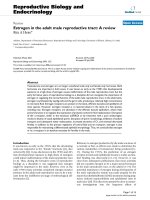
Figure 1. Photomicrograph of the anatomy (A-B) and histology (C-I) of the reproductive system of male Polistes vesicolor. A.
Reproductive system showing the testes (T), seminal vesicles (sv) and accessory glands (g) separated by the broken line,
deferent duct (dd) and the ejaculatory duct and B. showing the follicles (F) and the vesicle-gland complex involved by a single
capsule (circle of broken lines). C. Transverse section of the testes showing six testicular follicles (F) and the testicular capsule
(arrow). D. Inset: a cyst with 95 spermatozoa. E. Longitudinal section of a follicle (F), where the bundle of spermatozoa
(arrow) is observed being released from the follicles (F) to the efferent duct (ed). F. Transverse section of the seminal vesicle
showing the thick muscular layer (m), the epithelium (ep) and the lumen (L) with spermatozoa. G. Detail of the seminal
vesicle’s wall showing the epithelium comprising prismatic cells with spherical and basal nuclei (white arrow) and at the apical
third, some vesicular inclusions (circle of broken lines); the epithelium is separated from the external muscular layer (m) by a
thick basal membrane (black arrow). H. Longitudinal section of the accessory gland (g) and seminal vesicle (sv). I. Transverse
section of the accessory gland completely filled with secretions (black arrow). Note the epithelium with basal nuclei (white
arrow). Bars: A and B = 500 µm; C, F and I = 150 µm; D = 5 µm; E, G, H and J = 50 µm. High quality figures are available
online.
Journal of Insect Science: Vol. 10 | Article 71 Araújo et al.
Journal of Insect Science | www.insectscience.org 5
The epithelium of the seminal vesicle consists
of prismatic cells with spherical, basal nuclei.
In the apical third of these cells, some
vesicular inclusions (Figure 1G) can be
observed. In mature males, the vesicular
lumen is completely filled with spermatozoa.
The epithelium is separated from the external
muscular layer by a thick basal membrane
(Figures 1F-G).
The accessory glands are oval (Figures 1H-I),
and their epithelium consists of prismatic cells
with spherical and basal nuclei and large
secretory vesicles in the apical cell portion
(Figures 1I and 2A). The lumen is filled with
granular secretions (Figures 1H-I).
The deferent duct epithelium is formed by
cubical cells with a fairly evident striated
border (Figures 2A-B). The deferent duct
opens into the ejaculatory duct, whose
epithelium, consisting of cubical cells, is
completely covered by a thin cuticle (Figure
2C).
The spermatozoa of P. v. vesicolor measure
about 110 µm in length (Figure 2D), and the
nucleus is about 17 µm in length (Figure 2E).
Discussion
Variation in the morphology of the
reproductive system in males of Polistes was
observed between European (Bordas 1895)
and North American species (Dirks and
Sternburg 1972). In general, the reproductive
system of P. versicolor (species sampled in
South America) is morphologically similar to
that described for the North American species,
P. metricus (Say), P. exclamans and P.
annularis (Dirks and Sternburg 1972) as well
as the European species P. gallicus (Bordas
1895). However, the vesicle-accessory gland
complex observed in P. versicolor was
described only for the North American species
(Dirks and Sternburg 1972). These authors
suggest that the geographical isolation of
ancestral forms could have originated the
anatomical pattern found in North American
species. Our results point out that P.
versicolor and the species of North American
Polistes likely share a more recent ancestry
when compared to the European species P.
gallicus.
Although the reproductive systems in the
different Polistes species are very similar,
they differ markedly from those observed in
Ancistrocerus antilope (Eumeninae) (Bushrow
et al. 2006). In A. antilope, a single capsule
involving the testicles and the seminal
vesicles was reported – a pattern also
observed in several species of bees of the
subfamily Colletinae, Megachilinae, and
Apinae (Ferreira et al. 2004). Many species of
bees described by Ferreira et al. (2004) have
shown conspicuous variation in the
reproductive system of males, even within a
single family.
In P. v. versicolor, the presence of an average
of 95 spermatozoa per cyst indicates that there
are, at least, six mitotic cycles in the
spermatogonial proliferation phase. This
number was observed in other species of
Polistinae, such Mischocyttarus sp. (Brito et
al. 2005) and Agelaia vicina (personal
observation). In the family Sphecidae, six
mitotic cycles were also observed in
Sceliphrinae Sceliphron fistularium (Zama et
al. 2005). In Eumenes sp. (Eumeninae), there
are five mitotic cycles (personal observation).
In the wasps of the family Crabronidae, there
are also five mitotic cycles in
Pemphredoninae (Microstigmus) (Zama et al.
2007). There are four cycles in Crabroninae
Trypoxylon (Moreira et al. 2008) and in
Sphecinae Isodontia fuscipennis (Zama et al.
Journal of Insect Science: Vol. 10 | Article 71 Araújo et al.
Journal of Insect Science | www.insectscience.org 6
2007). Thus, at least in Hymenoptera, the
number of mitotic events during the
spermatogonial proliferation phase is
consistent within subfamilies and genera, but
not in taxa above these categories.
The spermatozoa bundle released from the
testes to the seminal vesicles, observed in P. v.
versicolor, is a phenomenon common to
Hymenoptera in general (Quicke et al. 1992;
Moreira et al. 2004; Lino-Neto et al. 2008a).
The observation of only mature cells at the
final stage of the spermiogenesis and
spermatozoa in the testes of adult P. v.
versicolor, as well as in other species of
Polistes (Dirks and Sternburg 1972) and in A.
antilope (Bushrow et al. 2006), indicates that
in these insects spermatogenesis begins in the
pupal stage and that they produce
spermatozoa only once. This has been
observed in ants (Ball and Vison 1984) and
bees (Dallacqua and Cruz-Landim 2003;
Araújo et al. 2005a) is therefore common
among the social Hymenoptera. In these
species, the process of testis degeneration
begins after the migration of the spermatozoa
to the seminal vesicles. The same process
possibly happens with social wasps. However,
it was not observed in P. v. versicolor because
the individuals were obtained from the nests
and were, therefore, not yet sexually mature.
The continuous production of spermatozoa is
commonly observed in species that mate
throughout the entire adult phase (Brockmann
1992; Garcia and Adis 1995; Coville et al.
2000; Buschini 2007; Moreira et al. 2008).
This phase is longer for such species than for
those that produce sperm only once.
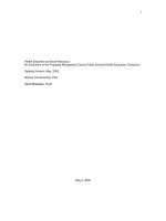
Figure 2. A-C. Histology of the male reproductive system of Polistes vesicolor. D-E. Photomicrograph of the spermatozoa
and of the nucleus stained with DAPI, respectively. A. Longitudinal section from the region of the insertions of the deferent
ducts (dd) into the accessory gland (g). Note the difference between the epithelium of the deferent duct (dd), showing basal
and spherical nuclei (arrow), and the striated border (arrowhead) and the epithelium of the accessory gland (g) with several
secretory vesicles at the apical portion (circle of broken lines). B. Transverse section of the deferent ducts (dd) showing the
striated border (arrow). C. Transverse section of the ejaculatory duct (ej), showing the cuticle (arrow). D. Photomicrograph
of the spermatozoon. E. Head region, stained with DAPI. Bars: A and D = 10 µm; B and C = 25 µm; E = 5 µm. High quality
figures are available online.
Journal of Insect Science: Vol. 10 | Article 71 Araújo et al.
Journal of Insect Science | www.insectscience.org 7
As in most insects, the ejaculatory duct of P.
versicolor is single, median and presents a
cuticle, demonstrating its ectodermic origin.
In A. antilope (Vespidae), the presence of two
ejaculatory ducts was verified. They begin at
the base of the accessory glands and later join
to form the common ejaculatory duct
(Bushrow et al. 2006). However, because
these authors did not mention the presence or
absence of a cuticle, it is not clear whether the
ducts consist entirely of an ejaculatory duct or
if they correspond to what we call the
posterior region of the deferent ducts,
followed by the ejaculatory duct.
In Hymenoptera, it has been observed that the
morphology of the spermatozoa varies even
among very closely related species. The
spermatozoon of P. v. versicolor, as in the
majority of the insects, is linear and slender.
However, such cells are spiralled in some
parasitic wasps, as in Chalcidoidea and
Platygastroidea (Lino-Neto et al. 2000; Lino-
Neto and Dolder 2001). The spermatozoa
length of P. versicolor (110 µm) is within the
very wide range in size observed for
Vespidae, which may vary from 13 to 577 µm
(Quicke et al. 1992; Bushrow et al. 2006;
Mancini et al. 2006; 2008).
This study supports the use of anatomical
differences of the male reproductive system as
a tool for the phylogenetic analysis among
families of Hymenoptera or higher taxa. The
number of spermatozoa per cyst may be used
only when compared within the levels
subfamily or genus. Nevertheless, the length
of the spermatozoa may be helpful in
taxonomic studies.
Acknowledgements
This research was supported by Fundação de
Amparo a Pesquisa de Minas Gerais
(FAPEMIG) Conselho Nacional de
Desenvolvimento Científico e Tecnológico
(CNPq) and Coordenação de
Aperfeiçoamento de Pessoal de Nível
Superior (Capes). We would like to thank
Mary Anne Heidi Dolder for critical reading
of the manuscript.
References
Araújo VA, Zama U, Neves CA, Dolder H,
Lino-Neto J. 2005a. Ultrastructural,
histological and histochemical characteristics
of the epithelial wall of the seminal vesicle of
mature males of Scaptotrigona xanthotricha
Moure (Hymenoptera, Apidae, Meliponini).
Brazilian Journal Morphological Science
22(4): 193-201.
Araújo VA, Zama U, Dolder H, Lino-Neto J.
2005b. Morphology and ultrastructure of the
spermatozoa of Scaptotrigona xanthotricha
Moure (Hymenoptera, Apidae, Meliponini).
Brazilian Journal Morphological Science
22(3): 137-141.
Baccetti B, Bairati A. 1964. Indagini
comparative sull’ultrastrutture delle cellule
germinale maschili in Dacus oleae Gmel ed in
Drosophila melanogaster Meig. (Ins.
Diptera)’. Redia 49: 1-29.
Ball DE, Vinson SB. 1984. Anatomy and
histology of the male reproductive system of
the fire ant, Sonelopsis invicta Buren
(Hymenoptera: Formicidae). International
Journal Morphology and Embriology 13(4):
283-294.
Bordas MS. 1895. Appareil genital male des
Hymenopteres. Annual Science and Nature
20: 101-184.
Journal of Insect Science: Vol. 10 | Article 71 Araújo et al.
Journal of Insect Science | www.insectscience.org 8
Brito P, Moreira J, Lino-Neto J. 2005.
Morphology of the male reproductive system
and sperm of Mischocyttarus sp.
(Hymenoptera: Vespidae: Polistinae).
Brazilian Journal of Morphological Sciences
22(Suppl.): 175-176.
Brockmann HJ. 1992. Male behavior,
courtship and nesting in Trypoxylon
(Trypargilum) monteverdeae (Hymenoptera:
Sphecidae). Journal of the Kansas
Entomological Society 65: 66-84.
Buschini MLT. 2007. Life-history and sex
allocation in Trypoxylon (syn. Trypargilum)
lactitarse (Hymenoptera; Crabronidae).
Journal Zoological Systematic and Evolution
Research 45: 206-213.
Bushrow ES, Fuller CL, Cowan DP, Byrd CA.
2006. Anatomy of the male reproductive
system and sperm morphology in the
caterpillar-hunting wasp Ancistrocerus
antilope (Hymenoptera,Vespidae).
Invertebrate Biology 125(4): 354-362.
Carpenter JM. 1991. Phylogenetic
relationships and the origin of social behavior
in the Vespidae. In: Ross KG, Matthews RW,
editors. The Social Biology of Wasps, pp. 7-
32. Cornell University Press.
Carpenter JM. 2003. On “Molecular
phylogeny of Vespidae (Hymenoptera) and
the evolution of sociality in wasps”. American
Museum Novitates 3389: 1-20.
Coville RE, Griswold C, Coville PL. 2000.
Observations on the nesting biology and
behavior of Trypoxylon (Trypargilum)
vagulum (Hymenoptera: Sphecidae) in Costa
Rica. Pan-Pac Entomology 76: 28-48.
Cruz-Landim C. 2001. Organization of the
cysts in bee (Hymenoptera, Apidae) testis:
number of spermatozoa per cyst. Iheringia
91:183-189.
Dallacqua RP, Cruz-Landim C. 2003.
Ultrastructure of the ducts of the reproductive
tract of males of Melipona bicolor Lepeletier
(Hymenoptera, Apidae, Meliponini).
Anatomy, Histology and Embryology 32: 276-
281.
Dirks TF, Sternburg JG. 1972. Male
reproductive system of three species of
Polistes (Hymenoptera: Vespidae).
International Journal of Insect Morphology
and Embryology 1(4): 315-320.
Evans ME. 1959. The evolution of social life
in wasps. Proceedings of the 10
th
International Congress of Entomology 449-
457.
Ferreira A, Abdalla FC, Kerr WE, Cruz-
Landim C. 2004. Comparative anatomy of the
male reproductive internal organs of 51
species of bees. Neotropical Entomology 33:
569-576.
Garcia MVB, Adis J. 1995. Comportamento
de nidificação de Trypoxylon (Trypargilum)
rogenhoferi Kohl (Hymenoptera, Sphecidae)
em uma floresta inundável de várzea na
Amazônia Central. Amazoniana 13: 259-282.
Gauld ID, Hanson PE. 1995. The evolution,
classification and identification of the
Hymenoptera. In: Hanson PE, Gauld ID,
editors. The Hymenoptera of the Costa Rica,
pp.138-156. Oxford University Press.
Lindsley DL, Tokuyasu KT. 1980.
Spermatogenesis. In: Ashbuner M, Wright
Journal of Insect Science: Vol. 10 | Article 71 Araújo et al.
Journal of Insect Science | www.insectscience.org 9
TR, editors. The genetics and biology of
Drosophila. pp. 225-295. Academic Press.
Lino-Neto J, Báo SN, Dolder H. 2000.
Structure and ultrastructure of the
spermatozoa of Trichogramma pretiosum
Riley and Trichogramma atopovirilia Oatman
and Platner (Hymenoptera:
Trichogrammatidae). Acta Zoologica
(Stockholm) 81: 205-211.
Lino-Neto J, Dolder H. 2001. Ultrastructural
characteristics of the spermatozoa of
Scelionidae (Hymenoptera; Platygastroidea)
with phylogenetic considerations. Zoologica
Scripta 30: 89-96.
Lino-Neto J, Dolder H, Mancini K, Mercati D,
Dallai R. 2008a. The short spermatodesm of
Arge pagana (Hymenoptera: symphyta).
Tissue & Cell 40: 185-193.
Lino-Neto J, Araújo VA, Dolder H. 2008b.
Inviability of the spermatids with little
cytoplasm in bees (Hymenoptera, Apidae).
Sociobiology 51(1): 163-172.
Mancini K, Lino-Neto J, Campos LAO,
Dolder H. 2006. Sperm ultrastructure of the
wasp Agelaia vicina (Hymenoptera,
Vespidae). Insectes Sociaux 53: 333-338.
Mancini K, Lino-Neto J, Dolder H, Dallai R.
2008. Sperm ultrastructure of the European
hornet Vespa crabro (Linnaeus, 1758)
(Hymenoptera: Vespidae). Arthropod
Structure & Development 38: 54-59.
Moreira J, Zama U, Lino-Neto J. 2004.
Release, behavior and phylogenetic
significance of spermatozoa in bundles in the
seminal vesicle during sexual maturation in
Aculeata (Hymenoptera). Brazilian Journal of
Morphological Science 21(4): 185-189.
Moreira PA, Araújo VA, Zama U, Lino-Neto
J. 2008. Morphology of male reproductive
system in three species of Trypoxylon
(Trypargilum) Richards (Hymenoptera:
Crabronidae). Neotropical Entomology 37(4):
429-435.
Oguma Y, Kurokawa H. 1984. The least cell
number of first spermatocytes per cyst found
in Drosophila kanekoi. Japanese Journal of
Genetics 59: 263-265.
Quicke DLJ, Ingram SN, Baillie HS, Gaitens
PV. 1992. Sperm structure and ultrasctructure
in the Hymenoptera (Insecta). Zoological
Scripta 21: 381-402.
Richards OW. 1978. The social wasps of the
Americas, excluding the Vespinae, pp. 436-
524. London, British Museum (Natural
History).
Schiff N, Flemming AJ, Quicke DLJ. 2001.
Spermatodesmata of the sawflies
(Hymenoptera: symphyta): evidence for
multiple increases in sperm bundle size.
Journal of Hymenoptera Research 10: 119-
125.
Schmitz J, Moritz RFA. 1998. Molecular
phylogeny of Vespidae (Hymenoptera) and
the evolution of sociality in wasps. Molecular
Phylogenetics and Evolution 9: 183-191.
Zama U, Brito P, Lino-Neto J, Campos LAO,
Dolder H, Báo SN. 2005. The sperm
morphology of mud dauber Sceliphron
fistularium Dahlbom (Hymenoptera: Apoidea:
Sphecidae), as an indicative of bees relation.
Journal of Submicroscopic Cytology and
Pathology 37(3-4): 91-99.
Zama U, Moreira JCS, Báo SN, Campos
Journal of Insect Science: Vol. 10 | Article 71 Araújo et al.
Journal of Insect Science | www.insectscience.org 10
LAO, Dolder H, Lino-Neto J. 2007.
Morphology of testicular and post-testicular
spermatozoa in Microstigmus arlei Richards,
1972 and M. nigrophthalmus Melo, 1992
(Hymenoptera: Apoidea: Pemphredoninae)
with phylogenetic consideration. Arthropod
Structure & Development 36: 304-316.