Báo cáo khoa học: Solution and membrane-bound chaperone activity of the diphtheria toxin translocation domain towards the catalytic domain" doc
Bạn đang xem bản rút gọn của tài liệu. Xem và tải ngay bản đầy đủ của tài liệu tại đây (347.14 KB, 10 trang )
Solution and membrane-bound chaperone activity of the
diphtheria toxin translocation domain towards the
catalytic domain
Anne Chassaing
1
, Sylvain Pichard
1
, Anne Araye-Guet
1
, Julien Barbier
1
, Vincent Forge
2
and
Daniel Gillet
1
1 Commissariat a
`
l’Energie Atomique (CEA), Institut de Biologie et Technologies de Saclay (iBiTecS), Service d’Inge
´
nierie Mole
´
culaire des
Prote
´
ines (SIMOPRO), Gif sur Yvette, France
2 Commissariat a
`
l’Energie Atomique (CEA), Institut de Recherche en Technologies et Sciences pour le Vivant (IRTSV), Laboratoire de
Chimie Biologie des Me
´
taux (LCBM), Grenoble, France
Introduction
Diphtheria toxin is a protein secreted by Corynebacte-
rium diphtheriae as a single polypeptide chain of
58 kDa [1]. During cell intoxication, it is cleaved by
furin into two fragments, the A chain, corresponding
to the catalytic (C) domain, and the B chain, corre-
sponding to the translocation (T) and receptor-binding
domains. The C and T domains remain covalently
linked by a disulfide bond. Following binding to its
cell surface receptor, diphtheria toxin is internalized
through the clathrin-coated pathway. The acidic pH in
the endosome triggers a conformational change lead-
ing to the insertion of the toxin in the membrane.
The C domain is then translocated across the en-
dosomal membrane into the cytosol. The C domain
Keywords
diphtheria toxin; membrane interaction;
molten globule; protein folding; translocation
Correspondence
D. Gillet, Commissariat a
`
l’Energie Atomique
(CEA), Institut de Biologie et Technologies
de Saclay (iBiTecS), Service d’Inge
´
nierie
Mole
´
culaire des Prote
´
ines (SIMOPRO),
F-91191 Gif sur Yvette, France
Fax: +33 1 69 08 90 71
Tel: +33 1 69 08 76 46
E-mail:
(Received 15 November 2010, revised 20
January 2011, accepted 15 February 2011)
doi:10.1111/j.1742-4658.2011.08053.x
During cell intoxication by diphtheria toxin, endosome acidification trig-
gers the translocation of the catalytic (C) domain into the cytoplasm. This
event is mediated by the translocation (T) domain of the toxin. Previous
work suggested that the T domain acts as a chaperone for the C domain
during membrane penetration of the toxin. Using partitioning experiments
with lipid vesicles, fluorescence spectroscopy, and a lipid vesicle leakage
assay, we characterized the dominant behavior of the T domain over the
C domain during the successive steps by which these domains interact with
a membrane upon acidification: partial unfolding in solution and during
membrane binding, and then structural rearrangement during penetration
into the membrane. To this end, we compared, for each domain, isolated
or linked together in a CT protein (the toxin lacking the receptor-binding
domain), each of these steps. The behavior of the T domain is marginally
modified by the presence or absence of the C domain, whereas that of the
C domain is greatly affected by the presence of the T domain. All of the
steps leading to membrane penetration of the C domain are triggered at
higher pH by the T domain, by 0.5–1.6 pH units. The T domain stabilizes
the partially folded states of the C domain corresponding to each step of
the process. The results unambiguously demonstrate that the T domain
acts as a specialized pH-dependent chaperone for the C domain. Interest-
ingly, this chaperone activity acts on very different states of the protein: in
solution, membrane-bound, and membrane-inserted.
Abbreviations
Br-PC, 1-palmitoyl-2-stearoyl(6,7)dibromo-sn-glycero-3-phosphocholine; EPA, phosphatidic acid; EPC,
L-a-phosphatidylcholine; k
max
, maximum
emission wavelength; LUV, large unilamellar vesicle; MG, molten globule; SRB, sulforhodamine B.
4516 FEBS Journal 278 (2011) 4516–4525 ª 2011 The Authors Journal compilation ª 2011 FEBS
ADP-ribosylates elongation factor 2, blocking protein
translation and leading to cell death.
The translocation process by which the C domain
crosses the membrane remains poorly characterized.
Several models have been proposed [1,2]. One suggests
that the C domain is translocated through a pore
formed by the B chain. Other studies have shown that
both the C and T domains are in contact with the
bilayer, and suggest that the hydrophilic surfaces of
the C domain are hidden from the hydrophobic core
of the membrane by its unfolding or ⁄ and by the
B chain [3], without translocating through the ion
channel formed by the T domain. Most studies have
focused on the pH-dependent conformational changes
of the isolated T or C domains [1–9], or the entire
toxin [10,11], and their propensity to penetrate into the
bilayer. It has been proposed that the T domain acts
as a chaperone for the C domain [12–15]. Indeed, the
T domain at acidic pH in solution or in membranes
was shown to bind proteins in a molten globule (MG)
state or hydrophobic peptides [14,15]. However, it was
concluded that the chaperone model had not been for-
merly demonstrated [15]. Also, only limited pH condi-
tions were explored instead of a continuous range of
pH values; the latter is indispensable for monitoring
all of the successive steps and structural transitions of
the toxin domains leading to membrane penetration.
In the present study, our aim was to determine step
by step how each of the C and T domains influences
the membrane interaction and the associated confor-
mational changes of the other domain. We compared
the pH sensitivities and the membrane interactions of
the C and T domains, isolated or within the protein
CT, in which the C domain is covalently linked to the
T domain. To this end, two CT proteins were pro-
duced, mutated at both Trp positions of either the
T domain or the C domain [11]. It was shown previ-
ously [11] that these mutations introduced into the
whole diphtheria toxin do not affect the native confor-
mation of the toxin or its ability to bind ApUp in its
catalytic site. In addition, low-pH conformational
changes and membrane insertion were only marginally
affected. Here, the conformational changes of the CT
proteins in solution and upon interaction with lipid
vesicles were measured as a function of pH, by fluores-
cence spectroscopy, as well as membrane binding and
penetration into the acyl chain regions of the lipid
bilayer.
The data showed that the T domain, by its own con-
formational changes, stabilizes the conformational
changes of the C domain that are responsible for its
membrane binding and penetration into the lipid
bilayer.
Results
Recombinant proteins
Five recombinant proteins were used in this study: C
and T, corresponding to the isolated C and T domains,
CT, corresponding to a truncated diphtheria toxin
lacking the R domain, and two mutant forms of CT in
which the Trp residues of either the T or C domain
were mutated to Phe [7]. These mutant CTs were pro-
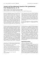
duced for Trp fluorescence experiments. CT contains
four Trp residues. Trp50 and Trp153 are located in the
C domain, in b-strand CB3 and just after strand CB7,
respectively, according to the crystal structure of diph-
theria toxin [16–18] (Fig. 1). Trp206 and Trp281 are
located in the T domain in helices TH1 and TH5,
respectively. Mutant CT
W50 ⁄ 153F
, in which the Trp
residues of the C domain were replaced by Phe,
allowed monitoring of the conformational changes of
the T domain within CT. Mutant CT
W206 ⁄ 281
, in which
the Trp residues of the T domain were replaced by
Phe, allowed monitoring of the conformational
changes of the C domain within CT.
Within the CTs, the C and T domains were folded
at basic pH and adopted their known MG state at
acidic pH
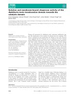
We studied, by CD spectropolarimetry in the far-UV
and near-UV, the secondary and tertiary structures of
the five recombinant proteins at pH 7.2. At this pH, the
toxin is considered to be in its native state [19]. The CD
spectra obtained for C in the far-UV, featuring low
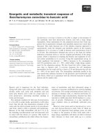
Fig. 1. Structure of CT (left) extracted from diphtheria toxin Protein
Data Bank file 1F0L (right). Red: C domain. Gray: T domain. Green:
receptor-binding domain. Blue: connecting loop. The Trp residues
are indicated in yellow.
A. Chassaing et al. Membrane interaction of diphtheria toxin
FEBS Journal 278 (2011) 4516–4525 ª 2011 The Authors Journal compilation ª 2011 FEBS 4517
signals at 190, 210 and 222 nm as compared with the
other proteins, indicated a mixed content of a-helices
and b-sheets (Fig. 2A, red curve). The far-UV CD spec-
tra of T (Fig. 2A, black curve) indicated a secondary
structure mainly composed of a-helices, also in agree-
ment with the crystal structure of the toxin. The spectra
of CT and its two mutants were identical, and showed a
mixed content of a and b structures compatible with a
contribution of the C and T domains.
In the near-UV, the CD spectra of C showed a small
positive signal between 280 and 300 nm, which can be
attributed to Trp constrained in a rigid environment.
Similarly, a double peak in the 260–270-nm region of
the spectra can be attributed to Phe side chains. The
spectra of T showed a strong peak at 292 nm, attrib-
uted to Trp, as described previously [4,5]. The spectra
of CT and its mutants exhibited both the signals of
Phe from C and of Trp from C and ⁄ or T.
The secondary and tertiary structures of the five
proteins were then studied at pH 3.5, at which both C
[13,14] and T [1,4,5] are known to adopt an MG state.
In the far-UV, the spectra of C (Fig. 2C, red curve)
appeared to be modified, with a loss of signal at
222 nm. This suggested some loss of a-helical content,
in agreement with previous observations [20]. The
spectra of T (Fig. 2C, black curve) were similar to that
recorded at pH 7.2 [4,5,21], with a slight loss of a-heli-
cal content. The spectra of the three CTs were mainly
unchanged, except for a small difference for the non-
mutated CT, probably because of some aggregation.
In the near-UV, the signals found at pH 3.5 were
greatly reduced (Fig. 2D). This indicated a release of
the tertiary constraints on the aromatic residues of the
proteins.
Altogether, the data suggested that all five recombi-
nant proteins were folded at pH 7.2 and exhibited a
native-like structure. At acidic pH, the loss of tertiary
structure signals in the near-UV region of the CD
spectra together with the mainly unchanged secondary
structure signals in the far-UV confirmed that C [13]
and T [4,5,21,22] adopted an MG conformation at
acidic pH. This was also the case within the CTs, and
showed that the mutations introduced did not alter
this behavior.
The T domain favored the acid-induced MG
transition of the C domain in solution when the
domains were covalently linked together
The maximum emission wavelength (k
max
) of the Trp
fluorescence was recorded to monitor the acid-induced
conformational changes of the recombinant proteins
(Fig. 3). All proteins showed a pH-dependent transi-
tion towards higher k
max
, indicating exposure of their
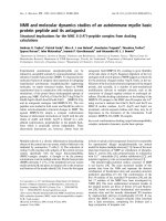
Fig. 2. Far-UV (A, C) and near-UV (B, D) CD spectra of C (red), T
(black), CT (light blue), CT
W206 ⁄ 281F
(orange), CT
W50 ⁄ 153F
(dark blue)
and C mixed with T (green) in solution at pH 7.2 or 3.5; h is the
molar ellipticity in degÆcm
2
Ædmol
)1
. For far-UV spectra, h is the
mean residue molar ellipticity. When the light blue curve corre-
sponding to CT cannot be seen, it is overlapped by the dark blue
curve corresponding to CT
W50 ⁄ 153F
.
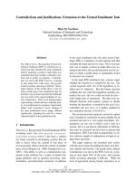
Fig. 3. Conformational changes of C, T, CT
W206 ⁄ 281F
and
CT
W50 ⁄ 153F
monitored by Trp fluorescence as a function of pH.
Closed red triangles: C. Closed black circles: T. Open pink triangles:
CT
W206 ⁄ 281F
. Open blue circles: CT
W50 ⁄ 153F
. The best fit for each
transition is represented (continuous lines). For T, the fitting para-
meters are: initial k
max
= 335.8 nm, final k
max
= 341.2 nm,
pK
1 ⁄ 2
= 5.3, and a Hill coefficient of 5. For CT
W50 ⁄ 153F
, the parame-
ters are: initial k
max
= 334 nm, final k
max
= 341.5 nm, pK
1 ⁄ 2
= 5.4,
and a Hill coefficient of 2.8. For C, the parameters are: initial
k
max
= 338 nm, final k
max
= 343 nm, pK
1 ⁄ 2
= 4.1, and a Hill coeffi-
cient of 3.6. For CT
W206 ⁄ 281F
, the parameters are: initial
k
max
= 333 nm, final k
max
= 340.5 nm, pK
1 ⁄ 2
= 5.05, and a Hill
coefficient of 1.3.
Membrane interaction of diphtheria toxin A. Chassaing et al.
4518 FEBS Journal 278 (2011) 4516–4525 ª 2011 The Authors Journal compilation ª 2011 FEBS
Trp residues to the solvent. The k
max
of C shifted from
338 to 343 nm between pH 4.8 and 3.9 (pK
1 ⁄ 2
4.2)
(Fig. 3, closed red triangles). The k
max
of T shifted
from 336 to 341 nm between pH 5.5 and 4.5
(pK
1 ⁄ 2
5.3) (Fig. 3, closed black circles). Interest-
ingly, the transition of the C domain within
CT
W206 ⁄ 281F
was profoundly modified as compared
with C (Fig. 3, open orange triangles). The fluores-
cence shifted from 333 to 341 nm between pH 5.9 and
3.9 (pK
1 ⁄ 2
5.1). This indicated that the Trp residues
of the C domain were in a less polar environment
within CT than when C was isolated. This could be
explained by the proximity of the two domains in CT.
Most of all, the transition of the C domain towards
the MG state occurred at a pH that was 0.9 units
higher than when it was isolated and was less coopera-
tive. In contrast, the transition of the T domain within
CT
W50 ⁄ 153F
was very similar to that of T (Fig. 3, open
blue circles). The pK
1 ⁄ 2
was nearly identical. The k
max
in the native state was lower by 2 nm, indicating that
the Trp residues were less exposed to the solvent,
probably because of the proximity of the C domain.
The fluorescence transitions monitored for the non-
mutated CT and for a mix of C and T were more
difficult to interpret (not shown). This was because of
the concomitant measurement of the fluorescence of
four Trp residues, each contributing differently in
terms of k
max
and fluorescence intensity [8]. In the
case of C and T mix, two separate transitions could
be seen, corresponding roughly to the respective
transition of each domain monitored separately. In
the case of CT, two overlapping transitions were
detected. The second transition, occurring at the low-
est pH values and probably corresponding to the
C domain, was shifted towards higher pH, as com-
pared with that of C.
Altogether, the results indicated that, within CT, the
native to MG transition of the T domain was similar
to that of the isolated T, whereas the native to MG
transition of the C domain was shifted to 0.9 pH units
higher than when it was isolated. Thus, the T domain
enabled the transition of the C domain to occur at
higher pH. This effect was possible only if the C and
T domains were covalently linked. Also, the results
strongly suggested that the two domains interacted
during the transitions.
The T domain favored the interaction of the
C domain with the membrane
We then studied the interaction of the recombinant
proteins with anionic large unilamellar vesicles (LUVs)
as a function of pH (Fig. 4). Binding was monitored
according to physical partition between the LUVs and
the solvent, by centrifugation and Trp fluorescence
measurements.
C and T bound to the LUVs from pH 6.0 to 4.5
and from pH 6.8 to 6.0, respectively (Fig. 4A), indicat-
ing preferential binding of T over C. CT
W50 ⁄ 153F
and
CT
W206 ⁄ 281F
bound to the LUVs from about pH 7.0 to
5.0. Thus, they started their binding transition at about
the same pH as T, but it occurred over two pH units
instead of one, showing reduced cooperativity. How-
ever, for these proteins, one cannot determine from
these data which domain bound first to the membrane:
T, C, or both.
These results indicated that isolated C bound the
membrane at about one pH unit lower than T. In con-
trast, the presence of the T domain covalently linked
with the C domain favored the interaction of C with
the membrane (at least through the binding of T), at a
pH higher than when it was isolated.
The T domain facilitated the insertion of the
C domain in the membrane
To better characterize the environment of the Trp resi-
dues of the C and T domains within CT during the
interaction with the membrane, we measured the
quenching of the Trp fluorescence of T, C and the CT
mutants by use of anionic LUVs containing brominat-
ed phospholipids as a function of pH (Fig. 4B). We
used 1-palmitoyl-2-stearoyl(6,7)dibromo-sn-glycero-3-
phosphocholine (Br-PC) lipids with bromine atoms
covalently bound at positions C6 and C7 of the oleoyl
chains.
The Trp fluorescence of C was slightly quenched
below pH 4.6, and by up to 12% at pH 3.8 (Fig. 4B,
closed red triangles). This may indicate weak pene-
tration of C in the hydrophobic layer of the mem-
brane. In contrast, the fluorescence of T was strongly
quenched as the pH decreased below 6, by up to 48%
at pH 3.8 (pK
1 ⁄ 2
4.9) (Fig. 4B, closed black circles).
This confirmed the results of similar experiments [8]
indicating deep penetration of T into the bilayer.
Very similar values were obtained for CT
W50 ⁄ 153F
(pK
1 ⁄ 2
4.7) (Fig. 4B, open blue circles), strongly sug-
gesting that the T domain reached the same depth into
the hydrophobic layer of the membrane, isolated or
within CT.
An intermediate situation was found with
CT
W206 ⁄ 281F
(Fig. 4B, open pink triangles). Significant
fluorescence quenching was observed below pH 5.4, by
up to 25% at pH 3.8. This is twice the effect measured
for C alone at the same pH. Unfortunately, no plateau
was detected at the pH investigated here. As a
A. Chassaing et al. Membrane interaction of diphtheria toxin
FEBS Journal 278 (2011) 4516–4525 ª 2011 The Authors Journal compilation ª 2011 FEBS 4519
consequence, in the case of the C domain (both C and
CT
W206 ⁄ 281F
), the pH dependences of the quenching
could not be fitted for estimation of the pK
1 ⁄ 2
and the
maximum of quenching. However, it is clear, in the
pH range we explored, that the C domain penetrated
deeper inside the bilayer when it was covalently linked
to the T domain, and that this transition occurred at
higher pH than when it was isolated.
The T domain favored the structural transitions
of the C domain during interaction with the
membrane
In order to investigate the structural transitions under-
gone by the C and T domains during interaction with
the membrane, we monitored the fluorescence of the
four proteins in the presence of anionic LUVs as a
function of pH. The k
max
of C shifted from 338 nm to
343 nm between pH 4.9 and 4.1, and then from 343 to
340 nm between pH 4.1 and 3.5 (Fig. 4C, closed red
triangles). These two successive transitions have been
observed previously [3]. The increase of the k
max
observed during the first transition could be attributed
to increased exposure of the Trp residues of C to the
aqueous buffer. Thus, this first transition could corre-
spond to a partial unfolding of C, as is the case for T
[4,5,8]. The second transition, indicating burial of the
Trp residues in an apolar environment, could corre-
spond to the penetration of C in the membrane [3], as
is the case for T [4,5,8].
T interacted with the LUVs according to the two-
step process described previously [4,5,8] (Fig. 4C,
closed black circles). The first transition was attributed
to the binding of T to the membrane and its unfolding
with exposure of its N-terminal Trp residues to the
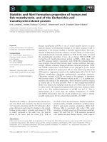
Fig. 4. (A) Partition of C (closed red triangles), T (closed black cir-
cles), CT
W206 ⁄ 281F
(open pink triangles) and CT
W50 ⁄ 153F
(open blue
circles) between the buffer and LUVs as a function of pH, studied
by ultracentrifugation. The best fit for each transition is represented
(continuous lines). For T, the fitting parameters are: pK
1 ⁄ 2
= 6.4
and a Hill coefficient of 3.2. For CT
W50 ⁄ 153F
, the parameters are:
pK
1 ⁄ 2
= 6.25 and a Hill coefficient of 2.0. For C, the parameters
are: pK
1 ⁄ 2
= 5.15 and a Hill coefficient of 2.7. For CT
W206 ⁄ 281F
, the
parameters are: pK
1 ⁄ 2
= 5.8 and a Hill coefficient of 1.9. (B)
Quenching of Trp fluorescence of C, T, CT
W206 ⁄ 281F
and CT
W50 ⁄ 153F
by LUVs containing Br-PC. The results are expressed as the relative
quenching efficiency as compared with the Trp fluorescence at
pH 7. The lower the value, the closer the Trp from the quencher. In
the case of T and CT
W50 ⁄ 153F
, the data could be fitted with Micha-
elis–Menten equations (continuous lines). For T, the fitting parame-
ters are: pK
1 ⁄ 2
= 4.9 and a final F ⁄ F
0
of 48%. For CT
W50 ⁄ 153F
, the
parameters are: pK
1 ⁄ 2
= 4.7 and a final F ⁄ F
0
of 49%. (C) Trp fluo-
rescence of C, T, CT
W206 ⁄ 281F
and CT
W50 ⁄ 153F
in the presence of
anionic LUVs as a function of pH. In the case of T and CT
W50 ⁄ 153F
,
the data could be fitted with the pK
1 ⁄ 2
values obtained from (A)
and (B) in order to estimate the k
max
of the various states of the
domain (continuous lines). For T, the initial k
max
is 335.5 nm, the
intermediate k
max
is 344.5 nm, and the final k
max
is 329.4 nm. For
CT
W50 ⁄ 153F
, the initial k
max
is 334 nm, the intermediate k
max
is
341 nm, and the final k
max
is 329 nm. (D) Permeabilization of anio-
nic LUVs by C, T and CT
W50 ⁄ 153F
. CT permeabilized LUVs as effi-
ciently as T and CT
W50 ⁄ 153F
.CT
W206 ⁄ 281F
permeabilized LUVs
slightly less efficiently (not shown).
Membrane interaction of diphtheria toxin A. Chassaing et al.
4520 FEBS Journal 278 (2011) 4516–4525 ª 2011 The Authors Journal compilation ª 2011 FEBS
buffer, and the second transition to penetration into
the bilayer.
The k
max
of CT
W50 ⁄ 153F
shifted from 334 to 340 nm
between pH 7.1 and 6.0, and then from 340 to 333 nm
between pH 6.0 and 4.3 (Fig. 4C, open blue circles),
thereby indicating two transitions very similar to those
of the isolated T (Fig. 4C, closed black circles).
Between these two transitions, the k
max
of CT
W50 ⁄ 153F
was 3 nm lower than that of T. The first transition
found for CT
W50 ⁄ 153F
correlated with membrane bind-
ing (Fig. 4A, open blue circles). Notably, this mem-
brane binding transition was less cooperative than that
of T (Fig. 4A, closed black circles). This may explain
the decreased k
max
found for CT
W50 ⁄ 153F
as compared
with T (Fig. 4C). Indeed, the first transition of the
T domain within CT
W50 ⁄ 153F
was not completed when
the second transition started. The second transition
correlated with the insertion in the membrane, which
was also monitored by fluorescence quenching
(Fig. 4B). The final k
max
was the same for T and
CT
W50 ⁄ 153F
, i.e. 329 nm. In both cases, the pH depen-
dence of k
max
could be fitted with the two values of
pK
1 ⁄ 2
obtained from the partition (Fig. 4A) and fluo-
rescence quenching (Fig. 4B) experiments (Fig. 4C,
continuous lines).
The k
max
of CT
W206 ⁄ 281F
shifted from 335 to 339 nm
between pH 6.4 and 5.0, and then from 339 to 336 nm
between pH 5.0 and 3.6 (Fig. 4B, open orange trian-
gles). Thus, although the C domain within CT fol-
lowed two transitions similar to those of the isolated
C, these transitions occurred at higher pH. This sug-
gested that the T domain favored the interaction of
the C domain with the membrane when C was cova-
lently linked to T. The k
max
of CT
W206 ⁄ 281F
was about
4 nm lower than that of C. Again, this suggested
proximity or contacts between the C and T domains
within CT, limiting exposure of the Trp residues to the
environment.
Overall, both domains underwent two structural
transitions upon binding and penetration into the
membrane. For T, the first transition corresponded to
binding and the second to membrane penetration, but
this is less obvious for C. Within CT, the presence
of the C domain did not affect the transitions of the
T domain, but the presence of the T domain favored
the transitions of the C domain at higher pH.
Anionic LUV permeabilization
Anionic LUV permeabilization was shown to be an
indicator of penetration of the T domain into the
membrane [4,5,23]. Whereas C did not permeabilize
LUVs significantly, CT permeabilized LUVs at least as
efficiently as T (Fig. 4D). This confirmed that the
T domain within CT was fully capable of penetrating
the lipid bilayer.
Discussion
Figure 5 summarizes the data collected in the present
study. Various methods were used to probe the inter-
actions of the C and T domains of diphtheria toxin
with the membrane, alone or covalently linked
together. Binding to the membrane was revealed
by centrifugation experiments (Figs 4A and 5, pink
arrows). Conformational changes of the C and T do-
mains were monitored by Trp fluorescence of CTs
mutated on the Trp of the C or T domains (Figs 4B
and 5, transitions C1, C2, and T1, T2). Penetration
into the fatty acid region of the bilayer was revealed
by quenching of the Trp fluorescence of the mutant
CTs by Br-PC (Figs 4C and 5, green arrows). Permea-
bilization of the membrane, which mainly coincided
with membrane penetration, was detected by fluores-
cent dye release from LUVs (Fig. 4D). On the basis of
all of the data, we describe the succession of steps
leading to membrane binding and membrane penetra-
tion of both domains of the protein. In addition, we
Fig. 5. Schematic representation of the successive steps followed
by C and CT when interacting with anionic LUVs, as a function of
pH. The binding and membrane-penetration transitions indicated as
pink and green arrows are from the curves of Fig. 4. The different
shapes named C1, C2, T1 and T2 symbolize the conformational
changes of the C and T domains associated with these transitions.
Binding (pink) data are from Fig. 4A. Penetration (green) data are
from Fig. 4B. Permeabilization data from Fig. 4D (not shown on this
scheme) mainly coincide with membrane penetration (green). Con-
formational changes of the protein domains observed by Trp fluo-
rescence are from Fig. 4C. The only difference found for T (not
shown in the scheme) as compared with CT is that the binding
transition is more cooperative and ends at pH 6.
A. Chassaing et al. Membrane interaction of diphtheria toxin
FEBS Journal 278 (2011) 4516–4525 ª 2011 The Authors Journal compilation ª 2011 FEBS 4521
show that the T domain behaves relatively indepen-
dently from the C domain, in solution (Fig. 3) and
during membrane interaction (Fig. 4), whereas the
C domain is highly influenced by the presence of the
T domain (Figs 4 and 5).
The T domain drives the successive steps by
which the C domain binds and penetrates the
membrane
From pH 7 to 5, CT binds to the membrane (Fig. 5).
The T domain is responsible for initiating binding,
because binding of T and CT starts at the same pH,
whereas binding of C starts at one pH unit lower.
Monitoring of the conformational changes C1 and T1
(Figs 4C and 5) associated with binding (Fig. 4A,B)
led to the same conclusion (see next section).
From pH 6 to 4 or below, both domains of CT pen-
etrate into the membrane (Figs 4C and 5, green
arrows). Again, the T domain leads the way for the
C domain. It is not influenced by the presence or
absence of the C domain, whereas the C domain is
clearly influenced by the presence of the T domain.
The T domain favors the conformational changes
adopted by the C domain during binding to, and
penetration into, the membrane
The structural behavior of the T domain interacting
with the membrane as a function of pH is quite similar
whether it is isolated or linked to C. The only differ-
ence found is that binding is less cooperative for CT
than for T (Fig. 4A). As a result, binding seems to
overlap both the unfolding of T (Fig. 5, structural
transition T1) and its rearrangement in the membrane
corresponding to penetration [4,8] (Fig. 5, structural
transition T2). However, in fact, a fraction of bound
molecules already starts to rearrange in the membrane
(T2) while a fraction of molecules have not fully
unfolded yet, owing to the decreased cooperativity of
the reaction.
In contrast, the structural behavior of the C domain
during interaction with the membrane is very different
when it is isolated or connected with T. When it is iso-
lated, its unfolding (Figs 4B and 5, structural transi-
tion C1) does not coincide with binding (Figs 4A and
5, pink arrow). This indicates that the C domain binds
first to the membrane without undergoing conforma-
tional change, and then unfolds in about the same pH
range as in solution (Fig. 3). Then, it progressively
penetrates the membrane to a shallow position
(Figs 4B and 5, green arrow), finishing unfolding
(Figs 4C and 5, transition C1) before a second rear-
rangement of its structure occurs (Figs 4C and 5, tran-
sition C2).
When the C domain is connected with the T
domain, the T domain clearly favors unfolding of the
C domain in solution (Fig. 3) and during binding to
the membrane (Fig. 4A,C and 5, structural transi-
tion C1 and pink arrow). Thus, the T domain favors
the interaction of the C domain with the membrane
because it stabilizes its partially unfolded state (Fig. 5,
C1). This strongly suggests that the C domain binds to
the membrane concomitantly with the T domain or at
a pH not more than 0.5 U lower than that driving the
binding of T. Then, the T domain helps the C domain
to penetrate into the hydrophobic core of the mem-
brane. During this step, the C domain finishes its
conformational change C1 (Figs 4C and 5), and then
undergoes conformational change C2 (Figs 4C and 5),
corresponding to deeper penetration into the acyl
chain layer of the membrane (Fig. 4B and 5, green
arrow), than in the absence of the T domain.
The T domain but not the C domain is
specialized to permeabilize the membrane
The T domain permeabilizes the membrane (Fig. 4D)
during the membrane-penetration step (Fig. 4B and 5,
green arrow). The deeper the T domain is inserted,
the stronger is the permeabilization. The results
clearly show that the T domain is absolutely required
for permeabilization, C alone being incapable of
doing so (Fig. 4D). Interestingly, the penetration of
the C domain in the membrane does not impair its
permeabilization by the T domain. This suggests that
the C domain does not plug the passageway formed
by the T domain in the bilayer. One cannot state,
however, whether or not this passageway is taken by
the C domain to cross the membrane. Nevertheless,
these results indicate that the T domain is specialized
to permeabilize the membrane but the C domain is
not, even though it is embedded in the bilayer. In
other words, the membrane is not destabilized by the
insertion of C.
The T domain acts as a chaperone for the
C domain
It has been proposed that the T domain acts as a
chaperone for the C domain, enabling its passage
through the membrane at acidic pH [12–15]. Indeed, T
at acidic pH in solution or in membranes was shown
to bind proteins in an MG state or hydrophobic pep-
tides [14,15]. However, it was concluded that the chap-
erone model had not been formerly demonstrated [15].
Membrane interaction of diphtheria toxin A. Chassaing et al.
4522 FEBS Journal 278 (2011) 4516–4525 ª 2011 The Authors Journal compilation ª 2011 FEBS
The present work demonstrates that the T domain in
its various pH-dependent conformations, in solution,
membrane-bound, and membrane-inserted, stabilizes
partially unfolded states of the C domain. In doing so,
the T domain favors membrane binding and mem-
brane penetration of the C domain. By definition, the
activity of a chaperone is the stabilization of a par-
tially folded (or unfolded) state of another protein.
Thus, we demonstrate that the T domain acts as a
chaperone for the C domain. A remarkable feature of
this chaperone activity is that it stabilizes at least three
different partially folded states of the C domain, each
corresponding to one of the successive steps of the ini-
tiation of translocation: conformational change in
solution, membrane binding, and membrane insertion.
How does the T domain exerts its chaperone activ-
ity on the C domain? The T domain adopts an MG
state displaying hydrophobic surfaces [4,21,22]. These
hydrophobic surfaces may offer an environment that
is propitious for the interaction with the hydrophobic
surfaces of the C domain, which are exposed only in
its MG state. Thus, the T domain in its MG state
must greatly displace the native to MG state equilib-
rium of the C domain in favor of the MG state. The
T domain favors the interaction of the C domain
with the membrane, because it brings the C domain
in its MG state into the vicinity of the bilayer, the
MG state of both domains being propitious for mem-
brane insertion and ⁄ or translocation [4,13,14,21]. The
membrane itself may also have a destabilizing effect
on the C domain: the interfacial pH is lower than in
the solvent and the hydrophobic acyl chains may
interact with hydrophobic regions of the protein.
Finally, the T domain imposes its rule on the C
domain because it is more sensitive to pH. Indeed, it
has an elaborate system for reacting to a wide range
of acidic pH values, starting just below pH 7, involv-
ing its six His residues [5].
It has been shown previously that, after transloca-
tion, the C domain and only the 63 N-terminal amino
acids of the T domain are present on the trans side of
the membrane [24,25]. The remaining 124 amino acids
of the T domain are left in the membrane. However, a
cytoplasmic chaperone, Hsp90, is involved in extrac-
tion of the C domain from the membrane and its
refolding [26]. The T domain seems to be no longer
needed for the last stages of translocation.
Our findings emphasize the importance of the physi-
cochemical properties that a protein should have in
order to interact with, and penetrate into, a mem-
brane. They should be taken into consideration to
evaluate or adapt the capacity of proteins to bind or
cross a membrane.
Experimental procedures
Recombinant proteins
Expression and purification of the recombinant T domain
containing mutation C201S (native diphtheria toxin number-
ing) has been described previously [21,23]. Two DNA
sequences coding for residues 1–380 of the native toxin (C and
T domains) and residues 1–193 (C domain) were prepared by
PCR and cloned into the pET-28a(+) vector (Novagen, Mad-
ison, WI, USA), using the NdeIandSalI restriction sites. The
two resulting plasmids, CTpET-28a(+) and CpET-28a(+),
encoded CT and C preceded by an N-terminal His tag
sequence. Cys186 in the C domain protein was mutated in
Ser. Mutations W50F and W153F, or W206F and W281F,
were introduced by PCR mutagenesis into plasmid CTpET-
28a(+). The sequences were checked by DNA sequencing.
Production and purification of recombinant C was per-
formed as described for T [21,23]. CT, CT
W50 ⁄ 153F
and
CT
W206 ⁄ 281F
were expressed at 37 °CinEscherichia coli
strain BL21(DE3) as inclusion bodies. The inclusion bodies
were solubilized in 8 m urea, 0.1 m Tris ⁄ HCl, and 0.1 mm
EDTA (pH 8), and the proteins were purified by immobi-
lized-nickel affinity chromatography. The proteins were
folded by dialysis against a 20 mm sodium phosphate buffer
at pH 8. The proteins were further purified on a Hi Load
Superdex 26 ⁄ 60 size exclusion column (GE Healthcare,
Orsay, France), and, finally, the buffer was exchanged with
NH
4
HCO
3
on a G25SF column before lyophilization and
storage at ) 20 °C.
Lipid vesicles
l-a-phosphatidylcholine (EPC), phosphatidic acid (EPA)
and Br-PC were from Avanti Polar Lipids (Alabaster, AL,
USA). Suspensions of anionic lipid bilayers at a lipid con-
centration of 20 mm were prepared in 5 mm citrate buffer
(pH 7.2) at an EPC ⁄ EPA molar ratio of 9 : 1. LUVs and
small unilamellar vesicles were prepared as described in [8].
In the presence of brominated lipids, the EPC ⁄ Br-PC ⁄ EPA
ratio was 5 : 4 : 1, and the LUVs were prepared at 37 °C.
CD spectropolarimetry
CD experiments on all of the recombinant proteins were
performed on a J-815 spectropolarimeter (Jasco, Tokyo,
Japan) as described previously [21], at pH 7.2 and pH 3.5.
Spectra were treated as previously described [21].
Fluorescence spectroscopy
Fluorescence measurements were performed with an FP-750
spectrofluorimeter (Jasco) as described previously [4]. Pro-
teins (1 lm) were added to 5 mm sodium citrate and 200 mm
NaCl at the indicated pH, and samples were incubated for
A. Chassaing et al. Membrane interaction of diphtheria toxin
FEBS Journal 278 (2011) 4516–4525 ª 2011 The Authors Journal compilation ª 2011 FEBS 4523
2 h at room temperature before measurements were per-
formed (excitation wavelength of 292 nm). Maximum emis-
sion wavelength (k
max
) represents the average of three values
obtained from emission spectra that were corrected for blank
measurements. For experiments with LUVs, proteins (1 lm)
were mixed with LUVs (500 lm)ina5mm citrate buffer at
the indicated pH values. The pH was always checked after
measurements. Physical binding measurements were moni-
tored as described in [4]. The control was obtained by centri-
fugation of the proteins at 350 000 g for 1.5 h without LUVs.
Fluorescence extinction in the presence of
brominated lipids
LUVs containing EPC, Br-PC and EPA (Avanti Polar Lip-
ids) at a 5 : 4 : 1 molar ratio were incubated for 2 h at
37 °C in the presence of 1 lm protein and 500 lm LUVs in
5mm citrate buffer at the indicated pH values. The fluores-
cence extinction of Trp was evaluated with the ratio F ⁄ F
0
,
where F and F
0
are the fluorescence intensities in the pres-
ence or in the absence of LUVs containing brominated lip-
ids, respectively. The results represent the average of five
measurements.
LUV leakage assay
LUVs containing 50 mm sulforhodamine B (SRB) (Molecu-
lar Probes, Eugene, OR, USA) were prepared in 5 mm cit-
rate buffer at pH 7.2. Unencapsulated SRB was removed
by size exclusion chromatography on a Sephadex G-25 col-
umn equilibrated with 5 mm citrate and 50 mm NaCl buffer
(pH 7.2). Release of SRB was monitored by measuring the
increase in fluorescence on a Jasco FP-750 spectrofluorime-
ter after addition of 9 nm protein to a 1.5-mL suspension
of 9 lm LUVs in 5 mm citrate buffer at different pH values
(excitation, 565 n; emission, 586 nm) with stirring. SRB
was selected as fluorescent probe because of its high quan-
tum yield independently of the pH. Fluorescence was nor-
malized as previously described [27]. The initial rate (V
0
)
was deduced from the slope at the origin of the curves.
Acknowledgements
We thank A. Lecoq for help with protein folding. This
work was supported by the Commissariat a
`
l’ Energie
Atomique (Signalization and Membrane Transport
Program of the Life Science Division). The authors
dedicate this work to the memory of A. Me
´
nez.
References
1 Chenal A, Nizard P & Gillet D (2002) Structure and
function of diphtheria toxin: from pathology to engi-
neering. J Toxicol Toxin Rev 21, 321–359.
2 London E (1992) Diphtheria toxin: membrane interac-
tion and membrane translocation. Biochim Biophys Acta
1113, 25–51.
3 Hayashibara M & London E (2005) Topography of
diphtheria toxin A chain inserted into lipid vesicles.
Biochemistry 44, 2183–2196.
4 Chenal A, Savarin P, Nizard P, Guillain F, Gillet D
& Forge V (2002) Membrane protein insertion
regulated by bringing electrostatic and hydrophobic
interactions into play. A case study with the translo-
cation domain of diphtheria toxin. J Biol Chem 277,
43425–43432.
5 Perier A, Chassaing A, Raffestin S, Pichard S,
Masella M, Menez A, Forge V, Chenal A & Gillet D
(2007) Concerted protonation of key histidines triggers
membrane interaction of the diphtheria toxin T domain.
J Biol Chem 282, 24239–24245.
6 Lai B, Zhao G & London E (2008) Behavior of the
deeply inserted helices in diphtheria toxin T domain:
helices 5, 8, and 9 interact strongly and promote pore
formation, while helices 6 ⁄ 7 limit pore formation.
Biochemistry 47, 4565–4574.
7 Wang Y, Malenbaum SE, Kachel K, Zhan H, Collier RJ
& London E (1997) Identification of shallow and deep
membrane-penetrating forms of diphtheria toxin T -
domain that are regulated by protein concentration and
bilayer width. J Biol Chem 272, 25091–25098.
8 Montagner C, Perier A, Pichard S, Vernier G, Menez A,
Gillet D, Forge V & Chenal A (2007) Behavior of the
N-terminal helices of the diphtheria toxin T domain
during the successive steps of membrane interaction.
Biochemistry 46, 1878–1887.
9 Kachel K, Ren J, Collier RJ & London E (1998) Identi-
fying transmembrane states and defining the membrane
insertion boundaries of hydrophobic helices in mem-
brane-inserted diphtheria toxin T domain. J Biol Chem
273, 22950–22956.
10 D’Silva PR & Lala AK (2000) Organization of diphthe-
ria toxin in membranes. A hydrophobic photolabeling
study. J Biol Chem 275, 11771–11777.
11 Wang Y, Kachel K, Pablo L & London E (1997) Use
of Trp mutations to evaluate the conformational behav-
ior and membrane insertion of A and B chains in whole
diphtheria toxin. Biochemistry 36, 16300–16308.
12 Papini E, Colonna R, Schiavo G, Cusinato F,
Tomasi M, Rappuoli R & Montecucco C (1987)
Diphtheria toxin and its mutant crm 197 differ in their
interaction with lipids. FEBS Lett 215, 73–78.
13 Zhao JM & London E (1988) Conformation and model
membrane interactions of diphtheria toxin fragment A.
J Biol Chem 263, 15369–15377.
14 Ren J, Kachel K, Kim H, Malenbaum SE, Collier RJ
& London E (1999) Interaction of diphtheria
toxin T domain with molten globule-like proteins and
Membrane interaction of diphtheria toxin A. Chassaing et al.
4524 FEBS Journal 278 (2011) 4516–4525 ª 2011 The Authors Journal compilation ª 2011 FEBS
its implications for translocation. Science 284, 955–
957.
15 Hammond K, Caputo GA & London E (2002) Interac-
tion of the membrane-inserted diphtheria toxin T -
domain with peptides and its possible implications for
chaperone-like T domain behavior. Biochemistry 41,
3243–3253.
16 Choe S, Bennett MJ, Fujii G, Curmi PM,
Kantardjieff KA, Collier RJ & Eisenberg D (1992) The
crystal structure of diphtheria toxin. Nature 357, 216–
222.
17 Bennett MJ, Choe S & Eisenberg D (1994) Refined
structure of dimeric diphtheria toxin at 2.0 A resolu-
tion. Protein Sci 3, 1444–1463.
18 Weiss MS, Blanke SR, Collier RJ & Eisenberg D (1995)
Structure of the isolated catalytic domain of diphtheria
toxin. Biochemistry 34, 773–781.
19 Tortorella D, Sesardic D, Dawes CS & London E
(1995) Immunochemical analysis of the structure of
diphtheria toxin shows all three domains undergo struc-
tural changes at low pH. J Biol Chem 270, 27439–
27445.
20 Wolff C, Wattiez R, Ruysschaert JM & Cabiaux V
(2004) Characterization of diphtheria toxin’s catalytic
domain interaction with lipid membranes. Biochim
Biophys Acta 1661, 166–177.
21 Chenal A, Nizard P, Forge V, Pugniere M, Roy MO,
Mani JC, Guillain F & Gillet D (2002) Does fusion of
domains from unrelated proteins affect their folding
pathways and the structural changes involved in their
function? A case study with the diphtheria toxin T
domain. Protein Eng 15, 383–391.
22 Zhan H, Choe S, Huynh PD, Finkelstein A,
Eisenberg D & Collier RJ (1994) Dynamic transitions
of the transmembrane domain of diphtheria toxin:
disulfide trapping and fluorescence proximity studies.
Biochemistry 33, 11254–11263.
23 Nizard P, Chenal A, Beaumelle B, Fourcade A &
Gillet D (2001) Prolonged display or rapid internali-
zation of the IgG-binding protein ZZ anchored to the
surface of cells using the diphtheria toxin T domain.
Protein Eng 14, 439–446.
24 Finkelstein A, Oh KJ, Senzel L, Gordon M, Blaustein
RO & Collier RJ (2000) The diphtheria toxin channel-
forming T-domain translocates its own NH2-terminal
region and the catalytic domain across planar
phospholipid bilayers. Int J Med Microbiol 290,
435–440.
25 Oh KJ, Senzel L, Collier RJ & Finkelstein A (1999)
Translocation of the catalytic domain of diphtheria
toxin across planar phospholipid bilayers by its
own T domain. Proc Natl Acad Sci USA 96, 8467–
8470.
26 Ratts R, Zeng H, Berg EA, Blue C, McComb ME,
Costello CE, vanderSpek JC & Murphy JR (2003) The
cytosolic entry of diphtheria toxin catalytic domain
requires a host cell cytosolic translocation factor com-
plex. J Cell Biol 160, 1139–1150.
27 Faudry E, Job V, Dessen A, Attree I & Forge V (2007)
Type III secretion system translocator has a molten
globule conformation both in its free and chaperone-
bound forms. FEBS J 274, 3601–3610.
A. Chassaing et al. Membrane interaction of diphtheria toxin
FEBS Journal 278 (2011) 4516–4525 ª 2011 The Authors Journal compilation ª 2011 FEBS 4525