Báo cáo khoa học: Molecular organization and force-generating mechanism of dynein pot
Bạn đang xem bản rút gọn của tài liệu. Xem và tải ngay bản đầy đủ của tài liệu tại đây (511.83 KB, 16 trang )
MINIREVIEW
Molecular organization and force-generating mechanism
of dynein
Hitoshi Sakakibara
1
and Kazuhiro Oiwa
1,2
1 National Institute of Information and Communications Technology, Kobe, Japan
2 Graduate School of Life Science, University of Hyogo, Japan
Introduction
A high molecular weight ATPase extracted from Tetra-
hymena cilia was the first microtubule-based force-gen-
erating ATPase to be discovered [1]. It was named
‘dynein’ after the cgs unit of force, the dyne [2].
Dynein is now known to consist of a functionally
diverse family of proteins, the members of which are
involved in a wide variety of essential cellular func-
tions in various cells. There are two major functional
classes of dynein: axonemal and cytoplasmic dyneins.
Axonemal dyneins are further classified into two sub-
classes, outer-arm and inner-arm dyneins, based on
their localization in the axoneme, while cytoplasmic
dynein contains two subclasses, dynein-1 and dynein-2
[3]. The latter is reported to be involved in intraflagel-
lar transport, which is a bidirectional transport of par-
ticles along axonemes in cilia and flagella. Although
discrimination into these classes was originally based
on function and localization, phylogenetic analyses of
full-length dynein heavy chain sequences have con-
firmed the existence of differences among the various
dyneins, and nine classes of dyneins (two cytoplasmic,
two outer-arm and five inner-arm) have been identified
[4,5].
The axonemal dynein is responsible for generating
the force required to drive movement of cilia and fla-
gella, while the cytoplasmic dynein is responsible for
Keywords
dynein; intracellular transport; microtubules;
molecular motor; processivity; retrograde
transport; single-molecule nanometry
Correspondence
K. Oiwa, National Institute of Information
and Communications Technology, Advanced
ICT Research Center, 588-2 Iwaoka,
Nishi-ku, Kobe 6512492, Japan
Fax: +81 78 969 2119
Tel: +81 78 969 2110
E-mail:
(Received 8 February 2011, revised 20 May
2011, accepted 1 July 2011)
doi:10.1111/j.1742-4658.2011.08253.x
Dynein, which is a minus-end-directed microtubule motor, is crucial to a
range of cellular processes. The mass of its motor domain is about 10 times
that of kinesin, the other microtubule motor. Its large size and the diffi-
culty of expressing and purifying mutants have hampered progress in
dynein research. Recently, however, electron microscopy, X-ray crystallog-
raphy and single-molecule nanometry have shed light on several key
unsolved questions concerning how the dynein molecule is organized, what
conformational changes in the molecule accompany ATP hydrolysis, and
whether two or three motor domains are coordinated in the movements of
dynein. This minireview describes our current knowledge of the molecular
organization and the force-generating mechanism of dynein, with emphasis
on findings from electron microscopy and single-molecule nanometry.
Abbreviations
BFP, blue fluorescent protein; FRET, Fo
¨
rster resonance energy transfer; GFP, green fluorescent protein; HC, heavy chain; IC, intermediate
chain; LC, light chain; LIC, light intermediate chain; MTBD, microtubule binding domain; Tctex1, T-complex testis-specific protein 1.
2964 FEBS Journal 278 (2011) 2964–2979 ª 2011 The Authors Journal compilation ª 2011 FEBS
intracellular transport, in which a wide variety of car-
gos including mRNA, receptor proteins, mitochondria
and several vesicles are transported along microtubule
tracks in cells (reviewed in [6–8]). Surprisingly, recent
studies have indicated that some viruses use the cyto-
plasmic dynein for their translocations in the cyto-
plasm following cell entry [9–11] (reviewed in [12]; also
see the minireview in this volume [13]). The cytoplas-
mic dynein also plays important physiological roles in
the maintenance of the Golgi apparatus [14,15], in
endosome recycling, in cytokinesis [15], in chromosome
separation during mitosis, and in the assembly and
maintenance of cilia and flagella [16]. The roles of
subunits of cytoplasmic dynein in distinct membrane-
trafficking processes have gradually been revealed
through RNA interference and cell imaging techniques
[15].
Despite their distinct roles in cells, cytoplasmic and
axonemal dyneins, forming large protein complexes,
are constructed along a similar basic plan: the com-
plexes contain heavy chains (HCs), several intermedi-
ate chains (ICs) with WD repeats involved in cargo
attachment (dynein adaptor proteins such as the p150
subunit of dynactin [17] and ZW10 subunit of Rod-
ZW-Zwilch [18] interact with ICs), and at least three
distinct classes of light chains (LCs), namely the highly
conserved LC8, and members of the roadblock ⁄ LC7
and T-complex testis-specific protein 1 (Tctex1) protein
families (Table 1; also see the minireview in this vol-
ume [19]). These LCs do not bind directly to the HCs
but are associated with the ICs at the base. The LCs
of cytoplasmic dynein work as mediators for interac-
tions with several dynein adaptor proteins such as
nuclear distribution protein E (NudE), NudE-like
(Nudel) and Bicaudal D that bind to LC8 (reviewed in
[20]).
Each dynein HC typically has a molecular mass of
500–540 kDa, consisting of approximately 4500 amino
acid residues (Fig. 1A). It contains a fundamental
motor domain in the C-terminal 380 kDa fragment
[50–53] (in budding yeast, 314 kDa), incorporating
sites for both ATP hydrolysis and microtubule bind-
ing, and a tail domain in the N-terminal, which medi-
ates dimerization of the HCs and also provides a
scaffold for ICs and light intermediate chains (LICs)
(Fig. 1B). While cytoplasmic dynein has identical HCs
that form homodimers, axonemal dynein is organized
with a few distinct HCs that form heterotrimers, hete-
rodimers or monomers together with ICs, LICs and
LCs. The number of HCs in axonemal dyneins
depends on the species of origin: outer-arm dyneins
from most sources consist of two distinct HCs
[39,40,44,54,55], whereas those from Tetrahymena and
Chlamydomonas [36,38] each contain three distinct
HCs. Inner-arm dyneins contain one or two HCs [56–
58] and at least seven subspecies were identified in
Chlamydomonas axonemes and termed a, b, c, d, e, f
(or known as I1) and g [21,27,31–33]. Studies on flagel-
lar mutants of Chlamydomonas have revealed that
inner-arm dyneins are responsible for determining the
size and shape of the flagellar bend [42,59]. Phenotypic
data demonstrate that dynein I1 ⁄ f may play key roles
in flagellar beating, and phylogenetic analysis shows
that dynein I1 ⁄ f is highly conserved. This dynein I1 ⁄ f
is composed of two distinct HCs, 1a [31] and 1b [32],
and three ICs, IC140, IC138 and IC97, and members
of LCs related to those of the outer arms: Tctex1,
Tctex2, roadblock ⁄ LC7 and LC8 [34,60]. The known
ICs and LCs in I1 ⁄ f dynein are not directly associated
with the motor domains. This is in contrast to the
LC1 subunit of the Chlamydomonas outer dynein arm
that interacts with the c HC motor domain [61,62].
The inner-arm dyneins, except I1 ⁄ f, consist of mono-
meric HCs, each of which associates with one actin
molecule and either the Ca
2+
-binding protein centrin
(dynein b, e and g) [21,29] or a dimer of the essential
LC termed p28 (dynein a, c and d) [24,26]. It is sug-
gested that actin plays a role in the proper assembly of
dynein subunits or attachment of the assembled com-
plex onto the doublet microtubules [63]; the function
of this actin subunit remains unknown. Among these
monomeric dyneins, dynein c has been intensively
studied by electron microscopy [64] and single-mole-
cule nanometry [65] because of its mono-disperse prop-
erty in solution and non-labile motility.
The bulkiness of the molecule and consequent diffi-
culties in expressing and purifying mutants in large
quantity have hampered the progress in structural
and mechanistic studies on dyneins. However, recent
success in expressing active cytoplasmic dyneins in
Dictyostelium discoideum [52], yeast [66] or insect cells
[67] have ushered in a new era of dynein research.
After more than 40 years of investigation since the
discovery of dynein, significant breakthroughs have
been achieved: the microtubule-binding domain
(MTBD) [68] and motor domains of yeast [69] and
Dictyostelium cytoplasmic dyneins [70] have been crys-
tallized, thus enabling new insights and research
direction. As well as X-ray crystallography, single-
molecule measurements and advanced electron micro-
copy, combined with protein engineering of dyneins,
have now shed light on key unsolved questions con-
cerning the organization of the molecule, the confor-
mational changes accompanying ATP hydrolysis, and
coordination among multiple motor domains during
their motions.
H. Sakakibara and K. Oiwa Dynein structure and its force-generating mechanism
FEBS Journal 278 (2011) 2964–2979 ª 2011 The Authors Journal compilation ª 2011 FEBS 2965
Table 1. Molecular composition of dyneins found in Chlamydomonas. MT, microtubule.
Dynein
Heavy
chain
Gene in
Chlamydomonas
Ortholog
in human
Intermediate
chains
Light
chains
Adaptor
proteins
MT
movements
[21–23,25,28,41]
Rotation of
MT [21,23,25,41]
Cytoplasmic
dynein
DHC1b cDHC1b [16] DYNC2H1 [14] IC
LIC
LC7, LC8
Tctex1
Dynactin
Lis1
NudE ⁄ Nudel
Bicaudal D
RZZ
1 lmÆs
)1
No rotation
Outer-arm
dynein
ODA11(a)
ODA4 (b)
ODA2 (c)
ODA11 [35,36]
ODA4 [36,37]
ODA2 ⁄ PF28 [37,38]
DNAH11(b) [39]
DNAH8(c) [40]
IC1
IC2
LC1, LC2,
LC3, LC4,
LC5, LC6,
LC7a, LC7b,
LC8, LC9,
LC10
DC1
DC2
DC3
ODA5
Lis1
ODA7
5 lmÆs
)1
at 25 °C No rotation
Inner-arm
dynein f ⁄ I1
I1a ⁄ DHC1
I1b ⁄ DHC10
DHC1 ⁄ IDA1
[31,42,43]
DHC10 ⁄ IDA2
[30,32,42]
DNAH10(a) [44]
DNAH2(b) [45]
IC140
IC138
IC97
FAP120 [46]
Tctex1
Tctex2b
LC7a
LC7b
LC8
ODA7 2 lmÆs
)1
at 24 °C
0.5 m
M ATP
No rotation
Inner-arm dynein
a DHC6 DHC6 [27,30] DNAH7 [47] None Actin [21]
p28 [21,24,25]
p38, p44(d) [48] 6 lmÆs
)1
at 23 °C 0.1 mM
ATP (after ADP activation)
8 lmÆs
)1
at 24 °C 0.5 mM ATP
5 lmÆs
)1
at
23 °C 0.1 m
M ATP
Rotation
c DHC9 DHC9 ⁄ IDA9
[30,33]
DNAH7
d DHC2 DHC2 [27,30] DNAH1 [45]
Inner-arm dynein
b DHC5 DHC5 [27,30] DNAH7 None Actin [21]
Centrin [21,29]
DRC(e) [49] 3 lmÆs
)1
at 23 °C 0.1 mM ATP
3 lmÆs
)1
at 23 °C 0.1 mM
ATP + 0.1 mM ADP
6 lmÆs
)1
at
23 °C 0.1 m
M ATP
Rotation
e DHC8 DHC8 [27,30] DNAH7
g DHC7 DHC7 [27,30] DNAH6 [44]
Inner-arm dyneins
not fully
characterized [27]
DHC3
DHC4
DHC11
DHC3
DHC4
DHC11
––––– –
Dynein structure and its force-generating mechanism H. Sakakibara and K. Oiwa
2966 FEBS Journal 278 (2011) 2964–2979 ª 2011 The Authors Journal compilation ª 2011 FEBS
Molecular organization of dyneins
AAA modules
Dynein is a member of the AAA+ ATPase superfam-
ily (AAA: ATPases associated with diverse cellular
activities), whose members mostly function as hexa-
meric rings [71,72]. However, it is quite unusual that
six non-identical AAA modules (AAA1–AAA6) are
linked in tandem in a single polypeptide (Fig. 1A).
Electron microscope observations show that these six
AAA modules form a ring-shaped head domain
approximately 13 nm in diameter with a complex mor-
phology [50,64,73–75] (Fig. 1B). Like other AAA hexa-
mers the ring has two different faces, suggesting that
the head is not a simple planar ring [64].
The first four AAA modules (AAA1–AAA4),
thought to bind nucleotide, contain a highly conserved
Walker A motif (GXXXXGKT, a so-called P-loop)
and a Walker B motif (DEXX) [76–79]. In contrast, the
sequences of the two AAA modules (AAA5 and AAA6)
most proximal to the C-terminal have highly degraded
Walker motifs. A principal site of ATP hydrolysis has
been mapped to the Walker A and B motifs of AAA1
by vanadate-mediated photocleavage [80] of the HC of
axonemal dynein. Further strong support for a func-
tional role of the Walker motif of AAA1 is provided by
molecular dissection of cytoplasmic dyneins in which
mutation of the Walker A motif eliminates their motor
activities in vivo [66,81] and in vitro [82].
It seems likely that the additional Walker motifs (in
AAA2–AAA4) act in a regulatory manner by binding
either ADP or ATP. In cytoplasmic dyneins, the AT-
Pase site in AAA3 plays important roles in motility,
since mutations in AAA3 ATP binding and hydrolysis
produce severe impairment in dynein motility [82,83].
Comparable mutations of the ATPase sites in AAA2
and AAA4 have more subtle effects on motility. In
C. reinhardtii axonemal dynein c
1
1000 2000 3000 4000 5000
Linker
#1
Stalk
C-domain
#2 #3 #4 #5 #6
Tail
Motor domain
Head ring
H1
H2
H3
H4
H5
H6
CC1
CC2
AAA1
AAA2
Linker
AAA3
AAA4
AAA5
AAA6
Buttress
Stalk
ab
AAA2
AAA3
AAA5
AAA6
AAA1
Linker
MTBD
Buttress
AAA4
C-domain
Stalk
A
B
CD
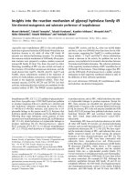
Fig. 1. Overview of the molecular organiza-
tion of dynein. (A) Linear map of the HC of
Chlamydomonas axonemal dynein c
(BAE19786, Chlamydomonas reinhardtii),
showing the domain structure: tail, linker,
AAA modules and MTBD. Amino acid num-
bers are shown at the bottom. (B) A sche-
matic drawing of the budding yeast dynein
HC in apo state. The six AAA modules are
arranged in a ring and the C-terminal domain
is on the ring. Each module is composed of
the N-terminal large domain and the C-termi-
nal small domain. Dynein has two distinct
faces. The linker face (a) corresponding to
the face seen in the left view [64] and the
C-terminal face (b) corresponding to that
seen in the right view [64]. (C) Crystal struc-
ture of the cytoplasmic dynein AAA mod-
ules (reproduced with permission from
Carter et al. [69]). The six individual AAA
modules are highlighted in color. (D) The
atomic model of the distal stalk and MTBD
of cytoplasmic dynein in the weakly binding
state (PDB accession code 3ERR; MMDB
ID 68163 [68]). The figure was prepared
using Cn3D provided by the National Center
for Biotechnology Information (http://
www.ncbi.nlm.nih.gov/Structure/CN3D/
cn3d.shtml).
H. Sakakibara and K. Oiwa Dynein structure and its force-generating mechanism
FEBS Journal 278 (2011) 2964–2979 ª 2011 The Authors Journal compilation ª 2011 FEBS 2967
some axonemal dyneins, the presence of ADP is
known to be essential for motility in vitro [22,84], and
in others ADP increases the gliding velocity of micro-
tubules driven by dyneins, indicating that ADP binds
to at least one of these AAA modules [22,28]. A hypo-
thetical atomic structure produced by homology
modelling of the dynein AAA modules suggests that
the nucleotide-binding Walker A motifs lie close to the
interface between adjacent modules [85]. Interactions
between adjacent AAA modules through their nucleo-
tide pockets supports the idea that they may act in
concert to produce a functional motor.
Recently, a crystal structure of the truncated motor
domain of the yeast cytoplasmic dynein HC (about
300 kDa) without nucleotide with 6 A
˚
resolution has
been reported [69]. Although 6 A
˚
resolution is not high
enough to resolve side-chains of amino acids, the crys-
tal shows virtually all of the helices and b sheets
(Fig. 1C). On the basis of features in the crystal struc-
ture, together with information from previous electron
microscopy studies as described above, the six AAA
modules, the mechanical element (termed the linker:
see below) and the base of the coiled-coil stalk were
assigned to the head ring. An individual AAA module
is composed of an N-terminal large domain with an
a ⁄ b Rossmann fold and a C-terminal a-helical domain
(small domain) (Fig. 1B). These AAA modules are
arranged asymmetrically in the motor domain; they
are oriented at different angles and have different
packing between adjacent AAA modules (Fig. 1B, C).
There is a large gap between AAA1 and AAA2 in
dynein crystallized without nucleotide. It is speculated
that if ATP bound to AAA1, it would draw the adja-
cent AAA2 closer and start hydrolysis of ATP [69].
Movement of AAA2 toward AAA1 starts conforma-
tional spread along the AAA modules. The fact that
the head domain of negative-stained dynein c with
ADP-V
i
is reported to be roughly circular whereas that
in the absence of nucleotide has a markedly different
shape [64] may support this speculation. The distortion
of the head ring might represent the signal pathway
between the MTBD of the stalk and the principal
ATPase site in AAA1.
As this paper was submitted for publication, another
crystal structure of the cytoplasmic dynein of Dictyos-
telium discoideum was reported at 4.5 A
˚
resolution
[70]. The structure contains the entire 380 kDa motor
domain including a whole stalk structure in the pres-
ence of Mg-ADP (in the post-power stroke state). The
Dictyostelium cytoplasmic dynein has a more symmet-
rical and planar motor domain than the yeast dynein
does, but a larger C-terminal domain, which is local-
ized on the face of the motor ring opposite to where
the linker resides. The large gap between AAA1 and
AAA2 was observed in Dictyostelium dynein motor
domain, but the gap between AAA5 and AAA6 that is
evident in the yeast structure was not [70].
Stalk
The head ring has two elongated flexible structures
called the stalk (about 15 nm long antiparallel coiled
coil) and the N-terminal tail (the cargo binding
domain, formerly known as the stem). The stalk
extends out from the head ring between AAA4 and
AAA5. It was predicted that a helix (CC1) coming out
of AAA4 and a helix (CC2) returning back to AAA5
form a coiled-coil stalk [86]. X-ray crystallography
showed that the stalk does not work as a bridge
between AAA4 and AAA5 but is the extension of heli-
ces in the small domain of AAA4 [69,70]. A MTBD is
localized at the tip of the stalk, forming a small globu-
lar domain (Fig. 1B, D). Although this globular
domain at the stalk tip has poor sequence conservation
[87], mutagenesis of conserved residues clearly inter-
feres with microtubule binding [51]. Microtubule bind-
ing of the stalk tip was also examined with a
recombinant stalk-tip peptide [88]. The stalk-tip pep-
tide was observed to bind to a microtubule with a peri-
odicity of 8 nm and to share the binding region on the
microtubule with kinesin [88].
During the mechanochemical cycle of dynein, bind-
ing of ATP to the primary ATPase site of AAA1
causes dissociation of dynein from the microtubule,
and binding of the MTBD to the microtubule acceler-
ates the dissociation of hydrolysis products from the
ATPase site [89,90]. Because the sites of microtubule
binding and primary ATP hydrolysis are spatially seg-
regated (about 25 nm), elucidation of the communica-
tion pathway between them is an important issue in
understanding dynein function, as described in the pre-
vious section. To investigate the communication mech-
anism of the stalk, Gibbons et al. [91] designed a series
of fusion constructs in which the MTBD, along with a
portion of its predicted coiled-coil stalk, is fused onto
a stable antiparallel coiled-coil base found in the native
structure of seryl-tRNA synthetase. They attempted to
identify the optimal alignment between the hydropho-
bic heptad repeats in the two strands of the coiled-coil
stalk. Alterations in the phase of the heptad repeats in
the CC1 changed the affinity of the MTBD to the
microtubules. Finally, they identified the pattern of
two alternative registries (a and b) having high and
low microtubule-binding affinity, respectively. On the
basis of these results, Gibbons et al. hypothesized that
during the mechanochemical cycle the two strands of
Dynein structure and its force-generating mechanism H. Sakakibara and K. Oiwa
2968 FEBS Journal 278 (2011) 2964–2979 ª 2011 The Authors Journal compilation ª 2011 FEBS
its coiled-coil stalk undergo a small amount of sliding
displacement as a means of communication between
the AAA core of the motor and the MTBD [91]. This
hypothesis is further supported by the use of an
expressed dynein motor domain in which the coiled
coil of the stalk was trapped at three specific registries
using oxidation to disulfides of paired cysteine residues
introduced into the two helices [92]. Coupling between
ATPase activity and the binding activity to microtu-
bules depend upon the registry of the coiled coil.
Carter et al. extended the research on the MTBD
and reported the 2.3 A
˚
resolution coordinates of the
MBTD in a weakly binding conformation (b registry)
and the distal portion of the coiled-coil stalk of mouse
cytoplasmic dynein [68] (Fig. 1D). As predicted, they
confirmed that the stalk is a coiled coil. The MTBD
consists of a bundle of six a-helices (H1–H6) and the
interface against microtubules is made up of three heli-
ces called H1, H3 and H6. The coiled coil of the stalk
is not straight but bent near the MTBD by a pair of
staggered highly conserved proline residues, with the
regular packing of hydrophobic residues in the coiled-
coil core being disrupted in the region between the
prolines. When the heptad registry resumes after the
prolines, the registry of CC1 has slipped by one half-
heptad relative to that of CC2. The distal portion of
CC2 makes extensive hydrophobic interactions with
H2, H4, H5 and H6, whereas CC1 makes only a few
contacts with H4 before joining directly into H1. It is
suggested that communication along the coiled coil of
the stalk is effected by interstrand sliding and this
asymmetry at the interface between the stalk and the
MTBD plays an important role in the dynein mecha-
nochemical cycle [68].
However, the concept that the sliding of the stag-
gered coiled coils relative to each other within the stalk
achieves two patterns of alternating registries has been
challenged by the crystal structure of dynein [69]. Since
the crystal structure suggests that two helices (CC1
and CC2) merge into the well-packed helices of the
AAA4 small domain, it is unlikely that either helix can
move at its base. Furthermore, in addition to the stalk
coiled coil, the crystal structure revealed the presence
of a second antiparallel coiled coil that emerges from
the small domain of AAA5 as a long extension of heli-
ces. The structure is called buttress [69] or strut [70]
and it extends toward and makes contact with the
stalk (Fig. 1B, C). Although the crystal of the yeast
dynein has no MTBD, owing to optimization of crys-
tallization, the interaction between the stalk and the
buttress ⁄ strut provides insight for the regulation mech-
anism of MTBD by the AAA modules in the head
ring. Through the interaction of the buttress ⁄ strut and
the stalk, the buttress ⁄ strut might relay rigid body
motion between AAA modules into shear motions
between the helices of the stalk coiled coil.
The crystallographic analysis of the motor domain
of Dictyostelium dynein provides the evidence for the
structural information pathway between AAA1 and
MTBD since the crystal unit contains a whole stalk
and two independent motor domains, which adopt dif-
ferent conformations [70]. The major structural differ-
ence is found in the stalk–buttress ⁄ strut structure. The
stalk of one motor domain is straight up to the tip,
while that of the other motor domain is kinked at the
region just beneath the contact site with the but-
tress ⁄ strut. This stalk tilting is accompanied by small
conformational changes of the strut. The kink of the
stalk while holding the basal portion in place could
induce interstrand sliding. Kon et al. thus hypothesize
that dynein coordinates AAA1 ATPase and MTBD by
switching the stalk-strut structure between the straight
and kinked conformations [70]. This hypothesis implies
a new communication pathway in which the structural
information could propagate from AAA1 to MTBD
through the C-terminal domain, AAA5 and then
buttress ⁄ strut [70].
Tail
The amino-terminal tail is involved in dimerization ⁄ tri-
merization of dynein HCs and acts as a scaffold for the
assembly of different ICs and LICs to form the dynein
complex. Since an expressed cytoplasmic dynein with-
out a native tail but with a substituted tail shows intact
processive movement in vitro, these subunits are not
essential for dynein motility in vitro [53]. However, they
may regulate dynein motility in vivo and recent data
indicate that the non-catalytic subunits link dynein to
cargos and to several adaptor proteins that regulate
dynein function [20] (see the minireviews [13,19]). For
example, missense mutations in the tail domain of cyto-
plasmic dynein in mice cause neurodegenerative disease.
The best characterized model of dynein dysfunction is
the Legs at odd angles (Loa) mouse [93]. This mutation
is thought to affect homodimerization of the dynein
HCs and ⁄ or the association of the HCs and ICs [94].
Single-molecule nanometry on the mutant dynein
showed that dynein purified from mutant mice has
lower processivity and shows more frequent bidirec-
tional motility along a microtubule and greater propen-
sity to sidestep to adjacent protofilaments than the
wild-type dynein does. These results suggest that muta-
tion in the tail domain of dynein causes increased flexi-
bility of the dynein molecule and diminished gating
between the motor domains [94].
H. Sakakibara and K. Oiwa Dynein structure and its force-generating mechanism
FEBS Journal 278 (2011) 2964–2979 ª 2011 The Authors Journal compilation ª 2011 FEBS 2969
While plus-end-directed transport in cells is carried
out by many kinesin family members with a wide
range of tail domains and despite the large repertoire
of cellular functions that dynein is involved in, all
minus-end-directed transports within the cytoplasm are
carried out by a single cytoplasmic dynein. The tail
domain is thus important to mediate interaction with
various types of cargo by recruiting specific and appro-
priate adaptor proteins.
Linker
The linker is a structure located in the portion of the
tail proximal to AAA1, which serves as a connection
between AAA1 and the main part of the tail (Fig. 1B,
C). The existence of the linker was first indicated in
images of negative-stained monomeric axonemal
dynein [64]. Although the linker is normally docked
onto the head ring, it is revealed as a relatively large
structure about 2 nm wide and 10 nm long when the
linker is undocked from the head ring [64]. The crystal
structure of dynein showed that the linker is composed
of helical bundles and does not sit flat on the head
ring but rather arches over it [69,70]. The linker is
composed of four predominantly helical subdomains
(from N-terminus, subdomain 1, 2, 3 and 4). The
C-terminus subdomain 4 interacts with AAA1 and part
of AAA6 and is connected into AAA1. The N-termi-
nal subdomain 1 contacts AAA5 in the yeast motor
domain [69] or AAA4 in the Dictyostelium head ring
[70], and this contact looks tenuous and may break
and dissociate from AAA4 or AAA5 during the AT-
Pase cycle. Although the significance of the difference
in the contact point remains unclear, it could represent
conformational changes upon ADP release during
dynein’s ATPase cycle [70].
It has been suggested that the linker is involved in
generation of force through its interaction with the
head ring [64]. Two-dimensional analysis on negative-
stained dynein c described the conformations of the
dynein molecule in two different nucleotide states
which mimic the post- and pre-power stroke conforma-
tions of the motor (Fig. 2). In the absence of nucleo-
tide (post-power stroke conformation, state I) the tail
emerges near the base of the stalk. In the presence of
ATP and vanadate, which forms a dynein–ADP–Vi
complex that mimics the dynein–ADP–Pi conforma-
tion (pre-power stroke conformation, state II), the tail
emerges further away from the stalk base. These obser-
vations were interpreted to originate from the swinging
of the linker relative to the head ring. The existence
and the movement of linker have subsequently been
confirmed in cytoplasmic dynein, identified as the
N-terminal region of the motor domain using green
fluorescent protein (GFP) and blue fluorescent protein
(BFP) tagged constructs by negative stain electron
microscopy and Fo
¨
rster resonance energy transfer
(FRET) [75,89]. In the absence of nucleotide or in the
presence of ADP, GFP inserted at the linker’s N-ter-
minus lies close to AAA4 (the crystal structure of the
yeast dynein [69] shows the N-terminus lies close to
AAA5, as described above), at the base of the stalk, in
the so-called un-primed position, whereas in the pres-
ence of ATP and vanadate the GFP lies close to
AAA2, in the primed position [75].
Dynamic measurements of the linker movement
were performed by measuring the FRET between a
GFP and a BFP both fused into a dynein construct
molecule [89]. A series of 380-kDa dynein constructs
from Dictyostelium were prepared that had a GFP
attached at the N-terminus and a BFP inserted into
various sites on the dynein head ring. The efficiency of
FRET was measured in each construct at various
nucleotide states under steady-state conditions. The
results showed two distinct values: a high FRET effi-
ciency and low FRET efficiency suggesting movement
of the N-terminus relative to the head ring. Using
mutants that were trapped in specific intermediate
states, it was shown that this movement is coupled to
ATPase steps [89].
Lever-arm model or winch model
The observations described above pose the model of
dynein force generation, which is the most widely cited
one: on binding of ATP to AAA1, the orientation of
the docked linker on the head ring causes the tail to
emerge far from the stalk (state II, top panel in
Fig. 2A). Upon release of products, the linker orienta-
tion on the ring changes, bringing the tail closer to the
stalk (state I, bottom panel in Fig. 2A). The linker
changes its orientation by switching between two differ-
ently docked positions on the head ring, thus producing
a rotation of the head ring that causes the stalk to
swing. In addition to this head ring motion, an ATP-
driven alteration in the coiled-coil registry may control
the affinity of the dynein HC for the microtubule. How-
ever, the stalk is not rigid since stiffness of a 15-nm
length of coiled-coil peptide clamped at one end can be
estimated to be about 0.4 pNÆnm
)1
. The range of con-
formations for an axonemal dynein molecule observed
in negatively-stained samples [64,73] also provides an
estimation of the stiffness of the stalk, which is
0.5 pNÆnm
)1
in apo molecules and 0.14 pNÆnm
)1
in
ATP-vanadate molecules [95]. These estimates imply
that stalk is too flexible to work as a rigid lever [96,97].
Dynein structure and its force-generating mechanism H. Sakakibara and K. Oiwa
2970 FEBS Journal 278 (2011) 2964–2979 ª 2011 The Authors Journal compilation ª 2011 FEBS
Cryo-electron-microscope images of whole dynein
molecules interacting with microtubules [98] have
recently revealed that, even though the tail and linker
shift relative to the head ring and stalk as in isolated
dynein molecules, the stalk orientation on the microtu-
bule remains fixed. The observation provides strong
evidence for the concept of dynein as an ATP-depen-
dent winch [96] (Fig. 2B).
Furthermore, winch-type motion was also observed
in an axoneme. Cryo-electron tomograms of axonemes
in the apo state were compared with those obtained in
the presence of ADP-vanadate [99]. Global changes of
dynein arm complexes were shown and several key
changes in dynein structures were found. Although the
stalks are not clearly visible in the tomograms, close
examination showed that the stalks typically tilt
towards the proximal end of the axoneme (the base of
the axoneme) in both nucleotide conditions. The
dynein head rings were observed to move 8 nm toward
the distal end of the axoneme upon release of the
nucleotide. Since the MTBD attached to the adjacent
microtubule, the movement results in dragging the
adjacent microtubule distally and producing the shear
[99].
The winch model explains the result of Carter et al.
[68], in which cytoplasmic dynein with its stalk coiled
coil either lengthened or shortened by seven heptads
moves towards the minus end of a microtubule, irre-
spective of the length of the stalk. This result is
remarkable since these stalk length changes would be
predicted to rotate the head ring by 180° and reverse
the direction of dynein movement according to the
lever-arm model. To explain the directionality of
dynein, it is proposed that the head ring does not elicit
a lever-like rotation of the linker domain perpendicular
to the stalk, but rather, contraction where the force
vector of the linker domain’s conformational change is
directed parallel to an angled stalk [68] (Fig. 2B).
Mechanical properties of dynein
Characterizations of the mechanical properties of
dyneins have been carried out using in vitro motility
assays, which enable the motility of dyneins along
S
AB
tate II
State I
State II
State I
Fig. 2. Proposed mechanisms of dynein’s power stroke. (A) Negative-stain electron microscopy followed by single-particle analysis suc-
ceeded in capturing two distinct conformations of dynein c molecules isolated from Chlamydomonas flagella in the ADP-V
i
(state II) and apo
(state I) state [64]. On ATP binding to AAA1, the tail emerges far from the stalk (state II, top panel). Upon product release, the tail emerges
closer to the stalk (state I, bottom panel), suggesting movement of the linker domain (and tail relative to head ring and stalk). This conforma-
tional change swings the stalk by 15 nm. This model is based upon [64] and [89]. (B) The winch model of dynein force generation
[68,96,98,99]. Product release from AAA1 leads to the contraction of the whole dynein molecule by the movement of the linker. As shown in
the electron microscope images of microtubules decorated with stalks [98], the stalk points toward the microtubule minus end and connects
the head ring to the microtubule. The microtubule is thus dragged by the contraction induced by the shift of the head ring relative to the tail.
H. Sakakibara and K. Oiwa Dynein structure and its force-generating mechanism
FEBS Journal 278 (2011) 2964–2979 ª 2011 The Authors Journal compilation ª 2011 FEBS 2971
microtubules to be reconstituted from purified and
characterized component molecules. Since experimental
conditions such as temperature, buffer compositions
and ATP concentrations are readily controlled, these
assays permit precise measurements of dyneins’
mechanical properties. Several important findings
about dyneins are listed below.
l
The minimal components of dynein motility are the
380 kDa domain of Dictyostelium dynein (314 kDa
domain of yeast dynein) which contains a linker,
six AAA domains and a stalk [52,53].
l
Each of the HCs of axonemal dyneins so far stud-
ied has distinct motile activity [21,22,41].
l
Some inner-arm dyneins can generate torques and
rotate a microtubule about its long axis while it is
moved on the dynein-coated surface [21,100].
l
Some inner-arm dyneins require a trace amount of
ADP for their microtubule motility [22,84].
l
Some inner-arm dyneins, even some single-headed
or heterodimeric motors, show high processivity
in vitro [25,65].
l
The three HCs of outer-arm dyneins are likely to
play distinct roles and regulate each other to
achieve coordinated force production [101].
l
Dynactin enhances the processivity of cytoplasmic
dynein [102–104]. Molecular dissections of dynactin
showed that the mechanism of processivity enhance-
ment is not due to anchoring the motor domain of
dynein to microtubules via dynactin but a mecha-
nism independent of microtubule tethering [104].
Furthermore, in vitro motility assays have paved the
way to single-molecule studies on dynein. In recent
decades, the development of a number of technologies,
such as atomic-force microscopy, optical-trap nanome-
try and fluorescence imaging with nanometer precision
have provided tools for studying the dynamics of sin-
gle molecules in situ over time scales from milliseconds
to seconds. The single-molecule sensitivities of these
methods permit studies to be made on conformational
changes and functions of dyneins that are masked in
ensemble-averaged experiments. Processivity, step size
and dwell-time distributions are among properties that
can be directly measured by single-molecule tech-
niques. Our understanding of the functions of dyneins
has benefited considerably from the application of sin-
gle-molecule techniques.
However, single-molecule measurements on the force
generation of dyneins have raised some questions. The
stall force generated by single dynein molecules varies
from measurement to measurement: for axonemal
dyneins, a value of 1–2 pN in single-headed inner-arm
dynein [65], 6 pN in an inner dynein arm in an
axoneme [105] and 4.7 pN in outer-arm dynein [106];
for cytoplasic dyneins, a value of 1 pN in bovine
cytoplasmic dynein [107,108], 3–4 pN [109] and 7–
8 pN [110] in porcine cytoplasmic dynein and 8 pN in
yeast cytoplasmic dynein [111]. The variation in force
may depend upon the type of dynein used and upon
distinct roles that dynein plays in vivo [53]. It is also
suggested that the geometry of the force measurements
may influence the force and the mode of motility [109].
Nucleotide concentrations may have an effect on the
force generation and modes of movement [106,109].
The precise measurements and direct comparison of
the force generated by cytoplasmic dyneins are now
required since they will provide important information
to reveal the mechanism of cargo transport by a num-
ber of or several types of motors mechanically coupled
to each other.
Processivity and modes of movement
Cytoplasmic dynein is known as a processive motor
that can take micrometer-scale movements along a
microtubule without dissociating. Through the creation
of a cytoplasmic dynein that can be converted between
monomeric and dimeric states by a small molecule,
rapamycin, it is demonstrated that processive motion
requires the dimerization of two motor domains,
although the endogenous dimerization domain (tail) is
not required for the processivity [53]. In addition, pro-
cessivity of cytoplasmic dynein in vitro does not
require any of the known dynein-associated subunits
[53] despite reports that the dynactin complex enhances
the processivity of cytoplasmic dyneins [102–104].
The step sizes and modes of movement of cytoplas-
mic dyneins are under debate. Cytoplasmic dynein
purified from bovine brain primarily takes large steps
(24–32 nm) at low loads, but decreases step size from
32 to 8 nm with increasing load to its stall force [107].
In addition, when multiple dynein molecules interact
with a microtubule and contribute to movement, the
dynein molecules move predominantly in 8-nm steps
[108]. In contrast, movement of single cytoplasmic
dynein molecules purified from porcine brain [110] and
a functional recombinant dimeric dynein of the bud-
ding yeast [53] were analyzed with a high spatial preci-
sion tracking technique and were stepwise with a
regular 8-nm step size, irrespective of the load.
Based upon these findings, Reck-Peterson et al.
proposed a molecular model (which they called the
‘alternating shuffling model’) to explain how processive
motion is achieved by cytoplasmic dynein [53] (Fig. 3).
The model is conceptually similar to the hand-over-
hand model proposed for processive kinesin motility:
two dynein heads alternate taking 16-nm steps while
Dynein structure and its force-generating mechanism H. Sakakibara and K. Oiwa
2972 FEBS Journal 278 (2011) 2964–2979 ª 2011 The Authors Journal compilation ª 2011 FEBS
the centroid position of the molecule moves by 8 nm
for each step. The large dimensions of the head ring
do not allow the head ring to alternately ‘swing’
forward at each step, but the two head rings always
overlap partially during the stepping motion. The
model thus requires coordination between two motor
domains. Optical-trap studies of dynein force produc-
tion suggest that this coordination is carried out by
strain transmitted through the linkage between the two
heads [111]. Besides the intramolecular strain, physical
contacts between two head rings during the movement
along a protofilament could mediate head–head coor-
dination [112].
However, given the apparently large size, the flexi-
bility of a dynein molecule and the magnitude of the
suggested power stroke [64] in relation to the tubulin
lattice, the 8-nm step size displayed by dimeric dyne-
ins is surprisingly small. This is in contrast to other
linear motors since the step size of a motor, except
myosin VI [113], can be predicted to be proportional
to the length of its power stroke [114,115]. One
possible explanation is that when a dimeric dynein
moves on a microtubule taking 8-nm steps, the mole-
cule could be compact and stiff with its two head
rings in close, intimate association as is seen in axo-
nemal dyneins in situ [116,117] and in electron micro-
scope observations of the phi (F) shaped structure of
cytoplasmic dynein in which the tails of the HCs are
close to each other and the head rings are partially
overlapped [118].
In contrast, in early studies of dynein motility, cyto-
plasmic-dynein-coated beads exhibited greater lateral
movements among microtubule protofilaments than
did kinesin [119]. Hence, dynein apparently does not
have to walk along a single protofilament. Precise
measurements showed that fluorescently labeled dynein
also displayed lateral stepwise movements, which
usually occurred simultaneously with forward stepping.
This shows that dynein has the reach or flexibility to
occasionally land on an adjacent protofilament [53].
Furthermore, as stated earlier, the Loa mutant
cytoplasmic dynein showed increased frequency of
A
B
C
Time
E
F
G
D
H
I
ATP
ADP
J
ATP
ADP
Fig. 3. Alternating shuffling model for processive movement of a dimeric dynein [53] with some modifications on the basis of the crystal
structure [69] and kinetics [90,125]. To simplify, we draw all presumed elastic elements in the tail domain as a simple spring that connects
two head rings. The stalk and the head ring are drawn as a rod and a large circle, respectively. The linker is drawn as a yellow curved bar,
which has a hinge as a yellow small circle. We construct linker motion swinging around the hinges (A). Binding of ATP to the trailing head
(red) releases the MTBD from the microtubule and then (B) the head is pulled forward by the strain stored in the connecting spring while
the leading head (blue) stays bound to the microtubule. (C) The linker is detached from the docking site on AAA5. Upon re-binding of the
MTBD to the microtubule (D), the head changes its conformation coupled with product release (E). (F)–(J) The trailing head carries out the
same mechanical process as shown in (A)–(E). Two dynein heads alternate taking 16 nm steps, whereas the position of the center of mass
of the molecule moves by 8 nm for each step. Note that, due to the large size of the head ring, two heads are partially overlapped during
stepping without changing the relation of their lateral positions.
H. Sakakibara and K. Oiwa Dynein structure and its force-generating mechanism
FEBS Journal 278 (2011) 2964–2979 ª 2011 The Authors Journal compilation ª 2011 FEBS 2973
sideways steps and backwards steps and decreased pro-
cessivity [94]. Modulation of the dynein tail domain
could affect coordination between two motor domains.
Although further studies will be necessary to clarify
the mechanism of the coordination, modulating the tail
domain could be one of the mechanisms of regulation
of dynein motility [94].
In addition, dynein shows large variation in the
mode of movements but the mode-switching mecha-
nism is not well understood. Several research groups
have shown that dynein can display a diffusive mode
of bidirectional motion along the microtubule as well
as processive stepwise motion [53,120,121]. Regulation
mechanisms may exist that switch from the stepping
mode to the diffusive mode and vice versa. Variation
of the modes may reflect physiological roles of dyneins
in vivo. Further work will be required to resolve these
reported differences in dynein behavior, with particular
attention paid to possible species variation, protein
preparation and assay conditions.
Conclusion and future perspectives
The characteristic feature of the dynein molecule is its
flexibility: both tail and stalk domains of isolated
dynein molecules are flexible [96]. Stalk flexibility
observed in axonemal dyneins in situ [122] suggests
important roles for the domain as compliant elements
within the motor. Although the compliant stalk is a
significant drawback for its acting as a stiff lever arm
that pushes against the microtubules, evidence for
both axonemal and cytoplasmic dyneins is now accu-
mulating that dynein molecules work as a winch
[68,96,98,99] which is well suited to generating
tension. In the winch-like mechanism, a large active
tension can be sustained by the stalk but it may be
buckled when negative forces are applied to the stalk.
This feature and the tilted stalk and dynein molecules
provide nonlinear elasticity against the direction of
external force; the nonlinearity may have advantages
when a large number of dyneins with distinct mechan-
ical properties work together on a single microtubule,
as seen in an axoneme. When the slower motors work
together with the faster motors on a microtubule, the
slower motors could work as a source of internal
drag [25]. However, the flexible stalks of the slower
motors can be buckled and may dissociate from
the microtubule rapidly to minimize the increase in
internal load.
In addition to the stalk, the neck subdomain of the
tail of dynein c, the part closest to the head and
8 nm long, has a similar width to the coiled-coil
stalk [64] and is very flexible. It might be another
important site of compliance within the dynein mole-
cule. Important questions thus arise concerning the
possible coordination between two motor domains
connected by this flexible tail as in cytoplasmic dynein,
dynein I1 ⁄ f or dyads of inner-arm dyneins [123]. Force
generation and the kinetics of individual motor
domains could be modulated by stress and strain gen-
erated by the activity of other motor domains in a
multimeric dynein molecule. Such modulation could
play important roles, especially in the processive move-
ment of cytoplasmic dyneins. Recent three-dimensional
reconstructions of in situ outer-arm dyneins have dem-
onstrated that their head rings are intimately associ-
ated with one another [116,117,124]. Reconstituted
outer arms are motile and can drive microtubules at
faster velocities than outer-arm dyneins randomly dis-
tributed on a glass surface in in vitro motility assays
[101]. In addition to these studies on the axonemal
dyneins, cytoplasmic dynein may require the compact
and stiff configurations of a molecule for stepwise pro-
cessive motion and the Loa mutant results [94] could
be explained by a disrupted coordination between the
two motor domains. The results now shed light on the
role of the tail for the gating mechanism.
Crystal structures of the dynein motor domain in
two different nucleotide states are now obtained
[69,70] and these structures are a great leap forward in
understanding dynein function. In addition to crystal
structures, dynamic measurements will also be vital,
supposing that the nature of dynein function is flexibil-
ity. In particular, a detailed mechanism of coordina-
tion between motor domains will await further analysis
using single-molecule techniques such as optical trap
measurements combined with genetically engineered
dynein molecules.
Acknowledgements
We thank Dr Hiroaki Kojima (National Institute of
Information and Communications Technology) for
helpful discussions and critical comments on the man-
uscript. This work is supported by Special Coordina-
tion Funds for Promoting Science and Technology
(K.O.) and a Grant-in-Aid for Scientific Research on
the Priority Area ‘Regulation of Nano-systems in
Cells’ by the Ministry of Education, Culture, Sports,
Science and Technology (K.O.).
References
1 Gibbons IR (1963) Studies on the protein components
of cilia from Tetrahymena pyriformis. Proc Natl Acad
Sci USA 50, 1002–1010.
Dynein structure and its force-generating mechanism H. Sakakibara and K. Oiwa
2974 FEBS Journal 278 (2011) 2964–2979 ª 2011 The Authors Journal compilation ª 2011 FEBS
2 Gibbons IR & Rowe AJ (1965) Dynein: a protein with
adenosine triphosphatase activity from cilia. Science
149, 424–426.
3 Pfister KK, Fisher EMC, Gibbons IR, Hays TS, Holz-
baur ELF, McIntosh JR, Porter ME, Schroer TA,
Vaughan KT, Witman GB et al. (2005) Cytoplasmic
dynein nomenclature. J Cell Biol 171, 411–413.
4Ho
¨
o
¨
k P & Vallee RB (2006) The dynein family at a
glance. J Cell Sci 119, 4369–4371.
5 Wickstead B & Gull K (2007) Dyneins across eukary-
otes: a comparative genomic analysis. Traffic 8, 1708–
1721.
6 Hirokawa N & Takemura R (2004) Molecular motors
in neuronal development, intracellular transport and
diseases. Curr Opin Neurobiol 14, 564–573.
7 Vallee RB & Stehman SA (2005) How dynein helps the
cell find its center: a servomechanical model. Trends
Cell Biol 15, 288–294.
8 Vallee RB, Williams JC, Varma D & Barnhart LE
(2004) Dynein: an ancient motor protein involved in
multiple modes of transport. J Neurobiol 58, 189–200.
9 Alonso C, Miskin J, Hernaez B, Fernandez-Zapatero
P, Soto L, Canto C, Rodriguez-Crespo I, Dixon L &
Escribano JM (2001) African swine fever virus protein
p54 interacts with the microtubular motor complex
through direct binding to light-chain dynein. J Virol
75, 9819–9827.
10 Greber UF & Way M (2006) A superhighway to virus
infection. Cell 124, 741–754.
11 Bremner KH, Scherer J, Yi J, Vershinin M, Gross SP
& Vallee RB (2009) Adenovirus transport via direct
interaction of cytoplasmic dynein with the viral capsid
hexon subunit. Cell Host Microbe 6, 523–535.
12 Do
¨
hner K, Nagel CH & Sodeik B (2005) Viral stop-
and-go along microtubules: taking a ride with dynein
and kinesins. Trends Microbiol 13, 320–327.
13 Merino-Gracia J, Garcı
´
a-Mayoral MF & Rodriguez-
Crespo I (2011) The association of viral proteins with
host cell dynein components during virus infection.
FEBS J 278, 2997–3011.
14 Vaisberg EA, Grissom PM & McIntosh JR (1996)
Mammalian cells express three distinct dynein heavy
chains that are localized to different cytoplasmic organ-
elles. J Cell Biol 133, 831–842.
15 Palmer KJ, Hughes H & Stephens DJ (2009) Specificity
of cytoplasmic dynein subunits in discrete membrane-
trafficking steps. Mol Biol Cell 20, 2885–2899.
16 Porter ME, Bower R, Knott JA, Byrd P & Dentler W
(1999) Cytoplasmic dynein heavy chain 1b is required
for flagellar assembly in Chlamydomonas. Mol Biol Cell
10, 693–712.
17 Vaughan KT & Vallee RB (1995) Cytoplasmic dynein
binds dynactin through a direct interaction between the
intermediate chains and p150 Glued. J Cell Biol 131,
1507–1516.
18 Vallee RB, Varma D & Dujardin DL (2006) ZW10
function in mitotic checkpoint control, dynein targeting
and membrane trafficking: is dynein the unifying
theme? Cell Cycle 5
, 2447–2451.
19 Rapali P, Szenes A, Radnai L, Bakos A, Pa
´
lG&
Nyitray L (2011) DYNLL ⁄ LC8: a light chain subunit
of the dynein motor complex and beyond. FEBS J 278,
2980–2996.
20 Kardon JR & Vale RD (2009) Regulators of the cytoplas-
mic dynein motor. Nat Rev Mol Cell Biol 10, 854–865.
21 Kagami O & Kamiya R (1992) Translocation and rota-
tion of microtubules caused by multiple species of
Chlamydomonas inner-arm dynein. J Cell Sci 103, 653–
664.
22 Yagi T (2000) ADP-dependent microtubule transloca-
tion by flagellar inner-arm dyneins. Cell Struct Funct
25, 263–267.
23 Kikushima K & Kamiya R (2008) Clockwise transloca-
tion of microtubules by flagellar inner-arm dyneins
in vitro. Biophys J 94, 4014–4019.
24 Yanagisawa HA & Kamiya R (2001) Association
between actin and light chains in Chlamydomonas
flagellar inner-arm dyneins. Biochem Biophys Res
Commun 288, 443–447.
25 Kotani N, Sakakibara H, Burgess SA, Kojima H &
Oiwa K (2007) Mechanical properties of inner-arm
dynein-f (dynein I1) studied with in vitro motility
assays. Biophys J 93, 886–894.
26 LeDizet M & Piperno G (1995) The light chain p28
associates with a subset of inner dynein arm heavy
chains in Chlamydomonas axonemes. Mol Biol Cell 6,
697–711.
27 Yagi T, Uematsu K, Liu Z & Kamiya R (2009) Identi-
fication of dyneins that localize exclusively to the prox-
imal portion of Chlamydomonas flagella. J Cell Sci 122,
1306–1314.
28 Kikushima K, Yagi T & Kamiya R (2004) Slow ADP-
dependent acceleration of microtubule translocation
produced by an axonemal dynein. FEBS Lett 563,
119–122.
29 Piperno G, Mead K & Shestak W (1992) The inner
dynein arms I2 interact with a ‘dynein regulatory com-
plex’ in Chlamydomonas flagella. J Cell Biol 118, 1455–
1463.
30 Porter ME, Knott JA, Myster SH & Farlow SJ (1996)
The dynein gene family in Chlamydomonas reinhardtii.
Genetics 144, 569–585.
31 Myster SH, Knott JA, O’Toole E & Porter ME (1997)
The Chlamydomonas Dhc1 gene encodes a dynein
heavy chain subunit required for assembly of the I1
inner arm complex. Mol Biol Cell 8, 607–620.
32 Perrone CA, Myster SH, Bower R, O’Toole ET & Porter
ME (2000) Insights into the structural organization of
the I1 inner arm dynein from a domain analysis of the
1b dynein heavy chain.
Mol Biol Cell 11, 2297–2313.
H. Sakakibara and K. Oiwa Dynein structure and its force-generating mechanism
FEBS Journal 278 (2011) 2964–2979 ª 2011 The Authors Journal compilation ª 2011 FEBS 2975
33 Yagi T, Minoura I, Fujiwara A, Saito R, Yasunaga T,
Hirono M & Kamiya R (2005) An axonemal dynein
particularly important for flagellar movement at high
viscosity. Implications from a new Chlamydomonas
mutant deficient in the dynein heavy chain gene
DHC9. J Biol Chem 280, 41412–41420.
34 Wirschell M, Hendrickson T & Sale WS (2007) Keep-
ing an eye on I1: I1 dynein as a model for flagellar
dynein assembly and regulation. Cell Motil Cytoskele-
ton 64, 569–579.
35 Sakakibara H, Mitchell DR & Kamiya R (1991) A
Chlamydomonas outer arm dynein mutant missing the
a heavy chain. J Cell Biol 113, 615–622.
36 Mitchell DR & Brown KS (1994) Sequence analysis of
the Chlamydomonas alpha and beta dynein heavy chain
genes. J Cell Sci 107, 635–644.
37 Kamiya R (1988) Mutations at twelve independent loci
result in absence of outer dynein arms in Chylamydo-
monas reinhardtii. J Cell Biol 107, 2253–2258.
38 Wilkerson CG, King SM & Witman GB (1994) Molec-
ular analysis of the c heavy chain of Chlamydomonas
flagellar outer-arm dynein. J Cell Sci 107, 497–506.
39 Bartoloni L, Blouin JL, Pan Y, Gehrig C, Maiti AK,
Scamuffa N, Rossier C, Jorissen M, Armengot M,
Meeks M et al. (2002) Mutations in the DNAH11
(axonemal heavy chain dynein type 11) gene cause one
form of situs inversus totalis and most likely primary
ciliary dyskinesia. Proc Natl Acad Sci USA 99, 10282–
10286.
40 Neesen J, Koehler MR, Kirschner R, Steinlein C, Kre-
utzberger J, Engel W & Schmid M (1997) Identification
of dynein heavy chain genes expressed in human and
mouse testis: chromosomal localization of an axonemal
dynein gene. Gene 200, 193–202.
41 Furuta A, Yagi T, Yanagisawa HA, Higuchi H &
Kamiya R (2009) Systematic comparison of in vitro
motile properties between Chlamydomonas wild-type
and mutant outer arm dyneins each lacking one of the
three heavy chains. J Biol Chem 284, 5927–5935.
42 Kamiya R, Kurimoto E & Muto E (1991) Two types
of Chlamydomonas flagellar mutants missing different
components of inner-arm dynein. J Cell Biol 112, 441–
447.
43 Myster SH, Knott JA, Wysocki KM, O’Toole E &
Porter ME (1999) Domains in the 1a dynein
heavy chain required for inner arm assembly and
flagellar motility in Chlamydomonas. J Cell Biol 146,
801–818.
44 Pazour GJ, Agrin N, Walker BL & Witman GB (2006)
Identification of predicted human outer dynein arm
genes: candidates for primary ciliary dyskinesia genes.
J Med Genet 43, 62–73.
45 Chapelin C, Duriez B, Magnino F, Goossens M, Escu-
dier E & Amselem S (1997) Isolation of several human
axonemal dynein heavy chain genes: genomic structure
of the catalytic site, phylogenetic analysis and chromo-
somal assignment. FEBS Lett 412, 325–330.
46 Ikeda K, Yamamoto R, Wirschell M, Yagi T, Bower
R, Porter ME, Sale WS & Kamiya R (2009) A novel
ankyrin-repeat protein interacts with the regulatory
proteins of inner arm dynein f (I1) of Chlamydomonas
reinhardtii. Cell Motil Cytoskeleton 66, 448–456.
47 Zhang YJ, O’Neal WK, Randell SH, Blackburn K,
Moyer MB, Boucher RC & Ostrowski LE (2002) Iden-
tification of dynein heavy chain 7 as an inner arm com-
ponent of human cilia that is synthesized but not
assembled in a case of primary ciliary dyskinesia.
J Biol Chem 277, 17906–17915.
48 Yamamoto R, Yanagisawa HA, Yagi T & Kamiya R
(2008) Novel 44-kilodalton subunit of axonemal dynein
conserved from Chlamydomonas to mammals. Eukaryot
Cell 7, 154–161.
49 Heuser T, Raytchev M, Krell J, Porter ME & Nicastro
D (2009) The dynein regulatory complex is the nexin
link and a major regulatory node in cilia and flagella.
J Cell Biol 187, 921–933.
50 Samso
´
M, Radermacher M, Frank J & Koonce MP
(1998) Structural characterization of a dynein motor
domain. J Mol Biol 276, 927–937.
51 Koonce MP & Tikhonenko I (2000) Functional ele-
ments within the dynein microtubule-binding domain.
Mol Biol Cell 11, 523–529.
52 Nishiura M, Kon T, Shiroguchi K, Ohkura R, Shima
T, Toyoshima YY & Sutoh K (2004) A single-headed
recombinant fragment of Dictyostelium cytoplasmic
dynein can drive the robust sliding of microtubules.
J Biol Chem 279, 22799–22802.
53 Reck-Peterson SL, Yildiz A, Carter AP, Gennerich A,
Zhang N & Vale RD (2006) Single-molecule analysis
of dynein processivity and stepping behavior. Cell 126,
335–348.
54 Hastie AT, Dicker DT, Hingley ST, Kueppers F, Hig-
gins ML & Weinbaum G (1986) Isolation of cilia from
porcine tracheal epithelium and extraction of dynein
arms. Cell Motil Cytoskeleton 6, 25–34.
55 King SM, Gatti JL, Moss AG & Witman GB (1990)
Outer-arm dynein from trout spermatozoa: substruc-
tural organization. Cell Motil Cytoskeleton 16, 266–
278.
56 Goodenough UW, Gebhart B, Mermall V, Mitchell
DR & Heuser JE (1987) High-pressure liquid chroma-
tography fractionation of Chlamydomonas dynein
extracts and characterization of inner-arm dynein
subunits. J Mol Biol 194, 481–494.
57 Piperno G & Luck DJ (1979) Axonemal adenosine
triphosphatases from flagella of Chlamydomonas
reinhardtii. Purification of two dyneins. J Biol Chem
254, 3084–3090.
58 Piperno G, Ramanis Z, Smith EF & Sale WS (1990)
Three distinct inner dynein arms in Chlamydomonas
Dynein structure and its force-generating mechanism H. Sakakibara and K. Oiwa
2976 FEBS Journal 278 (2011) 2964–2979 ª 2011 The Authors Journal compilation ª 2011 FEBS
flagella: molecular composition and location in the
axoneme. J Cell Biol 110, 379–389.
59 Kamiya R (1995) Exploring the function of inner and
outer dynein arms with Chlamydomonas mutants. Cell
Motil Cytoskeleton 32, 98–102.
60 Porter ME & Sale WS (2000) The 9 + 2 axoneme
anchors multiple inner arm dyneins and a network of
kinases and phosphatases that control motility. J Cell
Biol 151, F37–F42.
61 Patel-King RS & King SM (2009) An outer arm
dynein light chain acts in a conformational switch for
flagellar motility. J Cell Biol 186, 283–295.
62 Benashski SE, Patel-King RS & King SM (1999) Light
chain 1 from the Chlamydomonas outer dynein arm is a
leucine-rich repeat protein associated with the motor
domain of the c heavy chain. Biochemistry 38, 7253–
7264.
63 Kato-Minoura T, Hirono M & Kamiya R (1997)
Chlamydomonas inner-arm dynein mutant, ida5, has a
mutation in an actin-encoding gene. J Cell Biol 137,
649–656.
64 Burgess SA, Walker ML, Sakakibara H, Knight PJ &
Oiwa K (2003) Dynein structure and power stroke.
Nature 421, 715–718.
65 Sakakibara H, Kojima H, Sakai Y, Katayama E &
Oiwa K (1999) Inner-arm dynein c of Chlamydomonas
flagella is a single-headed processive motor. Nature
400, 586–590.
66 Reck-Peterson SL & Vale RD (2004) Molecular dissec-
tion of the roles of nucleotide binding and hydrolysis
in dynein’s AAA domains in Saccharomyces cerevisiae.
Proc Natl Acad Sci USA 101, 1491–1495.
67 Ho
¨
o
¨
k P, Mikami A, Shafer B, Chait BT, Rosenfeld SS
& Vallee RB (2005) Long range allosteric control of
cytoplasmic dynein ATPase activity by the stalk and
C-terminal domains. J Biol Chem 280, 33045–33054.
68 Carter AP, Garbarino JE, Wilson-Kubalek EM, Ship-
ley WE, Cho C, Milligan RA, Vale RD & Gibbons IR
(2008) Structure and functional role of dynein’s micro-
tubule-binding domain. Science 322, 1691–1695.
69 Carter AP, Cho C, Jin L & Vale RD (2011) Crystal
structure of the dynein motor domain. Science 331,
1159–1165.
70 Kon T, Sutoh K & Kurisu G (2011) X-ray structure of
a functional full-length dynein motor domain. Nat
Struct Mol Biol 18, 638–642.
71 Vale RD (2000) AAA proteins: lords of the ring. J Cell
Biol 150, F13–F19.
72 Ogura T & Wilkinson AJ (2001) AAA
+
superfamily
ATPases: common structure – diverse function. Genes
Cells 6, 575–597.
73 Burgess SA, Walker ML, Sakakibara H, Oiwa K &
Knight PJ (2004) The strucutre of dynein-c by
negative stain electron microscopy. J Struct Biol 146,
205–216.
74 Samso
´
M & Koonce MP (2004) 25 A
˚
resolution struc-
ture of a cytoplasmic dynein motor reveals a seven-
member planar ring. J Mol Biol 340, 1059–1072.
75 Roberts AJ, Numata N, Walker ML, Kato YS, Malk-
ova B, Kon T, Ohkura R, Arisaka F, Knight PJ, Sutoh
K et al. (2009) AAA
+
ring and linker swing mecha-
nism in the dynein motor. Cell 136, 485–495.
76 Gibbons IR, Gibbons BH, Mocz G & Asai DJ (1991)
Multiple nucleotide-binding sites in the sequence of
dynein b heavy chain. Nature 352, 640–643.
77 Ogawa K (1991) Four ATP-binding sites in the
midregion of the b heavy chain of dynein. Nature 352,
643–645.
78 Koonce MP, Grissom PM & McIntosh JR (1992)
Dynein from Dictyostelium: primary structure compari-
sons between a cytoplasmic motor enzyme and flagellar
dynein. J Cell Biol 119, 1597–1604.
79 Mikami A, Paschal BM, Mazumdar M and Vallee RB
(1993) Molecular cloning of the retrograde transport
motor cytoplasmic dynein (MAP 1C). Neuron 10, 787–
796.
80 Gibbons IR, Lee-Eiford A, Mocz G, Phillipson CA,
Tang WJ & Gibbons BH (1987) Photosensitized cleav-
age of dynein heavy chains. Cleavage at the ‘V1 site’
by irradiation at 365 nm in the presence of ATP and
vanadate. J Biol Chem 262, 2780–2786.
81 Silvanovich A, Li MG, Serr M, Mische S & Hays TS
(2003) The third P-loop domain in cytoplasmic dynein
heavy chain is essential for dynein motor function and
ATP-sensitive microtubule binding. Mol Biol Cell 14,
1355–1365.
82 Kon T, Nishiura M, Ohkura R, Toyoshima YY &
Sutoh K (2004) Distinct functions of nucleotide-
binding ⁄ hydrolysis sites in the four AAA modules
of cytoplasmic dynein. Biochemistry 43, 11266–
11274.
83 Cho C, Reck-Peterson SL & Vale RD (2008)
Regulatory ATPase sites of cytoplasmic dynein affect
processivity and force generation. J Biol Chem 283,
25839–25845.
84 Shiroguchi K & Toyoshima YY (2001) Regulation of
monomeric dynein activity by ATP and ADP concen-
trations. Cell Motil Cytoskeleton 49, 189–199.
85 Mocz G & Gibbons IR (2001) Model for the motor
component of dynein heavy chain based on homology
to the AAA family of oligomeric ATPases. Structure 9,
93–103.
86 Gee MA, Heuser JE & Vallee RB (1997) An extended
microtubule-binding structure within the dynein motor
domain. Nature 390, 636–639.
87 Asai DJ & Koonce MP (2001) The dynein heavy chain:
structure, mechanics and evolution. Trends Cell Biol
11, 196–202.
88 Mizuno N, Toba S, Edamatsu M, Watai-Nishii J,
Hirokawa N, Toyoshima YY & Kikkawa M (2004)
H. Sakakibara and K. Oiwa Dynein structure and its force-generating mechanism
FEBS Journal 278 (2011) 2964–2979 ª 2011 The Authors Journal compilation ª 2011 FEBS 2977
Dynein and kinesin share an overlapping microtubule-
binding site. EMBO J 23, 2459–2467.
89 Kon T, Mogami T, Ohkura R, Nishiura M & Sutoh K
(2005) ATP hydrolysis cycle-dependent tail motions
in cytoplasmic dynein. Nat Struct Mol Biol 12, 513–
519.
90 Imamula K, Kon T, Ohkura R & Sutoh K (2007) The
coordination of cyclic microtubule association ⁄ dissocia-
tion and tail swing of cytoplasmic dynein. Proc Natl
Acad Sci USA 104, 16134–16139.
91 Gibbons IR, Garbarino JE, Tan CE, Reck-Peterson
SL, Vale RD & Carter AP (2005) The affinity of the
dynein microtubule-binding domain is modulated by
the conformation of its coiled-coil stalk. J Biol Chem
280, 23960–23965.
92 Kon T, Imamula K, Roberts AJ, Ohkura R, Knight
PJ, Gibbons IR, Burgess SA & Sutoh K (2009) Helix
sliding in the stalk coiled coil of dynein couples AT-
Pase and microtubule binding. Nat Struct Mol Biol 16,
325–333.
93 Hafezparast M, Klocke R, Ruhrberg C, Marquardt A,
Ahmad-Annuar A, Bowen S, Lalli G, Witherden AS,
Hummerich H, Nicholson S et al. (2003) Mutations in
dynein link motor neuron degeneration to defects in
retrograde transport. Science 300, 808–812.
94 Ori-McKenney KM, Xu J, Gross SP & Vallee RB
(2010) A cytoplasmic dynein tail mutation impairs
motor processivity. Nat Cell Biol 12, 1228–1234.
95 Lindemann CB & Hunt AJ (2003) Does axonemal
dynein push, pull, or oscillate? Cell Motil Cytoskeleton
56, 237–244.
96 Burgess SA & Knight PJ (2004) Is the dynein motor a
winch? Curr Opin Struct Biol 14, 138–146.
97 Oiwa K & Sakakibara H (2005) Recent progress in
dynein structure and mechanism. Curr Opin Cell Biol
17, 98–103.
98 Ueno H, Yasunaga T, Shingyoji C & Hirose K (2008)
Dynein pulls microtubules without rotating its stalk.
Proc Natl Acad Sci USA 105, 19702–19707.
99 Movassagh T, Bui KH, Sakakibara H, Oiwa K &
Ishikawa T (2010) Nucleotide-induced global
conformational changes of flagellar dynein arms
revealed by in situ analysis. Nat Struct Mol Biol 17,
761–767.
100 Vale RD & Toyoshima YY (1988) Rotation and trans-
location of microtubules in vitro induced by dynein
from Tetrahymena cilia. Cell 52, 459–469.
101 Aoyama S & Kamiya R (2006) A dynein ⁄ microtubule
system that can partially mimic the motile properties of
flagellar axonemes. Seibutsu Butsuri 46, S215.
102 King SJ & Schroer TA (2000) Dynactin increases the
processivity of the cytoplasmic dynein motor. Nat Cell
Biol 2, 20–24.
103 Culver-Hanlon TL, Lex SA, Stephens AD, Quintyne
NJ & King SJ (2006) A microtubule-binding domain in
dynactin increases dynein processivity by skating along
microtubules. Nat Cell Biol 8, 264–270.
104 Kardon JR, Reck-Peterson SL & Vale RD (2009) Reg-
ulation of the processivity and intracellular localization
of Saccharomyces cerevisiae dynein by dynactin.
Proc
Natl Acad Sci USA 106, 5669–5674.
105 Shingyoji C, Higuchi H, Yoshimura M, Katayama E
& Yanagida T (1998) Dynein arms are oscillating force
generators. Nature 393, 711–714.
106 Hirakawa E, Higuchi H & Toyoshima YY (2000)
Processive movement of single 22S dynein molecules
occurs only at low ATP concentrations. Proc Natl
Acad Sci USA 97, 2533–2537.
107 Mallik R, Carter BC, Lex SA, King SJ & Gross SP
(2004) Cytoplasmic dynein functions as a gear in
response to load. Nature 427, 649–652.
108 Mallik R, Petrov D, Lex SA, King SJ & Gross SP
(2005) Building complexity: an in vitro study of cyto-
plasmic dynein with in vivo implications. Curr Biol 15,
2075–2085.
109 Walter WJ, Brenner B & Steffen W (2010) Cytoplasmic
dynein is not a conventional processive motor. J Struct
Biol 170, 266–269.
110 Toba S, Watanabe TM, Yamaguchi-Okimoto L,
Toyoshima YY & Higuchi H (2006) Overlapping
hand-over-hand mechanism of single molecular motility
of cytoplasmic dynein. Proc Natl Acad Sci USA 103,
5741–5745.
111 Gennerich A, Carter AP, Reck-Peterson SL & Vale
RD (2007) Force-induced bidirectional stepping of
cytoplasmic dynein. Cell 131, 952–965.
112 Shima T, Imamula K, Kon T, Ohkura R & Sutoh K
(2006) Head-head coordination is required for the
processive motion of cytoplasmic dynein, an AAA+
molecular motor. J Struct Biol 156, 182–189.
113 Okten Z, Churchman LS, Rock RS & Spudich JA
(2004) Myosin VI walks hand-over-hand along actin.
Nat Struct Mol Biol 11, 884–887.
114 Purcell TJ, Morris C, Spudich JA & Sweeney HL
(2002) Role of the lever arm in the processive stepping
of myosin V. Proc Natl Acad Sci USA 99, 14159–
14164.
115 Sakamoto T, Wang F, Schmitz S, Xu Y, Xu Q,
Molloy JE, Veigel C & Sellers JR (2003) Neck length
and processivity of myosin V. J Biol Chem 278, 29201–
29207.
116 Nicastro D, McIntosh JR & Baumeister W (2005) 3D
structure of eukaryotic flagella in a quiescent state
revealed by cryo-electron tomography. Proc Natl Acad
Sci USA 102, 15889–15894.
117 Ishikawa T, Sakakibara H & Oiwa K (2007) The archi-
tecture of outer dynein arms in situ. J Mol Biol 368,
1249–1258.
118 Amos LA (1989) Brain dynein crossbridges microtu-
bules into bundles. J Cell Sci 93, 19–28.
Dynein structure and its force-generating mechanism H. Sakakibara and K. Oiwa
2978 FEBS Journal 278 (2011) 2964–2979 ª 2011 The Authors Journal compilation ª 2011 FEBS
119 Wang Z, Khan S & Sheetz MP (1995) Single cytoplas-
mic dynein molecule movements: characterization and
comparison with kinesin. Biophys J 69, 2011–2023.
120 Mallik R & Gross SP (2004) Molecular motors: strate-
gies to get along. Curr Biol 14, R971–R982.
121 Ross JL, Wallace K, Shuman H, Goldman YE &
Holzbaur ELF (2006) Processive bidirectional motion
of dynein–dynactin complexes in vitro. Nat Cell Biol 8,
562–570.
122 Burgess SA (1995) Rigor and relaxed outer dynein
arms in replicas of cryofixed motile flagella. J Mol Biol
250, 52–63.
123 Bui KH, Sakakibara H, Movassagh T, Oiwa K &
Ishikawa T (2008) Molecular architecture of inner
dynein arms in situ in Chlamydomonas reinhardtii
flagella. J Cell Biol 183, 923–932.
124 Nicastro D, Schwartz C, Pierson J, Gaudette R, Porter
ME & McIntosh JR (2006) The molecular architecture
of axonemes revealed by cryoelectron tomography.
Science 313, 944–948.
125 Mogami T, Kon T, Ito K & Sutoh K (2007) Kinetic
characterization of tail swing steps in the ATPase cycle
of Dictyostelium cytoplasmic dynein. J Biol Chem 282,
21639–21644.
H. Sakakibara and K. Oiwa Dynein structure and its force-generating mechanism
FEBS Journal 278 (2011) 2964–2979 ª 2011 The Authors Journal compilation ª 2011 FEBS 2979