Báo cáo khoa học: N-glycosylation influences the structure and self-association abilities of recombinant nucleolin pot
Bạn đang xem bản rút gọn của tài liệu. Xem và tải ngay bản đầy đủ của tài liệu tại đây (394.07 KB, 13 trang )
N-glycosylation influences the structure and
self-association abilities of recombinant nucleolin
Marie-Estelle Losfeld
1,2,3
, Arnaud Leroy
1,2,3,4
, Bernadette Coddeville
1,2,3
, Mathieu Carpentier
1,2,3
,
Joe
¨
l Mazurier
1,2,3
and Dominique Legrand
1,2,3
1 Univ Lille Nord de France, Lille, France
2 USTL, UGSF, Villeneuve d’Ascq, France
3 CNRS, UMR, Villeneuve d’Ascq, France
4 EA 4529, Laboratoire de Biochimie applique
´
e, Faculte
´
de Pharmacie, Universite
´
Paris XI, Cha
ˆ
tenay-Malabry, France
Keywords
glycans; glycosylation; intermolecular
interactions; nucleolin
Correspondence
D. Legrand, Unite
´
de Glycobiologie
Structurale et Fonctionnelle, UMR CNRS
8576, IFR 147, Universite
´
des Sciences et
Technologies de Lille, 59655 Villeneuve
d’Ascq Cedex, France
Fax: +33 3 20436555
Tel: +33 3 20434430
E-mail:
(Received 28 January 2011, revised 5 April
2011, accepted 12 May 2011)
doi:10.1111/j.1742-4658.2011.08180.x
Nucleolin is a major nucleolar protein involved in fundamental processes of
ribosome biogenesis, regulation of cell proliferation and growth. Nucleolin is
known to shuttle between nucleus, cytoplasm and cell surface. We have pre-
viously found that nucleolin undergoes complex N- and O-glycosylations in
extra-nuclear isoforms. We found that surface nucleolin is exclusively gly-
cosylated and that N-glycosylation is required for its expression on the cells.
Interestingly, the two N-glycans are located in the RNA-binding domains
(RBDs) which participate in the self-association properties of nucleolin. We
hypothesized that the occupancy of RBDs by N-glycans plays a role in these
self-association properties. Here, owing to the inability to quantitatively pro-
duce full-size nucleolin, we expressed four N-glycosylation nucleolin variants
lacking the N-terminal acidic domain in a baculovirus ⁄ insect cell system. As
assessed by heptafluorobutyrate derivatization and mass spectrometry, this
strategy allowed the production of proteins bearing or not paucimannosidic-
type glycans on either one or two of the potential N-glycosylation sites. Their
structure was investigated by circular dichroism and fluorimetry, and their
ability to self-interact was analyzed by electrophoresis and surface plasmon
resonance. Our results demonstrate that all nucleolin-derived variants are
able to self-interact and that N-glycosylation on both RBD1 and RBD3, or
RBD3 alone, but not RBD1 alone, modifies the structure of the N-terminally
truncated nucleolin and enhances its self-association properties. In contrast,
N-glycosylation does not modify interaction with lactoferrin, a ligand of cell
surface nucleolin. Our results suggest that the occupancy of the N-glycosyla-
tion sites may contribute to expression and functions of surface nucleolin.
Structured digital abstract
l
NCT binds to NCT by surface plasmon resonance (View interaction)
l
R3CT binds to R3CT by surface plasmon resonance (View interaction)
l
RCT binds to RCT by surface plasmon resonance (View interaction)
l
RCT binds to RCT by blue native page (View interaction)
l
NCT binds to lactoferrin by surface plasmon resonance (View interaction)
l
R3CT binds to R3CT by blue native page (View interaction)
Abbreviations
HRP, horseradish peroxidase; MOI, multiplicity of infection; NBT ⁄ BCIP, 5-bromo-4-chloro-3-indolyl phosphate ⁄ nitroblue tetrazolium;
NCT, N-terminally deleted nucleolin; RBD, RNA-binding domain; RCT, N-terminally deleted nucleolin mutated on both Asn317 and Asn492
(N > A); R1CT, N-terminally deleted nucleolin mutated on Asn317 (N > A); R3CT, N-terminally deleted nucleolin mutated on Asn492 (N > A);
SPR, surface plasmon resonance.
2552 FEBS Journal 278 (2011) 2552–2564 ª 2011 The Authors Journal compilation ª 2011 FEBS
Introduction
Nucleolin is a major and ubiquitous nucleolar protein
of exponentially growing eukaryotic cells, which is
involved in several fundamental processes of ribo-
some biogenesis, regulation of cell proliferation and
growth [1,2]. In spite of its major nuclear localiza-
tion, nucleolin is also known to shuttle between the
nucleus and the cytoplasm and, during this traffick-
ing, it controls the organization of nucleolar chroma-
tin, packaging of pre-RNA, rDNA transcription and
ribosome assembly.
In addition to its traffic between the nucleus and
the cytosol, the presence of nucleolin at the surface
of cells was formerly reported [3,4]. Cell surface
expressed nucleolin has been reported as receptor or
co-receptor of many proteins and molecules. Nucleo-
lin interacts with apo-B and apo-E lipoproteins [4],
matrix laminin-1 [5,6], attachment factor J [7,8] and
endostatin [9], a domain of collagen XVIII, and it
acts as a receptor of the anti-HIV cytokine midkine
[10–12] and lactoferrin [13]. It has been proven that
nucleolin interacts with lactoferrin through medium
affinity and that nucleolin participates in endocytosis
and nuclear targeting of lactoferrin [13], as was
shown for midkine [10–12]. Furthermore, we recently
demonstrated that interaction of nucleolin with its
ligands may trigger signalization pathways [14]. HIV
[15,16] and microbes [17–19] may also opportunisti-
cally use surface nucleolin as an attachment molecule
on cells.
Knowledge of nucleolin structure is still patchy. Nu-
cleolin is composed of three main domains [2]: the
N-terminal domain is a highly acidic region with many
TPXKK motifs which are involved in the interactions
with histone H1 and chromatin; the central domain is
composed of four RNA-binding domains (RBDs) able
to interact with specific RNA targets like nucleolin rec-
ognition element or evolutionary conserved motif [20];
and finally the C-terminal domain is rich in arginin
and glycin residues and is able to interact with ribo-
somal proteins [21] or ligands like lactoferrin [13].
Interestingly, the central domain has been shown to be
involved in nucleolin self-interactions [22], a feature
whose role in the protein structure, functions and
trafficking is still unknown. Until now, the only three-
dimensional structure of a recombinant protein con-
sisting of domains RBD1 and RBD2 was resolved by
NMR [23,24].
Recently, we reported that cytosolic and cell sur-
face expressed nucleolins undergo complex N- and
O-glycosylations [25]. Using nucleolin purified from
Jurkat cells and techniques such as lectin recognition
and MALDI-MS, it was demonstrated that nucleolin
is glycosylated with two lactosaminic N-glycans local-
ized in domains RBD1 (Asn317) and RBD3
(Asn492) and two sialylated O-glycans among five
potential O-glycosylation sites in TPXKK motifs of
the N-terminal domain [25]. A third potentially
N-glycosylable site located between RBD1 and
RBD3 (Asn478) was not found glycosylated [25].
This glycosylation is atypical because nucleolin has
no signal sequence to direct it to the classical
secretion pathway.
It was not known until now how surface nucleolin,
which is not an integral membrane protein, is
expressed on the surface of cells and exerts its intracel-
lular signaling properties on ligand interactions [14].
The evidence that surface nucleolin is exclusively gly-
cosylated [14] strongly suggests that glycosylation is a
crucial requisite for the expression and functions of
nucleolin on cells. We thus hypothesized that glycosyl-
ation could modify both structure and (self-)interaction
properties of nucleolin.
To assess this hypothesis, four secreted proteins cor-
responding to human nucleolin deleted from its N-ter-
minal acidic domain were expressed on the milligram
scale by a baculovirus expression system. This strategy
was used owing to the inability to produce full-length
nucleolin. It also allows the convenient and quantita-
tive preparation of recombinant glycoproteins from the
culture medium. Protein mutations were designed to
allow occupancy with glycans of one, two or none of
the potentially N-glycosylable sites of nucleolin. These
recombinant glycoproteins were produced and charac-
terized by MALDI-TOF and GC ⁄ MS, CD and fluo-
rimetry, and their self-interaction properties were
investigated by PAGE and surface plasmon resonance
(SPR) assays.
Here, through monosaccharide composition and
glycopeptide analysis, we show the production at the
preparative level of nucleolin-derived proteins which
carry or not paucimannosidic fucosylated N-glycans.
This confirms the ability of the proteins to be gly-
cosylated on only two N-glycosylation sites previ-
ously evidenced on RBD1 and RBD3 in full-length
surface nucleolin [25]. Fluorimetry and CD analysis
demonstrate correct folding of the recombinant pro-
teins, and the influence of N-glycosylation site occu-
pancy in the structure of the proteins. SPR and
PAGE studies confirm the ability of the nucleolin-
derived proteins to self-interact, and show that this
ability is strengthened depending on their N-glycosyl-
ation status.
M E. Losfeld et al. Glycosylation enhances nucleolin self-interactions
FEBS Journal 278 (2011) 2552–2564 ª 2011 The Authors Journal compilation ª 2011 FEBS 2553
Results
Production and purification of recombinant
nucleolin-derived proteins, glycosylated or not,
by a baculovirus
⁄
insect cell system
Our previous attempts to produce recombinant full-
size nucleolin in a baculovirus system resulted into
low yield expression of intact proteins (not shown).
This may be attributed to the propensity of the N-ter-
minal highly acidic domain to cause nucleolin auto-
proteolysis [26]. This, together with the previous
evidence that the N-glycosylation sites are exclusively
located in RBD1 and RBD3 of the central domain
[25], led us to produce N-terminally truncated forms
of nucleolin. This shorter form of the proteins, which
encompasses residues 293–713 of nucleolin, also rids
the molecule of the O-glycosylation sites located
solely in the N-terminal domain of the protein [25].
To generate N-glycosylation variants of nucleolin, we
have designed the production of four nucleolin-
derived proteins in a baculovirus ⁄ insect cell system. In
that aim, the truncated cDNA of native nucleolin was
mutated for replacing Asn residues 317 and ⁄ or 492
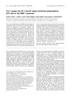
with Ala residues. As illustrated in Fig. 1, three
mutated proteins were obtained: R1CT mutated on
Asn317 (hence N-glycosylable only on Asn492),
R3CT mutated on Asn492 (hence N-glycosylable only
on Asn317) and RCT mutated on both Asn 317 and
492 (not N-glycosylable). NCT corresponds to the
non-mutated N-terminally truncated protein (hence
fully N-glycosylable). All constructs are based on
plasmid pAcSecG2T which contains a polyhedrin pro-
moter and the secretion signal of glycoprotein p67,
followed by the N-terminally truncated nucleolin
sequence.
Nucleolin-derived proteins were expressed using
recombinant baculoviruses prepared as described in
Materials and methods. Sf9 cells were used to propa-
gate baculoviruses, which were then used to infect
High Five cells. In these cells, a maximal level of
recombinant protein secreted in cell culture superna-
tant, estimated at 5–10 mgÆL
)1
of medium, was
observed after 5 days of infection (data not shown).
All four recombinant proteins were purified by
ion-exchange FPLC on an S-Sepharose Fastflow
column (GE Healthcare Pharmacia, Uppsala, Sweden)
as described in Materials and methods. Purified pro-
teins were concentrated and analyzed by SDS ⁄ PAGE.
As shown in Fig. 2 (Coomassie Blue staining), the
purified proteins migrated as thick single bands with
slight differences in their migrations, in the apparent
molecular mass order NCT > R1CT ‡ R3CT ‡ RCT.
This may be attributed to the presence of two glycans
(NCT), one glycan (R1CT, R3CT) or no glycan (RCT)
in the proteins. Immunostaining with anti-nucleolin
IgG confirms that these bands are related to nucleolin,
and also shows minor degradation products between
30 and 37 kDa (Fig. 2, Anti-nucleolin). Purity of pro-
teins in preparations was estimated at over 95%.
Characterization of the glycosylation of
recombinant proteins
In order to check the occupancy of glycosylation sites
and the structure of glycans, GC ⁄ MS, MALDI-TOF
Fig. 1. Schematic structural representation
of (A) human nucleolin and (B) the four
recombinant nucleolin-derived proteins
(NCT, R1CT, R3CT and RCT) produced by a
baculovirus ⁄ insect cell system. The different
domains of full-size human nucleolin are
represented: the acidic N-terminal
domain (N-terminal), RBD1–RBD4 and the
C-terminal RGG domain (C-terminal). The
nuclear localization sequence is represented
as NLS. The N-terminal domain was deleted
in all NCT, R1CT, R3CT and RCT recombi-
nant nucleolin-derived proteins. Positions of
the mutated (N > A) and non-mutated (N)
Asn residues of N-glycosylation sites are
indicated.
Glycosylation enhances nucleolin self-interactions M E. Losfeld et al.
2554 FEBS Journal 278 (2011) 2552–2564 ª 2011 The Authors Journal compilation ª 2011 FEBS
analyses and immunostaining assays were performed.
Monosaccharide composition of the glycans was deter-
mined using GC ⁄ MS analysis after heptafluorobutyric
anhydride treatment (Table 1). According to this
method [27], the N-acetylglucosamine (GlcNAc) resi-
due forming the N-glycosidic bond is converted to glu-
cosamine (GlcNH
2
). The GlcNH
2
peak (retention time
of 12.39 min) was used as a reference, and its corrected
area was considered as corresponding to one monosac-
charide residue. The results summarized in Table 1
suggest the presence of mannose (Man), fucose (Fuc)
and GlcNAc residues. Only three Man residues were
detected, for one GlcNH
2
residue, which suggests the
presence of paucimannosidic structures rather than
high mannose structures. We detected 1.1 GlcNAc that
may correspond to the GlcNAc residue linked to the
N-linked GlcNAc. Furthermore, we detected 0.8 or 0.9
residue of Fuc, which would correspond to a Fuc
linked in a1-3 on the N-glycan core, a feature specific
to insect glycans [28]. In the RCT protein, as expected,
no monosaccharide was found. This result also demon-
strates that the third consensus site (Asn478), not
mutated in any construction, is not occupied. This is
in accordance with the absence of glycosylation on
Asn478 observed in the glycosylated nucleolin isoforms
from human cells [25].
In order to verify the presence of the fucosylated
core, specific immunostaining of this structure was per-
formed with polyclonal anti horseradish peroxidase
(HRP) IgG as described by Kurosaka et al. [29]. As
shown in Fig. 2 (Anti-HRP), the presence of a fucosy-
lated core was revealed for the glycosylable NCT,
R1CT and R3CT forms, but not for non-glycosylable
protein RCT. Other faint bands probably correspond
to both nucleolin-derived degradation products (evi-
denced in Fig. 2, Anti-nucleolin) and contaminating
insect cell fucosylated proteins. Taken together, these
results strongly suggest the presence of fucosylated
paucimannosidic glycans on the potentially N-glycosy-
lable proteins. Lastly, occupancy of the glycosylation
sites was analyzed by MS. MALDI-TOF analysis
of glycopeptides from tryptic digest of recombinant
protein was used to localize the N-glycosylated sites
from the NCT protein, in order to verify, by compari-
son with RCT, that the third consensus sequence con-
taining Asn478 was not occupied. After enrichment of
glycopeptides as described in Materials and methods,
MS analysis (Fig. 3) of NCT tryptic peptides revea-
led mass peaks at 3373.3, 3520.3, 3629.4, 3679.4
and 3775.5 Da which could correspond to peptides:
Fig. 2. SDS ⁄ PAGE and immunoblotting of purified recombinant nucleolin-derived proteins NCT, R1CT, R3CT and RCT. Proteins were sepa-
rated by SDS ⁄ PAGE (10%) and processed as described in Materials and methods. Proteins were either stained with Coomassie Blue or
transferred onto nitrocellulose membranes for immunostaining with mouse monoclonal anti-nucleolin IgG (anti-C23, MS-3) by electrochemilu-
minescence (Anti-nucleolin), and with rabbit polyclonal anti-HRP IgG followed by NBT ⁄ BCIP staining (Anti-HRP). Anti-HRP staining reveals
a1–3 fucosylated glycans present in insect-expressed glycoproteins [29]. DF, dye front.
Table 1. Monosaccharide composition of the four nucleolin-derived
recombinant isoforms as determined by heptafluorobutyrate deriva-
tion and GC ⁄ MS.
Monosaccharide
Recombinant protein
NCT R1CT R3CT RCT
GlcNH
2
11 1 0
Man 3 2.9 2.9 0
Fuc 0.8 0.9 0.9 0
GlcNAc 1.1 1.2 1.2 0
M E. Losfeld et al. Glycosylation enhances nucleolin self-interactions
FEBS Journal 278 (2011) 2552–2564 ª 2011 The Authors Journal compilation ª 2011 FEBS 2555
298-VEGTEPTTAFNLFVGNLNFNK-318 ( 2312.16 Da)
and 487-TLVLSNLSYATEETLQEVFEK-508 (2501.
27 Da), not phosphorylated or tri-phosphorylated, and
carrying paucimannosidic mono-, di-fucosylated or
non-fucosylated N-glycans (Fig. 3A). We hypothesize
that phosphorylation of the recombinant proteins
would result from the presence in the cell culture med-
ium of kinases released from lysed baculovirus-infected
cells. The slight difference between the theoretical and
observed masses can be explained by the use of the
linear positive-ion mode for mass determination of
glycopeptides which could impair precision of this
determination. In contrast, no peptides between 1887
and 2049 Da were found (Fig. 3B, brackets) which
would correspond to a 995 Da peptide containing
Asn478 linked to 892–1054 Da paucimannose fuco-
sylated or non-fucosylated glycans. This result con-
firms that Asn478 is not glycosylated.
In conclusion, the structural analyses of the four
recombinant nucleolin-derived proteins have demon-
strated the presence of paucimannosidic fucosylated
glycans on both Asn317 and Asn492 of the NCT pro-
tein, on Asn492 of R1CT and on Asn317 of R3CT,
and their absence on the RCT protein.
Since directed mutagenesis can sometimes lead to
protein misfolding, CD and fluorimetry were used to
investigate the folding of the recombinant nucleolin-
derived proteins.
Interference of N-glycosylation with the structure
of the recombinant nucleolin-derived proteins
The two minima observed at 210 and 222 nm in the
CD spectra (Fig. 4) show a dominant a-helical content
in all four recombinant nucleolin isoforms with, how-
ever, significant ellipticity differences between the pro-
teins (Fig. 4). These differences can be attributed to
conformation discrepancies that result from the state
of glycosylation of the protein. Indeed, a minimal ellip-
ticity at 222 nm for the unmutated fully glycosylated
protein NCT and a maximal ellipticity at 222 nm for
the single mutated protein R3CT (glycosylated on
Asn317) can be observed (Fig. 4). Close and intermedi-
ary ellipticities on the whole spectrum can be observed
for R1CT (glycosylated on Asn492) and double
mutated unglycosylated RCT (Fig. 4). These differ-
ences in ellipticity of the proteins suggest that glycosyl-
ation on both Asn317 and Asn492 probably interfere
with conformation of the protein. Glycosylation of
Asn317 increases the a-helical content of R3CT
whereas glycosylation of Asn492 decreases the a-helical
content of both R1CT and NCT. These differences in
the measured ellipticity could also be interpreted as a
consequence of the glycosylation on the propensity of
NCT to oligomerization when glycosylated in position
492. This glycosylation may result, for example, in a
loss of a-helical structure for b-sheet structure
Fig. 3. MALDI-TOF mass spectra of the glycopeptides released
from tryptic digest of recombinant nucleolin-derived proteins NCT
and RCT. As described in Materials and methods, nucleolin iso-
forms were separated by SDS ⁄ PAGE. The bands were excised and
treated with trypsin. Glycopeptides were enriched by chromatogra-
phy on Sepharose 4B and, after purification, mixed with a sinapinic
acid matrix. Each sample was analyzed on a Voyager DE-STR
MALDI-TOF instrument in the linear positive-ion mode using an
accelerating voltage of 25 kV. (A) Spectra of glycopeptides from
NCT and RCT between 2500 and 4000 Da containing peptides
potentially glycosylated on Asn317 and Asn492: 3373.3 = 2312 Da
+ GlcNAc
2
Man
3
Fuc + Na
+
; 3520.3 = 2312 Da + GlcNAc
2
Man
3
Fuc
2
+Na
+
; 3629.4 = 2501 Da + GlcNAc
2
Man
3
triphosphate; 3679.4
= 2312 Da + GlcNAc
2
Man
4
Fuc
2
+Na
+
; 3775.5 = 2501 Da + Glc-
NAc
2
Man
3
Fuc triphosphate. (B) Spectra of glycopeptides from NCT
and RCT between 1000 and 2500 Da. The bar marks the region
potentially containing glycopeptide glycosylated on Asn478.
Glycosylation enhances nucleolin self-interactions M E. Losfeld et al.
2556 FEBS Journal 278 (2011) 2552–2564 ª 2011 The Authors Journal compilation ª 2011 FEBS
currently observed in self-associated proteins. We
therefore checked this hypothesis by studying the self-
association of the four recombinant isoforms (see the
role of N-glycosylation on the self-association proper-
ties of nucleolin later).
The nucleolin sequence possesses two tryptophan res-
idues at positions 481 and 644 [30]. Figure 5 shows the
fluorescence emission spectra of the four nucleolin-
derived proteins in the wavelength range 300–440 nm.
We can observe maximal emission between 340 and
360 nm. A similar tendency between the fluorescence
intensity and CD spectra is observed. A minimal inten-
sity of fluorescence at about 350 nm is observed for
NCT and a maximal intensity for R3CT. Close and
intermediary fluorescence intensities are observed for
R1CT and RCT (Fig. 5). Interestingly, the wavelength
of maximal emission for both R1CT and NCT (both
glycosylated on Asn492) undergoes a red shift com-
pared with the wavelength of maximum emission for
the double mutated unglycosylated RCT and for R3CT
(both unglycosylated on Asn492) (Fig. 5). This is not
surprising as one of the Trp residues (Trp481), which is
close to the glycosylated site Asn492, becomes probably
more hydrated and therefore more exposed to the sol-
vent when Asn492 is glycosylated.
This more exposed Trp observed when Asn492 is
glycosylated can explain the significantly modified
structures of NCT and R1CT, compared with R3CT
and RCT (Fig. 5). It also indicates that the occupancy
of Asn492 by an N-glycan may either modify the con-
formation of proteins or facilitate the oligomerization
of NCT and R1CT, or both (see the next section).
Studies of the role of N-glycosylation on the
self-association properties of the recombinant
nucleolin-derived proteins
To assess the influence of N-glycans on the self-associ-
ation properties of nucleolin, we investigated the abil-
ity of the four nucleolin isoforms to interact through
PAGE in native conditions and SPR assays.
In a first step, we investigated the PAGE behavior of
the recombinant nucleolin-derived proteins in native
conditions. Since the theoretical isoelectric point of the
proteins is estimated at 8.8 (calculated according to
PAGE was per-
formed at a lower pH (pH 7.0). As shown in Fig.6,
unlike R3CT and RCT which markedly migrated as sin-
gle major bands towards the cathode, NCT and R1CT
exhibited no migration or very poor migration on the
gel. Only faint bands can be observed in the upper part
of the gel. This suggests that both proteins may be self-
associated. The integrity of proteins separated in non-
denaturing conditions was checked by submitting the
proteins to a second electrophoresis on SDS ⁄ PAGE
with b-mercaptoethanol as described in Materials and
methods. All proteins migrated as single 45 kDa bands
(data not shown). Taken together, these results suggest
that the presence of a glycan on the RBD3 domain
modifies the propensity of nucleolin to self-associate.
Furthermore, our spectroscopic st udies (see a bove) s uggest
that the presence of a glycan located on Asn492 changes
the exposure to the solvent of the protein region close to
Trp481 that seems responsible for its self-association.
To confirm the results obtained by PAGE, SPR
studies were undertaken on a BIAcore 3000 system
using NCT, R1CT, R3CT and RCT immobilized on a
Fig. 4. CD spectra of the recombinant nucleolin-derived proteins
NCT, R1CT, R3CT and RCT. Spectra were obtained on 2 mg of
purified proteins with a 1 mm cell at 25 °C and recorded between
190 and 250 nm with an increment of 0.5 nm and an integration
time of 2 s.
Fig. 5. Fluorescence spectra of the recombinant nucleolin-derived
proteins NCT, R1CT, R3CT and RCT. Fluorescence of tryptophan
was recorded on purified proteins NCT, R1CT, R3CT and RCT
between 300 and 450 nm, as described in Materials and methods.
M E. Losfeld et al. Glycosylation enhances nucleolin self-interactions
FEBS Journal 278 (2011) 2552–2564 ª 2011 The Authors Journal compilation ª 2011 FEBS 2557
CM5 sensor chip (ligands) and injected as analytes.
Various concentrations between 0.25 and 4 lm were
injected. Figure 7 shows a representative experiment
from three sets of experiments performed on different
CM5 sensor chips. Interestingly, self-interactions
between all nucleolin-derived isoforms can be
observed. However, the affinity of NCT, R1CT or
R3CT self-interactions was about 100-fold higher than
that of RCT. Furthermore, although the K
d
of R3CT
self-interactions was similar to that of NCT and
R1CT, R3CT self-binding was about four-fold and
three-fold lower than that of NCT and R1CT, respec-
tively. These results are in agreement with the PAGE
results (Fig. 6) that demonstrate different migration
patterns for NCT and R1CT, compared with R3CT
and RCT. Hence, our results confirm the observation
that nucleolin is able to interact with itself and to
oligomerize [22], a property that could play an impor-
tant role for its receptor functions. We show here
that N-glycosylation, and most particularly RBD3
Fig. 6. Migration patterns of the recombinant nucleolin-derived pro-
teins NCT, R1CT, R3CT and RCT in PAGE in non-denaturing condi-
tions. Purified recombinant proteins (10 lg of each protein per lane)
were loaded onto a PAGE 7.5% gel and submitted to electrophore-
sis in non-denaturing conditions as described in Materials and
methods. Proteins were revealed by Coomassie Blue staining.
+, cathode of the generator; ), anode of the generator.
Fig. 7. Study by SPR (BIAcore) of the self-interactions of the
recombinant nucleolin-derived proteins NCT, R1CT, R3CT and RCT.
The SPR sensorgrams shown are from an experiment which is rep-
resentative of a set of three separate experiments on different sen-
sor chips with similar results. Recombinant nucleolin-derived
proteins NCT, R1CT, R3CT and RCT were immobilized onto a CM5
sensor chip. The same proteins were used as analytes at different
concentrations (250–4000 n
M) at a flow rate of 5 lLÆmin
)1
,as
described in Materials and methods. Association was studied during
3 min and dissociation during 10 min at a flow rate of 5 lLÆmin
)1
of
HBS. Constants were estimated using
BIAEVALUATION 3.1 with the
Langmuir 1 : 1 formula. The Scatchard plots derived from these
data at equilibrium are presented as inserts. RU, response unit.
Glycosylation enhances nucleolin self-interactions M E. Losfeld et al.
2558 FEBS Journal 278 (2011) 2552–2564 ª 2011 The Authors Journal compilation ª 2011 FEBS
N-glycosylation, strongly enhances self-interactions of
the nucleolin-derived proteins.
N-glycosylation does not influence the
interactions of the recombinant nucleolin-derived
proteins with lactoferrin
Cell-surface expressed nucleolin has been described as
receptor or co-receptor of many proteins and molecules
[1,2]. We previously demonstrated that nucleolin inter-
acts with lactoferrin with medium affinity
(K
d
= 0.24 lm) through the RGG C-terminal domain
of nucleolin, and that nucleolin participates, like midki-
ne [10–12], in endocytosis and nuclear targeting of its
ligand [13]. Since N-glycosylation interferes with
the structure and the self-interaction properties of the
nucleolin-derived proteins, we investigated the ability of
the different proteins to interact with lactoferrin using
SPR (BIAcore 3000). With this aim, NCT, R1CT,
R3CT and RCT were immobilized onto a CM5 sensor
chip and concentrations ranging from 50 to 800 nm of
human lactoferrin were injected. The results of a repre-
sentative study among three separate studies are shown
in Fig. 8. We observe that all nucleolin-derived proteins,
regardless of their N-glycosylation status, interact with
similar binding parameters, with an affinity of about
several hundred nanomoles, similar to that of native
full-size nucleolin reported by Legrand et al. [13].
These results suggest that N-glycosylation does not
interfere with the ability of the nucleolin-derived pro-
teins to interact with lactoferrin, one of its major sur-
face ligands.
Discussion
Intracellular nucleolin is a ubiquitous protein that par-
ticipates in important cellular events, like ribosome
biogenesis, chromatin organization, apoptosis and reg-
ulatory activity of transcription factors [1,2]. Cell-sur-
face-expressed nucleolin has been described as
interacting with extracellular components like laminin
[5,6] or proteoglycans [31], but also as a receptor for
apolipoproteins [4] or l-selectin [32] and as an internal-
izing receptor for lactoferrin [13], midkine [10–12] or
the gp120 protein of HIV [15,16]. However, many
aspects of the structure–function relationships of sur-
face nucleolin remain unknown. In fact, only the struc-
tures of RBD1 and RBD2 have been determined [24].
Our group has previously demonstrated that nucleolin
undergoes N- and O-glycosylations [25] and that
N-glycosylation is an essential requirement for its cell
surface expression [14]. Furthermore, we have recently
described the ability of nucleolin to trigger calcium
Fig. 8. Study by SPR (BIAcore) of the interactions of human lac-
toferrin with the recombinant nucleolin-derived proteins NCT,
R1CT, R3CT and RCT immobilized onto a CM5 sensor chip. The
SPR sensorgrams shown are from an experiment which is repre-
sentative of a set of three separate experiments on different sen-
sor chips with similar results. Recombinant nucleolin-derived
proteins NCT, R1CT, R3CT and RCT were immobilized onto a
CM5 sensor chip. Human lactoferrin was used as analyte at dif-
ferent concentrations (50–800 n
M) at a flow rate of 5 lLÆmin
)1
,as
described in Materials and methods. Association was studied dur-
ing 3 min and dissociation during 10 min at a flow rate of
5 lLÆmin
)1
of HBS. Constants were estimated using BIAEVALUATION
3.1 with the Langmuir 1 : 1 formula. The Scatchard plots derived
from these data at equilibrium are presented as inserts. RU,
response unit.
M E. Losfeld et al. Glycosylation enhances nucleolin self-interactions
FEBS Journal 278 (2011) 2552–2564 ª 2011 The Authors Journal compilation ª 2011 FEBS 2559
entry into cells through ligand binding, probably by
interacting with other still-unknown cell surface signal-
ing molecules [14]. The exact role(s) of N-glycans on
surface nucleolin remain(s) unspecified but we hypoth-
esized that they would interfere in the structure of
nucleolin and hence in its functions and interaction
abilities.
Here, in agreement with previous observations made
with recombinant nucleolin variants produced in Esc-
herichia coli [22], we confirm that all the nucleolin-
derived proteins are able to self-interact. Our results
also indicate that the absence of residues 1–292 does
not prevent the self-interactions, and hence support
previous evidence that the central domain of nucleolin
is involved in these interactions [22]. Most importantly,
we demonstrate that N-glycosylation strongly enhances
the self-interactions of nucleolin. This evidence was
gained by producing recombinant nucleolin isoforms
carrying or not paucimannosidic fucosylated N-glycans
by a baculovirus⁄ insect cell system. Although such
N-glycans are obviously different from those of natural
nucleolin [25], we hypothesized that their mere pres-
ence within the N-glycosylation sites would modify the
ability of the nucleolin-derived proteins to oligomerize.
The propensity of the nucleolin-derived proteins to
self-interact [22], regardless of their glycan content,
was ascertained through SPR which showed self-inter-
actions of the non-glycosylated isoform (RCT) with a
K
d
of 18.3 lm. Most interestingly, the fully N-glycosy-
lated isoform (NCT) or N-glycosylated on the sole
RBD3 (R1CT) exhibited 100-fold higher affinities than
RCT. Similar affinity but with a lower binding capac-
ity than NCT and R1CT was observed with the iso-
form N-glycosylated on the sole RBD1 (R3CT). This
suggests that the occupancy of the glycosylation sites,
and most particularly Asn492 (RBD3), is a requisite
for strong nucleolin self-interactions. These stronger
interactions of the NCT and R1CT isoforms, com-
pared with R3CT and RCT, were confirmed by native
PAGE analysis. To explain this enhancement of NCT
and R1CT self-interactions, and taking into account
CD and fluorimetry results, we hypothesize that N-gly-
cans, in particular the N-glycan on Asn492, modify
the structure of the protein. Such modification could
expose hydrophobic regions of the protein involved in
high affinity nucleolin self-interactions.
Whereas cell surface nucleolin appears to be located
near the lipid raft or associated with it [10], it is not
known how the molecule is presented at the cell sur-
face. Although nucleolin is not an integral membrane
protein, it is able to act as a receptor for a large num-
ber of molecules and to internalize some of them
[7,10–13] or, most unexpectedly, to induce cellular
events [7,14] by triggering calcium entry in cells [14].
To explain this later observation, we hypothesized that
nucleolin could act as a co-receptor able to interact
with cell surface signaling receptors following ligand
binding [14]. This implies a redistribution of nucleolin
at the surface of cells following ligand binding, and a
possible role of glycosylation in the topology of nucle-
olin on cells. In strong support of this hypothesis, our
results suggest that N-glycosylation probably influ-
ences the self-interaction properties of nucleolin, while
it does not affect the binding of ligands, such as lacto-
ferrin. It may also be hypothesized that whereas the
C-terminal domain is involved in the interactions of
surface nucleolin with most of its ligands, the RBD-
containing central domain is involved in the protein
self-interactions.
Interestingly, preliminary studies show that N-glyco-
sylation may modulate binding of the nucleolin-derived
protein to heparin (data not shown). This suggests that
glycosylation could modulate the interaction of nucleo-
lin with sulfated glycosaminoglycans, its distribution
on the surface of cells, and its ability to interact with
signaling molecules. Of course, we should bear in mind
that both composition and length of the N-glycans of
insect cells are different from human ones. It would
thus be interesting to transpose our system in human-
ized insect cells [33].
In conclusion, many questions remain to be resolved
concerning the expression of nucleolin at the surface of
cells and its trafficking, but also concerning the involve-
ment of glycosylation in these processes. As demon-
strated previously [14], glycosylation is an essential
requirement for cell surface expression. Here, our results
with N-terminally-truncated nucleolin suggest that N-
glycosylation may influence the structure of natural nu-
cleolin in a way that enhances its ability to self-interact.
The exact roles of high affinity nucleolin self-interac-
tions of surface nucleolin, in particular in the activation
of signaling partners, are still to be elucidated.
Materials and methods
Cells
Spodoptera frugiperda (Sf9) and Trichoplusia ni (High Five)
insect cells were purchased from Invitrogen (Carlsbad, CA,
USA). Sf9 and High Five cells were respectively grown
in Max-XP medium (BD Biosciences, Le Pont de Claix,
France) containing 1% (v ⁄ v) ultraglutamine (Lonza, Basel,
Switzerland), 1% fetal bovine serum (Cambrex, Emerain-
ville, France) and 50 lgÆmL
)1
gentamycin, and in Express
Five medium (Cambrex) containing 10% (v ⁄ v) ultragluta-
mine (Lonza) at 27 °C.
Glycosylation enhances nucleolin self-interactions M E. Losfeld et al.
2560 FEBS Journal 278 (2011) 2552–2564 ª 2011 The Authors Journal compilation ª 2011 FEBS
DNA, plasmids, site-directed mutagenesis and
baculovirus preparation
Human nucleolin cDNA was previously obtained from
MDA-MB231 cells (ATCC) in our laboratory and cloned
into pTRE-HA plasmid from Clontech (Takara Bio Europe,
Saint-Germain-en-Laye, France) [13]. Oligonucleotide prim-
ers were synthesized by Eurogentec (Lie
`
ge, Belgium) and
restriction enzymes were from New England Biolabs (Evry,
France). pAcSecDGST was obtained from plasmid pAc-
SecG2T (Pharmingen-BD Biosciences, Le Pont de Claix,
France) by inserting a BamH1 site before the GST sequence
in order to remove it (Directed mutagenesis Quickchange XL
II kit, Stratagene-Agilent, Santa Clara, CA, USA). The
primers used were 5¢-CCTTTGCGGATCTGATGTCCCCT
GGATCCGC-3¢ and 5¢-GCACAAGGCCCTTAATTTTC
CAATAACCGGA-3¢.
pTRE-HA containing the coding region of nucleolin
cDNA was mutated, or not, on the glycosylation sites (Direc-
ted mutagenesis Quickchange XL II kit). Mutation of
Asn317 (N > A) was achieved with primers 5¢-CAATCTCT
TTGTTGGAAACCTAAACTTTCAGAAATCTGCTCCT
GAATTAAAAACTGG-3¢ and 5¢-CCAGTTTTTAATTCA
GGAGCAGATTTCTGAAAGTTTAGGTTTCCAACAA
AGAGATTG-3¢. Mutation of Asn492 (N > A) was achie-
ved with primers 5¢-GGTGAATCAAAAACTCTGGTTTT
AAGCCAGCTCTCCTACAGTGCAACAGAAGAAACTC-3¢
and 5¢-GAGTTTCTTCTGTTGCACTGTAGGAGAGCTG
GCTTAAAACCAGAGTTTTTGATTCACC-3¢.
The coding region of nucleolin cDNAs corresponding to
the N-terminal truncated nucleolin sequence mutated or
not on glycosylation sites was amplified by PCR with con-
current introduction of BamH1 and EcoR1 sites at the 5¢
and 3¢ terminus, respectively. The primers used were 5¢-G
CCAAACAGAAAGCAGCTCCTGGATCCAAGAAACA
G-3¢ and 5¢-GTGCCTTCCACTTTCTGTTTCTTGGATCC
AGGAGCTGCTTTC-3¢. After sequence verification, the
BamH1- and EcoR1-digested inserts were cloned into sim-
ilarly digested pAcSecDGST. The resulting constructs were
used to co-transfect Sf9 cells with linearized baculovirus
BD BaculoGoldÔ Bright (Pharmingen-BD Bio-sciences)
by addition of 5 lL of FlyfectinÔ (OZ Biosciences,
Marseille, France) for 500 ng of recombinant plasmid and
100 ng of linearized baculovirus. After plasmid recombina-
tion and circularization of baculovirus, the infected cells
are able to express the green fluorescent protein. The level
of infection was thus verified by flow cytofluorimetry on
a FACScalibur cytometer (BD Bioscience). The super-
natant of transfected cells containing recombinant bacul-
oviruses was collected after 5 days. Viruses were amplified
by successive infections for obtaining a high multiplic-
ity of infection (MOI) (‡ 2 · 10
5
virusÆlL
)1
). Expression
of recombinant nucleolin-derived proteins was achieved
by infecting High Five cells with the baculovirus
suspensions.
Expression and purification of recombinant
nucleolin-derived proteins
Production of recombinant proteins was done in High Five
cells grown in 175 cm
2
flasks. Cells were infected by addi-
tion of 4% (v ⁄ v) of supernatant at high MOI in the culture
medium of 70% confluent cells. After 5 days, the superna-
tant was collected and stored at ) 20 °C.
A chromatography procedure was used to purify the
recombinant forms of nucleolin from insect cell superna-
tants. The purification was made by FPLC (GE Healthcare
Pharmacia, Uppsala, Sweden) using an ion exchange
S-Sepharose Fast Flow 5 · 2 cm column (GE Healthcare
Pharmacia). The supernatant were diluted in buffer A
[Tris ⁄ HCl 50 mm pH 7.5; MgCl
2
5mm; EDTA 0.1 mm;
Pefabloc 1 mm (Roche Diagnostics, Meylan, France)] with
addition of 5% (v ⁄ v) of EDTA 100 mm and 2% (v ⁄ v) of
NaOH 1 m. The elution was performed with a gradient
from 0% to 60% of buffer B (Tris ⁄ HCl 50 mm pH 7.5;
MgCl
2
5mm; EDTA 0.1 mm; NaCl 1 m; Pefabloc 1 mm)
for 60 min at a rate of 1 mLÆmin
)1
, followed by a gradient
from 60% to 100% of buffer B for 15 min at a rate of
1mLÆmin
)1
. The protein content of fractions was concen-
trated by centrifugation (3000 g) on a Vivaspin (Sartorius
AG, Goettingen, Germany) concentrator and dialyzed in
ammonium bicarbonate 200 mm.
GC
⁄
MS analysis of nucleolin monosaccharides
The four nucleolin isoforms were separated by SDS ⁄ PAGE
(4 ⁄ 7.5%, 2 lgÆlane
)1
) and electro-transferred on poly(vinyli-
dene difluoride) membrane. After Ponceau S staining, the
bands of nucleolin were cut out, washed and dried. The
preparation of samples was as described previously [25].
Samples were submitted to methanolysis (20 h at 80 °Cin
500 lL of anhydrous methanol containing 0.5 m gaseous
HCl). After the samples were dried under a stream of nitro-
gen, they were added to 200 lL of acetonitrile and 25 lL
of heptafluorobutyric anhydride and heated for 30 min at
150 °C.
After evaporation of the solvents, the samples were dis-
solved in 200 lL of dried acetonitrile and 1 lL was injected
in the Ross injector (260 °C) of a Carlo Erba GC 8000 gas
chromatograph (Carlo Erba, Sabadell, Spain) equipped
with a 25 m · 0.32 mm CP-Sil5 CB low bleed ⁄ Ms capillary
column, 0.25 lm film phase (Chrompack, Les Ulis, France).
The sample was analyzed using a program starting at 90 °C
for 3 min, followed by an increase (50 °CÆmin
)1
) until
260 °C was reached. The column was coupled to a Finni-
gan Automass II mass spectrophotometer (Thermo Finni-
gan, Courtaboeuf, France). Analysis was performed in the
electron impact mode (ionization energy 70 eV; source tem-
perature 150 °C). Quantitation of various constituents was
performed using the total ion count of the MS detector and
the xcalibur software (Thermo Finnigan).
M E. Losfeld et al. Glycosylation enhances nucleolin self-interactions
FEBS Journal 278 (2011) 2552–2564 ª 2011 The Authors Journal compilation ª 2011 FEBS 2561
Glycopeptide characterization by MS
To determine the glycosylated peptides present in recombi-
nant proteins, NCT and RCT proteins were submitted to
reduction alkylation and digested by trypsin. Glycopeptides
were isolated and analyzed by MS. Reduction was realized
on 100 lg of each protein using 25 lL ammonium bicar-
bonate 50 mm, pH 8, and 5 lL of dithiothreitol (DTT)
100 mm for 1 h at 37 °C. Proteins were alkylated by addi-
tion of 6 lL iodoacetamide 200 mm for 1 h at 37 °C
shielded from the light. Alkylation was stopped by 1 lLof
DTT 100 mm during 20 min at 37 °C. Proteins were then
digested by addition of 10 lL trypsin and 2 lL CaCl
2
at
37 °C during 16 h. Reaction was stopped by 2 lL trifluoro-
acetic acid 5%.
As described in [34], the glycopeptides obtained were
enriched on 15 lL Sepharose 4B in a butanol ⁄ etha-
nol ⁄ water (4 : 1 : 1, v ⁄ v ⁄ v) buffer. After 45 min, the sam-
ples were added to microtubes Minisorb (Nunc AS,
Roskilde, Denmark), centrifuged at 11 000 g, and incubated
for 30 min in the presence of 300 lL ethanol 50%. Super-
natants were dried under N
2
flux.
The peptide-containing solution was mixed with 1 lLof
matrix solution suitable for glycopeptides obtained by satu-
rating a water ⁄ acetonitrile 50 : 50 (v ⁄ v) and trifluoroacetic
acid 3% (v ⁄ v) solution with sinapinic acid.
A total of 1 l L of each sample was spotted onto the tar-
get, air-dried and analyzed on a Voyager DE-STR
MALDI-TOF instrument in the linear positive-ion mode by
delayed extraction using an accelerating voltage of 25 kV.
PAGE analysis
For SDS ⁄ PAGE, purified proteins (3 lg) were treated for
10 min at 100 °Cby3· buffer [Tris ⁄ HCl 100 mm,pH
6.8; SDS 6% (w ⁄ v); sucrose 30% (w ⁄ v); b-mercaptoetha-
nol 15% (v ⁄ v); bromophenol 0.015% (v ⁄ v)]. Samples were
separated on 10% SDS ⁄ PAGE gels (Mini-Protean 3 sys-
tem, Bio-Rad, Marnes la Coquette, France). Migration
was performed at 120 V in running buffer [Tris
25 mm ⁄ glycin 0.192 m, pH 8.3, SDS 0.1% (w ⁄ v)]. Proteins
were stained with Coomassie Blue [Coomassie Blue 0.25%
(w ⁄ v); methanol 50% (v ⁄ v); acetic acid 10% (v ⁄ v)]. De-
staining was done in the presence of ethanol 20% (v ⁄ v)
and acetic acid 7.5% (v ⁄ v).
For PAGE in non-denaturing conditions, proteins were
separated at pH 7.0. Vertical PAGE [acrylamide ⁄ bisacryla-
mide (30 ⁄ 1) 7.5% (v ⁄ v), Tris ⁄ HCL 50 mm, pH 7.0; ammo-
nium persulfate 1.5% (v ⁄ v), riboflavin 0.004% (w ⁄ v),
tetramethylethylenediamine 0.1% (v ⁄ v)] was performed on
a Protean II (Bio-Rad) system. 15 lg of each protein were
loaded in 25 lL of running buffer with 10% glycerol (v⁄ v)
with b-mercaptoethanol 15% (v ⁄ v). Proteins were submit-
ted to migration at 40 V in a Tris ⁄ sodium diethylbarbitu-
rate 50 m m, pH 7.0, running buffer. Proteins were stained
with Coomassie Blue [Coomassie Blue 0.25% (w ⁄ v); metha-
nol 50% (v ⁄ v); acetic acid 10% (v ⁄ v)]. Destaining was done
in the presence of ethanol 20% (v ⁄ v) and acetic acid 7.5%
(v ⁄ v).
For bidimensional electrophoresis, 15 lg of proteins were
loaded onto two lanes of PAGE in non-denaturing condi-
tions at pH 7.0 (as described above). After migration, one
of the two lanes was cut and stained with Coomassie Blue.
The second lane was cut and boiled in 3 · sample buffer
with b-mercaptoethanol for 30 min. After treatment, the
lane was set at the top of a 7.5% SDS ⁄ PAGE. Migration
was performed for 16 h at 40 V in running buffer [Tris
25 mm ⁄ glycin 0.192 m, pH 8.7; SDS 0.1% (w ⁄ v)]. Proteins
were stained with Coomassie Blue [Coomassie Blue 0.25%
(w ⁄ v); methanol 50% (v ⁄ v); acetic acid 10% (v ⁄ v)]. De-
staining was done in the presence of ethanol 20% (v ⁄ v) and
acetic acid 7.5% (v ⁄ v).
Immunoblotting and fucosylated core detection
NCT, R1CT, R3CT and RCT proteins were separated by
SDS ⁄ PAGE (10%) as described above and electro-trans-
ferred onto nitrocellulose membranes. Immunostaining with
anti-human nucleolin IgG was achieved with a mouse
monoclonal IgG anti-C23 (MS-3; Santa Cruz Biotechnol-
ogy, Santa Cruz, CA, USA) at dilution 1 ⁄ 500, and a sec-
ondary goat anti-mouse IgG conjugated to HRP (Sigma
Aldrich, Saint-Louis, MO, USA) at dilution 1 ⁄ 5000. Signals
were detected by autoradiography using the ECL Plus
detection kit (GE Healthcare Amersham, Buckinghamshire,
UK) according to the manufacturer’s instructions.
To specifically detect fucosylated N-glycans, membranes
were incubated first in blocking buffer [Tris ⁄ HCl 20 mm,
pH 7.5; NaCl 0.145 m; freeze-dried skimmed milk 3%
(w ⁄ v); Tween-20 1% (v ⁄ v)] for 2 h, and then incubated
overnight at 4 °C in the same buffer with rabbit anti-HRP
primary IgG (Sigma Aldrich) at 1 ⁄ 2000 dilution. Mem-
branes were then incubated for 1 h at ambient temperature
with goat anti-rabbit IgG coupled to alkaline phosphatase
(Jackson ImmunoResearch, Newmarket, UK) at 1 ⁄ 10 000
dilution [29]. Staining was achieved using 5-bromo-4-
chloro-3-indolyl phosphate ⁄ nitroblue tetrazolium (NBT ⁄
BCIP) (Roche Diagnostics) in Tris ⁄ HCl 0.1 m, pH 9.5;
NaCl 0.1 m ; MgCl
2
0.05 m. Image acquisition was perfor-
med using the gs800 calibrated imaging densitometer
and quantity one software (Bio-Rad).
CD and fluorimetry
CD analysis was performed on a model CD6 Jobin Yvon
ISA (Longjumeau, France) spectropolarimeter with a
1 mm path length quartz cell at 25 °C. Purified proteins
were concentrated at 5 lm in 20 mm phosphate buffer,
pH 7.4. The ellipticity was scanned from 195 to 250 nm
with an increment of 0.5 nm, an integration time of 2 s,
Glycosylation enhances nucleolin self-interactions M E. Losfeld et al.
2562 FEBS Journal 278 (2011) 2552–2564 ª 2011 The Authors Journal compilation ª 2011 FEBS
and a constant band-pass of 2 nm. The signal from the
blank scan was subtracted from the corresponding sample
scan.
Steady-state fluorescence of tryptophan was monitored
on a Fluoromax-2 (Jobin Yvon SPEX) spectrometer at
25 °C. An excitation wavelength of 295 nm was used and
the emission spectrum was scanned from 300 to 450 nm.
The excitation and emission slit widths were set to 3 nm.
The purified proteins were concentrated at 5 lm in 20 mm
sodium phosphate buffer, pH 7.4.
SPR
All materials and chemicals were from BIAcore (GE
Healthcare Europe Gmbh, Uppsala, Sweden). Analyses
were performed at 25 °C on a BIAcore 3000 biosensor.
Hepes-buffered saline (HBS: Hepes ⁄ NaOH 10 mm, pH 7.4;
NaCl 0.15 m; EDTA 3 mm) was used as a running buffer
at 5 lLÆmin
)1
and for the dilution of ligands and analytes.
For the binding assays, N-terminal truncated recombi-
nant forms of nucleolin were immobilized onto the BIAcore
sensor chip CM5 using an amine-coupling kit (BIAcore)
according to the manufacturer’s instructions. Recombinant
nucleolin was immobilized at a concentration of 1–
3 lgÆmL
)1
in 10 mm sodium acetate, pH 3.8, at a 5 lLÆ-
min
)1
flow rate of HBS. Covalent binding resulted in a sig-
nal of 1000 ± 100 resonance units. An empty flow cell was
used as a control for non-specific binding and bulk effects.
For the studies of self-interactions of nucleolin isoforms,
the ligands were injected at several concentrations (ranging
from 0.5 to 4 lm in HBS), at a 5 lLÆmin
)1
flow rate during
3 min. Dissociation was studied during 10 min at a flow
rate of 5 lLÆmin
)1
of HBS. After the dissociation phase,
the sensor chip was regenerated by injection of 10 lLof
NaOH 20 mm at a 5 lLÆmin
)1
flow rate.
For the study of lactoferrin binding to the nucleolin iso-
forms, human lactoferrin was injected at concentrations
ranging from 50 to 800 nm in HBS at a 5 lLÆmin
)1
flow
rate during 3 min. Regeneration was done by injection of
10 lL of NaOH 20 mm at a 5 lLÆmin
)1
flow rate.
The dissociation constant (K
d
) and R
max
± SEM were cal-
culated for each analysis using a method based on the Lang-
muir 1 : 1 binding model (biaevaluation 3.1 software).
Acknowledgements
This work was supported by the Universite
´
des Sci-
ences et Technologies de Lille (Institut Fe
´
de
´
ratif de
Recherche 147, Director Dr J. Mazurier) and the Cen-
tre National de la Recherche Scientifique (CNRS)
(UMR no. 8576; Director Dr J C. Michalski). The
mass spectrometry facility used in this study was
funded by the European Community (FEDER), the
Re
´
gion Nord-Pas de Calais (France), the CNRS and
the Universite
´
des Sciences et Technologies de Lille.
We thank Frank Wien of the synchrotron facilities
SOLEIL at Saint Aubin, France, for his advice and
fruitful scientific discussions for the circular dichroism
experiments.
References
1 Mongelard F & Bouvet P (2007) Nucleolin: a multifac-
eted protein. Trends Cell Biol 17, 80–86.
2 Srivastava M & Pollard HB (1999) Molecular dissection
of nucleolin’s role in growth and cell proliferation: new
insights. FASEB J 13 , 1911–1922.
3 Hovanessian AG, Puvion-Dutilleul F, Nisole S, Svab J,
Perret E, Deng JS & Krust B (2000) The cell-surface-
expressed nucleolin is associated with the actin cytoskel-
eton. Exp Cell Res 261, 312–328.
4 Semenkovich CF, Ostlund RE Jr, Olson MO & Yang
JW (1990) A protein partially expressed on the surface
of HepG2 cells that binds lipoproteins specifically is nu-
cleolin. Biochemistry 29, 9708–9713.
5 Kibbey MC, Johnson B, Petryshyn R, Jucker M &
Kleinman HK (1995) A 110-kD nuclear shuttling pro-
tein, nucleolin, binds to the neurite-promoting IKVAV
site of laminin-1. J Neurosci Res 42, 314–322.
6 Yu D, Schwartz MZ & Petryshyn R (1998) Effect of
laminin on the nuclear localization of nucleolin in rat
intestinal epithelial IEC-6 cells. Biochem Biophys Res
Commun 247, 186–192.
7 Larrucea S, Cambronero R, Gonzalez-Rubio C, Fraile
B, Gamallo C, Fontan G & Lopez-Trascasa M (1999)
Internalization of factor J and cellular signalization
after factor J-cell interaction. Biochem Biophys Res
Commun 266, 51–57.
8 Larrucea S, Gonzalez-Rubio C, Cambronero R, Ballou
B, Bonay P, Lopez-Granados E, Bouvet P, Fontan G,
Fresno M & Lopez-Trascasa M (1998) Cellular adhe-
sion mediated by factor J, a complement inhibitor. Evi-
dence for nucleolin involvement. J Biol Chem 273,
31718–31725.
9 Shi H, Huang Y, Zhou H, Song X, Yuan S, Fu Y &
Luo Y (2007) Nucleolin is a receptor that mediates anti-
angiogenic and antitumor activity of endostatin. Blood
110, 2899–2906.
10 Hovanessian AG (2006) Midkine, a cytokine that inhib-
its HIV infection by binding to the cell surface
expressed nucleolin. Cell Res 16, 174–181.
11 Said EA, Krust B, Nisole S, Svab J, Briand JP & Hov-
anessian AG (2002) The anti-HIV cytokine midkine
binds the cell surface-expressed nucleolin as a low affin-
ity receptor. J Biol Chem 277, 37492–37502.
12 Take M, Tsutsui J, Obama H, Ozawa M, Nakayama
T, Maruyama I, Arima T & Muramatsu T (1994)
Identification of nucleolin as a binding protein for
M E. Losfeld et al. Glycosylation enhances nucleolin self-interactions
FEBS Journal 278 (2011) 2552–2564 ª 2011 The Authors Journal compilation ª 2011 FEBS 2563
midkine (MK) and heparin-binding growth
associated molecule (HB-GAM). J Biochem 116,
1063–1068.
13 Legrand D, Vigie K, Said EA, Elass E, Masson M,
Slomianny MC, Carpentier M, Briand JP, Mazurier J &
Hovanessian AG (2004) Surface nucleolin participates
in both the binding and endocytosis of lactoferrin in
target cells. Eur J Biochem 271, 303–317.
14 Losfeld ME, Khoury DE, Mariot P, Carpentier M,
Krust B, Briand JP, Mazurier J, Hovanessian AG &
Legrand D (2009) The cell surface expressed nucleolin
is a glycoprotein that triggers calcium entry into mam-
malian cells. Exp Cell Res 315, 357–369.
15 Callebaut C, Blanco J, Benkirane N, Krust B, Jacotot
E, Guichard G, Seddiki N, Svab J, Dam E, Muller S
et al. (1998) Identification of V3 loop-binding proteins
as potential receptors implicated in the binding of HIV
particles to CD4(+) cells. J Biol Chem 273, 21988–
21997.
16 Nisole S, Krust B, Callebaut C, Guichard G, Muller S,
Briand JP & Hovanessian AG (1999) The anti-HIV
pseudopeptide HB-19 forms a complex with the cell-
surface-expressed nucleolin independent of heparan
sulfate proteoglycans. J Biol Chem 274, 27875–27884.
17 Barel M, Hovanessian AG, Meibom K, Briand JP,
Dupuis M & Charbit A (2008) A novel receptor–ligand
pathway for entry of Francisella tularensis in monocyte-
like THP-1 cells: interaction between surface nucleolin
and bacterial elongation factor Tu. BMC Microbiol 8,
145.
18 de Verdugo UR, Selinka HC, Huber M, Kramer B,
Kellermann J, Hofschneider PH & Kandolf R (1995)
Characterization of a 100-kilodalton binding protein for
the six serotypes of coxsackie B viruses. J Virol 69,
6751–6757.
19 Sinclair JF & O’Brien AD (2002) Cell surface-localized
nucleolin is a eukaryotic receptor for the adhesin inti-
min-gamma of enterohemorrhagic Escherichia coli
O157:H7. J Biol Chem 277, 2876–2885.
20 Ginisty H, Amalric F & Bouvet P (2001) Two different
combinations of RNA-binding domains determine the
RNA binding specificity of nucleolin. J Biol Chem 276,
14338–14343.
21 Bouvet P, Diaz JJ, Kindbeiter K, Madjar JJ & Amalric
F (1998) Nucleolin interacts with several ribosomal
proteins through its RGG domain. J Biol Chem 273,
19025–19029.
22 Hanakahi LA, Bu Z & Maizels N (2000) The C-termi-
nal domain of nucleolin accelerates nucleic acid anneal-
ing. Biochemistry 39, 15493–15499.
23 Allain FH, Bouvet P, Dieckmann T & Feigon J (2000)
Molecular basis of sequence-specific recognition of pre-
ribosomal RNA by nucleolin. EMBO J 19, 6870–6881.
24 Allain FH, Gilbert DE, Bouvet P & Feigon J (2000)
Solution structure of the two N-terminal RNA-binding
domains of nucleolin and NMR study of the interaction
with its RNA target. J Mol Biol 303 , 227–241.
25 Carpentier M, Morelle W, Coddeville B, Pons A,
Masson M, Mazurier J & Legrand D (2005) Nucleolin
undergoes partial N- and O-glycosylations in the extra-
nuclear cell compartment. Biochemistry 44, 5804–5815.
26 Chen CM, Chiang SY & Yeh NH (1991) Increased
stability of nucleolin in proliferating cells by inhibition
of its self-cleaving activity. J Biol Chem 266, 7754–7758.
27 Pons A, Richet C, Robbe C, Herrmann A, Timmerman
P, Huet G, Leroy Y, Carlstedt I, Capon C & Zanetta
JP (2003) Sequential GC ⁄ MS analysis of sialic acids,
monosaccharides, and amino acids of glycoproteins
on a single sample as heptafluorobutyrate derivatives.
Biochemistry 42
, 8342–8353.
28 Hollister J, Grabenhorst E, Nimtz M, Conradt H &
Jarvis DL (2002) Engineering the protein N-glycosyla-
tion pathway in insect cells for production of bianten-
nary, complex N-glycans. Biochemistry 41,
15093–15104.
29 Kurosaka A, Yano A, Itoh N, Kuroda Y, Nakagawa T
& Kawasaki T (1991) The structure of a neural specific
carbohydrate epitope of horseradish peroxidase recog-
nized by anti-horseradish peroxidase antiserum. J Biol
Chem 266, 4168–4172.
30 Srivastava M, Fleming PJ, Pollard HB & Burns AL
(1989) Cloning and sequencing of the human nucleolin
cDNA. FEBS Lett 250, 99–105.
31 Joo EJ, ten Dam GB, van Kuppevelt TH, Toida T, Lin-
hardt RJ & Kim YS (2005) Nucleolin: acharan sulfate-
binding protein on the surface of cancer cells. Glycobiol-
ogy 15, 1–9.
32 Harms G, Kraft R, Grelle G, Volz B, Dernedde J &
Tauber R (2001) Identification of nucleolin as a new
L-selectin ligand. Biochem J 360, 531–538.
33 Harrison RL & Jarvis DL (2006) Protein N-glycosyla-
tion in the baculovirus-insect cell expression system
and engineering of insect cells to produce ‘mammalian-
ized’ recombinant glycoproteins. Adv Virus Res 68,
159–191.
34 Wada Y, Tajiri M & Yoshida S (2004) Hydrophilic
affinity isolation and MALDI multiple-stage tandem
mass spectrometry of glycopeptides for glycoproteo-
mics. Anal Chem 76, 6560–6565.
Glycosylation enhances nucleolin self-interactions M E. Losfeld et al.
2564 FEBS Journal 278 (2011) 2552–2564 ª 2011 The Authors Journal compilation ª 2011 FEBS