Báo cáo khoa học: On the aggregation properties of FMRP – a link with the FXTAS syndrome? pot
Bạn đang xem bản rút gọn của tài liệu. Xem và tải ngay bản đầy đủ của tài liệu tại đây (756.53 KB, 10 trang )
On the aggregation properties of FMRP – a link with the
FXTAS syndrome?
Ljiljana Sjekloc
´
a*, Kris Pauwels and Annalisa Pastore
MRC National Institute for Medical Research, London, UK
Introduction
The fragile X mental retardation protein (FMRP) is
an 70 kDa human protein encoded by the X-linked
gene FMR1 which is expressed in different organs,
most prominently in brain and gonads [1–3]. FMRP is
a multi-domain protein which contains two Tudor
domains connected to two protein K homology (KH)
domains by an ca. 80 amino acids residue long linker.
This region is followed by a putative intrinsically
unstructured region which contains also arginine- and
glycine-rich (RGG) motifs [4,5]. Co-presence of differ-
ent nucleic acid binding domains in FMRP suggests
that the protein has a prominent capacity to bind
nucleic acids, in particular RNA, as experimentally
confirmed both in vitro and in vivo [6,7].
The cellular role of FMRP is not well understood.
Experimental evidence shows that FMRP binds co-
transcriptionally to certain messenger RNAs forming
messenger ribonucleoprotein (mRNP) particles, which
are exported from the nucleus to the cytoplasm [8]. In
the cytoplasm FMRP associates to microtubules, to
polysomes and to mRNPs and permits the mRNP par-
ticles to be delivered to distal dendrite sites [9]. It has
Keywords
aggregation; fragile X mental retardation
syndrome; fragile X related tremor ataxia
syndrome (FXTAS); protein misfolding;
structure
Correspondence
A. Pastore, MRC National Institute for
Medical Research, The Ridgeway, London
NW7 1AA, UK
Fax: +44 20 8905 4477
Tel: +44 20 8816 2630
E-mail:
*Present address
Department of Cell and Molecular Biology,
Karolinska Institutet, Stockholm SE-171 77,
Sweden
(Received 2 February 2011, revised 20
March 2011, accepted 25 March 2011)
doi:10.1111/j.1742-4658.2011.08108.x
Fragile X mental retardation protein (FMRP) is an RNA binding protein
necessary for correct spatiotemporal control of neuronal gene expression in
humans. Lack of functional FMRP causes fragile X mental retardation,
which is the most common inherited neurodevelopmental disorder in
humans. In a previous study, we described the biochemical and biophysical
aggregation properties of constructs spanning the conserved region of
FMRP and of two other human fragile X related (FXR) proteins, FXR1P
and FXR2P. Here, we show that the same regions have an intrinsic ten-
dency to aggregate and spontaneously misfold towards b-rich structures,
also under non-destabilizing conditions. These findings pave the way to
future studies of the mechanism of formation of FXR-containing ribonu-
cleoprotein granules and suggest a possible link with the as yet poorly
understood FXR proteins’ associated pathologies.
Structured digital abstract
l
FXR2P binds to FXR2P by fluorescence technology (View interaction)
l
FMRP binds to FMRP by electron microscopy (View interaction)
l
FXR1P binds to FXR1P by electron microscopy (View interaction)
Abbreviations
CD, circular dichroism; FMRP, fragile X mental retardation protein; FXR, fragile X related; FXS, fragile X syndrome; FXTAS, fragile X
associated tremor ataxia syndrome; KH, K homology; mRNP, messenger ribonucleoprotein; NDF, N-terminal domain; NES, nuclear export
signal; rCGG, cytosine guanine triribonucleotide; ThT, thioflavin T.
1912 FEBS Journal 278 (2011) 1912–1921 ª 2011 The Authors Journal compilation ª 2011 FEBS
also been shown that messenger RNAs bound to
FMRP are translationally repressed and that, in neu-
rons, FMRP acts in an activity-dependent manner as
an inhibitor of translation initiation ([10] and refer-
ences therein).
Most studies on FMRP are related to its functions
in brain neurons for two reasons. First, the lack of
functional FMRP, due to transcriptional silencing of
FMR1 gene, causes a neurodevelopmental disorder,
fragile X mental retardation syndrome (FXS), the most
common inherited mental disorder in humans. FXS is
characterized by mild to severe mental retardation,
autistic behaviour and, in male patients, macro-orchi-
dism [11]. Second, alteration of FMRP expression,
characterized by increased levels of FMR1 mRNA and
decreased protein levels, can lead to a late onset neuro-
degenerative disorder, the fragile X associated tremor
ataxia syndrome (FXTAS), with symptoms similar to
Parkinsonism, and to premature ovarian failure in
females [12,13]. Two other proteins, fragile X related
(FXR) proteins 1 and 2 (FXR1P and FXR2P), can
compensate partially for lack of FMRP in some
organs of FXS patients, but not in brain and in
gonads, thus emphasizing the crucial role of FMRP in
correct spatiotemporal control of neuronal gene
expression and for normal sexual maturation. FXR1P
and FXR2P are structurally and functionally related
to FMRP and they all together form the FXR protein
family which includes members from different phyla
[14]. All FXR proteins show a high degree of amino
acid sequence conservation in their amino terminus
and central region comprising Tudor and KH
domains, whereas the carboxyl terminus has low
sequence similarity with the only common denomina-
tor being the presence of RGG motifs.
FXR proteins are components of different types of
nuclear and cytoplasmic ribonucleoprotein granules in
which they often co-localize [1,15–19]. FXR proteins
can form hetero-oligomers in vitro and when over-
expressed in cellular systems [20–22], although in
mammalian cells they are believed to preferentially
homo-oligomerize [21]. The oligomerization properties
of FXR proteins are likely to have significant impor-
tance for regulation of their cellular functions as
shown in different model systems. In Drosophila neu-
rons, for instance, the mobility of certain mRNAs is
controlled by FMRP in a concentration-dependent
manner [23]. High levels of transfected Fmrp in mouse
embryonic Fmr1 KO STEK cells induce formation of
cytoplasmic stress granules in which mRNAs are
trapped into repressed mRNP granules [24,25]. In a
recent study, we investigated the oligomerization
properties of human FXR proteins and showed that,
in vitro, multi-domain constructs from the highly con-
served N-terminus have an elevated tendency to aggre-
gate [26]. They self-assemble not just by forming
dimers but through a more complex pattern of self-
association which proceeds in a continuous way from
the monomer to large molecular weight aggregates via
formation of dimeric species. We proposed that this
behaviour is typical of ‘complex-orphan proteins’, i.e.
proteins which exist in the cell as part of large molecu-
lar assemblies. When produced in isolation, they have
an elevated tendency to self-associate.
To further characterize the nature of aggregation of
FXR proteins, we have carried out a study of their
aggregation properties using different approaches. We
identified by in silico analysis potential hot-spots of
aggregation ⁄ fibrillation and showed that they all
cluster in the protein N-terminus. We then studied the
aggregation behaviour of various constructs from
FMRP, FXR1P and FXR2P using complementary
biophysical techniques. We demonstrate that not only
do all constructs have an intrinsic tendency to aggre-
gate but they also undergo an irreversible conforma-
tional transition towards b-enriched structures which
are typical of amyloidogenic diseases such as Alzhei-
mer’s, Parkinson’s and Huntington’s diseases. The
transition occurs spontaneously also under native-like
conditions without any need for fold destabilization.
We propose that our findings could be relevant for
understanding granule formation and could have a link
with the pathogenesis of FXTAS.
Results
FXR proteins present aggregation and
amyloidogenic hot-spots
To understand the aggregation properties of FXR pro-
teins, we first analysed their sequence for predicting
potential aggregative and amyloidogenic hot-spots
in polypeptides. The results all suggest the presence of
several aggregation hot-spots which cluster in the
highly conserved N-terminal 400 amino acids
(Fig. 1A) whereas no sequence was identified in the
C-terminal region of any of the FXR sequences, includ-
ing the most ancestral FMRP homologue dFMR1 from
Drosophila melanogaster. Two putative amyloidogenic
regions are present in the NDF of the human FXR
proteins (sequences YVIEYA and TYNEIV). Two
more hot-spots amongst the nine detected in FMRP by
waltz with reliability higher than 90% correspond to
the residues 166–174 located in the linker region con-
necting the Tudor and KH domains and to residues
303–308 (LIQEIV) in the second KH domain.
L. Sjekloc
´
a et al. Aggregation properties of fragile X related proteins
FEBS Journal 278 (2011) 1912–1921 ª 2011 The Authors Journal compilation ª 2011 FEBS 1913
Taken together, this in silico analysis suggests the
presence of several potential aggregation foci all
grouped in the highly conserved N-terminus.
Temperature induces a conformational transition
of FMRP domains
We tested experimentally the role of the different
regions in aggregation ⁄ fibrillation using single-and
multiple-domain constructs from human FXR proteins
which we knew from previous extensive characteriza-
tion are stable to degradation [26] (Fig. 2). Although
monodispersed at sufficiently low concentrations, we
had previously demonstrated that they all have a
strong tendency to aggregate [26]. To check whether
aggregation is associated with misfolding, we probed
their secondary structure as a function of temperature
by far-UV circular dichroism (CD). We first analysed
the secondary structure content at different tempera-
tures of FMRP Nt-KH1 (residues 1–280), the longest
of the FMRP fragments we could obtain in a stable
form (Fig. 3A). The spectrum of Nt-KH1 is typical of
an a–b fold at 20 °C, as expected from the presence in
the construct of Tudor and KH domains [4,28]. At
higher temperatures (40–45 °C), the conformation
starts to change (Fig. 3B). At 55 °C, the spectrum
becomes typical of an all-b protein indicating a
profound structural rearrangement with a minimum
around 215 nm. The transition is irreversible, as
the CD spectra of samples treated at 45 °C remain
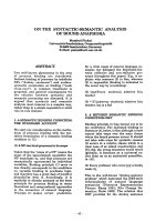
Fig. 1. Sequence alignment of FXR proteins and indication of fibrillogenic regions. The alignment was produced and colour coded according
to
CLUSTALW2 [27]. Extra rows were added below for the rulers relative to human FMRP, FXR1P and FXR2P. The regions predicted as fibrillo-
genic by the
WALTZ software [40] are indicated as red crosses.
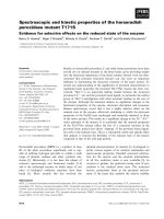
Fig. 2. Summary of the modular structure of FXR proteins (A) and
of the constructs used in this study (B) using a
SMART-like [41] rep-
resentation. NDF stands for the N-terminal domain which contains
two Tudor domains. KH stands for protein K homology domain.
Linker indicates the region between the second Tudor domain and
KH1. NES stands for nuclear export signal. The domain boundaries
used are the same as in [26].
Aggregation properties of fragile X related proteins L. Sjekloc
´
a et al.
1914 FEBS Journal 278 (2011) 1912–1921 ª 2011 The Authors Journal compilation ª 2011 FEBS
unaltered after the temperature is decreased to 37 or
to 20 ° C.
The spectra of shorter FMRP fragments lacking the
KH domain, NDF (residues 1–134) and Nt (residues
1–217), were examined to assess the role of the
individual domains (Fig. 3C,D). These fragments also
undergo structural rearrangements characteristic of an
irreversible increase of b content above 45 °C, suggest-
ing that they are individually able to misfold (Fig. 3B–
D). To test whether pre-incubation at a fixed moderate
temperature could also cause the observed b-enriched
structural rearrangement, the three FMRP fragments,
at the same protein concentration (5 lm), were inde-
pendently incubated at different temperatures. In a
time course measurement at 45 °C, NDF underwent a
conformational change after 3 h pre-incubation
(Fig. 3E). For comparison, the two longer constructs
Nt and Nt-KH1 incubated at 45 °C did not reach,
over the same time, the minimum CD signal (Fig. 3E)
observed for the corresponding samples at 55 °C. A
similar experiment was performed at 50 °C and
resulted in a faster conformational transition compared
with 45 °C (Fig. 3F). At this temperature, the intensi-
ties of the NDF and Nt-KH1 spectra reached a maxi-
mum after 40 and 120 min, respectively. Nt underwent
a conformation transition at 50 °C which was not,
however, complete during the time course of the exper-
iment (3 h). This suggests that, under the same experi-
mental conditions, the region C-terminal to the NDF,
comprising the linker between NDF and KH1, has a
prominent role in aggregation.
Taken together, these data show that different
domains of the conserved region of FXR proteins rear-
range their structure upon temperature treatment. In
all cases examined this rearrangement occurs with very
similar modalities and results in a significant enrich-
ment of the b content.
Tendency to misfold is a conserved feature of
FXR proteins
To extend our studies to other FXR proteins, we used
the human FMRP paralogues FXR1P and FXR2P.
The secondary structure of FXR1P Nt-NES was first
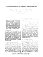
Fig. 3. Spectroscopic study of temperature-induced conformational changes of FMRP. (A) Far-UV CD spectra of Nt-KH1 of FMRP recorded
at 20 °C (black line) or 55 °C (dotted line) and expressed in molar ellipticity of Nt-KH1. (B) Temperature course at 220 nm, in molar ellipticity,
of FMRP NDF (curve a), Nt (curve b) and Nt-KH1 (curve c). The rate of temperature increase was 1 °CÆmin
)1
. (C), (D) Far-UV CD spectra at
20 °C (black lines) or 55 °C (dotted lines) of NDF and Nt respectively. (E), (F) Time course of the a-to-b transition of FMRP NDF (curve a),
Nt (curve b) and Nt-KH1 (curve c) induced by incubation at 45 and 50 °C, respectively. All spectra were recorded at protein concentrations of
5 l
M.
L. Sjekloc
´
a et al. Aggregation properties of fragile X related proteins
FEBS Journal 278 (2011) 1912–1921 ª 2011 The Authors Journal compilation ª 2011 FEBS 1915
examined at different temperatures since for this pro-
tein we could obtain a long construct which spans the
whole conserved region (residues 1–380). The far-UV
CD spectrum of this construct shows that an a-to-b
conformational transition occurs under experimental
conditions similar to those used for the FMRP
domains (Fig. 4A). Interestingly, at the protein concen-
trations used for the assay (5 lm), the b-enriched con-
formation of FXR1P Nt-NES persists up to 80 °C.
This behaviour is similar to that observed for FMRP
Nt-KH1 and suggests that the KH2 and NES regions
do not significantly influence solubility. Similar studies
with shorter FXR1P fragments as well as with FXR2P
fragments all underwent similar a-to-b conformational
transitions, although overall FXR2P seemed to be
more prone to aggregation (Fig. 4B).
These data indicate that not only FMRP but also
the other human FXR paralogues have a strong and
well conserved tendency to misfold.
Misfolding of FMRP domains occurs also under
non-destabilizing conditions
Since the duration of temperature treatment plays a
role in the observed process, we tested if prolonged
incubation could lead to a-to-b conformational transi-
tion also at 37 °C, i.e. close to the physiological tem-
perature at which FMRP functions in human cells.
Initially, incubation of FMRP Nt-KH1 (5 lm)at
37 °C did not cause a significant secondary structure
perturbation, but a conformational transition to a
b-enriched structure was observed after a 45-h incuba-
tion (Fig. 5A). The lag time decreased to 16 h at con-
centrations threefold or sixfold higher (15 and 30 lm
respectively), as expected for a concentration-depen-
dent phenomenon such as aggregation (Fig. 5B,C).
The final intensity of the recorded CD signal is very
similar to that recorded at 55 °C, suggesting not
only that a similar process takes place at both temper-
atures but also that the final states are structurally
comparable.
Freshly prepared FMRP Nt-KH1 samples (30 lm)
are monomeric and monodispersed, and if stored at
4 °C they remain mainly monomeric with a small but
detectable increase of dimeric species as time pro-
ceeds, i.e. after 16 h incubation. The size exclusion
chromatograms of these samples incubated at 37 °C
over the same time show the appearance of high
molecular weight species which are absent both in
fresh samples and in samples incubated at 4 °C
(Fig. 5D). We can conclude that recombinant Nt-
KH1 of FMRP has an intrinsic tendency to aggregate
in vitro also at physiological temperature in native-like
conditions.
FMRP has an intrinsic tendency to form
protofibrils
To characterize the nature of the aggregates, we exam-
ined the longest fragments from FMRP (Nt-KH1) and
from FXR1P and FXR2P (Nt-NES) using the fluores-
cence signal of thioflavin (ThT) dye that is indicative
of formation of amyloid-like structures [29]. After
addition of ThT to diluted FXR protein solutions
(5 lm), samples were incubated for 15 min at increas-
ing temperatures using 5 °C intervals and their fluores-
cence was monitored. No fluorescence could be
detected during incubation at temperatures below
65 °C. At this temperature, a small increase of fluores-
cence emission at 482 nm was observed. Treated sam-
ples were then incubated at room temperature and
fluorescence was measured at different time points
reaching a maximal emission at 482 nm after 12 h
(Fig. 6A). The following measurement after 24 h did
AB
Fig. 4. Spectroscopic studies of FXR1P and FXR2P. (A) Far-UV CD spectra expressed in molar ellipticity of the FXR1P Nt-NES at 20 °C (con-
tinuous line) or 55 °C (dotted line) using 5 l
M protein concentration. (B) Temperature course of FXR1P Nt-NES at 220 nm, using 5 lM con-
centrations. The recording rate was 1 °CÆmin
)1
.
Aggregation properties of fragile X related proteins L. Sjekloc
´
a et al.
1916 FEBS Journal 278 (2011) 1912–1921 ª 2011 The Authors Journal compilation ª 2011 FEBS
not show further increase and a net decrease was
observed after 96 h, probably caused by fibre sedimen-
tation (data not shown).
To verify the morphology of the end-states of aggre-
gation, we used transmission electron microscopy and
examined samples after the conformational transition
Fig. 5. Following the conformational transi-
tion of FMRP Nt-KH1 at 37 °C and different
incubation times as a function of protein
concentration. (A), (B), (C) Comparison of
the FMRP Nt-KH1 CD spectra before (con-
tinuous line) and after (dotted line) incuba-
tion at 37 °C using 5, 15 and 30 l
M protein
concentrations, respectively. (D) Size exclu-
sion chromatography elution profile of
FMRP Nt-KH1: the continuous line chro-
matogram derives from freshly prepared
Nt-KH1, the broken line chromatogram is
the profile of the same sample kept at 4 °C
for 16 h, and the dotted line chromatogram
corresponds to a sample incubated at 37 °C
for 16 h.
A
B
C
D
Fig. 6. Testing the fibrillogenic properties of
FXR proteins. (A) ThT fluorescence assay on
FXR2P Nt-NES treated over the temperature
range 20–65 °C, increasing the temperature
by 5 °C every 15 min and using 5 l
M
protein concentration. The fluorescence
observed at 20 °C (black curve) decreases
at temperatures between 45 and 55 °C
(red curve), and increases after exposure to
65 °C (orange). The fluorescence signal
reached a maximum after 12 h (green
curve). (B) Transmission electron micrograph
of negatively stained FMRP Nt (1–217)
protofibrils pre-incubated at 50 °C for 3 h.
(C) Negatively stained aggregates were
observed for the FMRP Nt-KH1 construct
(1–280) that was incubated for an extended
time at 37 °C. (D) Electron micrograph of
FXR1P Nt-NES aggregates obtained after
2 h incubation at 50 °C. The scale bars
correspond to 100 nm.
L. Sjekloc
´
a et al. Aggregation properties of fragile X related proteins
FEBS Journal 278 (2011) 1912–1921 ª 2011 The Authors Journal compilation ª 2011 FEBS 1917
achieved either by direct exposure to 50 °C or by pro-
longed incubation at 37 °C, at two different protein
concentrations (5 and 30 lm). Negatively stained pro-
tofibrillar and fibrillar assemblies were observed for
FMRP Nt, FMRP Nt-KH1 and FX1RP Nt-NES
(Fig. 6B–D). The FMRP Nt protofibrils appeared
homogeneous, with an apparent uniform diameter
(7 nm) and variable lengths that very rarely exceeded
100 nm (Fig. 6B). Interestingly, we also observed dense
networks of long linear and unbranched fibrils with a
10-nm diameter, which displayed repeating segments
and twists. The FMRP Nt-KH1 samples contained
globular particles with an average diameter of 24 nm,
often decorated with stain, as well as clustered deposits
of fibrils with an average diameter of 6 nm (Fig. 6C).
FX1RP Nt-NES aggregates have a curved appearance,
with an apparent average diameter of 10 nm (Fig. 6D).
They also clustered together and were often found to
be decorated with the uranyl acetate stain. Taken
together these results confirm a marked tendency of
FXR constructs to fibrillation.
Discussion
We have shown here that different fragments of FXR
proteins not only have a strong tendency to aggregate
as previously described [26] but also undergo an irre-
versible conformational transition which leads to a sig-
nificant increase in their b-structure content. Several
conserved putative aggregation and amyloidogenic
hot-spots were predicted by in silico analysis of the
FXR amino acid sequences. They are all grouped in
the highly conserved (more than 70–80% identity and
80–90% similarity) N-terminal half of the proteins
which is also the region involved in most of the inter-
actions with the FXR cellular partners [30], suggesting
that the aggregation hot-spots could have a prominent
role in determining the hetero- and self-assembly
behaviours of the full-length proteins. By combining
CD spectroscopy and size exclusion chromatography,
we have established a clear link between FMRP aggre-
gation and misfolding, as observed by the concentra-
tion dependence of the conformational transition.
Relatively small variations of protein concentration
also lead to an increase of the rates at which the con-
formational transition occurs. An appreciable ThT
fluorescence increase, irreversible b-enriched structural
transitions and electron microscopy analysis support
formation of ordered fibrillar aggregates. We observe a
very similar behaviour for the two FMRP paralogues,
FXR1P and FXR2P, for which the conformational
transition occurs with modalities very similar to
FMRP.
Interestingly, the observed transition towards b-en-
riched conformations occurs also at physiological tem-
perature under non-destabilizing conditions. This
behaviour is very interesting for a protein such as
FMRP which contains multiple globular domains.
While understanding how and when misfolding occurs
is easier for intrinsically unfolded proteins, such as the
Alzheimer Ab peptides or a-synuclein, studies of globu-
lar proteins have traditionally involved the use of
ad hoc mutations and ⁄ or destabilizing conditions, such
as high temperature, molecular crowding or high pres-
sure. These conditions lead to destabilization of the
structure and access to fibrillogenic regions normally
buried in the hydrophobic core. Only recently a small
but steadily increasing number of examples are being
described in which misfolding occurs in native-like con-
ditions. This is the case for instance of the globular Jo-
sephin domain of ataxin-3, the protein responsible for
the misfolding Machado–Joseph disease: we have
recently shown that Josephin aggregation and misfold-
ing is promoted by exposed hydrophobic patches
involved in recognition of its natural partner ubiquitin,
thus suggesting a link between normal function and
misfolding [31]. Likewise, the globular domain of the
prion protein contains a seeding region, H2H3, which
retains its fold during the early stages of unfolding [32].
It has been suggested that in many proteins related to
conformational diseases aggregation ⁄ amyloidogenic
regions coincide with interaction surfaces [33–35].
Our results bear a number of interesting conse-
quences. First, the strong tendency to aggregate of
FXR proteins could help us to understand the driving
forces that lead to granular formations and eventually
understand more about their functional role. The find-
ings presented in this study also suggest interesting
possibilities for the ability of this family of proteins to
contribute to both early life syndromes such as FXS
(for instance through destabilizing mutations) and
aggregation-related neurodegeneration later in life;
such could be the case of FXTAS. The latter is a par-
ticularly interesting possibility since it could shed new
light onto a still poorly understood syndrome:
although RNA aggregation is thought to be an impor-
tant driving force for formation of the pathological
neuronal intranuclear RNP inclusions observed in
FXTAS patients, little is known about the factors
which determine their formation and stability [36]. The
current view is that FXTAS is the end-point of a pro-
cess that begins in early development and reaches its
maximum late in life [37]. rCGG expansion in the
5¢UTR region of FMR1 mRNA is required for forma-
tion of neuronal inclusions in FXTAS patients, which
consist also of other mRNAs and of different proteins
Aggregation properties of fragile X related proteins L. Sjekloc
´
a et al.
1918 FEBS Journal 278 (2011) 1912–1921 ª 2011 The Authors Journal compilation ª 2011 FEBS
amongst which FXR1P and FXR2P [38]. Although
FMRP, which is expressed in FXTAS patients, has not
so far been identified amongst the components of the
inclusions, we cannot exclude at this stage that its
absence is not simply due to lack of sensitivity of the
detection methods used. We suggest as a working
hypothesis that the aggregative and misfolding ten-
dency of one or more of the FXR proteins could con-
tribute to pathology, thus adding FXTAS to the
family of misfolding diseases. While more work needs
to be done to test this hypothesis, we expect that
important information may come from cellular studies
of the effects of molecular crowding [39] on the FXR
folding, homo- and hetero-association when sur-
rounded by other cellular components.
Experimental procedures
Bioinformatic analysis
The amino acid sequences of human (Q06787, P51114,
P51116), mouse (P35922, Q61584, Q9WVR4), chicken
(Q5F3S6), frog (P51113, Q6GLC9, P51115), zebra fish
(Q7SYM7, Q7SXA0, Q6NY99) and fruit fly (Q9NFU0)
FXR proteins were aligned using program clustalw2 [27].
These sequences were also searched for putative determi-
nants of aggregation and amyloidogenesis by the following
consensus prediction tools: aggrescan (http://bioinf.
uab.es/aggrescan/) for prediction of hot-spots for aggrega-
tion in polypeptides; pasta ( />pasta/) for prediction of amyloid-like structure aggregation;
amylpred ( to
predict features related to the formation of amyloid fibrils;
tango ( for prediction of
sequence-dependent and mutational effects on the aggrega-
tion of the peptides and proteins; and waltz (http://
waltz.switchlab.org/) for predicting amyloidogenic regions
in protein sequences.
Cloning, protein expression and purification
The constructs studied in this paper were produced accord-
ing to procedures previously described [26]. In short, clones
of human FMR1, FXR1 and FXR2 were used as templates
for DNA amplification by PCR. PCR amplicons encoding
different fragments of the conserved region of FXR pro-
teins were cloned into a modified pET-24 vector (Novagen,
Gibbstown, NJ, USA) encoding an amino terminal Trx
(thioredoxin)-His6-tag and a tobacco etch virus (TEV) pro-
tease cleavage site.
Escherichia coli BL21 STAR (DE3) cells transformed
with plasmids encoding different FXR fragments were
grown at 37 °C in Luria–Bertani medium containing appro-
priate antibiotic. Protein over-expression was induced with
0.2 mm isopropyl thio-b-d-galactoside after the cell culture
had reached D
600nm
= 0.8; the growth was continued for
an additional 5 h at 28 °C. The cells were harvested by cen-
trifugation, resuspended in a lysis buffer containing 20 mm
Tris ⁄ HCl (pH 8.0), 150 mm NaCl, 10 mm imidazole, 0.2%
Igepal CA-630 (Sigma–Aldrich, St Louis, MO, USA),
2mm b-mercaptoethanol, supplemented with the Complete
EDTA-free protease inhibitor cocktail (Roche, Indianapo-
lis, IN, USA), and lysed by ultrasonication. The recombi-
nant peptides were then purified from the soluble fraction
of the centrifuged cell lysate by immobilized metal-affinity
chromatography (IMAC) using Ni-NTA (Ni
2+
-nitrilotriac-
etate) metal-affinity chromatography matrix (Qiagen,
Yokyo, Japan), and dialyzed against 50 mm Tris ⁄ HCl (pH
8.0), 1 mm dithiothreitol, 0.5 mm EDTA; the recombinant
Trx-His6-tag was removed by cleavage with TEV protease
(Invitrogen, Carlsbad, CA, USA). The FXR peptides were
further purified by IMAC, anion exchange chromatography
(MonoQ HR 5 ⁄ 5) and size exclusion chromatography
(Superdex 200 HR 16 ⁄ 60 or Superdex 200 10 ⁄ 30) using elu-
tion buffer consisting of 50 mm Tris ⁄ HCl (pH 8.0), 2 mm
b-mercaptoethanol. The purity of the recombinant peptides
was higher than 95% as verified by SDS ⁄ PAGE and by
mass spectrometry. Protein concentration was measured by
UV absorbance at 280 nm using theoretical extinction coef-
ficients calculated by
ProtParam.
Aggregation studies by CD and size exclusion
chromatography
CD spectra were recorded using a Jasco J-715 spectropola-
rimeter equipped with a thermostatted cell holder controlled
by a Jasco Peltier element, at different temperatures, over a
wavelength range from 260 to 190 nm in quartz cuvettes
(Hellma) of path length appropriate to protein concentra-
tion of the samples, i.e. 1 mm for 5 lm (0.15 mgÆmL
)1
),
0.2 mm for 15 lm (0.5 mg ÆmL
)1
) and 0.1 mm for 30 lm
(1 mgÆmL
)1
). Thermally induced denaturation transitions
were monitored by CD absorption at 220 nm from 10 to
95 °C, in 1-°C steps and with an equilibration time of
1 minÆ°C
)1
. Reversibility was tested by performing an
inverse temperature scan. The purified recombinant proteins
were in 20 mm Tris ⁄ HCl (pH 8.0), 1 mm b-mercaptoetha-
nol. To monitor progression of protein aggregation over
time, protein samples were incubated at 37 °C and CD
spectra were recorded at different time points (1, 6, 16, 24,
45, 72 h, 1 week).
Analytical size exclusion chromatography was carried out
by injecting 100 lL of samples (30 lm) into a Superdex 200
10 ⁄ 300 GL column.
ThT fluorescence assays
The ThT assays were performed by consecutively incubat-
ing at 20, 30, 40, 50, 55, 60 and 65 °C, for 15 min at each
L. Sjekloc
´
a et al. Aggregation properties of fragile X related proteins
FEBS Journal 278 (2011) 1912–1921 ª 2011 The Authors Journal compilation ª 2011 FEBS 1919
temperature without shaking, the purified protein solution
diluted to 5 lm in a buffer containing 20 lm ThT, 20 mm
Tris ⁄ HCl (pH 8.0), 1 mm b-mercaptoethanol. After the last
heating step, at 65 °C, the sample was kept at room tem-
perature (20 °C) and spectra were recorded after 1, 2, 3, 4,
12, 24 and 96 h. Fluorescence was measured using an ISS
PC1 (Interconnect Systems Solution) spectrofluorimeter. All
measurements were carried out at 20 °C over a 60 s time
course with excitation at 440 nm (0.4 nm slit width) and
emission at 482 nm (1.5 nm slit width). For each measure-
ment 10 scans were recorded. The measurement of the fluo-
rescence of the reaction buffer treated at 65 °C showed
only a weak peak at 520 nm.
Transmission electron microscopy
A sample volume of 4 lL was spotted onto freshly pre-
pared carbon-coated and glow-discharged copper grids
(FormVar). Upon adsorption to the grid surface for 30 s,
the sample was washed briefly with milli-Q water and sub-
sequently stained with 1% (w ⁄ v) uranyl acetate for 30 s.
Micrographs of negatively stained areas were taken with a
JEOL 1200 transmission electron microscope operating at
100 kV and at a magnification of 27 800· on electron
microscope films (Kodak) and developed with Phenisol
developer (Ilford) and Hypam fixer (Ilford) for 5 min each.
Acknowledgements
We thank Steve Martin for help with CD and fluores-
cence studies, Lesley Calder for support with electron
microscopy analysis and Steve Howell for mass spec-
trometry analysis. We are grateful to Cesira de Chiara
and Laura Masino for critical discussion and assis-
tance in graphic elaboration of CD results. We
acknowledge support from the MRC (Grant ref.
U117584256). Kris Pauwels is the recipient of an
EMBO long-term postdoctoral fellowship (ALTF 512-
2008).
References
1 Bakker CE, de Diego Otero Y, Bontekoe C, Raghoe P,
Luteijn T, Hoogeveen AT, Oostra BA & Willemsen R
(2000) Immunocytochemical and biochemical character-
ization of FMRP, FXR1P, and FXR2P in the mouse.
Exp Cell Res 258, 162–170.
2 Devys D, Lutz Y, Rouyer N, Bellocq JP & Mandel JL
(1993) The FMR-1 protein is cytoplasmic, most abun-
dant in neurons and appears normal in carriers of a
fragile X permutation. Nat Genet 4, 335–340.
3 Tamanini F, Willemsen R, van Unen L, Bontekoe C,
Galjaard H, Oostra BA & Hoogeveen AT (1997) Differ-
ential expression of FMR1, FXR1 and FXR2 proteins
in human brain and testis. Hum Mol Genet 6, 1315–
1322.
4 Ramos A, Hollingworth D, Adinolfi S, Castets M,
Kelly G, Frenkiel TA, Bardoni B & Pastore A (2006)
The structure of the N-terminal domain of the fragile X
mental retardation protein: a platform for protein–
protein interaction. Structure 14, 21–31.
5 Siomi H, Siomi MC, Nussbaum RL & Dreyfuss G
(1993) The protein product of the fragile X gene,
FMR1, has characteristics of an RNA-binding protein.
Cell 74, 291–298.
6 Ashley CT Jr, Wilkinson KD, Reines D & Warren ST
(1993) FMR1 protein: conserved RNP family domains
and selective RNA binding. Science 262, 563–566.
7 Brown V, Small K, Lakkis L, Feng Y, Gunter C,
Wilkinson KD & Warren ST (1998) Purified recombi-
nant Fmrp exhibits selective RNA binding as an
intrinsic property of the fragile X mental retardation
protein. J Biol Chem 273, 15521–15527.
8 Kim M, Bellini M & Ceman S (2009) Fragile X mental
retardation protein FMRP binds mRNAs in the
nucleus. Mol Cell Biol 29, 214–228.
9 De Diego Otero Y, Severijnen LA, van Cappellen
G, Schrier M, Oostra B & Willemsen R (2002)
Transport of fragile X mental retardation protein via
granules in neurites of PC12 cells. Mol Cell Biol 22,
8332–8341.
10 Napoli I, Mercaldo V, Boyl PP, Eleuteri B, Zalfa F,
De Rubeis S, Di Marino D, Mohr E, Massimi M,
Falconi M et al. (2008) The fragile X syndrome protein
represses activity-dependent translation through
CYFIP1, a new 4E-BP. Cell 134, 1042–1054.
11 Bassell GJ & Warren ST (2008) Fragile X syndrome:
loss of local mRNA regulation alters synaptic develop-
ment and function. Neuron 60, 201–214.
12 Brouwer JR, Willemsen R & Oostra BA (2009) The
FMR1 gene and fragile X-associated tremor ⁄ ataxia syn-
drome. Am J Med Genet B Neuropsychiatr Genet 150B,
782–798.
13 Oostra BA & Willemsen R (2009) FMR1: a gene with
three faces. Biochim Biophys Acta 1790, 467–477.
14 Guduric-Fuchs J, Mohrlen F, Frohme M & Frank U
(2004) A fragile X mental retardation-like gene in a
cnidarian. Gene 343, 231–238.
15 Anderson P & Kedersha N (2006) RNA granules. J Cell
Biol 172, 803–808.
16 Christie SB, Akins MR, Schwob JE & Fallon JR (2009)
The FXG: a presynaptic fragile X granule expressed in
a subset of developing brain circuits. J Neurosci 29,
1514–1524.
17 Huot ME, Bisson N, Davidovic L, Mazroui R, Labelle
Y, Moss T & Khandjian EW (2005) The RNA-binding
protein fragile X-related 1 regulates somite formation in
Xenopus laevis. Mol Biol Cell 16
, 4350–4361.
Aggregation properties of fragile X related proteins L. Sjekloc
´
a et al.
1920 FEBS Journal 278 (2011) 1912–1921 ª 2011 The Authors Journal compilation ª 2011 FEBS
18 Moser JJ & Fritzler MJ (2010) Cytoplasmic ribonucleo-
protein (RNP) bodies and their relationship to GW ⁄ P
bodies. Int J Biochem Cell Biol 42, 828–843.
19 Tassone F, Iwahashi C & Hagerman PJ (2004) FMR1
RNA within the intranuclear inclusions of fragile X-
associated tremor ⁄ ataxia syndrome (FXTAS). RNA Biol
1, 103–105.
20 Ceman S, Brown V & Warren ST (1999) Isolation of an
FMRP-associated messenger ribonucleoprotein particle
and identification of nucleolin and the fragile X-related
proteins as components of the complex. Mol Cell Biol
19, 7925–7932.
21 Tamanini F, Van Unen L, Bakker C, Sacchi N, Galj-
aard H, Oostra BA & Hoogeveen AT (1999) Oligomeri-
zation properties of fragile-X mental-retardation protein
(FMRP) and the fragile-X-related proteins FXR1P and
FXR2P. Biochem J 343, 517–523.
22 Zhang Y, O’Connor JP, Siomi MC, Srinivasan S, Dutra
A, Nussbaum RL & Dreyfuss G (1995) The fragile X
mental retardation syndrome protein interacts with
novel homologs FXR1 and FXR2. EMBO J 14, 5358–
5366.
23 Estes PS, O’Shea M, Clasen S & Zarnescu DC (2008)
Fragile X protein controls the efficacy of mRNA
transport in Drosophila neurons. Mol Cell Neurosci 39,
170–179.
24 Didiot MC, Subramanian M, Flatter E, Mandel JL &
Moine H (2009) Cells lacking the fragile X mental retar-
dation protein (FMRP) have normal RISC activity but
exhibit altered stress granule assembly. Mol Biol Cell
20, 428–437.
25 Mazroui R, Huot ME, Tremblay S, Filion C, Labelle Y
& Khandjian EW (2002) Trapping of messenger RNA
by fragile X mental retardation protein into cytoplasmic
granules induces translation repression. Hum Mol Genet
11, 3007–3017.
26 Sjekloca L, Konarev PV, Eccleston J, Taylor IA,
Svergun DI & Pastore A (2009) A study of the ultra-
structure of fragile-X-related proteins. Biochem J 419,
347–357.
27 Larkin MA, Blackshields G, Brown NP, Chenna R,
McGettigan PA, McWilliam H, Valentin F, Wallace
IM, Wilm A, Lopez R et al. (2007) Clustal W and
Clustal X version 2.0. Bioinformatics 23, 2947–2948.
28 Musco G, Stier G, Joseph C, Castiglione Morelli MA,
Nilges M, Gibson TJ & Pastore A (1996) Three-dimen-
sional structure and stability of the KH domain:
molecular insights into the fragile X syndrome. Cell 85,
237–245.
29 LeVine H III (1993) Thioflavine T interaction with syn-
thetic Alzheimer’s disease beta-amyloid peptides: detec-
tion of amyloid aggregation in solution. Protein Sci 2,
404–410.
30 Bardoni B, Davidovic L, Bensaid M & Khandjian EW
(2006) The fragile X syndrome: exploring its molecular
basis and seeking a treatment. Expert Rev Mol Med 8,
1–16.
31 Masino L, Nicastro G, Calder L, Vendruscolo M &
Pastore A (2010) Functional interactions as a survival
strategy against abnormal aggregation. FASEB J 25,
45–54.
32 Adrover M, Pauwels K, Prigent S, de Chiara C, Xu Z,
Chapuis C, Pastore A & Rezaei H (2010) Prion fibrilliza-
tion is mediated by a native structural element that com-
prises helices H2 and H3. J Biol Chem 285 , 21004–21012.
33 Castillo V & Ventura S (2009) Amyloidogenic regions
and interaction surfaces overlap in globular proteins
related to conformational diseases. PLoS Comput Biol
5, e1000476.
34 Espargaro A, Castillo V, de Groot NS & Ventura S
(2008) The in vivo and in vitro aggregation properties of
globular proteins correlate with their conformational
stability: the SH3 case. J Mol Biol 378, 1116–1131.
35 Frousios KK, Iconomidou VA, Karletidi CM &
Hamodrakas SJ (2009) Amyloidogenic determinants are
usually not buried. BMC Struct Biol 9, 44.
36 Sellier C, Rau F, Liu Y, Tassone F, Hukema RK,
Gattoni R, Schneider A, Richard S, Willemsen R,
Elliott DJ et al. (2010) Sam68 sequestration and partial
loss of function are associated with splicing alterations
in FXTAS patients. EMBO J 29, 1248–1261.
37 Garcia-Arocena D & Hagerman PJ (2010) Advances in
understanding the molecular basis of FXTAS. Hum
Mol Genet 19, R83–R89.
38 Iwahashi CK, Yasui DH, An HJ, Greco CM, Tassone
F, Nannen K, Babineau B, Lebrilla CB, Hagerman RJ
& Hagerman PJ (2006) Protein composition of the
intranuclear inclusions of FXTAS. Brain 129, 256–271.
39 Ellis RJ & Minton AP (2006) Protein aggregation in
crowded environments. Biol Chem 387, 485–497.
40 Maurer-Stroh S, Debulpaep M, Kuemmerer N, de la
Paz ML, Martins IC, Reumers J, Morris KL, Copland
A, Serpell L, Serrano L et al. (2010) Exploring the
sequence determinants of amyloid structure using posi-
tion-specific scoring matrices. Nat Methods 7, 237–242.
41 Letunic I, Doerks T & Bork P (2009) SMART 6: recent
updates and new developments. Nucleic Acids Res 37,
D229–D232.
L. Sjekloc
´
a et al. Aggregation properties of fragile X related proteins
FEBS Journal 278 (2011) 1912–1921 ª 2011 The Authors Journal compilation ª 2011 FEBS 1921