Báo cáo khoa học: Cys126 is a completely conserved residue in triosephosphate isomerase that docx
Bạn đang xem bản rút gọn của tài liệu. Xem và tải ngay bản đầy đủ của tài liệu tại đây (756.35 KB, 12 trang )
Probing the role of the fully conserved Cys126 in
triosephosphate isomerase by site-specific
mutagenesis – distal effects on dimer stability
Moumita Samanta
1
, Mousumi Banerjee
1
, Mathur R. N. Murthy
1
, Hemalatha Balaram
2
and Padmanabhan Balaram
1
1 Molecular Biophysics Unit, Indian Institute of Science, Bangalore, India
2 Molecular Biology and Genetics Unit, Jawaharlal Nehru Centre for Advanced Scientific Research, Bangalore, India
Introduction
The conserved amino acids in enzymes are, most often,
associated with the key steps of substrate recognition
and catalysis. The availability of rapidly expanding
databases of enzyme sequences may be effectively used
to identify key residues. Triosephosphate isomerase
(TIM) is an extremely well-studied enzyme [1–4], and
Keywords
dimer interface; dimer stability;
Plasmodium falciparum; thermal stability;
triosephosphate isomerase
Correspondence
P. Balaram, Molecular Biophysics Unit,
Indian Institute of Science, Bangalore-
560012, India
Fax: +91 80 2360 0535
Tel: +91 80 2293 3000
E-mail:
(Received 2 February 2011, revised 22
March 2011, accepted 28 March 2011)
doi:10.1111/j.1742-4658.2011.08110.x
Cys126 is a completely conserved residue in triosephosphate isomerase that
is proximal to the active site but has been ascribed no specific role in catal-
ysis. A previous study of the C126S and C126A mutants of yeast TIM
reported substantial catalytic activity for the mutant enzymes, leading to
the suggestion that this residue is implicated in folding and stability [Gonz-
alez-Mondragon E et al. (2004) Biochemistry 43, 3255–3263]. We re-exam-
ined the role of Cys126 with the Plasmodium falciparum enzyme as a
model. Five mutants, C126S, C126A, C126V, C126M, and C126T, were
characterized. Crystal structures of the 3-phosphoglycolate-bound C126S
mutant and the unliganded forms of the C126S and C126A mutants were
determined at a resolution of 1.7–2.1 A
˚
. Kinetic studies revealed an
approximately five-fold drop in k
cat
for the C126S and C126A mutants,
whereas an approximately 10-fold drop was observed for the other three
mutants. At ambient temperature, the wild-type enzyme and all five
mutants showed no concentration dependence of activity. At higher tem-
peratures (> 40 °C), the mutants showed a significant concentration
dependence, with a dramatic loss in activity below 15 l
M. The mutants also
had diminished thermal stability at low concentration, as monitored by far-
UV CD. These results suggest that Cys126 contributes to the stability of
the dimer interface through a network of interactions involving His95,
Glu97, and Arg98, which form direct contacts across the dimer interface.
Database
Structural data are available in the Protein Data Bank under the accession numbers
3PVF,
3PY2, and 3PWA.
Structured digital abstract
l
Tim binds to Tim by x-ray crystallography (View interaction)
Abbreviations
GAP, glyceraldehyde 3-phosphate; DHAP, dihydroxyacetone phosphate; PDB, Protein Data Bank; Pf TIM, Plasmodium falciparum
triosephosphate isomerase; PGA, phosphoglycolate; TIM, triosephosphate isomerase; T
m
, melting temperature.
1932 FEBS Journal 278 (2011) 1932–1943 ª 2011 The Authors Journal compilation ª 2011 FEBS
provides a good model system for exploring the role of
residues that are completely conserved or minimally
replaced during evolution. Examination of a dataset of
503 sequences of TIM from different organisms reveals
only nine fully conserved residues: Lys12, Thr75,
His95, Glu97, Cys126, Glu165, Pro166, Gly209, and
Gly228 [the numbering scheme used here corresponds
to that for Plasmodium falciparum TIM (Pf TIM),
and, for all of the fully conserved residues, this is iden-
tical to that of yeast TIM]. Of these, Lys12, His95,
Glu97 and Glu165 surround the substrate, with the
carboxylate of Glu165 acting as the base for abstrac-
tion of a proton from the C2 position of glycer-
aldehyde 3-phosphate (GAP) and dihydroxyacetone
phosphate (DHAP) [5–8]. Lys12 and His95 are
involved in substrate ⁄ transition state binding and pro-
ton transfer, respectively [6,9,10]. Pro166 is a hinge res-
idue located in loop 6, which undergoes dynamic
interconversion between open and closed states, with
the latter corresponding to the catalytically competent
form [11–15]. Gly209 is located near the active site in
the highly conserved 208–212 segment. Gly228 adopts
a backbone conformation accessible only for Gly resi-
dues, enabling appropriate positioning of the facing
208–209 segment by backbone–backbone hydrogen
bonds. Thr75 is a critical residue at the dimer interface
[16]; the side chain of this residue from one subunit
makes key hydrogen bonding contacts with Asn10 and
Glu97 of the other subunit, which are proximal to
the active site. Cys126 is a completely conserved resi-
due that is spatially proximal to the active site residue
Glu165 (Fig. S1).
Interestingly, a preliminary analysis of a dataset of
over 800 putative TIM sequences extracted from a
dataset of bacterial sequences of marine origin [17]
also revealed the occurrence of Cys at position 126.
Inspection of several 3D structures of TIM from
diverse organisms available in the Protein Data Bank
(PDB) does not immediately suggest a structural expla-
nation for the complete conservation of this residue.
Indeed, an earlier investigation of the C126S and
C126A mutants of yeast TIM revealed that their activ-
ity remained undiminished, with the mutants display-
ing a significantly lower degree of thermal stability.
This study suggested that Cys126 may be required for
efficient folding and stability rather than being
involved in maintaining catalytic activity [18]. A recent
treatise on enzymology highlights Cys126 in a discus-
sion of TIM [19]. As part of a program directed
towards understanding the role of conserved residues,
we describe the characterization of five Cys126
mutants of Pf TIM. The mutants studied were C126S,
C126A, C126V, C126M, and C126T. We describe
crystal structures of unliganded forms of the C126S
and C126A mutants, and the liganded form of the
C126S mutant. Temperature-dependent activity mea-
surements and spectroscopic studies suggest that
Cys126 may be involved in maintaining the structural
integrity of the active site in the temperature range 40–
50 °C. Furthermore, the residue also contributes to the
thermal stability of the dimer interface through an
extended interaction network involving His95, Glu97
and Thr75 of the neighboring subunit, all of which are
fully conserved residues.
Results
Analysis of crystal structures
Diffraction-quality crystals were obtained for the
C126S mutant complexed with phosphoglycolate
(PGA) and the unliganded C126S mutant. For the
C126A mutant, a structure could be determined only
for the unliganded form. PGA was bound to the active
site of the C126S mutant structure in a manner similar
to that for wild-type Pf TIM, whereas the C126A
mutant structure had no ligand bound to the active
site after cocrystallization. The difference in electron
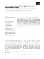
density at the ligand position is shown in Fig. 1A.
Figures were generated with pymol (http://www.
pymol.org). The active site loop 6 was in the ‘closed’
form in the structure of the C126S–PGA complex. In
the unliganded forms of the C126S and C126A
mutants, both of which contained a dimer in the asym-
metric unit, the active site loop 6 was in the ‘open’
conformation. In the C126S mutant unliganded struc-
ture, the active site was occupied by an ethylene glycol
molecule and a single water molecule in one subunit.
In addition, a proximal sulfate ion, derived from the
lithium sulfate in the crystallization medium, could
also be identified near the active site. The other sub-
unit in the C126S mutant and both subunits in the
C126A mutant contained two water molecules in the
active site, along with a distal sulfate ion. The electron
density maps (2F
o
) F
c
, contoured at 1.0r) surround-
ing the residues at position 126 for the mutants are
shown in Fig. 1.
Figure S2 compares the relationships between the
active site residues and Cys ⁄ Ser126 in the unliganded
and liganded forms of the wild-type enzyme and the
C126S mutant. The most notable difference is in the
orientation of the Ser side chain, with the hydroxyl
group forming a hydrogen bond with the carboxylate
of Glu165 in the liganded form. A change of v
1
from
)62.5° in the unliganded form to )170.8° in the ligan-
ded form is observed. In contrast, the Cys126 side
M. Samanta et al. Cys126 in triosephosphate isomerase
FEBS Journal 278 (2011) 1932–1943 ª 2011 The Authors Journal compilation ª 2011 FEBS 1933
chain remains unchanged in orientation upon ligand
binding. Interestingly, both the unliganded forms
contain two invariant water molecules, which form
hydrogen bonds with one another. Water 512 in the
wild-type enzyme (PDB ID: 1LYX) and water 349 in
the C126S mutant also form hydrogen bonds with the
fully conserved His95 and highly conserved Asn10
(Asn in 465 out of 470 sequences) side chains.
Water 558 in wild-type TIM (PDB ID:1LYX) and
water 36 in the C126S mutant form hydrogen bonds
with the side chain of the active site Glu165 and the
backbone CO of the fully conserved Gly209. These
two invariant water molecules form a similar network
of interactions in the C126A unliganded structure and
also in the previously reported, unliganded yeast struc-
ture (PDB ID:1YPI) [20]. Ligand binding and loop
closure result in the expulsion of these water molecules
and a change in the backbone conformational angles
for the highly conserved Gly209-Gly210-Ser211 seg-
ment. This results in a change in orientation of the
Gly209 backbone CO group.
Kinetic parameters
The kinetic parameters determined for Pf TIM and the
five mutants at position 126 are listed in Table 1. The
parameters determined for the wild-type yeast enzyme
and the C126S and C126A mutants by Gonzalez-
Mondragon et al. [18] are also shown for comparison.
In the earlier study of the yeast enzyme, the wild-type
and the Cys126 mutant enzymes had comparable kinetic
parameters, with a small reduction in k
cat
(approxi-
mately four-fold). Temperature-dependent activity mea-
surements were not reported in that study. In the
present study of Pf TIM, an approximately 5.8-fold
drop in k
cat
was observed for both the C126S and
C126A mutants. The other three mutants, C126V,
C126M, and C126T, showed significantly lower k
cat
values, corresponding to a reduction of approximately
10-fold in catalytic activity. These results suggest that
all five Cys126 mutants show a high degree of catalytic
activity, despite the fact that a completely conserved
residue, proximal to the active site Glu165 and His95
side chains, has been replaced by residues of varying
size and hydrogen-bonding ability. Figure 2A compares
the temperature dependence of the specific activity of
wild-type Pf TIM and the five Cys126 mutants, at a
protein concentration of 3.7 nm. For the wild-type
enzyme, there was the expected increase in activity over
the temperature range 25–40 °C, with a leveling off
between 40 °C and 50 °C. In sharp contrast, all five
mutants showed a dramatic reduction in activity in
the temperature range 40–50 °C, with essentially com-
plete absence of activity at 50 °C. The activities of the
wild-type enzyme and the five Cys126 mutants were also
measured as a function of protein concentration at
50 °C. The results summarized in Fig. 2B establish that
all of the Cys126 mutants exhibited a pronounced fall in
activity upon lowering of the protein concentration to
below 20 lm. Indeed, a fall in activity of approximately
10–1000-fold was observed on the change from 30 lm
to 1 lm. The pronounced concentration dependence of
A
B
C
Glu165
Ser126
His95
Lys12
Glu165
Ser126
His95
Lys12
Glu165
Ala126
His95
Lys12
Fig. 1. The electron density maps (2F
o
) F
c
contoured at 1.0r)at
position 126 and the active site residues in: (A) the Pf TIM C126S
PGA-bound structure; (B) the Pf TIM C126S-unliganded structure
with the molecule ethylene glycol (cryoprotectant) at the active
site; and (C) the Pf TIM C126A-unliganded structure.
Cys126 in triosephosphate isomerase M. Samanta et al.
1934 FEBS Journal 278 (2011) 1932–1943 ª 2011 The Authors Journal compilation ª 2011 FEBS
activity in the Cys126 mutants is suggestive of
diminished stability of the dimeric protein at high
temperature.
Structural stability
Figure 3A,B show the far-UV CD and fluorescence
emission spectra of Pf TIM and the five Cys126
mutants, determined at a protein concentration of
3 lm. The near identity of the observed spectra estab-
lished that there were no dramatic structural conse-
quences of the mutations at position 126. The far-UV
CD spectra also remained unchanged over the concen-
tration range 0.5–15 lm at 25 °C, suggesting the
absence of any concentration-dependent structural
effects at ambient temperature. Figure 3C shows a
comparison of the thermal melting profiles for wild-
type Pf TIM and the five mutants, obtained by moni-
toring the CD ellipticity at 222 nm as a function of
temperature, at a protein concentration of 15 lm. The
sharp reduction in CD ellipticity at temperatures
greater than 60 °C corresponds to unfolding, aggrega-
tion, and precipitation. The wild-type and the mutant
enzymes behaved in a very similar way under these
conditions. These results suggest that replacement of
Cys at position 126 does not significantly perturb the
overall folded structure of the protein or its thermal
stability, at this relatively high protein concentration.
However, when the protein concentration was reduced
to 0.5 lm, the melting curves determined using the fall
in ellipticity at 222 nm (shown in Fig. 3D) were dra-
matically different for the wild-type enzyme and the
mutants. The melting temperature (T
m
) for wild-type
Pf TIM was unaffected by lowering the concentration,
whereas the mutants melted at a significantly lower
temperature (midpoint of transition, 50 °C). This con-
centration dependence of protein thermal stability is
consistent with the fall in enzyme activity of the
mutants at low concentration and high temperature.
The reversibility of the thermal unfolding transition
was investigated by measurements of ellipticity at
222 nm upon cooling from a temperature of 55 °C for
the mutants and 60 °C for the wild-type enzyme, at a
protein concentration of 0.5 lm. Under these condi-
tions, aggregation and irreversible precipitation of the
thermally unfolded protein structure was minimized.
Figure 4 summarizes the results obtained for the heat-
ing and cooling cycles for the wild-type enzyme and
the five Cys126 mutants. Wild-type Pf TIM recovered
almost 90% of the original ellipticity upon cooling to
20 °C. The observed hysteresis in the cooling cycle has
also been previously noted for the wild-type enzyme
from Saccharomyces cerevisiae [18,21]. In the case of
all five mutants, only 60% of the CD ellipticity was
recovered upon cooling. These results correspond well
with those reported in the previous study of yeast TIM
C126S and C126A mutants. Gonzalez-Mondragon
et al. have noted that the reduction in T
m
observed for
the C126S and C126A mutants of the yeast enzyme
‘should be taken as an indication of diminished kinetic,
rather than thermodynamic, stability of the native
dimer’ [18]. They have also presented evidence for the
dependence of refolding rates of the C126S yeast
mutant at 30 °C and enzyme concentrations of 1.1 lm
and 1.9 lm. More rapid refolding is observed at higher
protein concentrations [18]. Our present study points
to a greater tendency of the Cys126 mutants than of
Fig. 2. (A) Temperature dependence of specific activity of wild-type
Pf TIM (TWT) and the Cys126 mutants. (B) Concentration depen-
dence of specific activity of the five Cys126 mutants at 50 °C.
M. Samanta et al. Cys126 in triosephosphate isomerase
FEBS Journal 278 (2011) 1932–1943 ª 2011 The Authors Journal compilation ª 2011 FEBS 1935
the wild-type enzyme to dissociate at low concentra-
tions and high temperatures.
The relative stability of the wild-type enzyme and
the five mutants with respect to guanidinium chloride-
induced and urea-induced perturbation was probed by
measuring the position of fluorescence maxima.
Unfolding results in a shift in the emission maximum
from 328 to 355 nm. It is evident from the data in
Fig. 5 that all five mutants were significantly less stable
to urea-induced and guanidinium chloride-induced
Fig. 3. CD and fluorescence spectra of
wild-type Pf TIM (TWT) and the five Cys126
mutants. (A) Far-UV CD, protein concentra-
tion 15 l
M,25°C. (B) Fluorescence
spectra, protein concentration 3 l
M,25°C.
(C) Thermal melting profile monitored at
222 nm, pathlength 1 mm, and protein
concentration 15 l
M. (D) Thermal melting
profile monitored at 222 nm, pathlength
1 cm, and protein concentration 0.5 l
M. All
spectra were recorded in 20 m
M Tris ⁄ HCl
(pH 8.0).
C126S
C126ATWT
C126V C126M C126T
Fig. 4. Thermal unfolding and refolding study on wild-type Pf TIM and the five Cys126 mutants. A protein concentration of 0.5 lM was used
in 20 m
M Tris ⁄ HCl (pH 8.0). Ellipticity changes at 222 nm were monitored with heating and cooling rates of 0.5 °CÆmin
)1
. The cooling cycle
was started immediately after completion of the denaturation transition. Black line: unfolding. Gray line: refolding.
Cys126 in triosephosphate isomerase M. Samanta et al.
1936 FEBS Journal 278 (2011) 1932–1943 ª 2011 The Authors Journal compilation ª 2011 FEBS
denaturation. The observed C
m
values (midpoint of
transition) for guanidinium chloride-induced denatur-
ation were 1.7 m for wild-type Pf TIM and 1.0 m
for all five Cys126 mutants; in the case of urea-induced
denaturation, the C
m
for wild-type Pf TIM was
>8m, and that for all five Cys126 mutants was
4 m. The precise nature of the side chain at posi-
tion 126 did not appear to have a significant influence,
with all of the mutants exhibiting very similar unfold-
ing transitions, suggesting that the Cys side chain is
unique in imparting local stability.
Discussion
We began this study with the intention of establishing
the role of the completely conserved Cys126 in the
structure and function of TIM. In a previously
reported study of S. cerevisiae TIM, Gonzalez-
Mondragon et al. had concluded that Cys126 ‘is
required not for enzymatic activity but for folding and
stability’ [18] Their studies of the C126S and C126A
mutants of the yeast enzyme established that these
mutations had little effect on enzymatic activity, but
resulted in greater susceptibility to thermal denatur-
ation. In addition, the mutations slowed down the
folding rate by a factor of 10. We have now re-exam-
ined the C126S and C126A mutants of Pf TIM, and
determined their 3D structures by X-ray diffraction, in
order to gain further insights into the structural conse-
quences of mutations at position 126. We have also
compared the kinetic and biophysical properties of
three additional mutants: C126V, C126M, and C126T.
The C126S and C126A mutants show a five-fold drop
in k
cat
, whereas the other three mutants show a 10-fold
drop. The observation of significantly high catalytic
rates in all five mutants suggests that the conservation
of Cys126 cannot be directly attributed to the impera-
tives of catalysis.
Our results clearly establish that the temperature
dependence of enzyme activity is strongly concen-
tration-dependent. At a temperature of 50 °C, the
measured activity of all of the mutants show a concen-
tration dependence over the range 1–20 lm. At low
concentrations (3.7 nm), whereas the wild-type enzyme
does not show marked temperature dependence over
the range 40–50 °C, all of the mutants show a sharp
loss in activity beyond 40 °C. Biophysical studies
also confirm a concentration dependence of thermal
stability, as probed with CD ellipticities at 222 nm.
The mutants are significantly less stable with respect to
thermal unfolding at low protein concentrations. Fur-
thermore, the mutants are also much more structurally
labile at appreciably lower concentrations of the dena-
turants urea and guanidinium chloride than the wild-
type enzyme. These results lead to the conclusion that
mutation at position 126 must cause a destabilization
of subunit interactions, despite the apparent nonin-
volvement of this residue in any direct contacts across
the dimer interface. We therefore turned to a re-exami-
nation of the structures of wild-type Pf TIM and
the C126S and C126A mutants and the yeast enzyme
DHAP complex reported by McDermott et al. (PDB
ID: 1NEY) [22].
From Fig. S2, it can be seen that Cys126 closely
approaches two active site residues, His95 and Glu165.
The shortest contact distances lie between 4.0 and
4.5 A
˚
in the case of the Pf TIM–PGA complex. In the
ligand-bound C126S mutant structure, the serine OH
group swings away from His95, in order to form a
hydrogen bond with the carboxylate of Glu165.
Figure 6 provides a view of the environment of
Cys126, illustrating a network of interactions that con-
nect this site to key residues at the subunit interface.
The Cys126 backbone CO and NH groups are held by
a pair of hydrogen bonds to the Arg99 guanidine side
chain and the backbone CO of Ile93, respectively.
Fig. 5. Unfolding study on wild-type Pf TIM
(TWT) and the five Cys126 mutants in the
presence of urea and guanidinium chloride,
by fluorescence. Protein at a concentration
of 3 l
M was incubated with different con-
centrations of urea and guanidinium chloride
in 20 m
M Tris ⁄ HCl (pH 8.0) for 45 min.
The data for unfolding were normalized by
taking the spectroscopic parameter to be
100% in the absence of any denaturant.
M. Samanta et al. Cys126 in triosephosphate isomerase
FEBS Journal 278 (2011) 1932–1943 ª 2011 The Authors Journal compilation ª 2011 FEBS 1937
The CO group of the fully conserved Gly94 is also
held by a second guanidine group on the side chain of
Arg99. The Cb methylene group of Cys126 is in close
proximity to Gly94 (3.71 A
˚
). Arg 99 is also a very
highly conserved residue, and is found in as many as
464 of 470 bacterial and eukaryotic sequences. Crucial
hydrogen bond interactions across the subunit inter-
face are made between the carboxylate of the fully
conserved Glu97 and the Cb hydroxyl of the fully con-
served Thr75 from the other subunit. The guanidine
group of Arg98 of one subunit also forms hydrogen
bonds with the backbone CO of Thr75 and the side
chain carboxylate of Glu77. The residues at posi-
tions 98 and 77 are also strongly conserved. Arg98
occurs in 441 of 470 sequences in our dataset, whereas,
at position 77, Glu is observed in 409 examples and
Asp in 51 examples from 470 sequences.
Figure 7 shows a view of the environment of the
Cys126 side chain. The thiol group of Cys126 does not
appear to be involved in any significant hydrogen-
bonding interaction. The closest potential hydrogen
bond acceptors are the backbone carbonyl oxygen
atoms of Ile93 (S–O=C: 4.12 A
˚
) and Ile124 (S–O=C:
4.39 A
˚
). A similar observation has been made in the
atomic resolution structure of Leishmania mexicana
TIM (0.83 A
˚
), where the distances are as follows:
3.91 A
˚
for S(Cys126)–O=C(Leu93); and 4.17 A
˚
for
S(Cys 126)–O=C(Ile124) [23]. No evidence for the
involvement of the Cys126 thiol group in strongly
Arg99
2.87
2.81
Gly94
2.88
Cys126
Ile93
His95
3.57
Glu97
2.72
Thr75
2.85
2.88
Glu77
Arg98
Fig. 6. Environment of Cys126 in Pf TIM (PDB ID: 1O5X), showing
the important network of hydrogen bond interactions involving sub-
unit interface residues. Thr75 and Glu77 are from the other
subunit.
A
B
C
D
Val91
Gly94
3.71
Cys126
4.03
4.10
4.68
4.13
4.68
Ile92
His95
Glu165
Glu97
Ser126
Gly94
Glu165
2.80
4.29
6.23
4.56
4.26
4.66
His95
5.60
Glu97
Ile92
Val91
Val91
Ala126
5.49
4.71
4.23
4.68
His95
4.06
Glu97
Glu165
Gly94
Ile92
Gly94
Ser126
4.75
4.58
4.10
4.69
His95
Glu97
3.04
Glu165
4.48
Ile92
Val91
Fig. 7. View of the Cys ⁄ Ser ⁄ Ala126 side chain with 92, 94, 95 and 165 residues. (A) Wild-type Pf TIM PGA-bound structure (PDB ID: 1LYX).
(B) C126S PGA-bound structure. (C) C126S-unliganded structure. (D) C126A-unliganded structure.
Cys126 in triosephosphate isomerase M. Samanta et al.
1938 FEBS Journal 278 (2011) 1932–1943 ª 2011 The Authors Journal compilation ª 2011 FEBS
directional hydrogen bond interactions is obtained
from the crystal structures of TIMs from diverse
organisms. The three proximal side chains are those of
Glu165, His95, and Ile92. The closest distances of
approach involving the thiol sulfur atom are 3.85 A
˚
for S(Cys126)–OOC(Glu165), 4.21 A
˚
for S(Cys126)–
Cd2(His95), (Fig. S1) and 4.10 A
˚
for S(Cys126)–C
c2
H
3
(Ile 92) (Fig. 7). The corresponding residues are shown
in the same orientation in the C126S–PGA complex
structure. It is evident that the only difference is with
respect to the orientation of the Ser126 hydroxyl
group. The absence of any significant change in the
relative orientations of His95 and Glu165 is consistent
with the relatively high k
cat
values determined for the
mutants at ambient temperature. However, creation of
a cavity at position 126 in the case of the mutants (as
shown in Fig. 7) may be expected to result in enhanced
flexibility of the fully conserved Gly94-His95 segment,
with the possibility of greater variability of the His95
side chain conformations upon heating.
The structural data provide a possible explanation
for the observed instability of the dimeric structure in
the Cys126 mutants at elevated temperature. Perturba-
tion of dimer interface contacts may be mediated by
altered interactions between His95 and Glu97, and also
through the Arg98-Arg99 segment (Fig. 6). The space-
filling interactions involving the side chain of Cys126
(Fig. 7) appear to be critical in maintaining the
observed network of hydrogen-bonding interactions,
which must contribute to the stability of both active
site residue orientation and subunit interface structure.
Complete conservation of Cys126 suggests that selec-
tive pressures for optimal dimer stability at low con-
centrations and physiological temperatures may have
been operative during the evolution of TIM sequences.
Experimental procedures
Mutagenesis
The Pf TIM gene was cloned into the pTrc99A vector
pARC1008 [24]. The protein was overexpressed in Escheri-
chia coli strain AA200, which has a null mutation for the
host TIM gene [25]. For the present study, the five single
mutants at position 126 were constructed by site-directed
mutagenesis with the single primer method [26]. A single
primer was sufficient to generate mutant ssDNA, which
was subsequently transformed into E. coli DH5a cells to
finally obtain the plasmid DNA with the desired mutation.
As only one primer was used to achieve the mutation, the
mutation site lies in the middle of a stretch of oligonucleo-
tides, with sufficient flanking residues to obtain a high T
m
,
close to 78 °C. A primer length of 35-mer to 40-mer was
successfully used to obtain the required mutations. The
thermostable proofreading polymerase enzyme Pfu was
used. The PCR mixture contained, in a total volume of
25 lL: template DNA, 150 ng; mutagenic primer, 20 pmol;
thermostable polymerase buffer (· 10), 2.5 lL; dNTPs,
6 lL of a solution containing 2.5 mm each dNTP; and
polymerase, 2.5 U. The cycling conditions for the PCR
were as follows. The PCR tube was initially taken to 95 ° C
for 5 min, and then 40 cycles consisting of 1 min at 95 °C,
annealing at 45 °C for 1 min and extension at 72 °C for
10 min were applied. Following this, a final extension at
72 °C for 20 min was applied. One microliter of DpnI
(equivalent to 10 U) was directly added to the reaction mix-
ture and incubated for 6–8 h at 37 °C, to digest the methy-
lated template (parent) DNA. Ten microliters of the
reaction mix was directly transformed into chemically com-
petent DH5a cells, after which the presence of mutations
was confirmed by restriction digestion and sequencing. In
this study, five mutations were constructed at the same
position. Because of the absence of a restriction site at the
desired mutation position, a two-step process was followed:
step 1, generating an intermediate clone, C126int, with the
introduction of EcoRV restriction site at the desired muta-
tion position; and step 2, taking C126int as the template
and generating the mutant clones C126S, C126A, C126V,
C126M, and C126T, with the subsequent removal of the
EcoRV restriction site at the desired mutation position. The
primer used for generating the C126int clone, with the
introduction of the EcoRV restriction site, was 5¢-TAAT
TTAAAAGCCGTGATATCTTTTGGTGAATCTT-3¢, and
the primers used for generating the five mutants were:
C126S, 5¢-TAATTTAAAAGCCGTTGTATCCTTTGGT
GAATCTT-3¢; C126A, 5¢-TAATTTAAAAGCCGTTGT
AGCTTTTGGTGAATCTT-3¢; C126V, 5¢-TAATTTAAAA
GCCGTTGTAGTTTTTGGTGAATCTT-3¢; C126M, 5¢-T
AATTTAAAAGCCGTTGTAATGTTTGGTGAATCTT-5¢;
and C126T, 5¢-TAATTTAAAAGCCGTTGTAACTTTT
GG TGAATCTT-3¢.
Protein expression and purification
The TIM gene carrying the mutation was expressed in
E. coli AA200 (a null mutant for the inherent TIM gene)
cells carrying the pTrc99A recombinant vector. Cells were
grown at 37 °C in Terrific broth, containing 100 lgÆmL
)1
ampicillin. Cells were induced with 300 lm isopropyl thio-
b-d-galactoside at a D
600 nm
of 0.6–0.8, harvested by centri-
fugation at 4 °C, resuspended in lysis buffer containing
20 mm Tris ⁄ HCl (pH 8.0), 1 mm EDTA, 0.01 mm phen-
ylmethanesulfonyl fluoride, 2 mm dithiothreitol, and 10%
glycerol, and disrupted by sonication. After centrifugation
(7245 g, 15 min, 4 °C) and removal of cell debris, the super-
natant was fractionated with ammonium sulfate. The pro-
tein fraction containing TIM was precipitated between 60%
M. Samanta et al. Cys126 in triosephosphate isomerase
FEBS Journal 278 (2011) 1932–1943 ª 2011 The Authors Journal compilation ª 2011 FEBS 1939
and 80% ammonium sulfate saturation. The precipitate was
obtained by centrifugation (19 320 g, 45 mins, 4 °C), and
after resuspension in buffer A (20 mm Tris⁄ HCl (pH 8.0),
2mm dithiothreitol, and 10 % glycerol), the following steps
were followed. Firstly, it was subjected to gel filtration
chromatography (Sephacryl-200), equilibrated with the
same buffer A. The fractions containing the protein were
pooled and further purified by anion exchange (Q-Sephar-
ose) chromatography, with a linear gradient of 0-1 m NaCl.
The purified protein obtained was then extensively dialyzed
overnight against buffer A at 4 °C. Protein purity was
checked by 12% SDS ⁄ PAGE. Mutations were confirmed
by ESI MS: m
obs
(m
calc
): wild-type TIM, 27 831 Da
(27 831 Da); C126S, 27 815.7 Da (27 815 Da); C126A,
27 799.8 Da (27 799 Da); C126V, 27 827.2 Da (27 827 Da);
C126M, 27 859.6 Da (27 859 Da); and C126T, 27 829 Da
(27 829 Da) (Fig. S3). The protein concentration was
determined with the Bradford method [27], using BSA as a
standard.
Enzyme activity
Enzyme activity was measured by a coupled assay
method. The conversion of GAP to DHAP by TIM was
monitored in the presence of the coupling enzyme, a-glyc-
erol phosphate dehydrogenase [28]. Enzymes were freshly
prepared in 100 mm triethanolamine-HCl (pH 7.6). The
reaction mixture contained (final volume, 1 mL) 100 mm
triethanolamine-HCl, 5 mm EDTA, 0.5 mm NADH and
20 lgÆmL
)1
a-glycerol phosphate dehydrogenase and GAP,
to which TIM was added to initiate the reaction. In the
case of the wild-type enzyme, the assay was started by
addition of 10 ng of protein, and in the case of the
Cys126 mutants 100 ng was used. Substrate concentrations
varied from 0.25 mm to 4.0 mm. The progress of the reac-
tion was monitored by the decrease in absorbance of
NADH at 340 nm. The extinction coefficient of NADH
was taken to be 6220 m
)1
Æcm
)1
at 340 nm [29]. The initial
rates showed a linear dependence on the enzyme concen-
tration in the range studied. This ensures the validity of
the assay [28]. The values for the kinetic parameters (K
m
,
k
cat
) were determined by fitting to the Michaelis–Menten
equation with graphpad prism (Version 5 for windows;
graphpad Software, San Diego, CA, USA; http://www.
graphpad.com).
Fluorescence spectroscopy
Fluorescence emission spectra were recorded on a HIT-
ACHI-250 spectroflorimeter. The protein samples were
excited at 295 nm, and the emission spectra were recorded
from 300 nm to 400 nm. Excitation and emission bandpasses
were kept as 5 nm and 10 nm, respectively. Denaturation
studies were performed by incubating 3 lm protein with
different concentrations of urea and guanidinium chloride
for 45 min. Spectra were acquired from 300 nm to 400 nm,
after excitation at 295 nm.
CD
Far-UV CD measurements were carried out on a JASCO-
715 spectropolarimeter equipped with a thermostatted cell
holder. The temperature of the sample solution in the cuv-
ette was controlled with a Peltier device. For thermal melting
studies, ellipticity changes at 222 nm were monitored. The
temperature was varied at a rate of 0.5 °CÆmin
)1
to follow
the unfolding and refolding transitions. Spectra were aver-
aged over four scans at a scanning speed of 10 nmÆmin
)1
.
The change of ellipticity was measured as a function of
temperature for thermal melting. Individual spectra
(250–200 nm) were averaged over four scans.
Table 1. Kinetic parameters of Pf TIM and its five Cys126
mutants.
Enzyme k
cat
(s
)1
) K
m
(mM)
k
cat
⁄ K
m
(mM
)1
Æs
)1
)
Wild type
a
(4.3 ± 0.3) · 10
3
0.35 ± 0.05 1.2 · 10
4
C126S
a
(7.5 ± 0.1) · 10
2
1.4 ± 0.20 5.4 · 10
2
C126A
a
(7.7 ± 0.2) · 10
2
1.5 ± 0.20 5.2 · 10
2
C126V
a
(1.6 ± 0.8) · 10
2
1.0 ± 0.10 1.6 · 10
2
C126M
a
(1.9 ± 0.3) · 10
2
1.5 ± 0.10 1.2 · 10
2
C126T
a
(3.3 ± 0.2) · 10
2
1.2 ± 0.20 2.8 · 10
2
Wild type
b
(4.7 ± 0.7) · 10
3
1.1 ± 0.4 4.3 · 10
3
C126S
b
(1.1 ± 0.2) · 10
3
0.3 ± 0.1 3.7 · 10
3
C126A
b
(3.1 ± 0.2) · 10
3
0.8 ± 0.3 3.9 · 10
3
a
P. falciparum (present study).
b
S. cerevisiae [18].
Table 2. Data collection statistics.
C126S-
liganded
C126S-
unliganded
C126A-
unliganded
PDB entry 3PVF 3PY2 3PWA
Space group C121 P2
1
2
1
2P2
1
2
1
2
Unit cell
a (A
˚
) 87.5 50.8 51.0
b (A
˚
) 63.1 173.8 175.4
c (A
˚
) 53.2 53.5 54.2
a (°)909090
b (°) 117.23 90 90
c (°)909090
Resolution range (A
˚
) 26.2–1.7 43.4–1.9 51.8–2.0
No. of reflections 53 371 208 875 297 253
No. of unique reflections 25 341 31 243 29 675
Completion (%)
a
94.2 (79.7) 85.6 (78.0) 93.2 (75.3)
Overall R merge(%)
a
3.3 (22.5) 8.2 (25.2) 8.2 (26)
Multiplicity
a
2.1 (2.1) 6.67(6.67) 10 (10.3)
<I> ⁄ <rI>
a
20.2 (5.3) 17.1 (8.3) 20.2 (8.9)
Average mosaicity 0.44 0.33 0.47
a
Values in parentheses correspond to the last resolution shell.
Cys126 in triosephosphate isomerase M. Samanta et al.
1940 FEBS Journal 278 (2011) 1932–1943 ª 2011 The Authors Journal compilation ª 2011 FEBS
Crystallization of Pf TIM Cys126 mutants
The Cys126 mutants were purified as described, and con-
centrated to approximately 10 mgÆmL
)1
. Crystals were
allowed to grow by the hanging drop method, at 23 °C
[30]. The C126S–PGA crystal was obtained under the fol-
lowing conditions: 20% poly(ethylene glycol), 1 m Hepes
buffer (pH 7.5), and 10 mm lithium sulfate. The unliganded
C126S crystal was obtained under the following conditions:
24% poly(ethylene glycol), 1 m Hepes buffer (pH 7.0), and
10 mm lithium sulfate. The unliganded C126A crystal was
obtained under the following conditions: 24% poly(ethylene
glycol), 1 m Hepes buffer (pH 7.0), and 10 mm lithium sul-
fate. The crystals appeared within 2 days, and grew to the
required sizes within 4–5 days.
Data collection and processing
Ethylene glycol (20%) was used as the cryoprotectant
before flash-freezing of the crystals. X-ray diffraction data
were collected with a Rigaku rotating anode generator and
a MAR Research image plate detector system. The data
were processed with mosflm and scala [31] of the ccp4
suite of programs [32]. The details of the datasets collected
and the data collection statistics are shown in Table 2.
Structure solution and refinement
The mutant structures were solved with the molecular
replacement program phaser of the ccp4 package [33]. The
native Pf TIM crystal structure (PDB ID: lLYX) was used
as the starting model for structure determination for the
datasets of C126S-liganded. The structure with the PDB ID
of 1O5X was used as the starting model in the case of the
datasets for C126S-unliganded and C126A. The coordinates
of 1LYX and of 1O5X were modified by removing the
loop 6 residues, ligand, water molecules, and alternative
conformations. Refinements of all the structures were car-
ried out with refmac [34], with an initial 20 cycles of rigid
body refinement followed by 50 cycles of restrained refine-
ment. The loop 6 residues, ligand and water molecules were
added on the basis of 2F
o
) F
c
and F
o
) F
c
maps con-
toured at 1r and 3r, respectively. Model building was per-
formed with coot [35]. One subunit in the case of the
C126S-liganded structure and two subunits in the case of
the C126S-unliganded and C126A structures were present
in the asymmetric unit. The existence of the C126S and
C126A mutations was confirmed from difference Fourier
maps. Water molecules were first located automatically by
coot, and validated if a peak was observed above 3r on a
difference map and above 1.5r on a double difference map.
The B-factors of all atoms were also refined, and alternative
conformations were included wherever necessary. All of
the structures were refined to reasonable R
work
and R
free
values and good geometry, and then validated with pro-
check [36] in the ccp4 package. The electron density maps
(2F
o
) F
c
contoured at 1.0r) surrounding the residues at
position 126 for the mutants are shown in Fig. 1. The
refinement statistics for the mutant structures are shown in
Table 3.
Acknowledgements
One of us (P. Balaram) is deeply indebted to N. V.
Joshi for his analysis of TIM sequences and helpful
discussions. M. Samanta was supported by a Senior
Research Fellowship from the Council of Scientific
and Industrial Research (India). X-ray diffraction and
MS facilities are supported by program grants from
the Department of Biotechnology (India).
References
1 Knowles JR (1991) Enzyme catalysis: not different, just
better. Nature 350, 121–124.
2 Wierenga RK (2001) The TIM-barrel fold: a versatile
framework for efficient enzymes. FEBS Lett 492, 193–198.
3 Cui Q & Karplus M (2003) Catalysis and specificity in
enzymes: a study of triosephosphate isomerase and
comparison with methyl glyoxal synthase. Adv Protein
Chem 66, 315–372.
Table 3. Refinement statistics.
C126S-
liganded
C126S-
unliganded
C126A-
unliganded
Resolution range (A
˚
) 26.2–1.7 43.4–1.9 51.8–2.0
Number of
subunits ⁄ asymmetric unit
12 2
Number of used reflections 25 341 31 297 29 723
% observed 94.4 85.8 93.1
R
work
(%) 15.2 17.2 18.6
R
free
(%) 19.0 22.0 23.0
Model quality
Number of atoms 2349 4537 4401
Number of water molecules 343 616 491
Number of ligand
atoms (PGA)
9– –
Average B-factor (A
˚
2
)
Protein 10.4 11.6 15.8
Water 20.5 24.8 29.1
Ligand (PGA) 9.5 – –
rmsd from ideal
Bond length (A
˚
) 0.026 0.006 0.006
Bond angle (°) 2.0 0.897 0.908
Ramachandran statistics
Most allowed region (%) 94.2 94.5 94.9
Allowed region (%) 5.8 5.5 5.1
Generously allowed
region (%)
00 0
Disallowed region (%) 0 0 0
M. Samanta et al. Cys126 in triosephosphate isomerase
FEBS Journal 278 (2011) 1932–1943 ª 2011 The Authors Journal compilation ª 2011 FEBS 1941
4 Wierenga RK, Kapetaniou EG & Venkatesan R (2010)
Triosephosphate isomerase: a highly evolved biocata-
lyst. Cell Mol Life Sci 67, 3961–3982.
5 Belasco JG, Herlihy JM & Knowles JR (1978) Critical
ionization states in the reaction catalyzed by triosephos-
phate isomerase. Biochemistry 17, 2971–2978.
6 Komives EA, Chang LC, Lolis E, Tilton RF, Petsko
GA & Knowles JR (1991) Electrophilic catalysis in
triosephosphate isomerase: the role of histidine-95.
Biochemistry 30, 3011–3019.
7 Rose IA, Fung WJ & Warms JV (1990) Proton diffu-
sion in the active site of triosephosphate isomerase.
Biochemistry 29, 4312–4317.
8 Raines RT, Sutton EL, Straus DR, Gilbert W & Know-
les JR (1986) Reaction energetics of a mutant triose-
phosphate isomerase in which the active-site glutamate
has been changed to aspartate. Biochemistry 25, 7142–
7154.
9 Lodi PJ, Chang LC, Knowles JR & Komives EA (1994)
Triosephosphate isomerase requires a positively charged
active site: the role of lysine-12. Biochemistry 33, 2809–
2814.
10 Go MK, Koudelka A, Amyes TL & Richard JP (2010)
Role of Lys-12 in catalysis by triosephosphate isomer-
ase: a two-part substrate approach. Biochemistry 49,
5377–5389.
11 Brown FK & Kollman PA (1987) Molecular dynamics
simulations of ‘loop closing’ in the enzyme triose phos-
phate isomerase. J Mol Biol 198, 533–546.
12 Joseph D, Petsko G & Karplus M (1990) Anatomy of a
conformational change: hinged ‘lid’ motion of the tri-
osephosphate isomerase loop. Science 249, 1425–1428.
13 Karplus M, Evanseck JD, Joseph D, Bash PA & Field
MJ (1992) Simulation analysis of triosephosphate isom-
erase: conformational transition and catalysis. Faraday
Discuss 93, 239–248.
14 Sampson NS & Knowles JR (1992) Segmental move-
ment: definition of the structural requirements for loop
closure in catalysis by triosephosphate isomerase. Bio-
chemistry 31, 8482–8487.
15 Casteleijn MG, Alahuhta M, Groebel K, Sayed IE,
Augustyns K, Lambeir AM, Neubauer P & Wierenga
RK (2006) Functional role of the conserved active site
proline of triosephosphate isomerase. Biochemistry 45,
15483–15494.
16 Schliebs W, Thanki N, Jaenicke R & Wierenga RK
(1997) A double mutation at the tip of the dimer inter-
face loop of triosephosphate isomerase generates active
monomers with reduced stability. Biochemistry 36,
9655–9662.
17 Yooseph S, Sutton G, Rusch DB, Halpern AL, Wil-
liamson SJ, Remington K, Eisen JA, Heidelberg KB,
Manning G, Li W et al. (2007) The Sorcerer II Global
Ocean Sampling Expedition: expanding the universe of
protein families. PLoS Biol 5, e16.
18 Gonzalez-Mondragon E, Zubillaga RA, Saavedra E,
Chanez-Cardenas ME, Perez-Montfort R & Hernandez-
Arana A (2004) Conserved cysteine 126 in triosephos-
phate isomerase is required not for enzymatic activity
but for proper folding and stability. Biochemistry 43,
3255–3263.
19 Purich DL (2010) Enzyme Kinetics: Catalysis & Control:
a Reference of Theory and Best-practice Methods.
Elsevier, Houston, TX.
20 Lolis E, Alber T, Davenport RC, Rose D, Hartman FC
& Petsko GA (1990) Structure of yeast triosephosphate
isomerase at 1.9-A resolution. Biochemistry 29, 6609–
6618.
21 Benı
´
tez-Cardoza CG, Rojo-Domı
´
nguez A & Herna
´
n-
dez-Arana A (2001) Temperature-induced denaturation
and renaturation of triosephosphate isomerase from
Saccharomyces cerevisiae: evidence of dimerization
coupled to refolding of the thermally unfolded protein.
Biochemistry 40 , 9049–9058.
22 Jogl G, Rozovsky S, McDermott AE & Tong L (2003)
Optimal alignment for enzymatic proton transfer: struc-
ture of the Michaelis complex of triosephosphate isom-
erase at 1.2-A resolution. Proc Natl Acad Sci USA 100 ,
50–55.
23 Kursula I & Wierenga RK (2003) Crystal structure of
triosephosphate isomerase complexed with 2-phospho-
glycolate at 0.83 A
˚
resolution. J Biol Chem 278, 9544–
9551.
24 Ranie J, Kumar VP & Balaram H (1993) Cloning of
the triosephosphate isomerase gene of Plasmodium falci-
parum and expression in Escherichia coli. Mol Biochem
Parasitol 61, 159–169.
25 Anderson A & Cooper RA (1970) Genetic mapping
of a locus for triosephosphate isomerase on the gen-
ome of Escherichia coli K12. J Gen Microbiol 62,
329–334.
26 Shenoy AR & Visweswariah SS (2003) Site-directed
mutagenesis using a single mutagenic oligonucleotide
and DpnI digestion of template DNA. Anal Biochem
319, 335–336.
27 Bradford MM (1976) A rapid and sensitive method for
the quantitation of microgram quantities of protein uti-
lizing the principle of protein-dye binding. Anal Bio-
chem 72, 248–254.
28 Plaut B & Knowles JR (1972) pH-dependence of the
triosephosphate isomerase reaction. Biochem J 129,
311–320.
29 Horecker BL & Kornberg A (1948) The extinction
coefficients of the reduced band of pyridine nucleotides.
J Biol Chem 175, 385–390.
30 Velanker SS, Ray SS, Gokhale RS, Suma S, Balaram
H, Balaram P & Murthy MRN (1997) Triosephosphate
isomerase from Plasmodium falciparum: the crystal
structure provides insights into antimalarial drug
design. Structure 5, 751–761.
Cys126 in triosephosphate isomerase M. Samanta et al.
1942 FEBS Journal 278 (2011) 1932–1943 ª 2011 The Authors Journal compilation ª 2011 FEBS
31 Evans PR (2005) Scaling and assessment of data qual-
ity. Acta Crystallogr D Biol Crystallogr 62, 72–82.
32 Collaborative Computational Project, Number 4 (1994)
The CCP4 Suite: programs for protein crystallography.
Acta Crystallogr D Biol Crystallogr 50, 760–763.
33 McCoy AJ, Grosse-Kunstleve RW, Adams PD, Winn
MD, Storoni LC & Read RJ (2007) Phaser crystallo-
graphic software. J Appl Crystallogr 40, 658–674.
34 Murshudov GN, Vagin AA & Dodson EJ (1997)
Refinement of macromolecular structures by the
maximum-likelihood method. Acta Crystallogr D Biol
Crystallogr 53, 240–255.
35 Emsley P & Cowtan K (2004) Coot: model-building
tools for molecular graphics. Acta Crystallogr D Biol
Crystallogr 60, 2126–2132.
36 Laskowski RA, MacArthur MW, Moss DS & Thornton
JM (1993) PROCHECK: a program to check the ste-
reochemical quality of protein structures. J Appl Crys-
tallogr 26, 283–291.
Supporting information
The following supplementary material is available:
Fig. S1. View of Cys126 and the active site residues in
the DHAP-bound, closed-loop yeast TIM structure
(PDB ID: 1NEY), with the key interaction distances.
Fig. S2. Relationship between the active site residues
and Cys ⁄ Ser126 in the liganded and unliganded struc-
tures of wild-type Pf TIM and the C126S mutant.
(A) Unliganded Pf TIM (PDB ID: 1YDV). (B) Unli-
ganded C126S mutant. (C) PGA-bound wild-type
Pf TIM (PDB ID: 1LYX). (D) PGA-bound C126S
structure.
Fig. S3. Mass spectra of the five Cys126 mutants:
C126S, C126A, C126V, C126M, and C126T.
This supplementary material can be found in the
online version of this article.
Please note: As a service to our authors and readers,
this journal provides supporting information supplied
by the authors. Such materials are peer-reviewed and
may be re-organized for online delivery, but are not
copy-edited or typeset. Technical support issues arising
from supporting information (other than missing files)
should be addressed to the authors.
M. Samanta et al. Cys126 in triosephosphate isomerase
FEBS Journal 278 (2011) 1932–1943 ª 2011 The Authors Journal compilation ª 2011 FEBS 1943