Báo cáo khoa học: Natriuretic peptide system: an overview of studies using genetically engineered animal models doc
Bạn đang xem bản rút gọn của tài liệu. Xem và tải ngay bản đầy đủ của tài liệu tại đây (261.75 KB, 12 trang )
MINIREVIEW
Natriuretic peptide system: an overview of studies using
genetically engineered animal models
Ichiro Kishimoto
1,2
, Takeshi Tokudome
1
, Kazuwa Nakao
3
and Kenji Kangawa
1
1 Department of Biochemistry, National Cerebral and Cardiovascular Center Research Institute, Osaka, Japan
2 Department of Endocrinology and Metabolism, National Cerebral and Cardiovascular Center, Osaka, Japan
3 Department of Medicine and Clinical Science, Kyoto University Graduate School of Medicine, Japan
Natriuretic peptides
The existence of an atrial factor with diuretic and
natriuretic activities has been postulated since 1981 [1].
In 1983–1984, the isolation and purification of such a
factor and determination of its amino acid sequence
were accomplished in rats and humans [2–7]. The fac-
tor is a peptide distributed mainly in the right and left
cardiac atria within granules of myocytes and thus
called atrial natriuretic factor or atrial natriuretic pep-
tide (ANP). The discovery of ANP revealed that the
heart is not only a mechanical pump driving the circu-
lation of blood but also an endocrine organ regulating
the cardiovascular–renal system. For instance, in situa-
tions of excessive fluid volume, cardiac ANP secretion
is stimulated, which causes vasodilatation, increased
renal glomerular filtration and salt ⁄ water excretion
and inhibition of aldosterone release from the adrenal
gland, which collectively result in a reduction of body
fluid volume.
Later, in 1988, a homologous peptide with similar
biological activities was isolated from porcine brain and
hence was named brain natriuretic peptide (BNP) [8].
However, it was soon found that brain BNP levels were
much lower in other species. It has since been shown
that BNP is mainly produced and secreted by the heart
ventricles [9]. Synthesis and secretion of BNP are regu-
lated differently from ANP [10], and the plasma con-
centration of BNP has been found to reflect the severity
of heart failure more closely than ANP [11].
In 1990, yet another type of natriuretic peptide
was isolated from porcine brain and named C-type
Keywords
bone; cardiac hypertrophy; guanylyl cyclase;
hypertension; natriuretic peptide
Correspondence
I. Kishimoto, Department of Biochemistry,
National Cerebral and Cardiovascular Center
Research Institute, 5-7-1 Fujishiro-dai, Suita,
Osaka 565-8565, Japan
Fax: +81 6 6835 5402
Tel: +81 6 6833 5012
E-mail:
(Received 16 August 2010, revised 11
March 2011, accepted 1 April 2011)
doi:10.1111/j.1742-4658.2011.08116.x
The mammalian natriuretic peptide system, consisting of at least three
ligands and three receptors, plays critical roles in health and disease. Exam-
ination of genetically engineered animal models has suggested the signifi-
cance of the natriuretic peptide system in cardiovascular, renal and skeletal
homeostasis. The present review focuses on the in vivo roles of the natri-
uretic peptide system as demonstrated in transgenic and knockout animal
models.
Abbreviations
ANP, atrial natriuretic peptide; BNP, brain natriuretic peptide; CNP, C-type natriuretic peptide; GC, guanylyl cyclase; MCIP1, myocyte-
enriched calcineurin-interacting protein; PAR, protease-activated receptor; PKG, cGMP-dependent protein kinase; RGS, regulator of G-protein
signaling.
1830 FEBS Journal 278 (2011) 1830–1841 ª 2011 The Authors Journal compilation ª 2011 FEBS
natriuretic peptide (CNP) [12]. CNP was initially
thought to function only in the brain but was later
shown to be produced in peripheral tissues such as the
vascular endothelium [13] and in smooth muscle cells
and macrophages [14]. Because CNP plasma levels are
considerably lower than those of ANP or BNP, CNP
is thought to mainly act locally as a paracrine factor
rather than as a circulating hormone.
Natriuretic peptide receptors
To date, three receptors for natriuretic peptides have
been identified. In 1988, one type of ANP receptor was
isolated from cultured vascular smooth muscle cells.
Using its partial amino acid sequence, the full-length
cDNA was cloned and the entire amino acid sequence
was deduced [15]. The receptor molecule consists of
496 amino acid residues and contains a large extracel-
lular domain, a putative single transmembrane helix
and a 37 amino acid residue cytoplasmic domain. It is
generally accepted that the role of this receptor is to
bind and remove natriuretic peptides and their frag-
ments from the circulation. Hence, this receptor is
termed natriuretic peptide clearance receptor (C recep-
tor). On the other hand, a signaling role of the C
receptor has also been suggested [16].
One of the earliest events following the binding of
ANP to its receptor is increase in the cytosolic cyclic
guanosine monophosphate (cGMP) levels. This finding
suggested that cGMP might act as the second messen-
ger mediating the physiological activities of ANP and
that the ANP receptor is coupled to guanylyl cyclase
(GC), the enzyme that catalyzes the generation of
cGMP. In 1989, a segment of the sea urchin GC
cDNA was used as a probe to screen various cDNA
libraries, which enabled cloning of the first mammalian
GC (thus called GC-A) from rats and humans [17].
Expression of the cloned enzyme confirmed that GC-A
is an ANP receptor. Soon after the discovery of GC-A,
cloning of a second mammalian GC (GC-B) was
reported [18,19]. GC-B also bound and was activated
by natriuretic peptides, demonstrating the diversity
within the natriuretic peptide receptor family. Since
these receptor proteins were first identified as GC fam-
ily members, we refer to them as GC-A or GC-B
throughout this paper.
Ligand selectivity
Subsequent studies revealed that GC-A preferentially
binds and responds to ANP, while GC-B preferentially
responds to CNP [20]. The relative effectiveness of the
three natriuretic peptides in stimulating cGMP produc-
tion via GC-A and GC-B has been reported [21]. The
rank order of potency for cGMP production via the
GC-A receptor was ANP ‡ BNP >> CNP. On the
other hand, cGMP response via GC-B was
CNP > ANP or BNP. Thus, the biological functions
of natriuretic peptides are mediated by two receptors:
GC-A (also known as the A-type natriuretic peptide
receptor, NPRA), which is selective for the cardiac
peptides ANP and BNP, and GC-B (also called the
B-type natriuretic peptide receptor, NPRB), which is
selective for CNP.
The binding affinities of ANP, BNP and CNP to the
human or rat C receptor have been reported [21]. Irre-
spective of the species examined, the rank order of
affinity for the C receptor was ANP > CNP > BNP.
This finding suggests that BNP is the least susceptible
to C-receptor-mediated clearance and is more stable in
the plasma.
Lessons from genetically engineered
animals
A variety of genetically engineered mice have been
generated to study the physiological function of each
component of the natriuretic peptide–receptor system
(summarized in Table 1).
Role of ANP- and BNP-mediated GC-A signaling
in blood pressure regulation
Transgenic animals, which constitutively express a
fusion gene consisting of the transthyretin promoter
and the ANP gene, have plasma ANP levels that are
higher than non-transgenic littermates by 5–10 fold
[22]. The mean arterial pressure in the transgenic ani-
mals was reduced by 24 mmHg, which was accompa-
nied by a 27% reduction in total heart weight. This
chronic reduction in blood pressure was due to a 21%
reduction in total peripheral resistance, whereas car-
diac output, stroke volume and heart rate were not sig-
nificantly altered. In 1994, transgenic mice carrying the
human serum amyloid P component ⁄ mouse BNP
fusion gene were generated so that the hormone
expression is targeted to the liver [23]. The animals
exhibited 10- to 100-fold increase in plasma BNP con-
centration and significantly lower blood pressure than
their non-transgenic littermates.
In 1995, ANP-deficient mice were generated, and
their blood pressure phenotype was reported [24]. The
mutant mice (homozygous null for the ANP gene) had
no circulating or atrial ANP, and their blood pressures
were significantly higher (8–23 mmHg) than the con-
trol mice when they were fed standard diets. When fed
I. Kishimoto et al. In vivo role of the natriuretic peptide system
FEBS Journal 278 (2011) 1830–1841 ª 2011 The Authors Journal compilation ª 2011 FEBS 1831
Table 1. Phenotypes of the genetically engineered animals for the natriuretic peptide system.
Mutated gene Targeting construct Targeted tissue Blood pressure phenotype Cardiac phenotype Other phenotypes
ANP overexpression
[22]
Mouse transthyretin
promoter ⁄ mouse ANP
fusion gene
Liver 25 mmHg lower than the
control
27% reduction in heart
weight
Plasma ANP elevated 8-fold
or more; 21% reduction in
peripheral resistance
ANP knockout [24] 11 bp in exon-2 replaced
with the neomycin
resistance gene
Systemic disruption Increase, 8–23 mmHg
(homozygotes); normal on
standard diet; 27 mmHg
increase on high-salt diet
(heterozygotes)
Heart to body weight ratio
1.4-fold higher than the
wild-type
Heterozygotes have normal
level of circulating ANP
BNP overexpression
[23]
Human serum amyloid P
component ⁄ mouse BNP
fusion gene
Liver 20 mmHg lower than
non-transgenic littermates
30% less heart weight
than non-transgenic
littermates
10- to 100- fold increase in
plasma BNP concentration;
skeletal overgrowth
BNP knockout [31] Exons 1 and 2 replaced with
the neomycin resistance
gene
Systemic disruption No signs of systemic
hypertension
No signs of ventricular
hypertrophy;
pressure-overload-induced
focal ventricular fibrosis
CNP overexpression
in the cartilage [63]
Col2a1 promoter
region ⁄ mouse CNP fusion
gene
Growth plate
cartilage
Not reported Not reported Longitudinal overgrowth of
bones (limbs, vertebrae,
skull)
CNP overexpression
in the liver [64]
Human serum amyloid P
component ⁄ mouse CNP
fusion gene
Liver Systolic blood pressure
unaffected
Heart weight unaffected Elongation of cartilage
bones; plasma CNP level is
84% higher than control
CNP overexpression
in the heart [65]
CNP gene fused
downstream of the murine
a-myosin heavy chain
promoter
Heart No change No change at baseline Ventricular hypertrophy after
myocardial infarction is
prevented
CNP knockout
(Kyoto) [59]
Exons 1 and 2 encoding
CNP replaced with the
neomycin resistance gene
Systemic disruption Not reported Not reported Severe dwarfism: impaired
endochondral ossification;
impaired nociceptive
neurons [62]
CNP knockout
(Berlin) [66]
Exon 1 replaced with a lacZ
expression cassette
Systemic disruption Not reported Not reported Lack of bifurcation of
sensory axons in the
embryonic dorsal root
entry zone
GC-A knock-in
overexpression [27]
Entire GC-A gene duplicated
with the neomycin
resistance gene in
between
Systemic
overexpression
Average 5.2 mmHg below
normal in F1 mice carrying
three copies of the GC-A
gene
No effect on heart weights
GC-A overexpression
in the heart [39]
GC-A gene fused
downstream of murine
a-myosin heavy chain
promoter
Heart Normal blood pressure Heart weight to body
weight ratio was
significantly less by 15%
In vivo role of the natriuretic peptide system I. Kishimoto et al.
1832 FEBS Journal 278 (2011) 1830–1841 ª 2011 The Authors Journal compilation ª 2011 FEBS
Table 1. (Continued).
Mutated gene Targeting construct Targeted tissue Blood pressure phenotype Cardiac phenotype Other phenotypes
GC-A knockout
(Dallas) [25]
Neomycin resistance gene
inserted in exon 4, which
encodes the
transmembrane domain
Systemic disruption Systolic blood pressure is
20–25 mmHg higher than
wild-type
Global cardiac hypertrophy
(40–60% increase in heart
weight); cardiac
contractility similar to that
in wild-type mice
Rapid increases in urine
output, urinary sodium and
cGMP excretion after
plasma volume expansion
are abolished; increased
susceptibility to
hypoxia-induced pulmonary
hypertension
GC-A knockout
(North Carolina) [26]
Exon 1, intron 1 and a
portion of exon 2 were
replaced with the
neomycin resistance gene
Systemic disruption 16 mmHg higher than the
control
Heart to body weight ratio
averaging185% (male) and
133% (female) of wild-type
Sudden death, with
morphological evidence
indicative of congestive
heart failure or of aortic
dissection; resistant to
LPS-induced fall in blood
pressure
GC-A conditional
knockout
Targeting vector contains
exons 1–13 and an
additional 3.8 kb of the 5¢
sequence of the GC-A
gene, a loxP-flanked
neomycin resistance
cassette (at )2.6 kb of
exon 1) and a third loxP
site in the middle of
intron 1
Cardiomyocytes
(by crossing with
cardiac a-myosin
heavy chain
promoter Cre
mice) [43]
7–10 mmHg below normal
(due to increased secretion
of cardiac natriuretic
peptides)
20% increase in heart to
body weight ratio
compared with floxed
GC-A mice; ventricular
collagen fractions
unaffected; preserved
cardiac contractility;
decreased cardiac
relaxation; markedly
impaired cardiac function
after pressure overload
2-fold increase in plasma
ANP concentration
Smooth muscle cells
(by crossing with
SM22-Cre mice) [33]
Normal; acute effect of
exogenous ANP on blood
pressure abolished
Heart weight and heart to
body weight ratio are not
different from wild-type
Exaggerated blood pressure
response to acute plasma
volume expansion; higher
vasodilatation sensitivity to
nitric oxide and enhanced
expression of soluble
guanylyl cyclase
Vascular endothelial
cells (by crossing
with Tie2
promoter ⁄ enhancer
Cre mice) [32]
Elevated systolic blood
pressure by 12–15 mmHg
20% increase in heart
weight
Plasma volume is increased
by 11–13%; increased
vascular permeability in
response to ANP is
abolished
GC-B dominant
negative
overexpression in
rat [67]
Dominant-negative mutant
for GC-B was fused with
the CMV promoter
Whole body No significant differences in
systolic, diastolic and mean
arterial pressure
Progressive cardiac
hypertrophy, which was
further enhanced in chronic
volume overload
Reduced bone growth;
modestly increased heart
rate
I. Kishimoto et al. In vivo role of the natriuretic peptide system
FEBS Journal 278 (2011) 1830–1841 ª 2011 The Authors Journal compilation ª 2011 FEBS 1833
a standard-salt (0.5% NaCl) diet, the heterozygotes
had normal circulating ANP levels and blood pres-
sures. However, on high-salt (8% NaCl) diets, they
were hypertensive, with 27 mmHg increases in systolic
blood pressure levels [24].
In the same year, disruption of the GC-A gene was
reported to result in chronically elevated blood pressure
(about 25 mmHg in systolic pressure) in mice on a
standard-salt diet [25]. Unlike mice heterozygous for
the ANP gene, blood pressures of GC-A heterozygotes
remained elevated and unchanged despite increasing
dietary salt intake. In 1997, another group reported
that the mice lacking functional Npr1 gene, which
encodes GC-A (denominated NPRA by the authors),
displayed elevated blood pressure and cardiac hypertro-
phy with interstitial fibrosis resembling that seen in
human hypertensive heart disease [26]. In a subsequent
paper, the blood pressures of one-copy F1 animals were
reported to be significantly higher on high-salt diet than
on low-salt diet [27]. The reason for the discrepancy
between the salt phenotypes of these two GC-A knock-
out mouse strains is still unknown. It is possible that
differences result from different targeting strategies or
the genetic background of the mouse strains used.
In 1999, the generation of mice in which the C
receptor was inactivated by homologous recombination
was reported [28]. C-receptor-deficient mice have less
ability to concentrate urine, exhibit mild diuresis and
tend to have depleted blood volume. C receptor homo-
zygous mutants have significantly lower blood pres-
sures (by 8 mmHg) than their wild-type counterparts.
The half-life of ANP in C-receptor-deficient mice is
two-thirds longer than that in wild-type mice, demon-
strating that C receptor plays a significant role in its
clearance. Moreover, C receptor modulates the avail-
ability of the natriuretic peptides to their target organs,
thereby allowing the activity of the natriuretic peptide
system to be tailored to specific local needs. In fact,
C receptor expression is tightly regulated by other sig-
naling molecules, such as angiotensin II [29] and cate-
cholamines [30]. Interestingly, the baseline levels of
ANP and BNP were not higher in the C-receptor-defi-
cient mice than in the wild-type mice, implying that
either the cardiac secretion or C-receptor-independent
clearance mechanism was altered in those mice.
In 2000, the targeted disruption of the BNP gene in
mice was reported. Multifocal fibrotic lesions were
found in the ventricles of BNP-deficient mice, suggest-
ing the protective role of BNP in pathological cardiac
fibrosis [31]. Interestingly, there were no signs of sys-
temic hypertension or ventricular hypertrophy, suggest-
ing that in the presence of ANP basal levels of BNP
are dispensable for these cardiovascular phenotypes.
Table 1. (Continued).
Mutated gene Targeting construct Targeted tissue Blood pressure phenotype Cardiac phenotype Other phenotypes
GC-B dominant
negative
overexpression in
mouse [60]
Dominant-negative mutant
for GC-B, fused with
promoter ⁄ enhancer
regions of murine pro-a
1(II) collagen gene (Col2a1)
Cartilage Not reported Not reported Significantly shorter
nasoanal length
GC-B knockout [60] Exons 3–7, encoding the
C-terminal half of the
extracellular ligand-binding
domain and the
transmembrane segment,
were replaced by the
neomycin resistance gene
Systemic disruption No significant differences in
blood pressure
Not reported Impaired endochondral
ossification, longitudinal
vertebra or limb-bone
growth; female infertility;
impaired female
reproductive tract
development
C receptor knockout
[28]
Most of exon 1 was
replaced by the neomycin
resistance gene
Systemic disruption 8 mmHg below normal Not reported Longer half-life of circulating
ANP; reduced ability to
concentrate urine; skeletal
deformities with increased
bone turnover
In vivo role of the natriuretic peptide system I. Kishimoto et al.
1834 FEBS Journal 278 (2011) 1830–1841 ª 2011 The Authors Journal compilation ª 2011 FEBS
To examine the tissue(s) responsible for the hyper-
tensive phenotype of systemic GC-A-null mice, a tar-
geting strategy was designed so that Cre recombinase
mediates the deletion of exon 1 of the GC-A gene.
Thus, in floxed GC-A mice, GC-A can be deleted in a
tissue-specific manner. Endothelium-specific deletion of
GC-A was achieved by crossing the floxed GC-A mice
with transgenic mice expressing Cre recombinase under
the control of the Tie2 promoter ⁄ enhancer. Endothe-
lium-specific GC-A-deficient mice display significantly
increased systolic blood pressure (by approximately
12–15 mmHg) and diastolic blood pressure (by
approximately 5–10 mmHg) than their control litter-
mates [32]. Interestingly, although the direct vasodila-
tation effects of exogenously administered ANP were
abolished, smooth-muscle-cell-restricted deletion of
GC-A did not affect the resting blood pressure [33],
indicating that endothelial cell GC-A, and not vascular
smooth muscle cell GC-A, is indispensable for chronic
regulation of blood pressure.
Overall, these results show the significance of the
endogenous natriuretic peptide system in the mainte-
nance of normal blood pressure.
Regulation of blood volume
Infusion of ANP results in substantial natriuresis and
diuresis in wild-type mice but fails to cause significant
changes in sodium excretion or urine output in GC-A-
deficient mice, indicating that GC-A is essential for
ANP-induced acute regulation of diuresis and natriure-
sis [34]. After experimental expansion of the plasma
volume, urine output as well as urinary sodium and
cGMP excretion increase rapidly and markedly in the
wild-type but not in systemic GC-A-deficient animals.
Nevertheless, plasma ANP levels are comparable or
even higher in CG-C-deficient animals [34]. On the con-
trary, the knock-in overexpression of GC-A (four-copy)
in mice results in augmented responses to volume
expansion in urinary flow and sodium excretion along
with rises in both glomerular filtration rate and renal
plasma flow, compared with wild-type (two-copy) mice
after volume expansion [35]. These results establish that
GC-A activation is the predominant mechanism medi-
ating the natriuretic, diuretic and renal hemodynamic
responses to acute blood volume expansion.
The plasma volumes of animals completely lacking
GC-A are expanded by 30%, suggesting the role of
GC-A in chronic regulation of the blood volume.
Interestingly, mice lacking GC-A specifically in the
vascular endothelium are volume expanded by 11–13%
[32], suggesting that GC-A in the endothelium at least
partly accounts for chronic blood volume regulatory
effects. Since previous experiments indicated that ANP
increased capillary permeability of the endothelium to
macromolecules like albumin [36], these data suggest
that the ANP ⁄ GC-A pathway regulates chronic trans-
vascular fluid balance by increasing microvascular per-
meability [37].
Cardiac remodeling and the local natriuretic
peptide system
Cardiac synthesis and secretion of ANP and BNP are
increased according to the severity of cardiac remodel-
ing in humans as well as in animal models [38]. Since
the two cardiac natriuretic peptides share a common
receptor (i.e. GC-A), the cardiac phenotype of mice
lacking GC-A revealed complete effects of the cardiac
natriuretic peptide signaling. Notably, targeted deletion
of the GC-A gene resulted in marked cardiac hypertro-
phy and fibrosis, which were disproportionately severe
[39,40] given the modest rise in blood pressure [25].
Since the chronic treatment of GC-A-deficient mice
with anti-hypertensive drugs, which reduce blood pres-
sure to levels similar to those seen in wild-type mice,
has no significant effect on cardiac hypertrophy [41],
these results imply that the natriuretic peptides ⁄ GC-A
system has direct anti-hypertrophic effects in the heart,
which are independent of its roles in blood pressure
and body fluid control.
More direct evidence of local anti-hypertrophic GC-A
signaling was obtained from animals in which the
GC-A gene was conditionally targeted. The GC-A gene
was selectively overexpressed in the cardiomyocytes of
wild-type or GC-A-null animals, and the effects were
examined [39]. Whereas introduction of the GC-A
transgene did not alter blood pressure or heart rate as
a function of genotype, it did reduce cardiomyocyte
size in both wild-type and null backgrounds. The
reduction in myocyte size was accompanied by a
decrease in cardiac ANP mRNA expression, which
suggests the existence of a local regulatory mechanism
that governs cardiomyocyte size and gene expression
via a GC-A-mediated pathway [42]. Conversely, the
GC-A gene was inactivated selectively in cardiomyo-
cytes by homologous loxP ⁄ Cre-mediated recombina-
tion, which circumvents the systemic hypertensive
phenotype associated with germline disruption of the
GC-A gene [43]. Mice with cardiomyocyte-restricted
GC-A deletion exhibited mild cardiac hypertrophy
with markedly increased transcription of cardiac
hypertrophy markers, including ANP. These observa-
tions are consistent with the idea that a local function
of the ANP ⁄ GC-A system is to moderate the molecu-
lar program of cardiac hypertrophy [44].
I. Kishimoto et al. In vivo role of the natriuretic peptide system
FEBS Journal 278 (2011) 1830–1841 ª 2011 The Authors Journal compilation ª 2011 FEBS 1835
Since the diuretic, natriuretic and vasorelaxant activ-
ities of ANP and BNP lead to reduction of the cardiac
pre- and after-load, these results suggest that the car-
diac natriuretic peptides ⁄ GC-A signaling exerts its car-
dioprotective actions in both an endocrine and an
autocrine ⁄ paracrine fashion. These mechanisms are
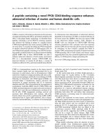
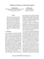
schematically depicted in Fig. 1.
The molecular mechanism of GC-A-mediated
inhibition of cardiac hypertrophy
To identify the molecular mechanism underlying car-
diac hypertrophy seen in GC-A-deficient mice, DNA
microarrays were used to identify genes upregulated in
the hypertrophied heart [45]. Among several genes
known to be upregulated in cardiac hypertrophy (e.g.
a-skeletal actin, ANP and BNP), it has been found
that the expression of the gene encoding myocyte-
enriched calcineurin-interacting protein (MCIP1) is
also increased. The MCIP1 gene is reportedly regu-
lated by calcineurin, a critical regulator of cardiac
hypertrophy. Thus, it was hypothesized that the calci-
neurin activity is enhanced in the heart of GC-A-defi-
cient mice. To test this hypothesis, cultured neonatal
cardiomyocytes were used to determine whether phar-
macological inhibition of GC-A would increase calci-
neurin activity, which it did not [45]. On the other
hand, stimulation of GC-A with ANP inhibited calci-
neurin activity, suggesting that it is by inhibiting the
calcineurin pathway that cardiac GC-A signaling (acti-
vated by locally secreted natriuretic peptides) exerts its
anti-hypertrophic effects. In fact, chronic treatment
with FK506, which in combination with FK506-bind-
ing protein inhibits the phosphatase activity of calci-
neurin, significantly reduces the heart weight to body
weight ratio, cardiomyocyte size and collagen volume
fraction in GC-A-deficient mice compared with the
wild-type mice [45]. A further study using microarray
analysis and real-time PCR analysis revealed that, in
addition to the calcineurin–nuclear factor of activated
T-cells (NFAT) pathway, the calmodulin–CaMK–
Hdac–Mef2 and PKC–MAPK–GATA4 pathways may
also be involved in the cardiac hypertrophy seen in the
GC-A-null mice [46].
Role of regulator of G-protein signaling in CG-A
cardioprotective actions
Recently, it has been elegantly demonstrated that
cGMP-dependent protein kinase (PKG) Ia attenuates
signaling by the thrombin receptor protease-activated
receptor (PAR) 1 through direct activation of regulator
of G-protein signaling (RGS) 2 [47]. PKG-Ia binds
directly to and phosphorylates RGS-2, which signifi-
cantly increases the GTPase activity of Ga
q
, thereby
terminating PAR-1 signaling. Given that cGMP is an
intracellular second messenger for natriuretic peptides,
RGS might mediate the cardioprotective effect of the
GC-A signaling. To test this hypothesis, the role of
RGS-4, which is the predominant RGS in cardiomyo-
cytes under physiological conditions, was examined. In
cultured cardiomyocytes, ANP stimulated the binding
of PKG-Ia to RGS-4 as well as the phosphorylation
of RGS-4 and its subsequent association with Ga
q
[48]. In addition, cardiomyocyte-specific overexpression
of RGS-4 in GC-A-null mice significantly rescued the
cardiac phenotype of these mice. On the contrary,
overexpression of a dominant-negative form of RGS-4
blocked the inhibitory effects of ANP on cardiac
hypertrophy [48]. Therefore, GC-A may activate car-
diac RGS-4, which then inhibits the activity of Ga
q
and its downstream hypertrophic effectors. The endog-
enous cardioprotective mechanism meditated by
ANP ⁄ BNP, GC-A and RGS-4 is depicted schemati-
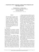
cally in Fig. 2.
Very recently, PKG activation reflecting chronic
inhibition of cGMP-selective phosphodiesterase 5 has
been shown to suppress maladaptive cardiac hypertro-
phy by inhibiting Ga
q
-coupled stimulation, and the
effect was not observed in mice lacking RGS-2 [49].
This suggests that RGS2 mediates the cardioprotective
actions of PKG in pathological conditions such as
‘Circulating hormones’
ANP
BNP
Vasodilatation
Natriuresis
GC-A
ANP
BNP
‘Local hormones’
Inhibition of
Cardiac remodeling
GC-A
Reduction of cardiac
p
re-and after-load
Fig. 1. ANP and BNP, the cardiac natriuretic peptides, protect the
heart in not only an endocrine but also a paracrine fashion. Because
ANP and BNP have potent diuretic, natriuretic and vasodilatory
actions, augmentation of the ANP and BNP ⁄ GC-A signaling leads to
a decrease in cardiac pre- and after-load, and their mobilization dur-
ing cardiac failure is considered one of the compensatory mecha-
nisms activated in response to heart damage. In addition to the
hemodynamic effects of their actions as circulating hormones,
recent evidence suggests that ANP and BNP also exert local cardio-
protective effects by acting as autocrine ⁄ paracrine hormones.
In vivo role of the natriuretic peptide system I. Kishimoto et al.
1836 FEBS Journal 278 (2011) 1830–1841 ª 2011 The Authors Journal compilation ª 2011 FEBS
pressure overload or excessive Ga
q
activation due to
hypertrophic stimuli. In fact, RGS-2 is also implicated
in the anti-hypertrophic action of cardiac GC-A [50].
The role of GC-A in myocardial infarction
It is well known that plasma levels of ANP and BNP
are dramatically elevated early after myocardial infarc-
tion [51]. To examine the significance of this upregula-
tion, experimental myocardial infarction by ligation of
the left coronary artery was induced in mice lacking
GC-A [52]. GC-A-deficient mice exhibited significantly
higher mortality rate than wild-type mice, reflecting a
higher incidence of acute heart failure. Four weeks
after infarction, left ventricular remodeling, including
myocardial hypertrophy and fibrosis, and impairment
of the left ventricular systolic function were signifi-
cantly more severe in mice lacking GC-A than in wild-
type mice [52]. GC-A activation by endogenous cardiac
natriuretic peptides may protect against acute heart
failure and attenuate chronic cardiac remodeling after
acute myocardial infarction.
Role of GC-A in peripheral arterial disease
A role of the natriuretic peptide system in peripheral
arterial diseases has also been suggested. Activation of
the natriuretic peptides–cGMP–PKG pathway was
found to accelerate vascular regeneration and blood
flow recovery in a murine model of peripheral arterial
disease, in which leg ischemia was induced by femoral
arterial ligation [53]. Recently, it has been reported
that intraperitoneal injection of carperitide, a recombi-
nant human ANP, accelerated blood flow recovery
with increasing capillary density in the ischemic legs
[54], indicating the role of exogenously administered
ANP and BNP in angiogenesis. When the hindlimb
ischemia model was performed in GC-A-deficient mice,
autoamputation or ulcers were more severe in GC-A-
deficient mice than in their wild-type counterparts [55].
Laser Doppler perfusion imaging revealed that the
recovery of blood flow in the ischemic limb was signifi-
cantly inhibited in GC-A-null mice compared with
wild-type mice. In addition, vascular regeneration in
response to critical hindlimb ischemia was severely
impaired [55]. Similar attenuation of ischemic angio-
genesis was observed in mice with conditional, endo-
thelial-cell-restricted GC-A deletion. On the other
hand, smooth-muscle-cell-restricted GC-A ablation did
not affect ischemic neovascularization [56], suggesting
that it is the endothelial GC-A that stimulates endo-
thelial regeneration after induction of ischemia. Taken
together, the evidence suggests that the natriuretic pep-
tide pathway significantly contributes to peripheral
vascular remodeling during ischemia.
Role of the CNP/GC-B pathway in bone
formation
In a 1998 study, mice with transgenic overexpression
of the BNP gene, especially those exhibiting high
expression levels, unexpectedly displayed deformed
bony skeletons characterized by kyphosis, elongated
limbs and paws, and crooked tails, which resulted
from a high turnover of endochondral ossification
accompanied by overgrowth of the growth plate [57].
Even after crossing with GC-A-null mice, transgenic
mice overexpressing BNP continued to exhibit marked
longitudinal growth of the vertebrae and long bones
[58]. Therefore, the effect of excess amount of BNP on
endochondral ossification is independent of GC-A,
and so signaling through another receptor was
suggested.
Fig. 2. Inhibitory mechanism of cardiac hypertrophy by the local
natriuretic peptide system. Cardiac hypertrophy agonists such as
angiotensin II, catecholamines and endothelins stimulate G-protein
coupled receptor. Subsequent production of inositol triphosphate
(IP3) promotes elevation of intracellular Ca
2+
levels, which results
in activation of the calcineurin ⁄ nuclear factor of activated T cells
(NFAT) pathway. Cooperatively with the family of GATA transcrip-
tion factors, NFAT activates the hypertrophic gene program, which
includes the ANP- and BNP-coding genes. In an autocrine or para-
crine fashion, ANP and BNP stimulate their receptor GC-A and
exert their anti-hypertrophic actions via the activation of the RGS,
which consequently results in an increase in the GTPase activity of
the a subunit of the guanine nucleotide binding protein (Ga
q
) and in
a decrease in the activity of the downstream signaling mediators
(adapted from [48]).
I. Kishimoto et al. In vivo role of the natriuretic peptide system
FEBS Journal 278 (2011) 1830–1841 ª 2011 The Authors Journal compilation ª 2011 FEBS 1837
In 2001, CNP-deficient mice were reported to show
severe dwarfism as a result of impaired endochondral
ossification [59], thus indicating that CNP acts locally
as a positive regulator of endochondral ossification. In
2004, the phenotype of mice lacking GC-B was
reported [60]. The GC-B-null animals exhibited dra-
matically impaired endochondral ossification and
attenuation of longitudinal vertebral or limb bone
growth. Therefore, it appears that GC-B is the recep-
tor mediating the CNP action in inducing longitudinal
bone growth. Furthermore, homozygous C-receptor-
null mice also have skeletal deformities associated with
a considerable increase in bone turnover [28], an oppo-
site phenotype to that observed in the mice deficient
for CNP. Since CNP is the only natriuretic peptide
expressed in bone, it is suggested that one function of
the C receptor is to clear locally synthesized CNP from
bone and modulate its effects.
Since pharmacological amounts of BNP can stimu-
late GC-B, these results suggest that activation of the
CNP ⁄ GC-B pathway in transgenic mice with elevated
plasma concentrations of BNP or in mice lacking the
C receptor for natriuretic peptides results in skeletal
overgrowth. By contrast, inactivation of the CNP ⁄ GC-
B pathway in mice lacking CNP, GC-B or cGMP-
dependent protein kinase II (a downstream mediator
of the CNP ⁄ GC-B pathway) results in dwarfism caused
by defects in endochondral ossification.
Summary
As stated above, studies using genetically engineered
animals revealed physiological and pathophysiological
roles of the natriuretic peptides ⁄ receptor signaling
pathways in the regulation of blood pressure ⁄ volume,
maintenance of the cardiovascular system, and devel-
opment of the longitudinal bone, acting as not only a
circulating hormonal system but also a local regulatory
system. Recent evidence also suggests roles for the
natriuretic peptide system in renal [61] and neuronal
[62] morphology and function. In addition, genetic
defects of each component of the system in humans
may cause diseases that are also observed in the geneti-
cally engineered animals. Furthermore, an interesting
hypothesis that needs verification is that these observed
phenomena could be the recapitulation of early devel-
opmental mechanisms. More studies at tissue, cellular
and molecular levels are needed to clarify the mecha-
nisms underlying the intriguing phenotypes observed in
transgenic animal models. In addition, more studies at
clinical and population levels are needed to elucidate
the potential importance of the natriuretic peptide sys-
tem in humans.
Acknowledgements
Our heartfelt appreciation goes to the late Dr Garbers, a
former professor of the University of Texas, whose com-
ments and suggestions were of inestimable value for our
study using GC-A knockout mice, to Professor Misono
of the University of Nevada School of Medicine, and to
the reviewers of the FEBS Journal, whose comments sig-
nificantly contributed to the writing of this review article.
Disclosures
The authors have nothing to disclose.
References
1 de Bold AJ, Borenstein HB, Veress AT & Sonnenberg
H (1981) A rapid and potent natriuretic response to
intravenous injection of atrial myocardial extract in
rats. Life Sci 28, 89–94.
2 Flynn TG, de Bold ML & de Bold AJ (1983) The
amino acid sequence of an atrial peptide with potent
diuretic and natriuretic properties. Biochem Biophys Res
Commun 117, 859–865.
3 Thibault G, Garcia R, Seidah NG, Lazure C, Cantin
M, Chre
´
tien M & Genest J (1983) Purification of three
rat atrial natriuretic factors and their amino acid com-
position. FEBS Lett 164, 286–290.
4 Kangawa K & Matsuo H (1984) Purification and com-
plete amino acid sequence of alpha-human atrial natri-
uretic polypeptide (alpha-hANP). Biochem Biophys Res
Commun 118, 131–139.
5 Misono KS, Fukumi H, Grammer RT & Inagami T
(1984) Rat atrial natriuretic factor: complete amino acid
sequence and disulfide linkage essential for biological
activity. Biochem Biophys Res Commun 119, 524–529.
6 Currie MG, Geller DM, Cole BR, Siegel NR, Fok KF,
Adams SP, Eubanks SR, Galluppi GR & Needleman P
(1984) Purification and sequence analysis of bioactive
atrial peptides (atriopeptins). Science 223, 67–69.
7 Kangawa K, Tawaragi Y, Oikawa S, Mizuno A, Saku-
ragawa Y, Nakazato H, Fukuda A, Minamino N &
Matsuo H (1984) Identification of rat atrial natriuretic
polypeptide and characterization of the cDNA encoding
its precursor. Nature 312, 152–155.
8 Sudoh T, Kangawa K, Minamino N & Matsuo H
(1988) A new natriuretic peptide in porcine brain.
Nature 332, 78–81.
9 Mukoyama M, Nakao K, Hosoda K, Suga S, Saito Y,
Ogawa Y, Shirakami G, Jougasaki M, Obata K, Yasue
H et al. (1991) Brain natriuretic peptide as a novel car-
diac hormone in humans. Evidence for an exquisite dual
natriuretic peptide system, atrial natriuretic peptide and
brain natriuretic peptide. J Clin Invest 87, 1402–1412.
In vivo role of the natriuretic peptide system I. Kishimoto et al.
1838 FEBS Journal 278 (2011) 1830–1841 ª 2011 The Authors Journal compilation ª 2011 FEBS
10 Nakagawa O, Ogawa Y, Itoh H, Suga S, Komatsu Y,
Kishimoto I, Nishino K, Yoshimasa T & Nakao K
(1995) Rapid transcriptional activation and early
mRNA turnover of brain natriuretic peptide in cardio-
cyte hypertrophy. Evidence for brain natriuretic peptide
as an ‘emergency’ cardiac hormone against ventricular
overload. J Clin Invest 96, 1280–1287.
11 Mukoyama M, Nakao K, Saito Y, Ogawa Y, Hosoda
K, Suga S, Shirakami G, Jougasaki M & Imura H
(1990) Increased human brain natriuretic peptide in
congestive heart failure. N Engl J Med 323, 757–758.
12 Sudoh T, Minamino N, Kangawa K & Matsuo H
(1990) C-type natriuretic peptide (CNP): a new member
of natriuretic peptide family identified in porcine brain.
Biochem Biophys Res Commun 168, 863–870.
13 Suga S, Itoh H, Komatsu Y, Ishida H, Igaki T,
Yamashita J, Doi K, Chun TH, Yoshimasa T, Tanaka
I et al. (1998) Regulation of endothelial production of
C-type natriuretic peptide by interaction between
endothelial cells and macrophages. Endocrinology 139,
1920–1926.
14 Naruko T, Ueda M, van der Wal AC, van der Loos
CM, Itoh H, Nakao K & Becker AE (1996) C-type
natriuretic peptide in human coronary atherosclerotic
lesions. Circulation 94, 3103–3108.
15 Fuller F, Porter JG, Arfsten AE, Miller J, Schilling JW,
Scarborough RM, Lewicki JA & Schenk DB (1988)
Atrial natriuretic peptide clearance receptor. Complete
sequence and functional expression of cDNA clones. J
Biol Chem 263, 9395–9401.
16 Anand-Srivastava MB, Sehl PD & Lowe DG (1996)
Cytoplasmic domain of natriuretic peptide receptor-C
inhibits adenylyl cyclase. Involvement of a pertussis
toxin-sensitive G protein. J Biol Chem 271, 19324–
19329.
17 Chinkers M, Garbers DL, Chang MS, Lowe DG, Chin
HM, Goeddel DV & Schulz S (1989) A membrane form
of guanylate cyclase is an atrial natriuretic peptide
receptor. Nature 338, 78–83.
18 Chang MS, Lowe DG, Lewis M, Hellmiss R, Chen E &
Goeddel DV (1989) Differential activation by atrial and
brain natriuretic peptides of two different receptor gua-
nylate cyclases. Nature 341, 68–72.
19 Schulz S, Singh S, Bellet RA, Singh G, Tubb DJ, Chin
H & Garbers DL (1989) The primary structure of a
plasma membrane guanylate cyclase demonstrates diver-
sity within this new receptor family. Cell 58, 1155–1162.
20 Koller KJ, Lowe DG, Bennett GL, Minamino N,
Kangawa K, Matsuo H & Goeddel DV (1991) Selective
activation of the B natriuretic peptide receptor by
C-type natriuretic peptide (CNP). Science 252, 120–
123.
21 Suga S, Nakao K, Kishimoto I, Hosoda K, Mukoyama
M, Arai H, Shirakami G, Ogawa Y, Komatsu Y, Nak-
agawa O et al. (1992) Receptor selectivity of natriuretic
peptide family, atrial natriuretic peptide, brain
natriuretic peptide, and C-type natriuretic peptide.
Endocrinology 130, 229–239.
22 Steinhelper ME, Cochrane KL & Field LJ (1990) Hypo-
tension in transgenic mice expressing atrial natriuretic
factor fusion genes. Hypertension 16, 301–307.
23 Ogawa Y, Itoh H, Tamura N, Suga S, Yoshimasa T,
Uehira M, Matsuda S, Shiono S, Nishimoto H & Nak-
ao K (1994) Molecular cloning of the complementary
DNA and gene that encode mouse brain natriuretic
peptide and generation of transgenic mice that overex-
press the brain natriuretic peptide gene. J Clin Invest
93, 1911–1921.
24 John SW, Krege JH, Oliver PM, Hagaman JR, Hodgin
JB, Pang SC, Flynn TG & Smithies O (1995) Genetic
decreases in atrial natriuretic peptide and salt-sensitive
hypertension. Science 267, 679–681.
25 Lopez MJ, Wong SK, Kishimoto I, Dubois S, Mach V,
Friesen J, Garbers DL & Beuve A (1995) Salt-resistant
hypertension in mice lacking the guanylyl cyclase-A
receptor for atrial natriuretic peptide. Nature 378,
65–68.
26 Oliver PM, Fox JE, Kim R, Rockman HA, Kim HS,
Reddick RL, Pandey KN, Milgram SL, Smithies O &
Maeda N (1997) Hypertension, cardiac hypertrophy,
and sudden death in mice lacking natriuretic peptide
receptor A. Proc Natl Acad Sci USA 94
, 14730–14735.
27 Oliver PM, John SW, Purdy KE, Kim R, Maeda N,
Goy MF & Smithies O (1998) Natriuretic peptide recep-
tor 1 expression influences blood pressures of mice in a
dose-dependent manner. Proc Natl Acad Sci USA 95,
2547–2551.
28 Matsukawa N, Grzesik WJ, Takahashi N, Pandey KN,
Pang S, Yamauchi M & Smithies O (1999) The natri-
uretic peptide clearance receptor locally modulates the
physiological effects of the natriuretic peptide system.
Proc Natl Acad Sci USA 96, 7403–7408.
29 Yoshimoto T, Naruse M, Naruse K, Shionoya K,
Tanaka M, Tanabe A, Hagiwara H, Hirose S, Muraki
T & Demura H (1996) Angiotensin II-dependent down-
regulation of vascular natriuretic peptide type C recep-
tor gene expression in hypertensive rats. Endocrinology
137, 1102–1107.
30 Kishimoto I, Yoshimasa T, Suga S, Ogawa Y, Komatsu
Y, Nakagawa O, Itoh H & Nakao K (1994) Natriuretic
peptide clearance receptor is transcriptionally down-reg-
ulated by beta 2-adrenergic stimulation in vascular
smooth muscle cells. J Biol Chem 269, 28300–28308.
31 Tamura N, Ogawa Y, Chusho H, Nakamura K, Nakao
K, Suda M, Kasahara M, Hashimoto R, Katsuura G,
Mukoyama M et al. (2000) Cardiac fibrosis in mice
lacking brain natriuretic peptide. Proc Natl Acad Sci
USA 97, 4239–4244.
32 Sabrane K, Kruse MN, Fabritz L, Zetsche B, Mitko D,
Skryabin BV, Zwiener M, Baba HA, Yanagisawa M &
I. Kishimoto et al. In vivo role of the natriuretic peptide system
FEBS Journal 278 (2011) 1830–1841 ª 2011 The Authors Journal compilation ª 2011 FEBS 1839
Kuhn M (2005) Vascular endothelium is critically
involved in the hypotensive and hypovolemic actions of
atrial natriuretic peptide. J Clin Invest 115, 1666–1674.
33 Holtwick R, Gotthardt M, Skryabin B, Steinmetz M,
Potthast R, Zetsche B, Hammer RE, Herz J & Kuhn M
(2002) Smooth muscle-selective deletion of guanylyl
cyclase-A prevents the acute but not chronic effects of
ANP on blood pressure. Proc Natl Acad Sci USA 99,
7142–7147.
34 Kishimoto I, Dubois SK & Garbers DL (1996) The
heart communicates with the kidney exclusively through
the guanylyl cyclase-A receptor: acute handling of
sodium and water in response to volume expansion.
Proc Natl Acad Sci USA 93, 6215–6219.
35 Shi SJ, Vellaichamy E, Chin SY, Smithies O, Navar LG
& Pandey KN (2003) Natriuretic peptide receptor A
mediates renal sodium excretory responses to blood vol-
ume expansion. Am J Physiol Renal Physiol 285,
F694–F702.
36 Potter LR, Abbey-Hosch S & Dickey DM (2006) Natri-
uretic peptides, their receptors, and cyclic guanosine
monophosphate-dependent signaling functions. Endocr
Rev 27, 47–72.
37 Curry FR (2005) Atrial natriuretic peptide: an essential
physiological regulator of transvascular fluid, protein
transport, and plasma volume. J Clin Invest 115, 1458–
1461.
38 Nakao K, Itoh H, Saito Y, Mukoyama M & Ogawa Y
(1996) The natriuretic peptide family. Curr Opin Neph-
rol Hypertens 5, 4–11.
39 Kishimoto I, Rossi K & Garbers DL (2001) A genetic
model provides evidence that the receptor for atrial
natriuretic peptide (guanylyl cyclase-A) inhibits cardiac
ventricular myocyte hypertrophy. Proc Natl Acad Sci
USA 98, 2703–2706.
40 Kuhn M, Holtwick R, Baba HA, Perriard JC, Schmitz
W & Ehler E (2002) Progressive cardiac hypertrophy
and dysfunction in atrial natriuretic peptide receptor
(GC-A) deficient mice. Heart 87, 368–374.
41 Knowles JW, Esposito G, Mao L, Hagaman JR, Fox
JE, Smithies O, Rockman HA & Maeda N (2001) Pres-
sure-independent enhancement of cardiac hypertrophy
in natriuretic peptide receptor A-deficient mice. J Clin
Invest 107, 975–984.
42 Kishimoto I, Tokudome T, Horio T, Garbers DL,
Nakao K & Kangawa K (2009) Natriuretic peptide
signaling via guanylyl cyclase (GC)-A: an endogenous
protective mechanism of the heart. Curr Cardiol Rev 5,
45–51.
43 Holtwick R, van Eickels M, Skryabin BV, Baba HA,
Bubikat A, Begrow F, Schneider MD, Garbers DL &
Kuhn M (2003) Pressure-independent cardiac hypertro-
phy in mice with cardiomyocyte-restricted inactivation
of the atrial natriuretic peptide receptor guanylyl
cyclase-A. J Clin Invest 111, 1399–1407.
44 Molkentin JD (2003) A friend within the heart: natri-
uretic peptide receptor signaling. J Clin Invest 111,
1275–1277.
45 Tokudome T, Horio T, Kishimoto I, Soeki T, Mori K,
Kawano Y, Kohno M, Garbers DL, Nakao K & Kang-
awa K (2005) Calcineurin-nuclear factor of activated T
cells pathway-dependent cardiac remodeling in mice
deficient in guanylyl cyclase A, a receptor for atrial and
brain natriuretic peptides. Circulation 111, 3095–3104.
46 Ellmers LJ, Scott NJ, Piuhola J, Maeda N, Smithies O,
Frampton CM, Richards AM & Cameron VA (2007)
Npr1-regulated gene pathways contributing to cardiac
hypertrophy and fibrosis. J Mol Endocrinol 38, 245–257.
47 Tang KM, Wang GR, Lu P, Karas RH, Aronovitz M,
Heximer SP, Kaltenbronn KM, Blumer KJ, Siderovski
DP, Zhu Y et al. (2003) Regulator of G-protein signal-
ing-2 mediates vascular smooth muscle relaxation and
blood pressure. Nat Med 9, 1506–1512.
48 Tokudome T, Kishimoto I, Horio T, Arai Y, Schwenke
DO, Hino J, Okano I, Kawano Y, Kohno M, Miyazato
M et al. (2008) Regulator of G-protein signaling sub-
type 4 mediates antihypertrophic effect of locally
secreted natriuretic peptides in the heart. Circulation
117, 2329–2339.
49 Tang KM, Wang GR, Lu P, Karas RH, Aronovitz M,
Heximer SP, Kaltenbronn KM, Blumer KJ, Siderovski
DP, Zhu Y et al. (2009) Regulator of G protein signal-
ing 2 mediates cardiac compensation to pressure over-
load and antihypertrophic effects of PDE5 inhibition in
mice. J Clin Invest 119, 408–420.
50 Klaiber M, Kruse M, Vo
¨
lker K, Schro
¨
ter J, Feil R,
Freichel M, Gerling A, Feil S, Dietrich A, London
˜
oJE
et al. (2010) Novel insights into the mechanisms mediat-
ing the local antihypertrophic effects of cardiac atrial
natriuretic peptide: role of cGMP-dependent protein
kinase and RGS2. Basic Res Cardiol 105, 583–595.
51 Morita E, Yasue H, Yoshimura M, Ogawa H, Jougasa-
ki M, Matsumura T, Mukoyama M & Nakao K (1993)
Increased plasma levels of brain natriuretic peptide in
patients with acute myocardial infarction. Circulation
88, 82–91.
52 Nakanishi M, Saito Y, Kishimoto I, Harada M, Kuwa-
hara K, Takahashi N, Kawakami R, Nakagawa Y,
Tanimoto K, Yasuno S et al. (2005) Role of natriuretic
peptide receptor guanylyl cyclase-A in myocardial
infarction evaluated using genetically engineered mice.
Hypertension 46, 441–447.
53 Yamahara K, Itoh H, Chun TH, Ogawa Y, Yamashita
J, Sawada N, Fukunaga Y, Sone M, Yurugi-Kobayashi
T, Miyashita K et al. (2003) Significance and therapeu-
tic potential of the natriuretic peptides ⁄ cGMP ⁄ cGMP-
dependent protein kinase pathway in vascular regenera-
tion. Proc Natl Acad Sci USA 100, 3404–3409.
54 Park K, Itoh H, Yamahara K, Sone M, Miyashita K,
Oyamada N, Sawada N, Taura D, Inuzuka M, Sono-
In vivo role of the natriuretic peptide system I. Kishimoto et al.
1840 FEBS Journal 278 (2011) 1830–1841 ª 2011 The Authors Journal compilation ª 2011 FEBS
yama T et al. (2008) Therapeutic potential of atrial
natriuretic peptide administration on peripheral arterial
diseases. Endocrinology 149, 483–491.
55 Tokudome T, Kishimoto I, Yamahara K, Osaki T,
Minamino N, Horio T, Sawai K, Kawano Y, Miyaz-
ato M, Sata M et al. (2009) Impaired recovery of
blood flow after hind-limb ischemia in mice lacking
guanylyl cyclase-A, a receptor for atrial and brain
natriuretic peptides. Arterioscler Thromb Vasc Biol 29,
1516–1521.
56 Kuhn M, Vo
¨
lker K, Schwarz K, Carbajo-Lozoya J,
Flo
¨
gel U, Jacoby C, Stypmann J, van Eickels M, Gam-
baryan S, Hartmann M et al. (2009) The natriuretic
peptide ⁄ guanylyl cyclase – a system functions as a
stress-responsive regulator of angiogenesis in mice.
J Clin Invest 119, 2019–2030.
57 Suda M, Ogawa Y, Tanaka K, Tamura N, Yasoda A,
Takigawa T, Uehira M, Nishimoto H, Itoh H, Saito Y
et al. (1998) Skeletal overgrowth in transgenic mice that
overexpress brain natriuretic peptide. Proc Natl Acad
Sci USA 95, 2337–2342.
58 Chusho H, Ogawa Y, Tamura N, Suda M, Yasoda A,
Miyazawa T, Kishimoto I, Komatsu Y, Itoh H, Tanaka
K et al. (2000) Genetic models reveal that brain natri-
uretic peptide can signal through different tissue-specific
receptor-mediated pathways. Endocrinology 141,
3807–3813.
59 Chusho H, Tamura N, Ogawa Y, Yasoda A, Suda M,
Miyazawa T, Nakamura K, Nakao K, Kurihara T,
Komatsu Y et al. (2001) Dwarfism and early death in
mice lacking C-type natriuretic peptide. Proc Natl Acad
Sci USA 98, 4016–4021.
60 Tamura N, Doolittle LK, Hammer RE, Shelton JM,
Richardson JA & Garbers DL (2004) Critical roles of
the guanylyl cyclase B receptor in endochondral ossifi-
cation and development of female reproductive organs.
Proc Natl Acad Sci USA 101, 17300–17305.
61 Das S, Au E, Krazit ST & Pandey KN (2010) Targeted
disruption of guanylyl cyclase-A ⁄ natriuretic peptide
receptor-A gene provokes renal fibrosis and remodeling
in null mutant mice: role of proinflammatory cytokines.
Endocrinology 151, 5841–5850.
62 Kishimoto I, Tokudome T, Horio T, Soeki T, Chusho
H, Nakao K & Kangawa K (2008) C-type natriuretic
peptide is a Schwann cell-derived factor for develop-
ment and function of sensory neurones. J Neuroendocri-
nol 20, 1213–1223.
63 Yasoda A, Komatsu Y, Chusho H, Miyazawa T, Ozasa
A, Miura M, Kurihara T, Rogi T, Tanaka S, Suda M
et al. (2004) Overexpression of CNP in chondrocytes
rescues achondroplasia through a MAPK-dependent
pathway. Nat Med 10, 80–86.
64 Kake T, Kitamura H, Adachi Y, Yoshioka T, Watana-
be T, Matsushita H, Fujii T, Kondo E, Tachibe T,
Kawase Y et al. (2009) Chronically elevated plasma
C-type natriuretic peptide level stimulates skeletal
growth in transgenic mice. Am J Physiol Endocrinol
Metab 297, E1339–E1348.
65 Wang Y, de Waard MC, Sterner-Kock A, Stepan H,
Schultheiss HP, Duncker DJ & Walther T (2007) Car-
diomyocyte-restricted over-expression of C-type natri-
uretic peptide prevents cardiac hypertrophy induced by
myocardial infarction in mice. Eur J Heart Fail 9,
548–557.
66 Schmidt H, Stonkute A, Ju
¨
ttner R, Koesling D, Friebe
A & Rathjen FG (2009) C-type natriuretic peptide
(CNP) is a bifurcation factor for sensory neurons. Proc
Natl Acad Sci USA 106, 16847–16852.
67 Langenickel TH, Buttgereit J, Pagel-Langenickel I,
Lindner M, Monti J, Beuerlein K, Al-Saadi N, Plehm
R, Popova E, Tank J et al. (2006) Cardiac hypertrophy
in transgenic rats expressing a dominant-negative
mutant of the natriuretic peptide receptor B. Proc Natl
Acad Sci USA 103, 4735–4740.
I. Kishimoto et al. In vivo role of the natriuretic peptide system
FEBS Journal 278 (2011) 1830–1841 ª 2011 The Authors Journal compilation ª 2011 FEBS 1841