Báo cáo khoa học: MicroRNA-373, a new regulator of protein phosphatase 6, functions as an oncogene in hepatocellular carcinoma pdf
Bạn đang xem bản rút gọn của tài liệu. Xem và tải ngay bản đầy đủ của tài liệu tại đây (398.71 KB, 11 trang )
MicroRNA-373, a new regulator of protein phosphatase 6,
functions as an oncogene in hepatocellular carcinoma
Nannan Wu*, Xuyuan Liu*, Xuemei Xu*, Xingxing Fan, Min Liu, Xin Li, Qiping Zhong and
Hua Tang
Tianjin Life Science Research Center and Basic Medical School, Tianjin Medical University, China
Introduction
Hepatocellular carcinoma (HCC) accounts for 80–90%
of liver cancers, and is one of the most prevalent carci-
nomas worldwide [1]. Liver cancer is a complex genetic
disease in which the expression of many specific genes,
known as oncogenes or tumor suppressor genes, is
abnormally changed. Previous studies have revealed
several genes related to human HCC. For example, the
cyclin G1 gene is upregulated in HCC [2], and the
phosphatase and tensin homolog (PTEN) gene is
downregulated in HCC [3]. Although focusing on
known genes has yielded much new information, previ-
ously unknown noncoding RNAs, such as microRNAs
(miRNAs), may also provide insights into the biology
of HCC. MicroRNAs are a group of noncoding single-
stranded RNAs, 22 nucleotides in length, that have
emerged as an important class of short endogenous
RNAs that post-transcriptionally regulate gene expres-
sion by base-paring with their target mRNA [4]. Sev-
eral lines of evidence have shown that the six to eight
nucleotides at the 5¢-end of miRNAs (positions 1–8)
Keywords
cell cycle; hepatocellular carcinoma; miRNA;
miRNA-373; protein phosphatase 6 catalytic
subunit (PPP6C)
Correspondence
H. Tang, Tianjin Life Science Research
Center and Basic Medical School, Tianjin
Medical University, Tianjin 300070, China
Fax: +86 22 23542503
Tel: +86 22 23542503
E-mail:
*These authors contributed equally to this
work
(Received 9 January 2011, revised 17 March
2011, accepted 5 April 2011)
doi:10.1111/j.1742-4658.2011.08120.x
MicroRNAs are a class of small noncoding RNAs that function as key reg-
ulators of gene expression at the post-transcriptional level. Recently, micr-
oRNA-373 (miR-373) has been found to function as an oncogene in
testicular germ cell tumors. In our study, we found that miR-373 is upregu-
lated in human hepatocellular carcinoma (HCC) tissues as compared with
adjacent normal tissues, and promotes the proliferation of the HCC cell
lines HepG2 and QGY-7703 by regulating the transition between G
1
-phase-
and S-phase. The gene encoding the protein phosphatase 6 catalytic sub-
unit (PPP6C ), a negative cell cycle regulator, was identified as a direct
target gene of miR-373 by use of a fluorescent reporter assay. The mRNA
and protein levels of PPP6C were both inversely correlated with the miR-
373 expression level. Overexpression of PPP6C abolished the regulation of
cell cycle and cell growth exercised by miR-373 in HepG2 cells. These
results indicate that miR-373 plays an important role in the pathogenesis
of HCC, and may be a new biomarker in HCC. Our results demonstrate
that miR-373 can regulate cell cycle progression by targeting PPP6C tran-
scripts and promotes the growth activity of HCC cells in vitro. The down-
regulation of PPP6C by miR-373 may explain why the expression of
miR-373 can promote HCC cell proliferation.
Abbreviations
ASO, antisense oligonucleotide; EGFP, enhanced green fluorescence protein; FACS, fluorescence-activated cell sorting; GAPDH,
glyceraldehyde-3-phosphate dehydrogenase; HCC, hepatocellular carcinoma; miRNA, microRNA; MTT, 3-(4,5-dimethylthiazol-2-yl)-2,5-
diphenyl-tetrazolium bromide; PI, proliferation index; PPP6C, protein phosphatase 6 catalytic subunit; SD, standard deviation; shRNA, small
hairpin RNA; siRNA, small interfering RNA.
2044 FEBS Journal 278 (2011) 2044–2054 ª 2011 The Authors Journal compilation ª 2011 FEBS
are important for target site recognition, and they have
been designated as the ‘seed’ region. Animal miRNAs
target mRNA 3¢-UTRs predominantly by seed
sequence complementarity, and are rarely fully comple-
mentary; therefore, they function through translational
repression rather than cleavage [5]. On the basis of
this, miRNAs could control as many as 30% of all
protein-coding genes [6]. MicroRNAs play important
roles in developmental timing, and participate in the
regulation of processes such as cell fate determination,
proliferation, differentiation, and cell death [7–10]. Pre-
vious studies have identified cancer-specific miRNAs in
many types of cancer, including B-cell chronic lympho-
cytic leukemia [11], colorectal cancer [12,13], lung can-
cer [14], breast cancer [15], and brain cancer [16,17].
A recent study described miR-373 as a tumor suppres-
sor gene in prostate cancer [18]; other studies provided
evidence that miR-373 was upregulated in breast can-
cer, testicular germ cell tumors, and human esophageal
cancer [19–21]. However, the regulatory effects of
miR-373 on the tumorigenesis of other cancers remain
to be elucidated.
In this study, we found, through real time reverse
transcription PCR (real time RT-PCR), that miR-373
was overexpressed in human HCC tissues as compared
with adjacent normal tissues, and identified the gene
encoding protein phosphatase 6 catalytic subunit
(PPP6C) as a direct target of miR-373. We also
observed that upregulation of miR-373 promoted cell
cycle progression through the G
1
⁄ S checkpoint in HCC
cells. Taken together, our results suggest that miR-373
regulates the proliferation of a human HCC cell line by
negatively regulating PPP6C expression.
Results
MicroRNA-373 is upregulated in HCC
To determine the expression of miR-373 in human
HCC tissues and adjacent normal tissues, we used
quantitative real time RT-PCR to detect 26 pairs of
HCC samples. It was shown that miR-373 expression
level was generally and significantly higher in cancer
tissues than in adjacent nontumor tissue (Fig. 1). Thus,
we speculated that miR-373 might be involved the
pathogenesis of HCC.
Alteration of miR-373 affects cell growth of HCC
in vitro
First, we transfected either miR-373 antisense oligonu-
cleotides (ASOs) or an miR-373 expression vector
(pcDNA3 ⁄ pri-miR-373, pri-373) into HCC cells, and
detected miR-373 levels by real time RT-PCR. Expres-
sion of miR-373 was increased 4.5-fold in the HepG2
cells transfected with pcDNA3 ⁄ pri-miR-373 as com-
pared with controls; miR-373 ASOs resulted in an
75% reduction of miR-373 levels (Fig. 2A). Cell via-
bility of HCC cells transfected with miR-373 ASOs or
pri-373 was evaluated with the 3-(4,5-dimethylthiazol-
2-yl)-2,5-diphenyl-tetrazolium bromide (MTT) assay;
miR-373 ASOs reduced cell viability at 48 or 72 h after
transfection, whereas pri-373 increased cell viability
(Fig. 2B). In parallel, we analyzed colony formation
and cellular proliferation to assess the effect of miR-
373 on the proliferative capacity of HCC cells. The
colony formation rate of HepG2 cells after transfection
with miR-373 ASOs was 30% lower than that of
HepG2 cells transfected with control oligomers. Con-
versely, transfection with pri-373 increased colony for-
mation by 29% in HepG2 cells (Fig. 2C,D). We
observed similar results in another HCC cell line,
QGY-7703 (Fig. 2). These results indicate that miR-
373 can promote the cell proliferation of HCC cells.
miR-373 facilitates the G
1
⁄
S-phase transition in
HepG2 cells
To explore whether the promotion of proliferation
caused by miR-373 in HCC cells is attributable to an
alteration in cell cycle progression, we performed
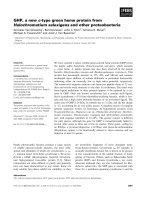
Fig. 1. Differential expression of miR-373 in HCC tissues. The
expression level of miR-373 in 26 pairs of HCC tissues (cancer) and
matched normal tissues (normal) was detected by real-time
RT-PCR. Box-plot lines represent medians and interquartile ranges
of the normalized threshold values; whiskers and spots indicate
10–90th percentiles and the remaining data points. The expression
of miR-373 is normalized to U6 small nuclear RNA (*P < 0.05).
N. Wu et al. MicroRNA-373 functions as an oncogene in HCC
FEBS Journal 278 (2011) 2044–2054 ª 2011 The Authors Journal compilation ª 2011 FEBS 2045
fluorescence-activated cell sorting (FACS) analysis.
Interestingly, in miR-373 ASO-treated HepG2 cells,
the percentage of cells in G
1
-phase increased to 54%,
whereas scramble ASO-treated cells had only 40% of
cells in G
1
-phase. The percentage of miR-373 ASO-
treated cells in S-phase decreased to 18%, as compared
with 31% in the control group (Fig. 3A). The prolifer-
ation index of miR-373 ASO-treated HepG2 cells was
85.2%, as compared with 150% in controls. In
contrast, after transfection with pri-373, the percentage
of HepG2 cells in G
1
-phase was 37%, as compared
with 50% in the scramble pcDNA3-treated cells, and
the percentage of cells in G
2
-phase was 20%, as com-
pared with 34% in scramble pcDNA3-treated cells
(Fig. 3B). These results indicate that miR-373 plays an
important role in the G
1
⁄ S-phase transition of the cell
cycle.
miR-373 targets PPP6C and negatively regulates
its expression
MicroRNAs regulate a variety of cellular activities
through regulation of the expression of target genes.
To determine the mechanism of miR-373-mediated cell
cycle dysregulation in HCC cells, we next identified
target genes that could be responsible for the effect of
miR-373. Taking into consideration that miR-373 was
upregulated in HCC tissues (Fig. 1), we reasoned that
its target genes should be correspondingly downregu-
lated. Twelve candidate genes were predicated by three
bioinformatics software packages (pictar, target-
scan, and microcosm ). Among these genes, the tumor
suppressor gene PPP6C, which was predicted to have
an miR-373-binding site in its 3¢-UTR (Fig. 4A), was
chosen for further study.
Fig. 2. Alteration of miR-373 levels affects
the growth of HCC cells. (A) The miR-373
expression level in HCC cells was effec-
tively altered by transfection of an miR-373
ASO vector or a pri-373 vector as detected
by real-time RT-PCR. U6 small nuclear RNA
was used for normalization. (B) HCC cells
were transfected with an miR-373 ASO vec-
tor or a pri-373 vector. The MTT assay was
used to determine relative cellular prolifera-
tion at 48 and 72 h. A
570 nm
is the absor-
bance of MTT measured at 570 nm. After
transfection with miR-373 or pri-373, the 48-
h and 72-h data points showed a statistically
significant difference (72-h data not shown).
(C) HCC cells were transfected with an
miR-373 ASO vector or a pri-373 vector.
(D) Cell growth was measured with colony
formation assays and proliferation curve
assays. The data represent the mean ± SD
of three different experiments (NC, negative
control; *P < 0.05, **P < 0.005,
#
P < 0.0005).
MicroRNA-373 functions as an oncogene in HCC N. Wu et al.
2046 FEBS Journal 278 (2011) 2044–2054 ª 2011 The Authors Journal compilation ª 2011 FEBS
To confirm whether miR-373 could bind to this
predicted region and suppress the expression of
PPP6C protein, we constructed an enhanced green flu-
orescence protein (EGFP) reporter vector (pcDNA3 ⁄
EGFP-PPP6C-3¢UTR), in which the 3¢-UTR fragment
of PPP6C, including the region encoding the putative
binding site, was inserted downstream of the EGFP
coding region. HepG2 cells were transfected with the
reporter vector together with either miR-373 ASOs or
pri-373. As shown in Fig. 4B, the intensity of EGFP
fluorescence was higher in the miR-373-blocked group
than in the control groups, whereas ectopic expression
of miR-373 decreased the intensity of EGFP fluores-
cence when compared with the control groups. In
addition, we constructed another EGFP reporter vec-
tor containing mutations in the regions encoding the
miR-373 binding sites (Fig. 4A). Neither blocking of
miR-373 with ASOs nor overexpression of miR-373
had any effect on the intensity of EGFP fluorescence
from the vector containing mutations in the region
encoding the miR-373 binding sites. These results
show that miR-373 binds directly to the 3¢-UTR of
the PPP6C transcript to repress gene expression.
To determine whether miR-373 negatively regulates
PPP6C expression at the mRNA or protein levels, we
assessed endogenous PPP6C expression in HepG2 cells
with altered miR-373 expression. HepG2 cells were
transfected with miR-373 ASOs or pcDNA3 ⁄ pri-miR-
373 to block or overexpress miR-373, respectively, and
the expression level of PPP6C mRNA was measured
by real time RT-PCR. When miR-373 was blocked,
PPP6C mRNA was elevated 3.8-fold as compared
with the control group, whereas overexpression of
miR-373 resulted in an 80% decrease in PPP6C mRNA
Fig. 3. miR-373 can promote the progres-
sion from G
1
-phase to S-phase in HepG2
cells. After transfection with an miR-373
ASO vector or a pri-373 vector, HepG2 cells
were detached, rinsed, fixed and stained as
described in Experimental procedures. Cell
cycle phase distribution was analyzed by
FACS. (A) The fraction of cells in G
1
-phase
was significantly increased in the miR-373
ASO group, and the fraction of cells in
S-phase was significantly decreased in the
miR-373 ASO group. (B) In the pri-373
group, the fraction of cells in G
1
-phase was
significantly decreased. The PI increased
noticeably in the pri-373 group. The data
represent the mean ± SD of three different
experiments (NC, negative control;
*P < 0.05, **P < 0.005).
Fig. 4. PPP6C is a direct target of miR-373. (A) The PPP6C 3¢-UTR
carries one potential miR-373-binding site. (B) The direct interaction
of miR-373 and PPP6C mRNA was confirmed by a fluorescent repor-
ter assay. HepG2 cells were transfected with an EGFP reporter vec-
tor together with an miR-373 ASO or a pri-373 vector, and the EGFP
intensity was measured. The data represent the mean ± SD of three
different experiments (NC, negative control; hsa, Homo sapiens;
*P < 0.05, **P < 0.005,
#
P < 0.0005). Similar results were obtained
in QGY-7703 cells (data not shown).
N. Wu et al. MicroRNA-373 functions as an oncogene in HCC
FEBS Journal 278 (2011) 2044–2054 ª 2011 The Authors Journal compilation ª 2011 FEBS 2047
(Fig. 5A). Western blot assay indicated that miR-373
ASOs resulted in a 2.3-fold increase in the PPP6C pro-
tein level, whereas overexpression of miR-373 reduced
the PPP6C protein level by 70% (Fig. 5C).
To confirm the results obtained from the cell lines, we
also examined the expression of PPP6C mRNA in 16
pairs of hepatocarcinoma tissue samples. Figure 5B
shows that, as compared with adjacent normal tissues,
PPP6C mRNA was consistently downregulated in HCC
tissue samples. These results suggest that miR-373 regu-
lates endogenous PPP6C expression through mRNA
degradation.
Knockdown of PPP6C promotes HCC cell growth
Sequence-specific small interfering RNA (siRNA) can
effectively suppress gene expression. We constructed a
plasmid expressing a small hairpin RNA (shRNA) tar-
geting PPP6C (pSilencer⁄ shRNA-PPP6C). Western
blot assay showed that the level of PPP6C expression
was reduced by 90% in HepG2 cells that were trans-
fected with pSilencer ⁄ shRNA-PPP6C, as compared
with HepG2 cells transfected with a control plasmid
(Fig. 6A). Inhibition of PPP6C expression increased
HCC cell growth as compared with the control group
(Fig. 6B–D), which was consistent with the effects of
miR-373 overexpression. Next, we examined the effects
of PPP6C knockdown on the cell cycle (Fig. 6E).
Knockdown of PPP6C resulted in a significant
decrease in the proportion of cells in G
1
-phase and an
increase in the proportion of cells in S-phase. These
findings indicate that PPP6C decreases the prolifera-
tion of HCC cells by inducing cell cycle arrest at the
G
1
⁄ S checkpoint, which is consistent with the effect of
miR-373 on HCC cells.
Overexpression of PPP6C counteracts the effects
of miR-373 expression on the G
1
⁄
S-phase
transition
PPP6C induces cell cycle arrest at the G
1
⁄ S checkpoint
in cancer cells [22]. We generated a plasmid (pcDNA3 ⁄
PPP6C, lacking the 3¢-UTR) to increase the protein
expression of PPP6C (Fig. 7A). We transfected HCC
cells with the plasmid, and analyzed cell growth and cell
cycle progression. In this experiment, ectopic expression
of PPP6C abrogated the promotion of cell growth
(Fig. 7B–D) and the increase in the rate of G
1
⁄ S-phase
transition (Fig. 7E) caused by miR-373 in HepG2 cells.
The overall effect of PPP6C overexpression was compa-
rable to that of miR-373 ASO treatment, suggesting
that PPP6C is a key mediator of the miR-373 regula-
tion of cell growth and cell cycle progression in HCC.
Discussion
MicroRNAs regulate diverse biological processes,
including tumorigenesis. It has been reported that
miR-373 functions as an oncogenic miRNA in testicu-
lar germ cell tumors [20] and in human esophageal
cancer [21]. We wanted to determine whether miR-373
also functions as an oncogenic miRNA in HCC cells.
To address this question, we first examined miR-373
expression in HCC tissues and matched adjacent non-
tumor tissues by real-time RT-PCR, as previously
described [23]. The results show that the level of
miR-373 is uncreased in tumor tissues as compared
with the matched normal tissues in 16 pairs of
matched specimens. It has been reported that the
upregulation of miR-373 promotes the growth of the
cell lines in many cancers, e.g. breast cancer [19], tes-
ticular germ cell tumors [20], and human esophageal
cancer [21]. We determined the effect of miR-373 on
the HCC cell lines HepG2 and QGY-7703 cells by gain
and loss of function approaches. MTT, colony forma-
tion and growth curve assays show that miR-373 can
increase the growth of those cells, and FACS analysis
indicates that miR-373 can promote progression of the
G
1
⁄ S-phase transition in the cell cycle. Thus, we
inferred that miR-373 might be a growth-promoting fac-
tor in HCC. In breast cancer, miR-373 can promote
invasion of the cancer cells [19]. The function of miR-373
on HCC cells needs to be elucidated in the future.
The fundamental function of miRNAs is to regulate
their targets by direct cleavage of mRNA or by inhibi-
tion of translation [18], depending on the degree of
complementarity with the 3¢-UTR of their target
genes. Computational algorithms were used to predict
miRNA targets, which are based mainly on base pair-
ing of miRNAs and the 3¢-UTRs of genes [6,24–26].
Twelve candidate genes were predicted by three bioin-
formatics software packages (pictar, targetscan,
and microcosm), which may be correlated with the
phenotype of the HCC cell lines caused by the alter-
ation of miR373. Among them, a tumor suppressor
gene, PPP6C, which regulates the G
1
⁄ S-phase transi-
tion [22], was selected for further study. It was pre-
dicted that the 3¢-UTR region of PPP6C transcript
would have an miR-373-binding site. Given that
miR-373 can target PPP6C mRNA, it may suppress
the expression of PPP6C. Western blot and real time
RT-PCR assays show that miR-373 decreases PPP6C
expression at the protein and mRNA levels in the
HCC cell lines as compared with the control
(Fig. 5A,C), and also that PPP6C expression is down-
regulated in HCC tissue as compared with normal tis-
sues (Fig. 5B), in which miR-373 is upregulated
MicroRNA-373 functions as an oncogene in HCC N. Wu et al.
2048 FEBS Journal 278 (2011) 2044–2054 ª 2011 The Authors Journal compilation ª 2011 FEBS
(Fig. 1). The regulation of genes by miRNA occurs
mainly through direct targeting of the 3¢-UTR region
[5]. We confirmed, with an EGFP-PPP6C-3¢UTR
reporter assay, that miR-373 can bind directly to the
PPP6C 3¢-UTR and negatively regulate PPP6C expres-
sion (Fig. 4B). A previous study has demonstrated the
direct regulation of CD4, a signal molecule involved in
cell growth and adhesion, by miR-373 [18]. Here, we
have enough evidence to confirm the tumor-suppress-
ing role of PPP6C in HCC cells, because knockdown
of PPP6C by siRNA promoted HCC cell proliferation,
whereas ectopic expression of PPP6C effectively allevi-
ated the miR-373-induced promotion of HCC cell
proliferation. Together, these findings indicate that
miR-373 might exert its effects in HCC mainly by tar-
geting PPP6C. However, Ivanov et al. [27] recently
reported that PPP6C can be targeted by miR-31, and
functions as an oncogene in mesothelioma, which is in
contrast to our results obtained in HCC. The possible
explanation is that the biological molecules have differ-
ent influences in different tumor cells. For example,
KLF4 was found to be an oncogene in breast cancer
[28], but Guan et al. [29] reported KLF4 as a tumor
suppressor in B-cell non-Hodgkin lymphoma and in
classic Hodgkin lymphoma. Also, miRNAs have dif-
ferent functions in different tissues; for instance, miR-9
is upregulated in breast cancer cells [30], but downreg-
ulated in human ovarian cancer. [31]. In addition, dif-
ferent miRNAs can target the same gene; for example,
CCND1 is directly regulated by the miR-16 family
[32], and miR-19a can also regulate the expression of
CCND1 [33]. Although miR-31 can target the PPP6C
transcript, the binding sites in nucleotides 1363–1369
of the 3¢-UTR are different from the miR-373-binding
sites in nucleotides 1338–1344 of the 3¢-UTR, which
do not overlap. Whether miR-373 and miR-31 can
simultaneously regulate PPP6C in HCC cells remains
to be elucidated.
In conclusion, miR-373 functions as an oncogene
and is upregulated in HCC tissues as compared with
adjacent normal tissues. Suppression of miR-373
repressed cell growth, possibly through inhibition of
the cell cycle by targeting PPP6C. Thus, the identifica-
tion of the oncogene, miR-373, and its target gene,
PPP6C, may help us to understand the molecular
mechanism of tumorigenesis in HCC and may have
potential diagnostic and therapeutic value in the
future.
Experimental procedures
Clinical specimen and RNA isolation
Twenty-six pairs of clinical specimens, including 26 human
HCC tissue samples and 26 matched normal liver tissue
samples, were obtained from the Tumor Bank Facility of
Tianjin Medical University Cancer Institute and Hospital
and the National Foundation of Cancer Research, with
patients’ informed consent, which was approved by the eth-
ics committee. The category of specimens was confirmed by
Fig. 5. The expression level of PPP6C is
inversely correlated with the level of miR-
373. (A) When miR-373 was blocked or
overexpressed, the mRNA level of PPP6C
was subsequently elevated or diminished,
respectively, as compared with the control
group. (B) Relative expression level of
PPP6C in HCC tissues or matched noncan-
cerous tissues, as measured by real-time
RT-PCR. The expression level of PPP6C is
normalized to b-actin. (C) When miR-373
was blocked or overexpressed, the protein
level of PPP6C was subsequently elevated
or diminished, respectively, as compared
with the control group (NC, negative control;
*P < 0.05,
#
P < 0.0005).
N. Wu et al. MicroRNA-373 functions as an oncogene in HCC
FEBS Journal 278 (2011) 2044–2054 ª 2011 The Authors Journal compilation ª 2011 FEBS 2049
pathological analysis. Large and small RNAs were isolated
from tissue samples with the mirVana miRNA Isolation
Kit (Ambion, Austin, TX, USA), according to the manu-
facturer’s instructions.
Cell culture and transfection
Two human HCC cell lines (HepG2 and QGY-7703) were
maintained in MEMa or RPMI-1640 (Gibco, Grand
Island, NY, USA), respectively, and supplemented with
10% fetal bovine serum, 100 IUÆmL
)1
penicillin, and
100 lgÆmL
)1
streptomycin. Cells were incubated at 37 °Cin
a humidified chamber supplemented with 5% CO
2
. Trans-
fection was performed with Lipofectamine 2000 Reagent
(Invitrogen, Carlsbad, CA, USA), following the manufac-
turer’s protocol.
Construction of expression vectors
To construct an miR-373-expressing vector (pcDNA3 ⁄ pri-
miR-373, pri-miR-373), we first amplified a 476-bp DNA
fragment carrying pri-miR-373 from genomic DNA; the
amplified fragment was then cloned into the pcDNA3 at
the XhoI and HindIII sites. For construction of EGFP-
PPP6C-3¢UTR reporter vectors (pcDNA3 ⁄ EGFP-PPP6C-
3¢UTR and pcDNA3 ⁄ EGFP-PPP6C-3¢UTR mutant), the
3¢-UTR and mutant 3¢-UTR fragments of PPP6C tran-
scripts amplified by RT-PCR were inserted into the vector
backbone downstream of the EGFP gene between the
BamHI and EcoRI sites of the pcDNA3 ⁄ EGFP vector, as
previously described [34]. The pSilencer ⁄ shRNA-PPP6C
plasmid expressing an siRNA targeting the PPP6C
transcript was constructed by annealing single-stranded
Fig. 6. Knockdown of PPP6C shows con-
cordant effects with miR-373 overexpres-
sion in HCC cells. (A) Western blot analysis
showed that the expression of PPP6C was
successfully suppressed by PPP6C siRNA.
(B–D) PPP6C was knocked down in HCC
cells, and cell growth ⁄ viability activity was
analyzed with the (B) MTT, (C) colony for-
mation and (D) proliferation curve assays.
(E) Cell cycle phase distribution was ana-
lyzed by FACS. The data represent the
mean ± SD of three different experiments
(NC, negative control; *P < 0.05,
**P < 0.005,
#
P < 0.0005).
MicroRNA-373 functions as an oncogene in HCC N. Wu et al.
2050 FEBS Journal 278 (2011) 2044–2054 ª 2011 The Authors Journal compilation ª 2011 FEBS
hairpin cDNA and inserting it into a pSilencer2.1 ⁄ neo vec-
tor (Ambion), using BamHI and HindIII sites. To construct
the PPP6C expression plasmid, the coding sequence (ORF)
without the 3¢-UTR of human PPP6C was amplified by
RT-PCR and inserted into the EcoRI and XhoI sites of
pcDNA3. All of the primers used are shown in Table 1.
Cell proliferation assay
Cells were seeded in 96-well plates at a density of 5000 cells
per well, and then transfected with pri-miR-373 or miR-373
ASOs on the next day. The MTT assay was used to deter-
mine relative cell viability at 48 and 72 h. Ten microliters
of MTT solution was added to 100 lL of culture medium,
and incubated for 4 h at 37 °C; the absorbance at 570 nm
(A
570
) was then measured.
For cell proliferation measurements, HCC cells were
seeded in 24-well plates at 10 000 cells per well (HepG2
cells) and 3000 cells per well (QGY-7703 cells) after trans-
fection with pri-miR-373 or miR-373 ASOs. Cell numbers
were then counted every day for 6 days. Each experiment
was performed in triplicate.
Fig. 7. Effects of PPP6C overexpression in
HCC cells. HCC cells were transfected with
pcDNA ⁄ 373 (pri-373). After 8 h, cells were
transfected with a pcDNA3 ⁄ PPP6C vector
or pcDNA3 empty vector. The
pcDNA3 ⁄ PPP6C vector did not contain the
3¢-UTR of PPP6C, and miR-373 could not
therefore regulate ectopic PPP6C expres-
sion. (A) At 48 h after transfection, PPP6C
expression was measured by western blot.
Cell growth ⁄ viability was analyzed with the
(B) MTT, (C) colony formation and (D) prolif-
eration curve assays. (E) Cell cycle phase
distribution was analyzed by FACS. The data
represent the mean ± SD of three different
experiments (*P < 0.05, **P < 0.005,
#
P < 0.0005).
N. Wu et al. MicroRNA-373 functions as an oncogene in HCC
FEBS Journal 278 (2011) 2044–2054 ª 2011 The Authors Journal compilation ª 2011 FEBS 2051
Colony formation assay
After transfection, cells were counted and seeded in 12-well
plates (in triplicate) at a density of 500 cells per well
(HepG2 cells) or 100 cells per well (QGY-7703 cells). The
culture medium was replaced every 3 days. Colonies were
counted only if they contained more than 50 cells, and the
cells were stained with crystal violet. The rate of colony for-
mation was calculated with the following equation: colony
formation rate = (number of colonies ⁄ number of seeded
cells) · 100%.
Flow cytometry analysis
At 48 h after transfection, the cells were detached from the
plates by trypsin incubation, rinsed with NaCl ⁄ P
i
, and fixed
in 95% (v ⁄ v) ethanol. The cells were then rehydrated in
NaCl ⁄ P
i
and incubated with RNase (100 lgÆmL
)1
) and pro-
pidium iodide (60 lg ÆmL
)1
) (Sigma-Aldrich, MO, USA).
Cells were analyzed with the FACS Calibur System (Beck-
man Coulter, Brea, CA, USA), and the cell cycle phase was
determined by cell quest analysis. The proliferation index
(PI) was calculated as follows: PI = (S + G
2
⁄ M) ⁄ G
1
(S,
G
2
⁄ M and G
1
refer to the percentages of cells in S-phase,
G
2
⁄ M-phase and G
1
-phase, respectively) [35].
Bioinformatics
The miRNA targets predicted by computer-aided algo-
rithms were obtained with pictar, targetscan, and
microcosm. For identification of the genes commonly pre-
dicted by the three different algorithms, the results of pre-
dicted targets were intersected with matchminer.
EGFP reporter assay
To confirm the direct interaction between miR-373 and
PPP6C mRNA, HCC cells were simultaneously transfected
with pri-miR-373 or control vector pcDNA3 and the repor-
ter vectors in 48-well plates. The red fluorescent protein
expression vector pDsRed2-N1 (Clontech) was used for
normalization. The cells were lysed with radioimmunopre-
cipitation assay buffer (150 mm NaCl, 50 mm Tris ⁄ HCl,
pH 7.2, 1% Triton X-100, 0.1% SDS) 72 h later, and the
proteins were harvested. The intensities of EGFP and red
fluorescent protein fluorescence were detected with an
F-4500 Fluorescence Spectrophotometer (Hitachi, Tokyo,
Japan).
Real time RT-PCR
The stem–loop real time RT-PCR method was performed
to detect the miRNA level, as previously described [23].
Real time RT-PCR was performed with SYBR Premix Ex
Taq (TaKaRa, Otsu, Shiga, Japan) on a 7300 Real Time
PCR system (ABI, Foster City, CA, USA). The relative
expression of miR-373 was defined as follows: quantity of
miR-373 ⁄ quantity of U6 in the same sample. The real time
RT-PCR results were analyzed and expressed as relative
expression of CT (threshold cycle) value, using the 2
)DDCT
method [36].
To detect the relative levels of PPP6C transcript, real time
RT-PCR was performed. Briefly, a cDNA library was gener-
ated through reverse transcription, using Moloney murine
leukemia virus reverse transcriptase (Promega, Madison,
WI, USA) with 5 lg of the large RNA, and used to amplify
the PPP6C gene (and the b-actin gene as an endogenous
control) by PCR. PCR primers were as follows: PPP6C sense
and PPP6C antisense as above; b-actin sense, 5¢-CGTGAC-
ATTAAGGAGAAGCTG-3¢; and b-actin antisense, 5¢-CT-
AGAAGCATTTGCGGTGGAC-3¢. PCR cycles were as
follows: 94 °C for 5 min, followed by 40 cycles at 94 °C for
1 min, 56 °C for 1 min, and 72 °C for 1 min. Real time
RT-PCR was performed as described above, and the relative
expression level of PPP6C was defined as follows: quantity
of PPP6C ⁄ quantity of b-actin in the same sample.
Table 1. The oligonucleotides used in this work.
Name Sequence (5¢-to3¢)
miR-373-sense GACGGCTCGAGGACCAAGGGGCTGTATGCAC
miR-373-antisense GCCAGAAGCTTCCTGCCCTGTTCATCTGCAGG
PPP6C-3¢UTR-sense CGGGATCCTCTTGTATTACCCTCTA
PPP6C-3¢UTR-antisense GCGAATTCTCCATCGTGCC
PPP6C-3¢UTR-mut-sense TTTTTATTGTGGAGTATGCTGCTGAAATG
PPP6C-3¢UTR-mut-antisense ATTTCAGCAGCATACTCCACAATAAAAAG
PPP6C-siR-Top GATCCGCTTTGTGTAAGTAATTTGATTCAAGAA
TCAAATTACTTACAAGTTTTTTGAATTCTCGAGA
PPP6C-siR-Bottom AGCTTCTCGAGAATTCAAAAAACTTTGTGTAAGTAATT
TGATCTCTTGAACAAATTACTTACACAAAGAG
PPP6C-forward AGGGAATTCATGGCGCCGCTAGACCTGGC
PPP6C-reverse GAGGCCTCGAGTCAAAGGAAATATGGCGTTG
MicroRNA-373 functions as an oncogene in HCC N. Wu et al.
2052 FEBS Journal 278 (2011) 2044–2054 ª 2011 The Authors Journal compilation ª 2011 FEBS
Western blot
HCC cells were transfected and lysed 48 h later with radio-
immunoprecipitation assay buffer, and proteins were har-
vested. All proteins were resolved on a 10% SDS denatured
polyacrylamide gel, and then transferred onto a nitrocellu-
lose membrane. Membranes were incubated with an
antibody against PPP6C or an antibody against glyceralde-
hyde-3-phosphate dehydrogenase (GAPDH) overnight at
4 °C. The membranes were washed and incubated with
horseradish peroxidase-conjugated secondary antibody.
Protein expression was assessed by enhanced chemilumines-
cence and exposure to chemiluminescent film. lab works
image acquisition and analysis software was used to quan-
tify band intensities. Antibodies were purchased from Tian-
jin Saier Biotech and Sigma-Aldrich.
Statistical analysis
Data are expressed as mean ± standard deviation (SD),
and P < 0.05 is considered to be statistically significant
with the Student–Newman–Keuls test.
Acknowledgements
This work was supported by the National Natural
Science Foundation of China (No. 30873017; No.
31071191) and the Natural Science Foundation of
Tianjin (No. 08JCZDJC23300 and No. 09JCZDJC
17500).
References
1 Di Bisceglie AM (2004) Issues in screening and surveil-
lance for hepatocellular carcinoma. Gastroenterology
127, S104–S107.
2 Gramantieri L, Ferracin M, Fornari F, Veronese A,
Sabbioni S, Liu CG, Calin GA, Giovannini C, Ferrazzi
E, Grazi GL et al. (2007) Cyclin G1 is a target of miR-
122a, a microRNA frequently down-regulated in human
hepatocellular carcinoma. Cancer Res 67, 6092–6099.
3 Meng F, Henson R, Wehbe-Janek H, Ghoshal K, Jacob
ST & Patel T (2007) MicroRNA-21 regulates expression
of the PTEN tumor suppressor gene in human hepato-
cellular cancer. Gastroenterology 133, 647–658.
4 Lim LP, Lau NC, Weinstein EG, Abdelhakim A, Yekta
S, Rhoades MW, Burge CB & Bartel DP (2003) The
microRNAs of Caenorhabditis elegans. Genes Dev 17,
991–1008.
5 Farazi TA, Juranek SA & Tuschl T (2008) The growing
catalog of small RNAs and their association with dis-
tinct Argonaute ⁄ Piwi family members. Development
135, 1201–1214.
6 Lewis BP, Burge CB & Bartel DP (2005) Conserved
seed pairing, often flanked by adenosines, indicates that
thousands of human genes are microRNA targets. Cell
120, 15–20.
7 Ambros V (2004) The functions of animal microRNAs.
Nature 431, 350–355.
8 Farh KK, Grimson A, Jan C, Lewis BP, Johnston WK,
Lim LP, Burge CB & Bartel DP (2005) The widespread
impact of mammalian microRNAs on mRNA repres-
sion and evolution. Science 310, 1817–1821.
9 Houbaviy HB, Murray MF & Sharp PA (2003) Embry-
onic stem cell-specific microRNAs. Dev Cell 5, 351–358.
10 Reinhart BJ, Slack FJ, Basson M, Pasquinelli AE, Bett-
inger JC, Rougvie AE, Horvitz HR & Ruvkun G
(2000) The 21-nucleotide let-7 RNA regulates develop-
mental timing in Caenorhabditis elegans. Nature 403,
901–906.
11 Calin GA, Liu CG, Sevignani C, Ferracin M, Felli N,
Dumitru CD, Shimizu M, Cimmino A, Zupo S, Dono
M et al. (2004) MicroRNA profiling reveals distinct
signatures in B cell chronic lymphocytic leukemias.
Proc Natl Acad Sci USA 101, 11755–11760.
12 Michael MZ, O’ Connor SM, van Holst Pellekaan NG,
Young GP & James RJ (2003) Reduced accumulation
of specific microRNAs in colorectal neoplasia. Mol
Cancer Res 1, 882–891.
13 Cummins JM, He Y, Leary RJ, Pagliarini R, Diaz LA,
Sjoblom T, Barad O, Bentwich Z, Szafranska AE, Lab-
ourier E et al. (2006) The colorectal microRNAome.
Proc Natl Acad Sci USA 103, 3687–3692.
14 Yanaihara N, Caplen N, Bowman E, Seike M, Kumam-
oto K, Yi M, Stephens RM, Okamoto A, Yokota J,
Tanaka T et al. (2006) Unique microRNA molecular
profiles in lung cancer diagnosis and prognosis. Cancer
Cell 9, 189–198.
15 Iorio MV, Ferracin M, Liu CG, Veronese A, Spizzo R,
Sabbioni S, Magri E, Pedriali M, Fabbri M, Campiglio
M et al. (2005) MicroRNA gene expression deregu-
lation in human breast cancer. Cancer Res 65, 7065–
7070.
16 Chan JA, Krichevsky AM & Kosik KS (2005)
MicroRNA-21 is an antiapoptotic factor in human
glioblastoma cells.
Cancer Res 65, 6029–6033.
17 Calin GA, Sevignani C, Dumitru CD, Hyslop T, Noch
E, Yendamuri S, Shimizu M, Rattan S, Bullrich F,
Negrini M et al. (2004) Human microRNA genes are
frequently located at fragile sites and genomic regions
involved in cancers. Proc Natl Acad Sci USA 101,
2999–3004.
18 Yang K, Handorean AM & Iczkowski KA (2009) Mi-
croRNAs 373 and 520c are downregulated in prostate
cancer, suppress CD44 translation and enhance invasion
of prostate cancer cells in vitro. Int J Clin Exp Pathol 2,
361–369.
19 Huang QH, Gumireddy K, Schrier M, Le Sage C,
Nagel R, Nair S, Egan DA, Li AP, Huang GH,
Klein-Szanto AJ et al. (2008) The microRNAs miR-373
N. Wu et al. MicroRNA-373 functions as an oncogene in HCC
FEBS Journal 278 (2011) 2044–2054 ª 2011 The Authors Journal compilation ª 2011 FEBS 2053
and miR-520c promote tumour invasion and metastasis.
Nat Cell Biol 10, 202–283.
20 Voorhoeve PM, le Sage C, Schrier M, Gillis AJ,
Stoop H, Nagel R, Liu YP, van Duijse J, Drost J,
Griekspoor A et al. (2006) A genetic screen implicates
miRNA-372 and miRNA-373 as oncogenes in testicular
germ cell tumors. Cell 124, 1169–1181.
21 Lee KH, Goan YG, Hsiao M, Lee CH, Jian SH, Lin JT,
Chen YL & Lu PJ (2009) MicroRNA-373 (miR-373)
post-transcriptionally regulates large tumor suppressor,
homolog 2 (LATS2) and stimulates proliferation in
human esophageal cancer. Exp Cell Res 315, 2529–2538.
22 Stefansson B & Brautigan DL (2007) Protein phospha-
tase PP6 N terminal domain restricts G1 to S phase pro-
gression in human cancer cells. Cell Cycle 6, 1386–1392.
23 Chen C, Ridzon DA, Broomer AJ, Zhou Z, Lee DH,
Nguyen JT, Barbisin M, Xu NL, Mahuvakar VR,
Andersen MR et al. (2005) Real-time quantification of
microRNAs by stem–loop RT-PCR. Nucleic Acids Res
33, e179.
24 Stark A, Brennecke J, Russell RB & Cohen SM (2003)
Identification of Drosophila microRNA targets. PLoS
Biol 1, E60.
25 Kiriakidou M, Nelson PT, Kouranov A, Fitziev P,
Bouyioukos C, Mourelatos Z & Hatzigeorgiou A (2004)
A combined computational–experimental approach pre-
dicts human microRNA targets. Genes Dev 18, 1165–
1178.
26 Lewis BP, Shih IH, Jones-Rhoades MW, Bartel DP &
Burge CB (2003) Prediction of mammalian microRNA
targets. Cell 115, 787–798.
27 Ivanov SV, Goparaju CM, Lopez P, Zavadil J,
Toren-Haritan G, Rosenwald S, Hoshen M, Chajut A,
Cohen D & Pass HI (2010) Pro-tumorigenic effects of
miR-31 loss in mesothelioma. J Biol Chem 285, 22809–
22817.
28 Al-Nedawi K, Meehan B, Micallef J, Lhotak V, May
L, Guha A & Rak J (2008) Intercellular transfer of the
oncogenic receptor EGFRvIII by microvesicles derived
from tumour cells. Nat Cell Biol 10, 619–624.
29 Guan HF, Xie LK, Leithauser F, Flossbach L, Moller
P, Wirth T & Ushmorov A (2010) KLF4 is a
tumor suppressor in B-cell non-Hodgkin lymphoma
and in classic Hodgkin lymphoma. Blood 116, 1469–
1478.
30 Ma L, Young J, Prabhala H, Pan E, Mestdagh P, Muth
D, Teruya-Feldstein J, Reinhardt F, Onder TT, Valast-
yan S et al. (2010) miR-9, a MYC ⁄ MYCN-activated
microRNA, regulates E-cadherin and cancer metastasis.
Nat Cell Biol 12, 247–256.
31 Li J & Wang CY (2008) TBL1-TBLR1 and beta-catenin
recruit each other to Wnt target-gene promoter for
transcription activation and oncogenesis. Nat Cell Biol
10, 160–169.
32 Chen RW, Bemis LT, Amato CM, Myint H, Tran H,
Birks DK, Eckhardt SG & Robinson WA (2008) Trun-
cation in CCND1 mRNA alters miR-16-1 regulation in
mantle cell lymphoma. Blood 112 , 822–829.
33 Qin X, Wang X, Wang Y, Tang Z, Cui Q, Xi J, Li YS,
Chien S & Wang N (2010) MicroRNA-19a mediates the
suppressive effect of laminar flow on cyclin D1 expres-
sion in human umbilical vein endothelial cells. Proc
Natl Acad Sci USA 107, 3240–3244.
34 Liu T, Tang H, Lang Y, Liu M & Li X (2009) Micro-
RNA-27a functions as an oncogene in gastric adeno-
carcinoma by targeting prohibitin. Cancer Lett 273,
233–242.
35 Seifer DB, MacLaughlin DT, Penzias AS, Behrman
HR, Asmundson L, Donahoe PK, Haning RV Jr &
Flynn SD (1993) Gonadotropin-releasing hormone ago-
nist-induced differences in granulosa cell cycle kinetics
are associated with alterations in follicular fluid mulle-
rian-inhibiting substance and androgen content. J Clin
Endocrinol Metab 76, 711–714.
36 Livak KJ & Schmittgen TD (2001) Analysis of relative
gene expression data using real-time quantitative PCR
and the 2(-Delta Delta C(T)) method. Methods 25,
402–408.
MicroRNA-373 functions as an oncogene in HCC N. Wu et al.
2054 FEBS Journal 278 (2011) 2044–2054 ª 2011 The Authors Journal compilation ª 2011 FEBS