Báo cáo khoa học: Cloning, expression and characterization of a new aspartate aminotransferase from Bacillus subtilis B3 docx
Bạn đang xem bản rút gọn của tài liệu. Xem và tải ngay bản đầy đủ của tài liệu tại đây (346.4 KB, 13 trang )
Cloning, expression and characterization of a new
aspartate aminotransferase from Bacillus subtilis B3
Hui-Jun Wu
1,
*, Yang Yang
1,
*, Shuai Wang
2,
*, Jun-Qing Qiao
1
, Yan-Fei Xia
1
, Yu Wang
1
,
Wei-Duo Wang
1
, Sheng-Feng Gao
1
, Jun Liu
1
, Peng-Qi Xue
1
and Xue-Wen Gao
1
1 Department of Plant Pathology, College of Plant Protection, Nanjing Agricultural University, Key Laboratory of Monitoring and Management
of Crop Diseases and Pest Insects, Ministry of Agriculture, China
2 Key Laboratory of Synthetic Biology, Institute of Plant Physiology and Ecology, Shanghai Institutes for Biological Sciences, Chinese
Academy of Sciences, China
Introduction
Aspartate aminotransferases (AAT; EC 2.6.1.1) cata-
lyze the reversible reaction of transamination between
four- and five-carbon dicarboxylic amino acids and
the corresponding a-keto-acids by a ping-pong, bi-bi
mechanism, with pyridoxal 5¢-phosphate (PLP) as an
essential cofactor [1]. The enzyme plays a key role in
the metabolic regulation of carbon and nitrogen
metabolism in all organisms [2]. In eukaryotes, AAT
along with malate dehydrogenase comprise a system
(i.e. the malate-aspartate shuttle) for transporting
reducing equivalents across organellar membranes [3].
In prokaryotes, AAT represents a central enzyme in
metabolism of the Krebs citric acid cycle intermedi-
ates. For example, AAT converts newly-formed
organic nitrogen to the nitrogen carriers, Glu and
Asp, and the formation of Asp is used to generate
several essential amino acids such as Asn, Met, Thr,
Lys and Ile. AATs regenerate the carbon skeletons
Keywords
aspartate aminotransferase; Bacillus subtilis;
conserved active residues; kinetic
parameters; protein sequence analysis
Correspondence
X W. Gao, Department of Plant Pathology,
College of Plant Protection, Nanjing
Agricultural University, Key Laboratory of
Monitoring and Management of Crop
Diseases and Pest Insects, Ministry of
Agriculture, Nanjing 210095, China
Fax: +86 25 84395268
Tel: +86 25 84395268
E-mail:
*These authors contributed equally to this
work
(Received 4 December 2010, revised 20
January 2011, accepted 11 February 2011)
doi:10.1111/j.1742-4658.2011.08054.x
In the present study, we report the identification of a new gene from the
Bacillus subtilis B3 strain (aatB3), which comprises 1308 bp encoding a 436
amino acid protein with a monomer molecular weight of 49.1 kDa. Phylo-
genetic analyses suggested that this enzyme is a member of the Ib subgroup
of aspartate aminotransferases (AATs; EC 2.6.1.1), although it also has
conserved active residues and thermostability characteristic of Ia-type
AATs. The Asp232, Lys270 and Arg403 residues of AATB3 play a key role
in transamination. The enzyme showed maximal activity at pH 8.0 and
45 °C, had relatively high activity over an alkaline pH range (pH 7.0–9.0)
and was stable up to 50 °C. AATB3 catalyzed the transamination of five
amino acids, with
L-aspartate being the optimal substrate. The K
m
values
were determined to be 6.7 m
M for L-aspartate, 0.3 mM for a-ketoglutarate,
8.0 m
M for L-glutamate and 0.6 mM for oxaloacetate. A 32-residue N-termi-
nal amino acid sequence of this enzyme has 53% identity with that of
Bacillus circulans AAT, although it is absent in all other AATs from differ-
ent organisms. Further studies on AATB3 may confirm that it is poten-
tially beneficial in basic research as well as various industrial applications.
Database
The nucleotide sequence data have been deposited in the GenBank database under accession
Numbers AY040867.1
Abbreviations
AAT, aspartate aminotransferase; PLP, pyridoxal 5¢-phosphate.
FEBS Journal 278 (2011) 1345–1357 ª 2011 The Authors Journal compilation ª 2011 FEBS 1345
(a-ketoglutarate) for further primary nitrogen assimi-
lation [4].
AATs from many species have been classified into
the aminotransferase family I and then divided into
two subgroups, Ia and Ib, on the basis of their amino
acid sequences [5,6]. The Ia subgroup contains AATs
from eubacteria and eukaryotes, such as Escherichia
coli, yeast, chickens and pigs, whereas Ib includes
those from thermophilic eubacteria and thermoacido-
philic archaebacteria, such as Thermus thermophilus
HB8 [6], Bacillus sp. YM-2 [7] and Rhizobium meliloti
[8]. More recently, a novel prokaryote-type AAT was
identified in plants belonging to the Ib subfamily in
eukaryotic organisms [2,9]. The amino acid sequence
identities between subgroups Ia and Ib are only
$ 15%. Up until now, the most extensively investi-
gated AATs, with studies reported on their structure
as well as their function, are those from subgroup Ia,
whereas much less is known about AATs from sub-
group Ib. Recently, the 3D structures of the subgroup
Ib AATs from T. thermophilus, Phormidium lapideum
and Thermotoga maritima were solved, showing that
the structures of the enzymes in subgroups Ia and Ib
are very similar [10–12] and that the active site residues
are well-conserved [6].
X-ray crystallographic studies in conjunction with
site-directed mutagenesis experiments have elucidated
the function of several conserved active residues of
AAT. The Tyr70 is hydrogen bonded to the phos-
phate group of the co-enzyme PLP and stabilizes the
transition state [13]. The Asn194 and Tyr225 residues
regulate the electron distribution through hydrogen-
bonding to O (3¢) of the co-enzyme PLP [14]. Asp222
serves as a protein ligand tethering the co-enzyme in
a productive mode within the active site and stabi-
lizes the protonated N(1) of the co-enzyme to
strengthen the electron-withdrawing capacity of the
co-enzyme [15]. The active site Lys258 transfers a
proton from the amino acid substrate to the cofactor
and forms an internal Schiff base with the cofactor
[16]. Arg292 of the large domain in subgroup Ia
AAT recognizes the distal carboxyl groups of dicarb-
oxylate substrates [17]; however, this residue is not
found in the corresponding regions of subgroup Ib,
and the Lys109 residue performs this function instead
in subgroup Ib [18]. Arg386 of the small domain
binding the a-COO
)
of the substrate plays a key role
in the activity of the enzyme [19,20]. The functions
of the above-mentioned conserved active residues
were all identified by using the AAT from E. coli as
the template, except for that of the Lys109 residue in
subgroup Ib, which was determined from the AAT
of T. thermophilus.
In Bacillus spp., AAT plays a very important role in
the Krebs cycle, which synthesizes aspartate from
oxaloacetate and is also involved in the synthesis of
several essential amino acids [21]. AATs have been iso-
lated and characterized from several Bacillus spp. In
B. subtilis 168, the AAT is encoded by the aspB gene,
which appears to be constitutively expressed [22].
However, there are four other putative AATs in B. sub-
tilis 168 based on whole genome analysis. The AAT
from alkalophilic Bacillus circulans contains an addi-
tional N-terminal sequence of 32 amino acid residues,
which functions to stabilize the structure over a wide
pH range and to prevent aromatic fluorophores from
quenching by water [23]. A preliminary X-ray structure
of the AAT from Bacillus sp. YM-2 has been obtained
[7]. More recently, aminotransferases were divided into
six subgroups and classified from B. subtilis as members
of the If subgroup instead of the Ia subgroup [24].
However, the generally accepted view is that AAT from
B. subtilis is a member of the Ib subgroup.
In the present study, a new gene aatB3 (accession
number AY040867) encoding an AAT was cloned from
the B. subtilis B3 strain and analyzed phylogenetically.
We also describe the expression in E. coli and charac-
terization of the recombinant enzyme by determining
the optimum pH and temperature, substrate specifici-
ties, kinetic parameters and the active-site residues.
Results
DNA and protein sequence analysis
The aatB3 gene and its regulatory element within a
3642 bp genomic region of B. subtilis B3 were previ-
ously sequence (accession number AY040867) [25]. By
analysis using software available online (as described in
the Materials and methods), the sequence of the aatB3
gene was shown to comprise 1308 bp, including an
ATG initiation codon and a TGA termination codon.
The G+C ratio of the ORF is 48.6%, which is $ 2%
and 6% higher than the genomic G+C ratio of Bacil-
lus amyloliquefaciens FZB42 (46.4%) and B. subtilis 168
(43.5%) [26], respectively. The deduced 436 amino acid
product of aatB3 was predicted to have a molecular
weight of 49.1 kDa, which is slightly lower than the
value obtained on SDS ⁄ PAGE ($ 55 kDa). This differ-
ence is the result of an additional 38 amino acid
sequence including a 6 · His tag fused to the N-termi-
nus of AATB3. The calculated isoelectric point of
AATB3 is $ 5.4. The putative promoter and ribosomal
binding site regions were found upstream of the aatB3
gene. The promoter has a typical )35, )10 and
transcription start site, and there is a rho-independent
Identification of a new aspartate aminotransferase H J. Wu et al.
1346 FEBS Journal 278 (2011) 1345–1357 ª 2011 The Authors Journal compilation ª 2011 FEBS
transcription terminator flanking the stop codon of the
aatB3 gene.
The amino acid sequence of AATB3 showed 97–
98% identities with the putative AATs from other
B. subtilis strains, although their enzymatic activities
have not been identified. From the protein sequence
alignment of AATB3 and ATTs from several other
organisms (Fig. 1), the AATB3 showed 56% identity
with B. circulans AAT, and 16% and 14% with Bacil-
lus. sp. YM-2 and T. thermophilus HB8 AATs, respec-
tively. The latter two AATs belong to subgroup Ib
[5,6], although AATB3 showed 12% and 10% identi-
ties, respectively, with the E. coli and pig cytosolic
AATs, which belong to subgroup Ia [5,6]. Therefore,
based on the results described above, it appeared that
the AATs from B. subtilis and B. circulans should
belong to subgroup Ib.
Expression and purification of AATB3 and its
mutants
To produce recombinant AATB3 and the three mutant
proteins, the aatB3 gene and its mutants were expressed
in E. coli. The recombinant proteins were purified by a
single chromatographic step using Ni
2+
-nitrilotriacetic
acid metal-chelating affinity chromatography as
described in the Materials and methods. The purified
enzyme and three mutants each migrated as a
single band on SDS ⁄ PAGE with a molecular weight of
$ 55.0 kDa (Fig. 2A), which is identical to the calcu-
lated value. The sizes of the AATB3 protein and its
mutant proteins were slightly larger than the natural
forms (49.1 kDa) as a result of the additional 38 amino
acids, including a 6 · His Tag sequence for affinity
chromatography fused to the N-terminus.
Activities and functions of AATB and its mutants
To determine whether this new AAT from B. subtil-
is B3 might also have AAT activity, the enzymatic
activity of the recombinant AATB3 expressed and
purified from E. coli was analyzed. Native PAGE anal-
ysis showed that the wild-type AATB3 had AAT acti-
vity when l-aspartate and the a-ketoglutarate were
used as amino donor and acceptor, respectively
(Fig. 2C). In the paper chromatography analysis of
amino acids (Fig. 3), the AATB3 also demonstrated
the ability to transfer the a-amino of the l-tryptophan
to a-ketoglutarate and oxaloacetate to produce l-glu-
tamate (Fig. 3A) and l-aspartate, respectively
(Fig. 3B). The results of the spectrophotometry analy-
sis showed that AATB3 also has weak l-tyrosine and
l-phenylalanine aminotransferase activities (Table 1).
To confirm which residues play key roles in the
interaction between B. subtilis B3 AAT and PLP, the
Asp232 and Lys270 residues (corresponding to Asp222
and Lys258 in E. coli AAT) were replaced with Asn
and His using site-directed mutagenesis to obtain the
mutants D232N and K270H, respectively. The
Asp232 fi Asn replacement led to a loss of the nega-
tive charge at position 232, and the Lys270 fi His
replacement introduced an imidazole ring into the
enzyme and changes the structure of the enzyme. No
enzymatic activities were determined on native gels for
the D232N and K270H mutant enzymes (Fig. 2C),
which is consistent with the spectrophotometry
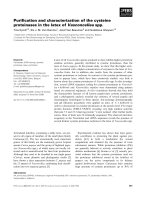
Fig. 1. Alignment of sequences of AATs. Alignment was per-
formed using
CLUSTAL X [29]. B.B3, B. subtilis B3; B.circ., B. circu-
lans; B.YM, Bacillus sp. YM2; T.th., T. thermophilus HB8; cPig, pig
cytosolic. Gaps in the alignment are shown by gray dashes. Identi-
cal residues are shown in black; similar residues are shown in gray.
H J. Wu et al. Identification of a new aspartate aminotransferase
FEBS Journal 278 (2011) 1345–1357 ª 2011 The Authors Journal compilation ª 2011 FEBS 1347
analysis. These two mutants also lost their transamina-
tion ability when using l-Trp and l-Phe as amino
donors (data not shown). To determine the exact role
of Arg403 (corresponding to Arg386 in E. coli AAT)
in B. subtilis B3, the R403Y mutant enzyme was con-
structed. The Arg403 fi Tyr replacement disrupted the
PLP-Asn194-Arg403 hydrogen-bond linkage system
and changed the conformation of the active center of
the enzyme. The enzyme activity analysis showed that
the R403Y mutant also lost transamination activity
(Fig. 2C). These results showed that the Asp232,
Lys270 and Arg403 residues of B. subtilis B3 AAT
play key roles in transamination.
Comparison and alignment of AAT sequences
To confirm the exact contributions of the Asp232,
Lys270 and Arg403 residues to the function of B. sub-
tilis B3 AAT, the deduced amino acid sequence was
compared with the five AATs identified from B. circu-
lans, pig cytosolic, E. coli, T. thermophilus HB8 and
Bacillus sp. YM-2. The alignment results revealed 19
invariant amino acids in these six AATs (Fig. 1).
Among these conserved residues, the Tyr70, Asn194,
Asp222, Tyr225, Lys258 and Arg266 residues in E. coli
AAT (numbered on the basis of the pig cytosolic
AAT) are involved in the binding of PLP, which acts
as the co-enzyme [19,27]. The Asp232 and Lys270 resi-
dues in B. subtilis B3 AAT correspond to Asp222 and
Lys258, respectively, in E. coli AAT. Together with
Fig. 2. Purification and functional analysis of the recombinant wild-
type (WT) and mutant AATB3 enzymes. (A) Aliquots of purified
enzyme for the wild-type and each AATB3 mutant were separated
by SDS ⁄ PAGE and stained with Coomassie Brilliant Blue. (B) Aliqu-
ots of purified enzyme for the wild-type and each AATB3 mutant
were separated by native PAGE and stained with Coomassie Bril-
liant Blue. (C) Native PAGE gel was stained with Fast Blue in accor-
dance with the method described by de la Torre et al. [9].
Fig. 3. Detection of L-tryptophan aminotransferase activity using
paper chromatography of amino acids. (A) the a-ketoglutarate was
used as the amino acceptor;
L-Glu, standard L-Glu; L-Try, standard
L-Try; 1-3, reaction sample. (B) The oxaloacetate was used as the
amino acceptor;
L-Asp, standard L-Asp; L-Try, standard L-Try; 1–3,
reaction sample.
Table 1. Activity of purified AATB3 towards different amino acids
and oxo acids. The reaction was performed at 25 °C for 20–40 min.
The activity was measured as described in the Materials and
methods.
Concentration
(m
M)
Relative
activity (%)
Amino donor
a
L-aspartate 30 100.0
L-glutamate 30 46.7
L-tryptophan 6 1.7
L-tyrosine 6 0.4
L-phenylalanine 6 0.3
Amino acceptor
b
a-ketoglutarate 10 100.0
Oxaloacetate 10 81.5
a
The AAT from B. subitilis B3 showed relative high activity toward
L-aspartate and L-glutamate, although the activities were very weak
toward three aromatic amino acid aminotransferases (
L-tryptophan,
L-tyrosine and L-phenylalanine). Therefore, 30 mM was used for
L-aspartate and L-glutamate, and 6 mM for the three aromatic amino
acid substrates. a-ketoglutarate (10 m
M) was used as amino group
acceptor except for the oxaloacetate (10 m
M) used for L-glutamate.
The activity of
L-aspartate was adjusted to 100.
b
30 mML-aspartate
was used as amino donor for a-ketoglutarate, and 30 m
ML-gluta-
mate was used as amino donor for oxaloacetate. The activity of
a-ketoglutarate was adjusted to 100.
Identification of a new aspartate aminotransferase H J. Wu et al.
1348 FEBS Journal 278 (2011) 1345–1357 ª 2011 The Authors Journal compilation ª 2011 FEBS
analysis of the activities of the mutants D232N and
K270H (Fig. 2C), we concluded that Asp232 in B. sub-
tilis B3 AAT, which corresponds to Asp222 in E. coli
AAT [15], is the residue that enhances the function of
the enzyme-bound co-enzyme PLP. The Lys270 residue
of B. subtilis B3 AAT serves the same function as
Lys258 in E. coli AAT, which binds to PLP and forms
an internal Schiff base [16].
The conserved residues Asn194 and Arg386 in
E. coli AAT participate in substrate binding [14,20],
which correspond to Asn199 and Arg403, respectively,
in B. subtilis B3 AAT. The loss of transamination
activity of the R403Y mutant confirmed that the
B. subtilis B3 AAT utilizes the Arg403 residue to bind
the a-COO
)
of the substrate, which is similar to the
role of Arg386 in E. coli AAT. The Arg292 residue,
which is the invariant residue in the subgroup Ia AATs
[17] identified in the primary structure of B. subtilis B3
and B. circulans AATs, interacts directly with the dis-
tal carboxyl groups of dicarboxylate substrates
(Fig. 1). However, this residue is not found in the cor-
responding regions of subgroup Ib. By contrast, the
conserved active residue Lys109 in subgroup Ib carries
out the function of recognizing the substrates as does
the Arg292 residue in subgroup Ia [18]. From the
alignment, the Thr109 was shown also to be conserved
in B. subtilis B3, B. circulans, E. coli and pig cytosolic
AATs, and the Trp140 invariant among the six AATs
(Fig. 1). These two residues provide hydrogen bonds
to the phosphate group and distal carboxyl group of
the substrate [27,28].
Molecular phylogeny
To examine the phylogenetic relationship of this new
bacteria gene with AAT genes from plants, animals,
protozoa, eubacteria and archeabacteria, a phylogram
was constructed using the Neighbor-joining method
with 44 full-length AAT amino sequences from Gen-
Bank. As shown in Fig. 4, the AATs were divided into
six main branches: animal mitochondrial, animal cyto-
plasmic, plant mitochondrial, plant cytoplasmic and
the two branches in bacteria. The AAT from B. subtil-
is B3, clustering together with the AAT from B. circu-
lans, is in the large branch of bacterial AATs. From
the phylogenetic tree analysis, the AATs from different
organisms can also be divided into two major sub-
groups according to the classification system estab-
lished by Jensen and Gu [5]. The Ia subgroup contains
eubacterial and eukaryotic AATs, including enzymes
from E. coli, Haemophilus influenzae, animals and
plants. The Ib subgroup consists almost exclusively of
AATs from prokaryotes, including AATs from proto-
zoa, archaebacteria and bacteria. Interestingly, plants
also have Ib subgroup-prokaryote-type AATs [2,9].
Although the AATs from B. subtilis B3 and B. circu-
lans belong to the Ib subgroup in our analysis, these
new AATs show significant differences from other Ib
subgroup-type AATs. They occupy a small separate
branch at a far phylogenetic distance from AATs
belonging to another large branch of the Ib subgroup.
From the homology analysis, the identity between the
two AATs from B. subtilis B3 and B. circulans was
$ 56%, and the AAT from B. subtilis B3 showed rela-
tively high identity ($ 19%) with the AAT from Syn-
echocystis sp. compared to other AATs from the Ib
subgroup.
Enzyme specificity and kinetics parameters
The purified AATB3 was optimally active at 45 °C (at
pH 7.2), and more than 80% of the maximum activity
was retained in the temperature range 25–55 °C
(Fig. 5A). After incubation at 50 °C for 30 min, the
enzyme had more than 85% of the maximum activity
(Fig. 5B). When incubated at 60 °C for 15 min, the
enzyme also had 65% activity, although increasing the
treatment time to 30 min caused the enzyme to lose
almost all activity. Above 65 °C, the stability of the
enzyme decreased rapidly (Fig. 5B). The optimal pH
for the enzyme activity was pH 8.0 at the optimal tem-
perature (45 °C) (Fig. 5C). The enzyme activity over
the pH range 7.0–8.6 was more than 80% of the maxi-
mum activity. From these results, we demonstrated
that AATB3 tended to have relatively high activity
and stability in alkaline environments.
Table 2 summarizes the effect of some metal ions on
the activity of the purified aminotransferase. At a low
concentration (1 mm), Cu
2+
and Mn
2+
could inhibit
the activity of the purified aminotransferase, and other
metal ions had no remarkable effects, although Ca
2+
and Co
2+
could promote the reaction to some extent.
Partial inhibition was observed in the presence of some
metal ions at 10 mm, and the order of the ions by
enzyme inhibitory activity was Zn
2+
>Cu
2+
>Mg
2+
>Mn
2+
. It could be concluded that the enzyme is not
metal ion-dependent because EDTA had no inhibitory
or stimulatory effects on the activity (Table 2).
AATB3 showed transamination activity between
various amino acids and a-ketoglutarate (Table 1),
with l-aspartate being the best substrate. Aromatic
amino acids such as l-tryptophan, l-tyrosine and
l-phenylalanine were weakly active as amino donors,
and the activity of transamination activity toward
l-tryptophan was relatively higher than the other two
residues.
H J. Wu et al. Identification of a new aspartate aminotransferase
FEBS Journal 278 (2011) 1345–1357 ª 2011 The Authors Journal compilation ª 2011 FEBS 1349
To further characterize the enzyme, the kinetic
parameters K
m
, V
max
and k
cat
were determined for the
purified AATB3. Values for K
m
and V
max
for both
amino donors (l-aspartate and l-glutamate) and ac-
ceptors (a-ketoglutarate and oxaloacetate) were calcu-
lated from the double-reciprocal plots. The K
m
values
of AATB3 were 6.7, 0.3, 8.0 and 0.6 mm for l-aspar-
tate, a -ketoglutarate, l-glutamate and oxaloacetate,
respectively. For the amino donors, AATB3 showed
more affinity for l-aspartate than l-glutamate,
whereas, for the amino acceptors, this enzyme had
more affinity for a-ketoglutarate (Table 3). The calcu-
lated V
max
for l-aspartate, a-ketoglutarate, l-gluta-
mate and oxaloacetate were 0.23, 0.21, 0.07 and
0.11 mmÆmin
)1
, respectively (Table 3). The k
cat
⁄ K
m
ratios listed in the Table 3, which represent the cata-
lytic efficiency, show that the enzyme had relative
higher catalytic efficiency for oxo acids than for amino
acids. The enzyme variants D232N, K270H and
R403Y were almost inactive (Fig. 2C), and therefore
no kinetic parameters could be determined.
Discussion
AATs that catalyze the tricarboxylic acid cycle inter-
mediates to amino acids have been studied in a variety
of organisms. These enzymes play a key role in aspar-
tate catabolism and biosynthesis as well as in linking
carbon metabolism with nitrogen metabolism. In the
present study, we cloned and characterized such an
AAT from the B. subtilis B3 strain. This enzyme con-
sists of 436 amino acid residues and is encoded by the
aatB3 gene. We found the typical promoter and termi-
nator regions upstream and downstream, respectively,
of this new gene.
To examine explicitly the phylogenetic relationship
between the AATB3 and other AATs from different
organisms, a phylogenetic tree was constructed using
Fig. 4. Phylogenetic tree of AATs from dif-
ferent organisms. The phylogenetic tree
was constructed with full-length AAT amino
acid sequences using the Neighbor-joining
method of
MEGA 4.0. Bootstrap values are
expressed as percentages of 1000 replica-
tions. Bar 0.1 sequence divergence. c, cyto-
solic; ch, chloroplastic; cy, cytoplastimic; p,
plastidic; m, mitochondrial. GenBank acces-
sion numbers of the AATs are shown. The
black circle represents the branch of AAT
from B. subtilis B3 and B. circulans; the
black triangle shows the prokaryote-type
AATs from plants.
Identification of a new aspartate aminotransferase H J. Wu et al.
1350 FEBS Journal 278 (2011) 1345–1357 ª 2011 The Authors Journal compilation ª 2011 FEBS
previously characterized AAT sequences from animals,
plants and prokaryotes. The AATs from B. subtilis B3
and B. circulans clustered together with other bacterial
AATs and appeared to be more closely related to the
Ib-type of bacterial AATs than to the Ia-type of other
bacterial AATs (Fig. 4). However, AATB3 showed low
identify with AATs from the Ib subgroup, and the
highest identity was only $ 19% compared to AAT
from Synechocystis sp. (Ib subgroup).
Multiple alignments, which were built using AATs
of distant species, clearly show that most of the resi-
dues interacting with the PLP and the substrates
[27,29] are conserved in AATB3 (Fig. 1). From this
comparison, the AATB3 tends to have more conserved
active residues that belong to the Ia subgroup but do
not exist in the Ib subgroup. For example, the Gly38,
Thr109 and Arg292 residues (numbered on the basis of
the pig cytosolic AAT), which are conserved in
AATB3 and Ia subgroup AATs, are not found in the
Ib subgroup AATs. These three residues are all
involved in the interaction with the substrate [27,28],
especially the Arg292 residue, which plays a key role
in recognizing the distal carboxylate of the substrate
[17]. In subgroup Ib, the same role appears to be car-
ried out by Lys109 [18]. Therefore, AATB3 is more
similar to the AATs from the Ia subgroup than the Ib
subgroup in structure.
We used site-directed mutagenesis to determine the
exact role of three residues in AATB3. The loss of the
activity from the mutations together with the multiple
alignment analysis indicated that the Asp232 residue
of AATB3 enhances the function of the enzyme-bound
coenzyme PLP and that the Lys270 residue mediates
binding of PLP, whereas the Arg403 residue is respon-
sible for recognizing the a-COO
)
of the substrate.
These functions are performed by the corresponding
residues of Asp222, Lys258 and Arg386 of the AAT
from E. coli [15,16,19,20].
We also described in detail the physicochemical and
catalytic properties of AAT from B. subtilis B3. The
purified enzyme was demonstrated to have an optimal
temperature at 45 °C and thermostability of only up to
Fig. 5. Characterization of the purified AATB3. (A) Effect of tempera-
ture on activity of AATB3 (pH 7.2). (B) Thermostability of AATB3. The
enzyme was pre-incubated at 40, 50, 60 or 65 °C for 5, 15 or 30 min
before the assay. (C) Effect of pH on activity of AATB3. The assay
was performed at 45 °C in buffers with pH in the range 4.4–10.2.
Table 2. Effect of metal ions on the activity of purified AATB3.
Values represent the means of triplicates relative to the untreated
control samples.
Chemicals
Relative activity (%)
1m
M 10 mM
None 100 ± 0.6 100 ± 0.6
MgCl
2
96.9 ± 4.4 34.7 ± 3.1
CaCl
2
115.5 ± 2.7 75.7 ± 3.0
MnSO
4
90.1 ± 2.3 42.8 ± 7.9
CuSO4 84.3 ± 2.0 4.9 ± 1.3
ZnSO4 99.7 ± 7.4 3.1 ± 1.3
CoCl2 122.1 ± 2.5 54.1 ± 4.5
EDTA 106.4 ± 10.1 99.3 ± 1.8
Table 3. Kinetic parameters for recombinant AATB3 from Bacil-
lus subtilis B3. Kinetic parameters were obtained from double reci-
procal plots as described in the Materials and methods. Values
represent the mean ± SD of three determinations.
Substrates
B. subtilis B3 AATB3
V
max
(mMÆL
)1
Æmin)
k
cat
(s
)1
) K
m
(mM)
k
cat
⁄ K
m
(mM
)1
Æs
)1
)
L-aspartate 0.23 ± 0.03 30 ± 3 6.68 ± 1.45 4.50
L-glutamate 0.07 ± 0.01 14 ± 2 8.00 ± 1.32 1.75
a-ketoglutarate 0.21 ± 0.01 27 ± 1 0.32 ± 0.08 84.38
Oxaloacetate 0.11 ± 0.01 22 ± 1 0.60 ± 0.06 36.67
H J. Wu et al. Identification of a new aspartate aminotransferase
FEBS Journal 278 (2011) 1345–1357 ª 2011 The Authors Journal compilation ª 2011 FEBS 1351
50 °C. These characteristics are similar to those of the
AAT from E. coli [30] and not of AATs from the Ib
subgroup, which usually have high thermostability. The
thermostability appears to be related to the amino acid
composition of the AAT. Okamoto et al. [6] reported
that the high Pro content of the Ib-type AAT from
T. thermophilus (6.5%) will render the enzyme rigid
and thermostable. The same features are also found in
other subgroup Ib AATs, such as Thermus aquaticus
YT1 AAT (7.0%) [31] and Phormidium lapdideum
(6.1%), as well as the newly found Ib-prokaryote-type
AAT in Pinus pinaster (6.4%) [9,11]. The Pro content
of B. subtilis B3 AAT is 4.1%, which is similar to that
of subgroup Ia E. coli AAT (3.8%) and is much lower
than that of subgroup Ib T. thermophilus AAT. For
this reason, the thermostability of B. subtilis B3 AAT is
similar to that of E. coli AAT and is lower than that of
T. thermophilus AAT by $ 20 °C [6].
We showed that the AAT from B. subtilis B3 had an
optimal pH at 8.0 and had relatively high activity over
a wide alkaline pH range (pH 7.0–9.0). This character-
istic is similar to that of the AAT from B. circulans.
The B. circulans AAT has been reported to have high
optimal pH and a wide pH stability range as a result
of the N-terminal two a-helical segments, which con-
tain an additional sequence of 32 acid residues not
found in many AATs [23]. Interestingly, B. subtilis B3
AAT also has a similar additional N-terminal sequence
of 32 acid residues (Fig. 1), which shows 53% identity
with that of B. circulans AAT, and the additional
N-terminus of B. subtilis B3 AAT appears to perform
the same function as that of B. circulans AAT.
The results obtained in the present study indicate
that the AAT from B. subtilis B3 can catalyze l-aspar-
tate, l-glutamate, l-tryptophan, l-tyrosine and l-phen-
ylalanine transamination, with l-aspartate being the
best substrate. However, the activity of AATB3
toward three aromatic amino acids were weak, similar
to that of AAT from Bacillus sp. YM-2 strain [32],
and was unlike AAT from E. coli, which was shown to
have 22% of the activity of the total tyrosine amino-
transferase [33]. The K
m
values for AATB3 were 6.7,
0.3, 8.0 and 0.6 mm for l-aspartate, a-ketoglutarate,
l-glutamate and oxaloacetate, respectively. Similar to
the other AATs, the K
m
values for oxo acids are lower
than that for the amino acids [9,32,34]. However, it is
worth noting that both k
cat
and k
cat
⁄ K
m
values are
lower than those of AAT from E. coli [35].
This new AAT phylogenetically belongs to subgroup
Ib of AAT, although it also has conserved active resi-
dues and thermostability characteristic of Ia-type
AATs. Although our combined results appear to be
contradictory, we propose that the B. subtilis gene
described in the present study may have arisen from
the interaction between the Ia-type and Ib-type aat
genes during evolution. A similar phenomenon is seen
when the genome segment of B. subtilis B3 is com-
pared with those of B. subtilis A1 ⁄ 3 and B. amylolique-
faciens FZB42. The aatB3 gene frequently appears in
the region between the srf operon and sfp gene. This
region is the putative regulatory region relevant to bio-
synthesis of the lipopeptides, especially for the sfp
gene, which is essential for biosynthesis of the lipopep-
tides [26]. We presume that the aat gene in this region
can regulate the biosynthesis of the lipopeptides. The
experiments performed in the present study showed
that this AAT can form Glu and Asp, and the forma-
tion of Glu and Asp is used to synthesize Gln and
Asn, respectively. These four residues are common
components in lipopeptides, such as surfactin, iturin
and fengycin. Another interesting observation was that
the B. subtilis B3 has another aat gene similar to aspB
outside this region. This could be explained by the
need to synthesize more AATs to provide adequate
nutrients (carbon and nitrogen sources) and lipopep-
tides so as to survive in complex environments and
deal with competitors.
In summary, a new AAT with an additional N-ter-
minal sequence was identified from B. subtilis B3.
Having both Ia-type and Ib-type characteristics and a
high activity over an alkaline pH range, this enzyme
may regulate the biosynthesis of lipopeptides and has
various potential industrial applications, such as
in the synthesis of l-tyrosine, l-phenylalanine and
l-homophenylalanine. A detailed characterization of
the role of B. subtilis B3 AAT and its structure are in
progress.
Materials and methods
Bacterial strains, plasmids and growth conditions
The bacterial strains and plasmids used in the present study
are described in Table 4. E. coli DH5a was used as the host
for amplification of all plasmids, and recombinant proteins
were expressed in E. coli BL21. B. subtilis B3 was used for
cloning the aatB3 gene. LB broth was used for the growth
of E. coli and B. subtilis strains. When required, antibiotics
were added at the final concentrations: ampicillin (Amp),
100 lgÆmL
)1
; kanamycin (Km), 50 lgÆmL
)1
.
DNA manipulation and transformation
The isolation and manipulation of recombinant DNA were
performed using standard techniques. All enzymes used in
the present study were purchased from Takara Bio Inc.
Identification of a new aspartate aminotransferase H J. Wu et al.
1352 FEBS Journal 278 (2011) 1345–1357 ª 2011 The Authors Journal compilation ª 2011 FEBS
(Otsu, Japan). The specific primers used for the PCR are
described in Table 5.
The original sequence of the aatB3 gene was obtained
through the B. subtilis B3 gene library constructed in a pre-
vious study (accession number AY040867) [25]. To express
the recombinant AATB3 protein in E. coli, the entire aatB3
ORF was amplified using primers P1 and P2 using B. sub-
tilis B3 chromosomal DNA as the template; the amplified
product was digested with KpnI and EcoRI, and cloned into
the same sites of the cloning vector pUC19 and expression
vector pET30a(+), resulting in the plasmids pUCAAT and
pETAAT, respectively. The entire cloned regions were con-
firmed by sequencing (Invitrogen Biotechnology Co., Ltd,
Shanghai, China).
Site-directed mutagenesis via PCR
Single mutations were introduced into the cloned AATB3
using the Takara MutanBEST Kit (Takara). Reactions were
carried out using the primer pairs: for D232N, D232N-F and
D232N-R; for K270H, K270H-F and K270H-R; and, for
R403Y, R403Y-F and R403Y-R. The pUCAAT vector was
used as a template. The introduced mutations in the aatB3
gene were confirmed by DNA sequencing. The resulting vec-
tors were designated pUCD232N, pUCK270H and
pUCR403Y, and the three different DNA fragments carrying
mutant aatB3 genes from these vectors were subcloned into
the KpnI and EcoRI restriction sites of the pET30a(+)
expression vector to obtain pETD232N, pETK270H and
pETR403Y, respectively.
Expression and purification of recombinant
wild-type and mutant AATB3 enzymes
The E. coli strain BL21 (DE3) was transformed with
pETAAT or the three expression plasmids carrying different
Table 4. Bacterial strains and plasmids used in the present study. Resistance markers were: Amp
r
, ampicillin resistance; Km
r
, kanamycin
resistance.
Strain or plasmid Relevant genotype or characteristics Source or reference
Strains
E. coli
DH5a F
)
F80dlacZ DM12 minirecA Stored in this laboratory
a
BL21(DE3) F
)
ompT hsdS
B
(r
B
)
m
B
)
) gal dcm(DE3) Stored in this laboratory
B. subtilis
B3 Wild-type; bacillomycin D and fengycin producer Present study
Plasmids
pET30a(+) T7 promoter-based expression vector; Km
r
Novagen (Merck KGaA,
Darmstadt, Germany)
pUC19 E. coli clone vector; lacZ; Amp
r
Stored in this laboratory
pETAAT The aatB3 fragment was inserted into KpnI and EcoRI sites of
pET30a(+) for the expression of protein AATB3; T7 promoter-based
expression vector; Km
r
Present study
pUCAAT The aatB3 fragment was inserted into KpnI and EcoRI sites of
pUC19 for construction the mutant of AATB3 protein; Amp
r
Present study
pUCD232N pUC19 carrying a fragment encoding the D232N mutant; Amp
r
Present study
pUCK270H pUC19 carrying a fragment encoding the K270H mutant; Amp
r
Present study
pUCR403Y pUC19 carrying a fragment encoding the R403Y mutant; Amp
r
Present study
pETD232N The fragment from pUCD232N was inserted into KpnI and EcoRI
sites of pET30a(+) for the expression of protein D232N; T7
promoter-based expression vector; Km
r
Present study
pETK270H The fragment from pUCK270H was inserted into KpnI and EcoRI
sites of pET30a(+) for the expression of protein K270H; T7
promoter-based expression vector; Km
r
Present study
pETR403Y The fragment from pUCR403Y was inserted into KpnI and EcoRI
sites of pET30a(+) for the expression of protein R403Y; T7
promoter-based expression vector; Km
r
Present study
a
Key Laboratory of Monitoring and Management of Crop Diseases and Pest Insects, Ministry of Agriculture, Nanjing, China.
Table 5. Oligo DNA primers used in the present study. Restriction
sites or mutation sites in primers are underlined.
Name Sequence of primers (5¢-to3¢)
P1
GGTACCATGAATGATGCAGCAAAAG (KpnI)
P2
GAATTCTCAGCCTGATATTTCCGCCT (EcoRI)
D232N-F CGTGCTCGTA
AACGATGCGTATTAC
D232N-R ACAATCTCTTTGCCGGCCTCCGC
K270H-F GGCGCGACG
CACGAAAATTACGC
K270H-R GTCTATTTTCACGCAAAGCACCCGGT
R403Y-F AAACCGATTTG
TACATCGCATTTTC
R403Y-R CATTAATGGATATCGTTCCGATTCC
H J. Wu et al. Identification of a new aspartate aminotransferase
FEBS Journal 278 (2011) 1345–1357 ª 2011 The Authors Journal compilation ª 2011 FEBS 1353
mutant aatB3 genes. The transformants were cultivated at
37 °C with shaking in LB medium containing 50 lgÆmL
)1
kanamycin until D
600
of 0.5–0.7 was reached. Flasks con-
taining the cultures were supplemented with isopropyl thio-
b-d-galactoside at a final concentration of 1 mm. After incu-
bation at 37 °C for a further 6 h with vigorous shaking, the
cells were harvested by centrifugation at 6000 g for 20 min.
The cell pellets were resuspended in a buffer containing
20 mm potassium phosphate, 500 mm NaCl, 5% glycerol
and 20 mm imidazole buffer at pH 7.3. Cells were lysed by
sonication, and cell debris was removed by centrifugation at
10 000 g for 20 min. The recombinant enzymes were purified
by a single chromatographic step using HisTrapHP (GE
Healthcare, Milwaukee, WI, USA). The column was loaded
with the bacterial cell lysate, and the non-adherent proteins
were removed by rinsing with 20 volumes of wash buffer
(20 mm potassium phosphate, pH 7.3, 5% glycerol, 500 mm
NaCl, 20 mm imidazole). The proteins were eluted with a
gradient of 10–500 mm imidazole in wash buffer. The
purified enzymes were stored at )20 ° C after salt removal
using the HiTrap Desalting columns (GE Healthcare). Pro-
tein concentrations were measured with a BCA-100 protein
quantitative analysis kit (Biocolor Biotech, Shanghai, China)
using BSA as the standard.
Determination of enzyme activities
AAT activity was assayed as described by Collier and
Kohlhaw [36]. The assay mixture contained (in 0.8 mL total
volume): 0.1 m potassium phosphate buffer (pH 7.2),
30 mml-aspartate, 10 mm a-ketoglutarate, 38 lm pyridoxal
5¢-phosphate and enzyme. The stock solution of a-ketoglu-
tarate was prepared daily, and its pH was adjusted to 7.2
with NaOH. The assay was performed at 25 °C for 20–
40 min, and the reaction was stopped with 0.1 mL of 10 m
NaOH. After 30 min at room temperature, the increase in
absorbance at 265 nm was measured for the test sample, as
well as a control to which NaOH had been added before
the addition of a-ketoglutarate. A molar extinction coeffi-
cient for oxaloacetate of 780 m
)1
Æcm
)1
was used, and one
unit of activity was defined as the amount of enzyme neces-
sary to form 1 lmolÆmin
)1
of oxaloacetate.
The aromatic amino acid aminotransferases were assayed
according to Mavrides and Orr [37]. The assay was estab-
lished for AAT except that aspartate was replaced with
6mm tryptophan, tyrosine or phenylalanine, and the con-
centration of the a-ketoglutarate was decreased to 10 mm.
The increase in absorbance of the reaction solution was
measured at 335, 330 and 315 nm. The molar extinction
coefficients for the reaction products indole pyruvate, q-hy-
droxyphenylpyruvate and phenylpyruvate were 10 000,
19 500 and 17 500 m
)1
Æcm
)1
, respectively. One unit of aro-
matic amino acid aminotransferase activity was defined as
the amount of enzyme necessary to form 1 lmol of indole
pyruvate, q-hydroxyphenylpyruvate or phenylpyruvate.
A paper chromatography assay for amino acids was also
used to detect the activity toward tryptophan. The reaction
was performed as described above, and a-ketoglutarate and
oxaloacetate were used as amino acceptors. At the end of
the reaction, 10 lL of the reaction solution was spotted
onto a filter paper and separated by chromatography
(n-butyl alcohol ⁄ ethanol ⁄ water at 4 : 1 : 1, v ⁄ v). Subse-
quently, the filter paper was sprayed with 0.1% ninhydrin.
After drying, the products of the amino acid on the filter
paper were displayed purple in color.
To determine the effects of pH, temperature and inhibi-
tors, l-aspartate and a-ketoglutarate were used as amino
donor and acceptor, respectively, and the reactions were
performed as described above. To investigate the effect of
pH at the optimum temperature (45 °C), three buffered
systems at a final concentration of 50 mm were used:
acetate ⁄ sodium acetate (pH 4.4–6.0), potassium phosphate
(pH 6.0–8.0) and glycine ⁄ sodium hydroxide (pH 8.0–10.2).
The temperature dependence was determined at pH 7.2, and
the stability of the enzyme was examined by keeping the pure
preparation for 5, 15 and 30 min at 40, 50, 60 and 65 °C
before the assay. The effect of inhibitors was established with
the reaction system containing different metal ions at final
concentrations of 1 and 10 mm. The specific activities for
amino acids were analyzed under similar conditions.
Kinetic experiments
For determination of kinetic parameters, an assay was
established by coupling with malate dehydrogenase as
described previously [38]. In the routine assay, the reaction
mixture contained 0.1 m potassium phosphate buffer
(pH 7.6), 25 lm pyridoxal 5 ¢-phosphate, 0.5 mm NADH,
0.08 U malate dehydrogenase and 0.5 lL of purified
enzyme in a reaction volume of 200 lL. The temperature
was 30 °C. The reaction was monitored by the decrease in
absorbance of NADH at 340 nm over 180 s with a Thermo
Multiskan Ascent (Thermo Fisher Scientific Inc., Waltham,
MA, USA) and the data were recorded every 20 s. AAT
substrate concentrations were varied in the range 1–20 mm
l-aspartate with a fixed concentration of 10 mm a-ketoglu-
tarate (for K
LÀasp
m
) and in the range 0.5–10 mm a-ketogluta-
rate with a fixed concentration of 20 mml-aspartate (for
K
aÀKG
m
). The kinetic parameters for l-glutamate and oxalo-
acetate were coupled to glutamate dehydrogenase [39]. Our
assay was established using the same methods, and the
200 lL reactions contained l-glutamate, oxaloacetate,
1mm NADH, 2 U of glutamate dehydrogenase and 12 mm
NH
4
Cl (as second substrate for glutamate dehydrogenase)
in 0.1 m potassium phosphate buffer (pH 7.6). AAT sub-
strate concentrations were varied in the range 1.0–27 mm
l-glutamate with a fixed concentration of 5 mm oxaloace-
tate (for K
LÀglu
m
) and in the range 0.5–20 mm oxaloacetate
with a fixed concentration of 12 mml-glutamate
(for K
OAA
m
). K
m
and V
max
values were estimated from the
Identification of a new aspartate aminotransferase H J. Wu et al.
1354 FEBS Journal 278 (2011) 1345–1357 ª 2011 The Authors Journal compilation ª 2011 FEBS
variation in initial reaction velocity with substrate concen-
tration using the Hanes transformation [40]. The k
cat
parameter was defined as V
max
divided by the enzyme con-
centration in the 200 lL reaction.
PAGE
PAGE was performed with a Mini Protean II cell
(Bio-Rad, Hercules, CA, USA) in accordance with the
manufacturer’s instructions. For SDS ⁄ PAGE, the separat-
ing gel was made with 12% acrylamide and the stacking gel
was made with 5% acrylamide. The Prestained Protein
Marker (Fermentas China Co., Ltd, Shenzhen, China) was
used as the molecular weight marker. Proteins were visual-
ized by Coomassie Brilliant Blue staining. Native PAGE
was carried out with discontinuous gels in which the sepa-
rating gel consisted of 8% acrylamide and the stacking gel
consisted of 5% acrylamide. The running buffer contained
25 mm Tris-HCl and 250 mm Gly (pH 8.3). The gels were
run at 15 mA for 90 min at 4 °C. They were then placed in
a bath containing 50 mL of AAT substrate solution with
gentle shaking for 5 min. AAT activity was detected when
the AAT substrate solution was supplemented with
1mgÆmL
)1
Fast Blue (Sigma-Aldrich Shanghai Trading
Co., Ltd, Shanghai, China). The composition of the AAT
substrate solution (pH 7.4) was 2.2 mm a-ketoglutarate,
8.6 mm Asp, 0.5% (w ⁄ v) polyvinylpyrrolidone-40, 1.7 mm
EDTA and 100 mm Na
2
HPO
4
[9].
Sequence analysis
Alignments of DNA and protein sequences were conducted
with blastn and blastp software, respectively (http://
www.ncbi.nlm.nih.gov/BLAST/). Genes were predicted using
genemark ( The pro-
moter and terminator were predicted using the online tools
neural network promoter prediction (http://www.
fruitfly.org/seq_tools/promoter.html) and findterm (http://
linux1.softberry.com/berry.phtml), respectively.
Additional aminotransferase sequences were obtained
from GenBank and aligned by using clustal x, followed
by manual adjustments [41]. Aligned sequences were visual-
ized with genedoc [42]. Phylogenetic trees were constructed
using the Neighbor-joining algorithm [43] in mega 4.0 [44],
with its reliability assessed by 1000 bootstrap repetitions.
Acknowledgements
This work was supported by grants from the National
Natural Science Fund of China (30570041); the
National 863 Program of China (2006AA10Z172;
2006AA10A203); the Special Nonprofit Scientific
Research Program, P. R. China (3-23); the Program of
International Science and Technology Cooperation
(2009DFA32740); the Specialized Research Fund for
the Doctoral Program of Higher Education, P. R.
China (20060307012); the National Transgenic Major
Program (2009ZX08009-055B); and Youth Science and
Technology Innovation Fund of Nanjing Agricultural
University (KJ09007).
References
1 Deu E, Koch KA & Kirsch JF (2002) The role of the
conserved Lys68*:Glu265 intersubunit salt bridge in
aspartate aminotransferase kinetics: multiple forced
covariant amino acid substitutions in natural variants.
Protein Sci 11, 1062–1073.
2 de la Torre F, De Santis L, Fernanda SM, Crespillo R
& Canovas FM (2006) Identification and functional
analysis of a prokaryotic-type aspartate aminotransfer-
ase: implications for plant amino acid metabolism.
Plant J 46, 414–425.
3 Wilkie SE, Lambert R & Warren MJ (1996) Chloro-
plastic aspartate aminotransferase from Arabidopsis
thaliana: an examination of the relationship between the
structure of the gene and the spatial structure of the
protein. Biochem J 319, 969–976.
4 Miesak BH & Coruzzi GM (2002) Molecular and physi-
oligical analysis of Arabidopsis mutants defective in
cytosolic or chloroplastic aspartate aminotransferase.
Plant Physiol 129, 650–660.
5 Jensen R & Gu W (1996) Evolutionary recruitment of
biochemically specialized subdivisions of Family I
within the protein superfamily of aminotransferases.
J Bacteriol 178, 2161–2171.
6 Okamoto A, Kato R, Masui R, Yamagishi A,
Oshima T & Kuramitsu S (1996) An aspartate
aminotransferase from an extremely thermophilic
bacterium, Thermus thermophilus HB8. J Biochem 119,
135–144.
7 Sung MH, Tanizawa K, Tanaka H, Kuramitsu S,
Kagamiyama H, Hirotsu K, Okamoto A, Higuchi T &
Soda K (1991) Thermostable aspartate aminotransferase
from a thermophilic Bacillus species. Gene cloning,
sequence determination, and preliminary x-ray charac-
terization. J Biol Chem 266, 2567–2572.
8 Watson RJ & Rastogi VK (1993) Cloning and nucleo-
tide sequencing of Rhizobium meliloti aminotransferase
genes: an aspartate aminotransferase required for
symbiotic nitrogen fixation is atypical. J Bacteriol 175,
1919–1928.
9 de la Torre F, Moya-Garcia AA, Suarez MF,
Rodriguez-Caso C, Canas RA, Sanchez-Jimenez F &
Canovas FM (2009) Molecular modeling and site-direc-
ted mutagenesis reveal essential residues for catalysis in
a prokaryote-type aspartate aminotransferase. Plant
Physiol 149, 1648–1660.
H J. Wu et al. Identification of a new aspartate aminotransferase
FEBS Journal 278 (2011) 1345–1357 ª 2011 The Authors Journal compilation ª 2011 FEBS 1355
10 Nakai T, Okada K, Akutsu S, Miyahara I,
Kawaguchi S, Kato R, Kuramitsu S & Hirotsu K
(1999) Structure of Thermus thermophilus HB8 aspartate
aminotransferase and its complex with maleate.
Biochemistry 38, 2413–2424.
11 Kim H, Nakaoka M, Yagi M, Ashida H, Hamada K,
Shibata H & Sawa Y (2003) Cloning, structural analysis
and expression of the gene encoding aspartate amino-
transferase from the thermophilic cyanobacterium
Phormidium lapideum. J Biosci Bioeng 95, 421–424.
12 Schwarzenbacher R, Jaroszewski L, von Delft F, Abd-
ubek P, Ambing E, Biorac T, Brinen LS, Canaves JM,
Cambell J, Chiu HJ et al. (2004) Crystal structure of an
aspartate aminotransferase (TM1255) from Thermoto-
ga maritima at 1.90 A
˚
resolution. Proteins 55, 759–763.
13 Inoue K, Kuramitsu S, Okamoto A, Hirotsu K, Higu-
chi T & Kagamiyama H (1991) Site-directed mutagene-
sis of Escherichia coli aspartate aminotransferase: role
of Tyr70 in the catalytic processes. Biochemistry 30,
7796–7801.
14 Yano T, Mizuno T & Kagamiyama H (1993) A hydro-
gen-bonding network modulating enzyme function:
asparagine-194 and tyrosine-225 of Escherichia coli
aspartate aminotransferase. Biochemistry 32, 1810–1815.
15 Yano T, Kuramitsu S, Tanase S, Morino Y & Kagam-
iyama H (1992) Role of Asp222 in the catalytic mecha-
nism of Escherichia coli aspartate aminotransferase: the
amino acid residue which enhances the function of
the enzyme-bound coenzyme pyridoxal 5¢-phosphate.
Biochemistry 31, 5878–5887.
16 Jeffery CJ, Gloss LM, Petsko GA & Ringe D (2000)
The role of residues outside the active site: structural
basis for function of C191 mutants of Escherichia coli
aspartate aminotransferase. Protein Eng 13, 105–112.
17 Almo SC, Smith DL, Danishefsky AT & Ringe D
(1994) The structural basis for the altered substrate
specificity of the R292D active site mutant of aspartate
aminotransferase from E. coli. Protein Eng 7, 405–412.
18 Nobe Y, Kawaguchi SI, Ura H, Nakai T, Hirotsu K,
Kato R & Kuramitsu S (1998) The novel substrate rec-
ognition mechanism utilized by aspartate aminotransfer-
ase of the extreme thermophile Thermus thermophilus
HB8. J Biol Chem 273, 29554–29564.
19 Matharu A, Hayashi H, Kagamiyama H, Maras B &
John RA (2001) Contributions of the substrate-binding
arginine residues to maleate-induced closure of the
active site of Escherichia coli aspartate aminotransfer-
ase. Eur J Biochem 268, 1640–1645.
20 Danishefsky AT, Onnufer JJ, Petsko GA & Ringe D
(1991) Activity and structure of the active-site mutants
R386Y and R386F of Escherichia coli aspartate amino-
transferase. Biochemistry 30, 1980–1985.
21 Belitsky BR (2002) Biosynthesis of amino acids of the
glutamate and aspartate families, alanine, and polyam-
ines. In Bacillus subtilis and its Closest Relatives
(Sonenshein AL ed), pp 203–231. ASM Press, Washing-
ton, DC.
22 Iijima T, Diesterhaft MD & Freese E (1977) Sodium
effect of growth on aspartate and genetic analysis of a
Bacillus subtilis mutant with high aspartase activity.
J Bacteriol 129, 1440–1447.
23 Kravchuk Z, Tsybovsky Y, Koivulehto M, Vlasov A,
Chumanevich A, Battchikova N, Martsev S & Korpela
T (2001) Truncated aspartate aminotransferase from al-
kalophilic Bacillus circulans with deletion of N-terminal
32 amino acids is a non-functional monomer in a par-
tially structured state. Protein Eng 14, 279–285.
24 Berger BJ, English S, Chan G & Knodel MH (2003)
Methionine regeneration and aminotransferases in
Bacillus subtilis, Bacillus cereus, and Bacillus anthracis.
J Bacteriol 185, 2418–2431.
25 Yao S, Gao X, Fuchsbauer N, Hillen W, Vater J &
Wang J (2003) Cloning, sequencing, and characteriza-
tion of the genetic region relevant to biosynthesis of the
lipopeptides iturin A and surfactin in Bacillus subtilis.
Curr Microbiol 47, 272–277.
26 Chen XH, Koumoutsi A, Scholz R, Eisenreich A,
Schneider K, Heinemeyer I, Morgenstern B, Voss B,
Hess WR, Reva O et al. (2007) Comparative analysis of
the complete genome sequence of the plant growth-pro-
moting bacterium Bacillus amyloliquefaciens FZB42.
Nat Biotechnol 25, 1007–1014.
27 Okamoto A, Higuchi T, Hirotsu K, Kuramitsu S & Ka-
gamiyama H (1994) X-ray crystallographic study of
pyridoxal 5¢-phosphate-type aspartate aminotransferases
from Escherichia coli in open and closed form. J Bio-
chem 116, 95–107.
28 Ja
¨
ger J, Moser M, Sauder U & Jansonius JN (1994)
Crystal structures of Escherichia coli aspartate amino-
transferase in two conformations: comparison of an
unliganded open and two liganded closed forms. J Mol
Biol 239, 285–305.
29 Rhee S, Silva MM, Hyde CC, Rogers PH, Metzler CM,
Metzler DE & Arnone A (1997) Refinement and com-
parisons of the crystal structures of pig cytosolic aspar-
tate aminotransferase and its complex with 2-
methylaspartate. J Biol Chem 272, 17293–17302.
30 Yagi T, Kagamiyama H, Motosugi K, Nozaki M &
Soda K (1979) Crystallization and properties of aspar-
tate aminotransferase from Escherichia coli B. FEBS
Lett 100, 81–84.
31 O’Farrell PA, Sannia G, Walker JM & Doonan S
(1997) Cloning and sequencing of aspartate aminotrans-
ferase from Thermus aquaticus YT1. Biochem Biophys
Res Commun 239, 810–815.
32 Sung MH, Tanizawa K, Tanaka H, Kuramitsu S,
Kagamiyama H & Soda K (1990) Purification and
characterization of thermostable aspartate aminotrans-
ferase from a thermophilic Bacillus species. J Bacteriol
172, 1345–1351.
Identification of a new aspartate aminotransferase H J. Wu et al.
1356 FEBS Journal 278 (2011) 1345–1357 ª 2011 The Authors Journal compilation ª 2011 FEBS
33 Mavrides C & Orr W (1974) Multiple forms of pluri-
specific aromatic: 2-oxoglutarate (oxaloacetate) amino-
transferase (transaminase A) in Escherichia coli and
selective repression by L-tyrosine. Biochem Biophys Acta
336, 70–78.
34 Wilkie SE & Warren MJ (1998) Recombinant expres-
sion, purification, and characterization of three isoen-
zymes of aspartate aminotransferase from
Arabidopsis thaliana. Protein Expr Purif 12 , 381–389.
35 Ko
¨
hler E, Seville M, Ja
¨
ger J, Fotheringham I, Hunter
M, Edwards M, Jansonius JN & Kirschner K (1994)
Significant improvement to the catalytic properties of
aspartate aminotransferase: role of hydrophobic and
charged residues in the substrate binding pocket.
Biochemistry 33, 90–97.
36 Collier RH & Kohlhaw G (1972) Nonidentity of the
aspartate and the aromatic aminotransferase compo-
nents of transaminase A in Escherichia coli. J Bacteriol
112, 365–371.
37 Mavrides C & Orr W (1975) Multispecific aspartate
and aromatic amino acid aminotransferases in
Escherichia coli. J Biol Chem 250, 4128–4133.
38 Yagi T, Kagamiyama H, Nozaki M & Soda K (1985)
Glutamate-aspartate transaminase from microorgan-
isms. Methods Enzymol 113, 83–89.
39 Turano FJ, Wilson BJ & Matthews BF (1990) Purifica-
tion and characterization of aspartate aminotransferase
isoenzymes from carrot suspension cultures. Plant Phys-
iol 92, 587–594.
40 Price NC & Stevens L (1989) Fundamentals of Enzymol-
ogy, 2nd edn, pp. 143–146. Oxford University Press,
New York.
41 Thompson JD, Higgins DG & Gibson TJ (1994)
CLUSTAL W: improving the sensitivity of progressive
multiple sequence alignment through sequence weight-
ing, position-specific gap penalties and weight matrix
choice. Nucleic Acids Res 22, 4673–4680.
42 Nicholas KB, Nicholas HB Jr & Deerfield DW II
(1997) GeneDoc: Analysis and Visualization of Genetic
Variation. EMBnet. news 4(2), 1–4.
43 Saitou N & Nei M (1987) The neighbor-joining method:
a new method for reconstructing phylogenetic trees.
Mol Biol Evol 4 , 406–425.
44 Tamura K, Dudley J, Nei M & Kumar S (2007)
MEGA4: Molecular Evolutionary Genetics Analysis
(MEGA) software, version 4.0. Mol Biol Evol 24,
1596–1599.
H J. Wu et al. Identification of a new aspartate aminotransferase
FEBS Journal 278 (2011) 1345–1357 ª 2011 The Authors Journal compilation ª 2011 FEBS 1357