Pediatric emergency medicine trisk 1001
Bạn đang xem bản rút gọn của tài liệu. Xem và tải ngay bản đầy đủ của tài liệu tại đây (131.95 KB, 4 trang )
aneurysms present as unruptured and incidental. Most pediatric aneurysms
are spontaneous, with the remainder related to high-energy head trauma
causing dissection, hypertension secondary to aortic coarctation, polycystic
kidney disease, Marfan syndrome, fibromuscular dysplasia, atherosclerosis,
moya moya disease, or aortic hypoplasia. Family history of cerebral
aneurysms, alcohol or tobacco use can also play a role in pediatric cerebral
aneurysms.
Diagnostic Imaging. Because computed tomography (CT) is noninvasive
and widely available, CT angiography (CTA) has been used for the
screening and diagnosis of vascular injuries ( Fig. 122.1 ). The main
disadvantage of CTA is related to bony artifact limiting its ability to identify
abnormalities in some areas such as carotid canal or transverse foramina.
However, current scanners are capable of rendering very high–resolution
images along with high-speed data acquisition.
Magnetic resonance imaging (MRI) and MR angiography (MRA) offers
a high-resolution noninvasive approach for diagnosis and follow-up of
vascular injuries. It is helpful in visualization of the arterial wall and
detection of intramural hematoma. However, the accuracy of MRA is
limited in detecting small intimal injuries (<25% luminal stenosis) and early
pseudoaneurysm formation. The resolution of MRA now approaches that of
conventional angiography.
Cerebral angiography remains the gold standard diagnostic modality. It is
currently the most accurate modality as it provides fine detail of vascular
anatomy and intimal injury near bony structures such as the skull base or
the transverse foramen. However, due to its invasive nature and associated
risk of iatrogenic injuries, it is advisable to reserve formal angiography for
confirmation of findings detected on a screening diagnostic examination.
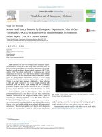
FIGURE 122.1 Axial CT of the brain shows hyperdensity consistent with aneurysm
associated with acute hemorrhage in the basal cisterns. The prominence of the temporal
horns of the lateral ventricles is also indicative of acute hydrocephalus.
Management. Spontaneous cerebral aneurysms presenting with bleeding
are uncommon but should be treated because of a poor natural history.
Imaging should be first acquired, and only if mass effect is not detected and
a high suspicion of aneurysmal rupture still exists should a lumbar puncture
be attempted. Aggressive surgical management with clipping, resection, or
trapping of intracranial dissecting aneurysms by surgical or endovascular
methods seems the most appropriate treatment.
Cavernous Malformation
Current Evidence. Cavernous malformations (CMs), also known as
cavernous angiomas or cavernomas, are compact lesions comprised of
sinusoidal vascular channels lined by a single layer of endothelium that
lacks the full complement of mature vessel wall components. Between the
vascular channels in the core of the lesion, there is loose connective tissue
stroma without intervening brain parenchyma. The prevalence of CMs has
been estimated to be between 0.4% and 0.9% of the population and 8% and
15% of all vascular malformations. They present with headache, seizure,
focal neurologic deficit, or as an incidental radiographic finding.
The majority of CMs are located supratentorially, typically in the white
matter of the cerebral hemispheres. The infratentorial CMs are located in
the cerebellum, pons, midbrain, and medulla. Less frequent locations of
CMs are the lateral and third ventricles, cranial nerves, and optic chiasm.
Acute hemorrhage from a chiasmal CM is a rare cause of permanent visual
loss. Of the extracerebral locations, the cavernous sinus, the orbits, and the
spinal cord are the most common.
Diagnostic Imaging. CT is more sensitive at detecting CMs, but its
specificity is low since most appear simply as high-density lesions (acute
hemorrhage) with little or no contrast enhancement. This is in contrast to
the high sensitivity and specificity of MRI for CMs. The MRI appearance
of CMs has been categorized into four types: a hyperintense core on T1and T2-weighted images representing subacute hemorrhage (Type I); a
“classic” picture of mixed-signal, reticulated core surrounded by a lowsignal rim (Type II); an iso- or hypointense lesion on T1 and markedly
hypointense lesion with hypointense rim on T2, which corresponds to
chronic hemorrhage (Type III); and punctate, poorly visualized hypointense
foci, which can be visualized only on gradient echo MRI, representing tiny
CM or telangiectasia (Type IV).
Management. With most asymptomatic CMs, particularly when the
diagnosis is relatively clear by MRI characteristics, the right approach for
the patient is conservative management with close follow-up. Type I and II
CMs are composed of acute or subacute hemorrhage and are more likely to
rebleed and may warrant closer follow-up. In contrast to a bleeding episode
from an AVM, a bleeding episode from a CM is rarely life threatening.
However, there is more controversy with symptomatic CMs which
hemorrhage in deep, difficult-to-access surgical locations.
Arteriovenous
Malformation.
Current
Evidence.
Arteriovenous
malformations (AVMs) are vascular abnormalities composed of a fistulous
connection of arteries and veins without a normal intervening capillary bed.
In the cerebral hemispheres, they frequently occur as cone-shaped lesions
with the apex of the cone reaching toward the ventricles. Nearly all AVMs
are thought to be congenital. Supratentorial location is the most common
(90%). The most common presentation of an AVM is intracerebral
hemorrhage (ICH). AVMs are responsible for 30% to 50% of hemorrhagic
strokes in children. After ICH, seizure is the second most common
presentation. Other presentations of AVMs include headache and focal
neurologic deficits which, including seizure, may be related to steal
phenomena or other alterations in perfusion in the tissue adjacent to the
AVM.
Size of AVM. In a series of 168 patients followed after presentation
without a prior hemorrhage, the size of the AVM was not found to be
predictive of future hemorrhage. However, other studies have found AVMs
of small size to be at higher risk of hemorrhage.
AVMs and Aneurysms. Prevalence of the association of AVMs with
aneurysms varies from 2.7% to 22.7%. This association seems to be
correlated with a higher risk of hemorrhage. Brown et al. studied 91
patients with unruptured AVMs and found the risk of ICH in patients with
coexisting aneurysm to be 7% at 1 year compared with 3% among those
with AVM alone. At 5 years, the risk persisted at 7% per year, while it
decreased to 1.7% per year in those with an AVM not associated with
aneurysms.
Diagnostic Imaging. A CT scan may be used as an initial screening tool
for patients presenting with neurologic sequelae related to unruptured or
ruptured AVMs. This study can be used quickly to determine location of the
lesion, acute hemorrhage, hydrocephalus, or areas of encephalomalacia
from previous surgery or rupture. A nonenhanced CT may show irregular
hyperdense areas frequently associated with calcifications in unruptured
AVMs or acute hemorrhage with ruptured AVMs ( Fig. 122.2 ). A contrastenhanced CT can demonstrate the nidus, feeding vessels, or dilated draining
veins.
MRI is superior to CT scan in delineating details of the macro
architecture of the AVM, except in the case of acute hemorrhage. These