Báo cáo khoa học: A region within the C-terminal domain of Ure2p is shown to interact with the molecular chaperone Ssa1p by the use of cross-linkers and mass spectrometry doc
Bạn đang xem bản rút gọn của tài liệu. Xem và tải ngay bản đầy đủ của tài liệu tại đây (516.88 KB, 12 trang )
A region within the C-terminal domain of Ure2p is shown
to interact with the molecular chaperone Ssa1p by the use
of cross-linkers and mass spectrometry
Virginie Redeker
1
, Jonathan Bonnefoy
1
, Jean-Pierre Le Caer
2
, Samantha Pemberton
1
,
Olivier Lapre
´
vote
2
and Ronald Melki
1
1 Laboratoire d’Enzymologie et Biochimie Structurales, CNRS, Gif-sur-Yvette, France
2 Institut de Chimie des Substances Naturelles, CNRS, Gif-sur-Yvette, France
Introduction
The aggregation of the prion Ure2p is at the origin of
the [URE3] trait in the baker’s yeast Saccharomyces
cerevisiae [1,2]. The propagation of the prion element
[URE3] is highly dependent on the expression of a
number of molecular chaperones from the Hsp100,
Hsp70 and Hsp40 protein families [3–5]. For example,
over-expression of the Hsp70 Ssa1p cures [URE3] [4]
and a mutation in the peptide-binding domain of Ssa2p
abolishes [URE3] propagation [5]. We have shown
in vitro that Ssa1p sequesters Ure2p in an assembly
incompetent state [6]. Affinity measurements performed
with full-length Ure2p and the compactly folded globu-
lar domain of the protein [7,8] revealed that Ssa1p
interacts with both full-length Ure2p and the C-termi-
Keywords
cross-linker; mass spectrometry; molecular
chaperone; oligomerization; Ure2p; prion
Correspondence
Virginie Redeker or Ronald Melki,
Laboratoire d’Enzymologie et Biochimie
Structurales, CNRS, Avenue de la terrasse,
91198 Gif-sur-Yvette, Cedex, France
Fax: +33 1 69 82 31 29
Tel: +33 1 69 82 34 60 or
+33169823503
E-mail: ,
(Received 28 July 2010, revised 5 October
2010, accepted 12 October 2010)
doi:10.1111/j.1742-4658.2010.07915.x
The propagation of yeast prion phenotypes is highly dependent on molecu-
lar chaperones. We previously demonstrated that the molecular chaperone
Ssa1p sequesters Ure2p in high molecular weight, assembly incompetent
oligomeric species. We also determined the affinity of Ssa1p for Ure2p,
and its globular domain. To map the Ure2p–Ssa1p interface, we have used
chemical cross-linkers and MS. We demonstrate that Ure2p and Ssa1p
form a 1 : 1 complex. An analytical strategy combining in-gel digestion of
cross-linked protein complexes, and both MS and MS ⁄ MS analysis of pro-
teolytic peptides, allowed us to identify a number of peptides that were
modified because they are exposed to the solvent. A difference in the expo-
sure to the solvent of a single lysine residue, lysine 339 of Ure2p, was
detected upon Ure2p–Ssa1p complex formation. These observations
strongly suggest that lysine 339 and its flanking amino acid stretches are
involved in the interaction between Ure2p and Ssa1p. They also reveal that
the Ure2p amino-acid stretch spanning residues 327–339 plays a central
role in the assembly into fibrils.
Structured digital abstract
l
MINT-8044534: Ure2p (uniprotkb:Q8NJQ9) and Ure2p (uniprotkb:Q8NJQ9) bind (MI:0407)
by cross-linking study (
MI:0030)
l
MINT-8044522: Ssa1p (uniprotkb:C8Z3H3) and Ssa1p (uniprotkb:C8Z3H3) bind (MI:0407)
by cross-linking study (
MI:0030)
l
MINT-8043971, MINT-8043985, MINT-8044494, MINT-8044548: Ure2p (uniprotkb:
Q8NJQ9) and Ssa1p (uniprotkb:C8Z3H3) bind (MI:0407)bycross-linking study (MI:0030)
Abbreviations
amu, atomic mass unit; EDC, 1-ethyl-3-(3-dimethylaminopropyl)carbodiimide hydrochloride; HXMS, hydrogen ⁄ deuterium exchange
measurement by mass spectrometry; LTQ, linear ion trap; NHS, N-hydroxysuccinimide; TFA, trifluoroacetic acid.
5112 FEBS Journal 277 (2010) 5112–5123 ª 2010 The Authors Journal compilation ª 2010 FEBS
nal domain of the protein [6]. The slightly higher affin-
ity of Ssa1p for full-length Ure2p was interpreted as
being the consequence of a preferential interaction with
the flexible N-terminal domain of Ure2p, critical for
assembly into fibrils. To further identify the regions
involved in Ure2p–Ssa1p interaction, we set up a chem-
ical cross-linking strategy coupled to the identification
of the chemically modified polypeptides by MS.
Covalent cross-linking approaches allow: (a) the iden-
tification of surface areas involved in protein-protein
interactions within protein complexes; (b) the character-
ization of the distance constraints within a protein com-
plex; and (c) the assessment of regions exposed or not
to the solvent within a protein [9–14]. Cross-linking of
protein complexes generates three types of products: (a)
mono-linked peptides when the cross-linker binds to a
reactive residue at one end, whereas the other reactive
group is hydrolyzed; (b) loop-linked peptides when both
ends of a cross-linker molecule bind to a single polypep-
tide chain; and (c) cross-linked peptides when the ends of
a cross-linker bind two distinct polypeptide chains.
Although far from straightforward [15], the proof of
protein–protein interactions comes from the identifica-
tion of cross-linked peptides [16–23]. The interfaces
involved in protein–protein interactions can be also
identified from the changes in the intensity of mono-
linked peptides, before and after complex formation
[13,24].
In the present study, we document Ure2p–Ssa1p
complex formation using two homo-bifunctional N-hy-
droxysuccinimide (NHS)-ester cross-linkers and the
zero length carbodiimide cross-linker 1-ethyl-3-(3-dim-
ethylaminopropyl) carbodiimide hydrochloride (EDC).
The stoichiometry of the Ure2p–Ssa1p complexes that
we generate is determined. Using MS after chemical
cross-linking and proteolysis, we map the solvent acces-
sibility of reactive residues on Ure2p and Ssa1p before
and after Ure2p–Ssa1p complex formation and identify
a region, located within the C-terminal domain of
Ure2p that interacts with Ssa1p. Because the C-termi-
nal domain of Ure2p is tightly involved in the assembly
of the prion into fibrils [25–28] and because Ssa1p
sequesters Ure2p in an assembly incompetent state, we
conclude that this region and its surroundings are
involved in the Ure2p fibrillar scaffold.
Results
Analysis of the intact cross-linked protein
complexes
The cross-linking conditions were optimized using
SDS ⁄ PAGE. The optimal Ure2p and Ssa1p concentra-
tions are 20 and 10 lm, respectively, compatible with
both a total inhibition of Ure2p assembly by Ssa1p
and the formation of high amounts of protein com-
plexes [6]. The two homo-bifunctional NHS-esters,
BS2G and BS3, were selected for their ability to cross-
link significant amounts of polypeptide chains at a
protein to cross-linker ratio of 1 : 20. Mixtures of deu-
terium labeled (d4) and unlabeled (d0) cross-linkers
were used to facilitate cross-linked peptide detection
and identification. The zero-length cross-linker EDC
was also used (not shown).
We previously demonstrated that Ssa1p–Ure2p inter-
action is nucleotide dependent [6]. We also showed
through assembly kinetic measurements that Ssa1p
binds a hexameric form of Ure2p in the presence of
ATP, whereas the form that is bound in the presence of
ADP is different, and probably dimeric [6]. We there-
fore performed Ure2p and Ssa1p cross-linking reactions
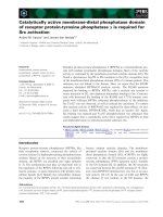
in the presence of ATP or ADP (Fig. 1A). Regardless
of the nucleotide present, two specific Ure2p–Ssa1p
complexes with apparent molecular masses of 120 and
160 kDa were observed. Western blot analysis con-
firmed the presence of Ure2p and Ssa1p in all protein
complexes (Fig. 1B). The extent of Ure2p–Ssa1p com-
plex formation was significantly higher in the presence
of ADP than in the presence of ATP, as seen by
SDS ⁄ PAGE, This is in agreement with the finding that
Ssa1p binds hexameric Ure2p in the presence of ATP,
whereas it binds dimeric Ure2p in the presence of ADP
[6]. Because Ssa1p efficiently inhibits Ure2p assembly in
the presence of ADP and as higher amounts of Ure2p–
Ssa1p cross-links are obtained in the presence of ADP,
all cross-linking reactions and subsequent analysis were
performed in the presence of ADP.
It should be noted that Ure2p cross-links into
dimers with distinct conformations, and thus different
mobilities (Fig. 1B). Similarly, nucleotide-dependent
conformational changes occurring within Ssa1p were
observed. Fast migrating monomeric and oligomeric
Ssa1p species, most likely corresponding to compact
Ssa1p species, were observed in the presence of ATP.
No change in the mobility of the Ure2p–Ssa1p com-
plexes was detected in the presence of ATP.
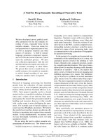
The stoichiometry of Ure2p and Ssa1p within the 120
and 160 kDa cross-linked complexes was assessed using
high-mass MALDI-TOF MS (Fig. 2). In agreement
with the SDS ⁄ PAGE, two Ure2p–Ssa1p complexes were
observed in the mass spectrum (Fig. 2C): one where
a single Ure2p is cross-linked to a single Ssa1p
(110 993 Da), and another where two Ure2p molecules
are bound to one Ssa1p (151 346 Da). Because the bind-
ing of the cross-linkers leads to an increase in the molec-
ular mass (Table S1), the number of cross-linkers bound
V. Redeker et al. Ure2p–Ssa1p interaction
FEBS Journal 277 (2010) 5112–5123 ª 2010 The Authors Journal compilation ª 2010 FEBS 5113
to Ure2p and Ssa1p can be estimate as 5 ± 1 and
10 ± 1 for BS2G and BS3, respectively.
Identification of modified and cross-linked
polypeptides
The analytical strategy used to characterize the
polypeptides involved in Ure2p–Ssa1p interaction is
schematized in Fig. S1. Cross-linked Ure2p, Ssa1p and
Ure2p–Ssa1p complexes resolved by SDS ⁄ PAGE were
treated with both trypsin and chymotrypsin to obtain
high protein sequence coverage (86% and 84.7% for
Ure2p and Ssa1p, respectively; Fig. S2). The modified
peptides were detected by MS using the 4.0247 atomic
mass unit (amu) mass difference conferred by t he binding
of the nondeuterated or deuterated cross-linkers
(Fig. 3A) [13,29]. Detection of modified peptides was
further confirmed using the 42.0469 amu mass differ-
ence as a result of the difference in the spacer arm
length of BS2G and BS3 (Fig. 3). A list of peptides
modified by BS2G or BS3 cross-linkers was derived
from MS analyses as described in the Materials and
methods and Fig. S1. Given the variety of theoretical
cross-links and modifications, exact mass measure-
ments were insufficient to unambiguously identify all
the peptides in our list using the available softwares
(gpmaw [30], xquest [31] and msx-3d [12]) with a
mass tolerance of 5 p.p.m. We therefore used MALDI-
TOF-TOF and ⁄ or nanoLC-Orbitrap tandem MS to
further identify peptides from this list. Twenty-five
mono-linked peptides and five loop-linked peptides
from Ure2p or Ssa1p (Table 1) were thus identified.
Most of the modified or loop-linked amino acid resi-
dues that we identified are exposed to the solvent as
shown on the 3D structure of Ure2p and Ssa1p
(Fig. 4). No intermolecular cross-links were detected.
This is probably a result of the low abundance of
cross-linked peptides and potential changes in their
ionization properties [32]. Because changes in the reac-
tivity of amino-acid residues to the cross-linkers can be
efficiently used to map conformational changes or pro-
tein–protein interaction interfaces [13,24], we further
compared the modified peptides derived from Ure2p
and Ssa1p alone and the two Ure2p–Ssa1p complexes.
Changes in the reactivity of Ure2p amino acid
residues upon Ure2p–Ssa1p complex formation
Most of the peptides originating from Ure2p were
found in both Ure2p–Ssa1p complexes. Their intensi-
ties were also similar. A similar observation was made
for peptides originating from Ssa1p. Two unique
differences were observed: one for Ure2p and one for
Ssa1p. The peptide spanning residues 337–343 from
Ure2p was found modified on lysine 339 (Fig.5) in
monomeric Ure2p and the Ure2p–Ssa1p complex with
an apparent molecular mass of 160 kDa but not that
of 120 kDa (Fig. S3). The finding that the Ure2p 337–
343 fragment is neither detected unmodified, nor modi-
fied, in the 120 kDa Ure2p–Ssa1p complex strongly
suggests that it is cross-linked to Ssa1p. Similarly,
*
*
A
B
160
120
Ssa1p
Ure2p
ADP
*
*
*
*
*
*
170 kDa
130 kDa
95 kDa
72 kDa
55 kDa
*
ATP ADP ATP
BS2G BS3
Anti-Ure2p Anti-His-tagged Ssa1p
*
12345 67 8 910111213
123 4 5 6 7 8 9101112 13 14
170 kDa
130 kDa
95 kDa
72 kDa
55 kDa
Fig. 1. SDS ⁄ PAGE analysis of cross-linked protein products. The
reaction products generated upon treatment of Ure2p, Ssa1p and
Ure2p in the presence of Ssa1p with the cross-linking agents BS2G
and BS3 were separated on a 7.5% acrylamide SDS ⁄ PAGE and
stained with Coomassie blue (A) or western blotted and stained
with antibodies directed against Ure2p or His-Tagged Ssa1p (B).
(A) A mixture of untreated Ure2p and Ssa1p (lane 1); Ure2p alone
(lanes 2, 5, 8 and 11), Ssa1p alone (lanes 3, 6, 9 and 12), and
Ure2p incubated in presence of Ssa1p (lanes 4, 7, 10 and 13) were
treated with BS2G (lanes 2–7) or BS3 (lanes 8–13), in the presence
of ADP 0.5 m
M (lanes 2–4 and 8–10) or 4 mM ATP and 5 mM
MgCl
2
(lanes 5–7 and 11–13). (B) Western blots of cross-linked
products obtained in the presence of 0.5 m
M ADP stained with
antibodies directed against Ure2p (lanes 1–7) and His-Tagged Ssa1p
(lanes 8–14); a mixture of untreated Ure2p and Ssa1p is seen in
lanes 3 and 10. Ure2p, Ssa1p and Ure2p incubated with Ssa1p
treated with BS2G are seen in lanes 1 and 8, 4 and 11 and 6 and
13, respectively. Similar samples treated with BS3 are seen in
lanes 2 and 9, 5 and 12 and 7 and 14, respectively. The arrows
show the cross-linked Ure2p–Ssa1p complexes with apparent
molecular masses of 120 and 160 kDa. Nucleotide-dependent
changes in Ssa1p conformation following BS3 treatment at the ori-
gin of electrophoretic modifications are labeled with stars.
Ure2p–Ssa1p interaction V. Redeker et al.
5114 FEBS Journal 277 (2010) 5112–5123 ª 2010 The Authors Journal compilation ª 2010 FEBS
lysine 325 was found to be modified in Ssa1p but not
in Ure2p–Ssa1p complexes.
These observations strongly suggest that the expo-
sure to the solvent of lysine 339 from Ure2p and lysine
325 from Ssa1p changes upon the formation of a 1 : 1
Ure2p–Ssa1p complex. Indeed, Ure2p is dimeric and
lysine 339 from each monomer within the dimer is
exposed to the solvent and can interact with Ssa1p.
When cross-linking occurs between Ure2p and Ssa1p,
a 120 kDa product is generated. When, in addition to
the latter covalent bond, the two monomers within
Ure2p dimer are cross-linked, a 160 kDa product is
observed. Additional complexes with apparent molecu-
lar weight higher than 200 kDa that are immuno
stained by both antibodies directed against Ure2p and
Ssa1p are also seen (Fig. 1B). The latter products cor-
respond to species where covalent bonds between
Ure2p monomers and each Ure2p monomer and Ssa1p
have been established. Ssa1p lysine 325 is not located
within the client binding pocket of the chaperone. Its
lack of modification upon complex formation can only
be attributed to a conformational rearrangement
within Ssa1p that occurs upon Ure2p–Ssa1p complex
formation.
Discussion
The propagation of the [URE3] trait is highly depen-
dent on the expression of molecular chaperones [3–5].
We recently showed that Ssa1p modulates the assem-
40 679
81 131
211.5
71 010
35 487
141 900
113.1
Intensity (%)
Mass (m/z)
70 702
40 408
80 870
35 044
897.5
132 000
Complex 1
(M
Ure2p
+ M
Ssa1p
)
110 993
151 346
141 193
Mass (m/z)
10
M
Ure2p
D
Ure2p
M
+2
Ssa1p
M
Ssa1p
D
Ssa1p
100
50
0
M
+2
Ssa1p
M
Ssa1p
D
Ssa1p
M
Ure2p
D
Ure2p
0
Complex 2
(D
Ure2p
+ M
Ssa1p
)
0
30 000
64 000 98 000 132 000 166 000
200 000
30 000 64 000 98 000 132 000
166 000 200 000
100
50
0
Intensity (%)
64 000 98 000 132 000 166 000 200 000
Intensity (%)
100
50
0
Mass (m/z)
Mass (m/z)
Counts
Counts
Counts
A
B
C
Fig. 2. High-mass MALDI-TOF mass spectra of the products generated upon cross-linking. Ure2p (A), Ssa1p (B) and Ure2p incubated with
Ssa1p (C) were cross-linked with BS3 in the presence of 0.5 m
M ADP. The mass of each peak and its identity are given (M, monomer; D,
dimer). The part of the spectrum containing the Ure2p–Ssa1p complexes is enlarged; inset in (C). The stoichiometry within the Ure2p–Ssa1p
complexes is indicated.
V. Redeker et al. Ure2p–Ssa1p interaction
FEBS Journal 277 (2010) 5112–5123 ª 2010 The Authors Journal compilation ª 2010 FEBS 5115
bly of Ure2p into protein fibrils in vitro and sequesters
Ure2p into assembly incompetent oligomeric species
[6]. Using fluorescence polarization, full-length Ure2p
and an Ure2p fragment spanning residues 94–354, we
assessed the affinity of Ssa1p for full-length Ure2p and
its compactly folded C-terminal domain (30 and
20 nm, respectively). The finding that Ssa1p binds with
slightly higher affinity to full-length Ure2p than its
compactly folded C-terminal domain was interpreted
as a consequence of the additional interaction between
Ssa1p and the flexible N-terminal moiety of Ure2p,
which is critical for assembly. An alternative explana-
tion that can account for this observation is that Ssa1p
binds with higher affinity a conformational state of
Ure2p as a result of the presence of the N-terminal
domain of the protein that slightly differs from that
adopted by its C-terminal moiety.
The only amino acid residue belonging to Ure2p
which exposure to the solvent is affected upon the
interaction of Ure2p with Ssa1p is lysine 339. This sug-
gests that lysine 339 and its flanking amino acid resi-
dues are involved in Ure2p–Ssa1p complex formation.
Because the binding of Ssa1p prevents Ure2p assem-
bly, it is reasonable to consider that the Ure2p region
centered on lysine 339 is involved in the assembly of
this prion into fibrils. Interestingly, hydrogen ⁄ deute-
rium exchange measurements by mass spectrometry
(HXMS) have revealed a decrease in the exposure to
the solvent of the amino acid stretch spanning residues
327–335 upon assembly of Ure2p into fibrils [33].
Thus, the binding of Ssa1p in the vicinity of this
stretch interferes with Ure2p assembly into fibrils
either because of a change in the conformation of this
stretch or the crowding of a surface interface involved
in intermolecular interactions within the fibrils or both.
Alternatively, the inability of Ure2p to form fibrils
upon binding of Ssa1p to Ure2p region centered on
lysine 339 could be a consequence of its incapacity to
acquire an assembly competent conformation. We
recently showed that the regions centered on residue 6
and 137 establish intramolecular interactions in assem-
bly competent Ure2p [26]. Interestingly, phenylalanine
137 and lysine 339 are located 27 A
˚
apart within the
same area in the 3D structure of Ure2p. Thus, the
binding of Ssa1p to the region centered on lysine 339
could abolish the acquisition by Ure2p of an assembly
competent state.
The results obtained in the present study provide a
rationale for the inhibition of Ure2p assembly by
Ssa1p and underline the key role of the C-terminal
domain of Ure2p in assembly into fibrils [25–28]. The
results, together with those previously obtained by
HXMS [33], further narrow the region critical either
for modulating Ure2p assembly into fibrils, or for the
establishment of intermolecular interactions between
Ure2p molecules within the fibrils, to that spanning
lysines 327–339.
The finding that lysine 325 from Ssa1p, which is
located at the interface between the nucleotide and cli-
864.0 874.2 884.4 894.6 904.8 915.0
Mass (m/z)
1.6E + 4
0
100
Intensity (%)
2677.1
0
50
100
Intensity (%)
2683.1
0
50
100
Intensity (%)
4400.4
0
50
100
Intensity (%)
7775.3
50
100
Intensity (%)
50
Cx160 BS2G-d0/d4
[312–318]
[596–602]
[241–247]
[327–333]
[186–192]
[13–20]
0
[596–602]
[596–602]
[596–602]
[241–247]
[241–247]
[241–247]
[13–20]
[312–318]
[13–20]
[312–318]
[13–20]
[312–318]
907.5220
Counts
911.5521
907.5096
911.5482
Counts
Counts
Counts
Counts
907.5172
911.5504
865.4789
869.5035
Cx160 BS3-d0/d4
Cx120 BS3-d0/d4
Ssa1p BS3-d0/d4
Ure2p BS3-d0/d4
A
B
C
D
E
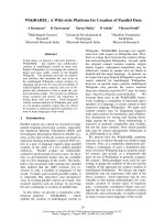
Fig. 3. Detection of chymotryptic peptides modified by nondeuter-
ated and deuterated cross-linkers by MALDI-TOF-TOF mass spec-
trometry. A selection of mass spectra illustrates how the
comparison of chymotryptic peptides from different cross-linked
complexes and protein controls allows the detection of modified
peptides. (A, B) Mass spectra of the 160 kDa Ure2p–Ssa1p com-
plex cross-linked with BS2G-d
0
⁄ d
4
and BS3-d
0
⁄ d
4
, respectively. The
mass spectra of the 120 kDa Ure2p–Ssa1p complex cross-linked
with BS3-d
0
⁄ d
4
and that of Ssa1p, and Ure2p after treatment with
BS3-d
0
⁄ d
4
are shown in (C–E). The curved arrows indicate the
4.0247 amu increase conferred by the binding of (light ⁄ heavy,
d
0
⁄ d
4
) cross-linkers. A 42.0469 amu mass difference between the
doublet peaks recorded upon binding of BS2G-d
0
⁄ d
4
(A) and BS3-
d
0
⁄ d
4
(B) is observed. The peptides indicated by arrowheads were
identified by exact mass measurement. The peptide labeled by
curved arrows was identified by tandem mass spectrometry as a
mono-linked Ure2p [S
100
RITK*F
105
] where K* is the mono-linked
residue.
Ure2p–Ssa1p interaction V. Redeker et al.
5116 FEBS Journal 277 (2010) 5112–5123 ª 2010 The Authors Journal compilation ª 2010 FEBS
Table 1. Mono- and loop-linked peptides list. The tryptic and chymotryptic peptides that were identified are denoted T and CT, respectively. The protonated monoisotopic experimental
masses (MH+exp) and the calculated mass difference (p.p.m.) with the theoretical monoisotopic mass of the identified peptide are given. The presence of the modified peptides in the
Ure2p, Ssa1p and Ure2p–Ssa1p complexes with apparent molecular masses 160 and 120 kDa tryptic and chymotryptic reaction products is indicated by an X. The amino acid sequences
and the modification sites are indicated. Loop-linked peptides are labeled (T1). ND, not determined.
BS2G BS3 Peptide detection Peptide identification in Ssa1p Peptide identification in Ure2p
MH+exp p.p.m. MH+exp p.p.m. Cx160 Cx120 Ssa1p Ure2p Sequence Site Sequence Site
CT 865.4783 0.5 907.5245 0.2 X X X S
100
RITKF
105
K104
CT 1125.6722 1.6 1167.722 1.2 X X X K
243
RKNKKDL
250
K243, K247 (T1)
CT 1407.646 2.6 1449.6946 0.9 X X X T
378
GDESSKTQDLL
389
K384
CT 1427.7954 1.1 1469.8446 1.1 X X X K
243
RKNKKDLSTN
253
K243, K247 (T1)
CT 1478.8682 0.7 1520.9146 1.3 X X X V
334
GGSTRIPKVQKL
346
K342, K345 (T1)
CT 1502.7592 0.6 1544.8064 0.3 X X X H
237
SQKIASAVERY
248
K240
CT 1616.7878 3.7 ND X X H
237
SQKIASAVERY
248
K240,
S243
CT 1842.9832 2.5 ND X R
319
DAKLDKSQVDEIVL
333
K322 or K325
CT 1973.9536 1.3 2015.9996 0.4 X X X N
149
DSQRQATKDAGTIAGLN
166
K157
T 1002.5442 2.4 1044.5926 1 X X X N
414
STIPTKK
421
K420
T 1075.5522 0.5 1117.5962 2.5 X X X L
507
SKEDIEK
514
K509
T 1103.5114 1.1 1145.5578 1.6 X X W
337
TKHMMR
343
K339
T 1119.5052 2.2 1161.5534 0.9 X X W
337
TKHMMR
343
(1M
ox
) K339
T 1131.5986 1.8 1173.6466 0.8 X X X I
498
TITNDKGR
506
K504
T 1135.5033 1 1177.5468 2.5 X X W
337
TKHMoxMoxR
343
(2M
ox
) K339
T 1136.7232 3.1 1178.7712 2 X X R
344
PAVIKALR
352
K349
T 1137.7082 2.1 1179.6844 7.3 X X R
344
PAVIKALR
352
K349
T 1299.664 1.1 1341.7112 0.8 X X X N
246
KKDLSTNQR
255
K247, K248 (T1)
T 1414.696 0.1 1456.742 1.1 X X X A
153
PEFVSVNPNAR
164
S158
T 1427.758 2.1 1469.8068 0.3 X X X K
245
NKKDLSTNQR
255
K247,K248 (T1)
T 1544.8222 0.2 ND X X L
234
VNHFIQEFKR
244
K243
T 1649.8272 1 1691.8724 2.7 X X X Y
235
FHSQKIASAVER
247
K240
T 1741.9448 0.5 1783.9916 0.4 X X X Q
154
ATKDAGTIAGLNVLR
169
K157
T 1915.0585 3.7 1957.1016 0.2 X X L
234
VNHFIQEFKRKNK
247
K243 or K245
T 1990.9105 3.6 ND X X X M
1
MNNNGNQVSNLSNALR
17
M1
T 2006.9006 1.2 ND X X M
1
MNNNGNQVSNLSNALR
17
(1Mox)
M1
T 2022.8986 1.8 2064.9464 2.7 X X M
1
MNNNGNQVSNLSNALR
17
(2Mox)
M1
T 2043.1486 1.2 ND X X L
234
VNHFIQEFKRKNKK
248
K245
T 2297.0602 0.1 ND X X M
515
VAEAEKFKEEDEKESQR
532
K521
T 2453.1048 1.3 2495.1526 2.1 X X X N
66
GSQNNDNENNIKNTLEQHR
85
K78
V. Redeker et al. Ure2p–Ssa1p interaction
FEBS Journal 277 (2010) 5112–5123 ª 2010 The Authors Journal compilation ª 2010 FEBS 5117
ent protein binding domains of Ssa1p, is modified in
Ssa1p but not in Ssa1p–Ure2p complexes exquisitely
illustrates the conformational rearrangements that
affect Ssa1p domains upon its interaction with Ure2p
with the burial of a Ssa1p stretch comprising lysine
325.
The results reported in the present study are consis-
tent with the view that subtle conformational changes
modulate the assembly of Ure2p into fibrils and
further highlight the involvement of the C-terminal
domain of Ure2p in the fibrillar scaffold. Mutagenesis
approaches targeting Ure2p stretch 325–340 will pro-
vide additional insight into the mechanism of Ure2p
assembly into fibrils and the manner with which
molecular chaperones modulate this process under
physiological conditions.
Materials and methods
Production of proteins
Ure2p was expressed in Escherichia coli, purified and stored
as described previously [34]. Ssa1p was expressed with an
N-terminal His-tag in S. cerevisiae, purified and stored as
M1
K78
K104
K249
K339
K349
ATPase
domain
Peptide binding
domain
ATPase
domain
Peptide binding
domain
180°
K243
K247
K157
K527
K504
K509
K420
K509
K325
K346
K342
K243
K247
K247
K248
K504
C
A
B
D
K240
S243
S243
K240
K339
S158
K349
K339
S158
K349
180°
K339
K349
K349
K104
K104
K339
S158
K339
Fig. 4. Location of the mono-linked and loop-linked lysines in Ure2p and Ssa1p. Peptides containing modified and loop-linked lysine are col-
ored magenta in Ssa1p (A) and Ure2p (B–D) structures. Loop-linked residues are colored blue. Mono-linked residues are colored orange. The
Ssa1p 3D model in (A) was built using the ATPase domain of bovine Hsc70 (P19120), and the peptide binding domain of E. coli DnaK
(P0A6Y8), Protein Data Bank accession numbers 3HSC and 1BPR, respectively. The two monomers constituting Ure2p dimer (Protein Data
Bank accession number 1G6Y) in (B) are colored green and blue. A model of full-length Ure2p is presented in (C) to map modified peptides.
This model was built from the X-ray structure of the C-terminal domain of Ure2p and integrates the finding that the N-terminal domain of
Ure2p is flexible. An enlargement of the region of Ure2p involved in the interaction with Ssa1p is shown in (D). Lysine 339 is shown in
orange; the region of Ure2p whereby exposure to the solvent was shown to change upon assembly into fibrils by HXMS [33] is colored red.
The figure was generated with
PYMOL ().
Ure2p–Ssa1p interaction V. Redeker et al.
5118 FEBS Journal 277 (2010) 5112–5123 ª 2010 The Authors Journal compilation ª 2010 FEBS
described previously [35]. Ure2p and Ssa1p concentrations
were determined as reported previously [6] and using the
Bradford dye assay, respectively.
Cross-linking reaction
Cross-linking reactions were carried out with mixtures of
deuterium labeled (d4) and unlabeled (d0) homo-bifunc-
tional sulfo-NHS esters cross-linker reagent: BS2G-d0 ⁄ d4
[bis(sulfosuccinimidyl) glutarate] with a 7.7 A
˚
spacer arm
and BS3-d0 ⁄ d4 [bis(sulfosuccinimidyl) suberate] with a
11.4 A
˚
spacer arm (Pierce, Waltham, MA, USA). Both
cross-linkers react with the e-amino group of lysine residues
and a-amino group from protein N-termini and, to a lesser
extent, with the hydroxyl groups of serine, threonine and
tyrosine residues [36]. The zero-length EDC cross-linker
cross-links carboxyl groups to primary amines. The pro-
teins were dialyzed for 2 h at 4 °C against cross-linking
buffer (40 mm Hepes-KOH, pH 7.5, 75 mm KCl) before
cross-linking. The samples were then spun for 10 min at
W T K H M M R
BS
2
G
y1y2y3y4
b2 b4 b6b5
y6 y5
b3
1.99E3
200 700 800 1100
Mass (m/z)
0
10
20
30
40
50
60
70
80
90
100
Intensity (%)
m
2H
+
y3
y6
b2
b3
b4
b5
b6
M
R
H
M
K–BS
2
G
WT H M
TM
•
y2
y1
y4
y5
*
*
300 400 500 600 900 1000
1100.0 1102.6 1105.2 1107.8 1110.4 1113.0
Mass (m/z)
168.3
0
50
100
Intensity (%)
1103.5170
<
<
x 4
<
<
x 4
1107.5415
B
A
K–BS
2
G
Fig. 5. NanoLC-LTQ-Orbitrap identification of the mono-linked Ure2p peptide [337–343]. Mass spectra of the tryptic peptide [337–343] from
Ure2p treated with BS2G-d
0
⁄ d
4
. (A) MALDI-TOF-TOF mass spectrum of the light (d0) and heavy (d4) precursor ions presenting a mass differ-
ence of 4.024 Da (indicated by the curved arrow). (B) Fragmentation mass spectrum of the double-charged d0 precursor ion obtained using
nanoLC-LTQ-Orbitrap MS ⁄ MS analysis in the LTQ. The sequence of the loop-linked peptide V
340
FGGSTRIPK*VQK*L
352
is presented with
the identified y and b fragment ions. Black and grey stars and black circles correspond to y
2+
,b
2+
and internal fragment ions respectively.
K* is the mono-linked residue.
V. Redeker et al. Ure2p–Ssa1p interaction
FEBS Journal 277 (2010) 5112–5123 ª 2010 The Authors Journal compilation ª 2010 FEBS 5119
15 000 g and 4 °C. To generate the Ure2p–Ssa1p com-
plexes, the Ure2p and Ssa1p concentrations were adjusted
to 20 and 10 lm, respectively. The reaction mixture con-
taining 0.5 mm ADP or 4 mm ATP and 5 mm MgCl
2
was
then incubated for 2 h at 10 °C under mild agitation. Con-
trol reactions consisted of incubating Ure2p and Ssa1p
individually under the same experimental conditions. The
NHS-ester cross-linkers (5 mm) were dissolved in dimethyl-
sulfoxide. A mixture of deuterated and nondeuterated
(1 : 1) cross-linkers were added to Ure2p, Ssa1p and Ure2p
incubated with Ssa1p, with up to 20-fold molar excess.
Cross-linking was performed at room temperature for
30 min and the reaction was terminated by the addition of
ammonium bicarbonate (50 mm). EDC cross-linking was
performed for 60 min in the presence of 4 m m EDC and
5mm sulfo-NHS (N-hydroxysulfosuccinimide). The reac-
tion was stopped by addition of b-mercaptoethanol and
hydroxylamine (20 and 10 mm, respectively). Samples for
SDS ⁄ PAGE analysis were immediately mixed (1 : 1 volume
ratio) with denaturing buffer and heated at 95 °C. For
high-mass MALDI-TOF MS, the samples were directly
spotted on the MALDI plate.
SDS/PAGE and western blotting
SDS ⁄ PAGE analysis was performed on 7.5% polyacryl-
amide gels (8 · 7 · 0.15 cm) as described by Laemmli [37].
Equal amounts of proteins (10 lg) were loaded in each
well. The gels were Coomassie blue stained, destained and
imaged using a Sony charge-coupled device camera (Sony
Corp., Tokyo, Japan). The proteins within the gels were
transferred to nitrocellulose membranes. Ure2p and Ssa1p
protein bands were probed with polyclonal antibody direc-
ted against full-length Ure2p and monoclonal anti-His-tag
serum for His-tagged Ssa1p (Sigma-Aldrich, St Louis, MO,
USA) and the membranes were developed with the enzyme-
coupled luminescence technique (ECL; GE Healthcare,
Milwaukee, WI, USA). All images were analyzed using nih
image software (available at: />nih-image/).
Peptide preparation
The protein bands resolved by SDS ⁄ PAGE and, corre-
sponding to monomeric Ure2p, monomeric Ssa1p and
Ure2p–Ssa1p complexes with apparent molecular masses of
120 and 160 kDa were excised. Each protein band was sub-
jected to in-gel enzymatic cleavage after reduction and
alkylation of cysteine residues in the presence of 10 mm
dithiothreitol and 55 mm iodocetamide [38]. Trypsin (Pro-
mega Gold; Promega, Madison, WI, USA) or Chymotryp-
sin (Roche, Basel, Switzerland) (12.5 ngÆlL
)1
) treatments
were performed overnight at 37 °C under mild agitation in
25 mm ammonium bicarbonate. Peptides were extracted in
100% acetonitrile following the incubation under agitation
of the reaction products with 5% formic acid at 37 °C for
15 min. The extracted peptides were vacuum dried, dis-
solved in 1% formic acid and stored at )20 °C until MS
analysis.
High mass MALDI-TOF MS
High-mass MALDI-TOF mass spectra of the intact protein
complexes were obtained using a MALDI-TOF mass spec-
trometer (Voyager DE STR; Applied Biosystems, Foster
City, CA, USA) equipped with an HM1 high-mass detec-
tion system (CovalX, Zu
¨
rich, Switzerland) [39]. The instru-
ment was operated in positive and linear mode with a
25 kV acceleration voltage, 85% grid voltage and 2000 ns
delayed extraction time. Mass spectra were obtained by
averaging 100–1000 shots. The instrument was externally
calibrated with enolase (10 lm) using the double-charged
monomer, and the single-charged monomer and dimer. Cal-
ibration was checked using noncross-linked Ure2p and
Ssa1p. The mass accuracy was 100–200 Da at 150 kDa.
One volume of cross-linked proteins was diluted with one
volume of 1% trifluoroacetic acid (TFA). This acidified
sample was mixed 1 : 1 (v ⁄ v) with a saturated solution of
sinapinic acid (10 mgÆmL
)1
in 30% acetonitrile and 0.1%
TFA).
MALDI-TOF-TOF MS
The samples were desalted (with 5% acetonitrile, 0.1%
TFA) and eluted from a C18 reversed-phase Zip-Tip
Ò
(Mil-
lipore, Billerica, MA, USA) in 40% acetonitrile, 0.1%
TFA. Peptides samples were mixed 1 : 5 to 1 : 20 (v ⁄ v) with
a-cyano-4-hydroxycinnamic acid (4 mgÆmL
)1
in 50% aceto-
nitrile, 10 mm ammonium citrate and 0.1% formic acid)
and spotted (0.5 l L) on a stainless steel MALDI target
(Opti-TOF; Applied Biosystems). MALDI-TOF-TOF MS
and MS ⁄ MS spectra were acquired with a MALDI-TOF ⁄ -
TOFÔ 4800 mass spectrometer (Applied Biosystems) in the
positive and reflector mode. An external calibration was
performed using standard peptide solution Cal Mix1 and
Cal Mix2 (Applied Biosystems) and an additional internal
calibration was performed during mass spectra analysis
using nonmodified peptides of both Ure2p and ⁄ or Ssa1p.
Acquisition and data analysis were performed using the
explorer 3.5.2 and data explorer 4.9 software from
Applied Biosystems.
NanoLC-linear ion trap (LTQ)-Orbitrap mass
spectrometry
Tryptic and chymotryptic peptide digests were analyzed by
NanoLC MS ⁄ MS using a HPLC system (Ultimate U3000;
Dionex, Sunnyvale, CA, USA) coupled online to a LTQ-
Orbitrap (ThermoScientific, Waltham, MA, USA) equipped
Ure2p–Ssa1p interaction V. Redeker et al.
5120 FEBS Journal 277 (2010) 5112–5123 ª 2010 The Authors Journal compilation ª 2010 FEBS
with a nanoelectrospray ion source after separation on a
reversed-phase C18 pepmap 100 column (75 lm inner
diameter, 5 lm particules of 100 A
˚
diameter, 15 cm length)
from Dionex. The peptides were loaded at a flow rate of
20 lLÆmin
)1
, and eluted at a flow rate of 200 nLÆmin
)1
by
a three step gradient: (a) 2–60% solvent B for 40 min;
(b) 60–100% solvent B for 1 min; and (c) 100% solvent B
for 20 min. Solvent A was 0.1% formic acid in water,
whereas solvent B was 0.1% formic acid in 100% acetonitrile.
NanoLC-MS ⁄ MS experiments were conducted in the data-
dependent acquisition mode. The mass of the precursors
was measured with a high resolution (60 000 FWHM) in
the Orbitrap. The four most intense ions, above an intensity
corresponding to 400 ions, were selected for fragmentation
in the LTQ.
The isotope label of cross-linked peptides results in
doublet signals with m ⁄ z differences of 4.0247, 2.0123 and
1.341 for mono-protonated, double or triple-protonated
peptides, respectively. This information was used for
LC-MS post-acquisition filtering using the software viper
( First, nanoLC-
MS ⁄ MS data were de-isotoped using the decon2ls soft-
ware (available at: />php). The resulting csv files were further analyzed with
viper [40]. A list with a delta m ⁄ z of 4.0247 corresponding
to labeled ion pairs with a maximum mass tolerance of
10 p.p.m. was generated. Mass deviation and peptide elu-
tion time were used to filter the list of peptide doublets,
corresponding to candidate cross-linked peptides. The list
of light and heavy precursor masses was further used either
to analyze the MS ⁄ MS spectra acquired in the data-depen-
dent acquisition analysis or to build an inclusion list with
the light and heavy precursor masses for cross-linked candi-
date peptides analysis. NanoLC-LTQ-Orbitrap data were
processed automatically as described as well as manually.
Acknowledgements
We are grateful to Luc Bousset for designing a pro-
gram for exploiting the MS data and for building the
Ssa1p 3D model. We thank Alain Brunelle for helpful
discussions about MALDI-TOF-HM1 MS. This work
was supported by the French Ministry of Education,
Research and Technology through the Centre
National de la Recherche Scientifique (CNRS), the
Institut National de la Sante
´
et de la Recherche Me
´
di-
cale (INSERM) and the Agence Nationale pour la
Recherche (ANR-06-BLAN-0266 and ANR-08-PCVI-
0022-02).
References
1 Masison DC, Maddelein ML & Wickner RB (1997)
The prion model for [URE3] of yeast: spontaneous
generation and requirements for propagation. Proc Natl
Acad Sci USA 94, 12503–12508.
2 Masison DC & Wickner RB (1995) Prion-inducing
domain of yeast Ure2p and protease resistance of
Ure2p in prion-containing cells. Science 270, 93–
95.
3 Moriyama H, Edskes HK & Wickner RB (2000)
[URE3] prion propagation in Saccharomyces cerevisiae:
requirement for chaperone Hsp104 and curing by over-
expressed chaperone Ydj1p. Mol Cell Biol 20, 8916–
8922.
4 Schwimmer C & Masison DC (2002) Antagonistic inter-
actions between yeast [PSI(+)] and [URE3] prions and
curing of [URE3] by Hsp70 protein chaperone Ssa1p
but not by Ssa2p. Mol Cell Biol 22, 3590–3598.
5 Roberts BT, Moriyama H & Wickner RB (2004)
[URE3] prion propagation is abolished by a mutation
of the primary cytosolic Hsp70 of budding yeast. Yeast
21, 107–117.
6 Savistchenko J, Krzewska J, Fay N & Melki R (2008)
Molecular chaperones and the assembly of the prion
Ure2p in vitro. J Biol Chem 283, 15732–15739.
7 Thual C, Bousset L, Komar AA, Walter S, Buchner J,
Cullin C & Melki R (2001) Stability, folding, dimeriza-
tion, and assembly properties of the yeast prion Ure2p.
Biochemistry 40, 1764–1773.
8 Bousset L, Belrhali H, Janin J, Melki R & Morera S
(2001) Structure of the globular region of the prion pro-
tein Ure2 from the yeast Saccharomyces cerevisiae.
Structure (Camb.) 9, 39–46.
9 Sinz A (2006) Chemical cross-linking and mass spec-
trometry to map three-dimensional protein structures
and protein-protein interactions. Mass Spectrom Rev
25, 663–682.
10 Lutter LC & Kurland CG (1975) Chemical determina-
tion of protein neighbourhoods in a cellular organelle.
Mol Cell Biochem 7, 105–116.
11 Cohen FE & Sternberg MJ (1980) On the use of chemi-
cally derived distance constraints in the prediction of
protein structure with myoglobin as an example. J Mol
Biol 137, 9–22.
12 Heymann M, Paramelle D, Subra G, Forest E, Marti-
nez J, Geourjon C & Dele
´
age G (2008) MSX-3D: a tool
to validate 3D protein models using mass spectrometry.
Bioinformatics 24, 2782–2783.
13 Pimenova T, Nazabal A, Roschitzki B, Seebacher J,
Rinner O & Zenobi R (2008) Epitope mapping on
bovine prion protein using chemical cross-linking and
mass spectrometry. J Mass Spectrom 43, 185–195.
14 Pimenova T, Pereira CP, Schaer DJ & Zenobi R (2009)
Characterization of high molecular weight multimeric
states of human haptoglobin and hemoglobin-based
oxygen carriers by high-mass MALDI MS. J Sep Sci
32, 1224–1230.
V. Redeker et al. Ure2p–Ssa1p interaction
FEBS Journal 277 (2010) 5112–5123 ª 2010 The Authors Journal compilation ª 2010 FEBS 5121
15 Kalkhof S & Sinz A (2008) Chances and pitfalls of
chemical cross-linking with amine-reactive N-hydroxy-
succinimide esters. Anal Bioanal Chem 392, 305–312.
16 Seebacher J, Mallick P, Zhang N, Eddes JS, Aebersold R
& Gelb MH (2006) Protein cross-linking analysis using
mass spectrometry, isotope-coded cross-linkers, and
integrated computational data processing. J Proteome
Res 5 , 2270–2282.
17 Gao QX, Doneanu CE, Shaffer SA, Adman ET, Good-
lett DR & Nelson SD (2006) Identification of the inter-
actions between cytochrome P450 2E1 and cytochrome
b(5) by mass spectrometry and site-directed mutagene-
sis. J Biol Chem 281, 20404–20417.
18 Kalkhof S, Ihling C, Mechtler K & Sinz A (2005) Chemi-
cal cross-linking and high-performance Fourier trans-
form ion cyclotron resonance mass spectrometry for
protein interaction analysis: application to a calmodu-
lin ⁄ target peptide complex. Anal Chem 77, 495–503.
19 Tang XT, Munske GR, Siems WF & Bruce JE (2005)
Mass spectrometry identifiable cross-linking strategy for
studying protein-protein interactions. Anal Chem 77,
311–318.
20 Sinz A, Kalkhof S & Ihling C (2005) Mapping protein
interfaces by a trifunctional cross-linker combined with
MALDI-TOF and ESI-FTICR mass spectrometry.
J Am Soc Mass Spectrom 16, 1921–1931.
21 Schmidt A, Kalkhof S, Ihling C, Cooper DMF & Sinz A
(2005) Mapping protein interfaces by chemical cross-
linking and Fourier transform ion cyclotron resonance
mass spectrometry: application to a calmodulin ⁄ adenylyl
cyclase 8 peptide complex. Eur J Mass Spectrom 11,
525–534.
22 Dimova K, Kalkhof S, Pottratz I, Ihling C, Rodriguez-
Castaneda F, Liepold T, Griesinger C, Brose N, Sinz A
& Jahn O (2009) Structural insights into the calmodu-
lin-Munc13 interaction obtained by cross-linking and
mass spectrometry. Biochemistry 48, 5908–5921.
23 Manolaridis I, Mumtsidu E, Konarev P, Makhov AM,
Fullerton SW, Sinz A, Kalkhof S, McGeehan JE, Cary
PD, Griffith JD et al. (2009) Structural and biophysical
characterization of the proteins interacting with the her-
pes simplex virus 1 origin of replication. J Biol Chem
284, 16343–16353.
24 Jaya N, Garcia V & Vierling E (2009) Substrate bind-
ing site flexibility of the small heat shock protein
molecular chaperones. Proc Natl Acad Sci USA 106,
15604–15609.
25 Bousset L, Redeker V, Decottignies P, Dubois S, Le
Mare
´
chal P & Melki R (2004) Structural character-
ization of the fibrillar form of the yeast Saccharomyces
cerevisiae prion Ure2p. Biochemistry 43, 5022–5032.
26 Fay N, Redeker V, Savischenko J, Dubois S, Bousset L
& Melki R (2005) Structure of the prion Ure2p in
protein fibrils assembled in vitro. J Biol Chem 280,
37149–37158.
27 Ranson N, Stromer T, Bousset L, Melki R & Serpell LC
(2006) Insights into the architecture of the Ure2p yeast
protein assemblies from helical twisted fibrils. Protein Sci
15, 2481–2487.
28 Bousset L, Bonnefoy J, Sourigues Y, Wien F & Melki R
(2010) Structure and assembly properties of the N-termi-
nal domain of the prion Ure2p in isolation and in its
natural context. PLoS ONE 5, e9760.
29 Maiolica A, Cittaro D, Borsotti D, Sennels L, Ciferri C,
Tarricone C, Musacchio A & Rappsilber J (2007) Struc-
tural analysis of multiprotein complexes by cross-linking,
mass spectrometry, and database searching. Mol Cell
Proteomics 6, 2200–2211.
30 Peri S, Steen H & Pandey A (2001) GPMAW – a soft-
ware tool for analyzing proteins and peptides. Trends
Biochem Sci 26, 687–689.
31 Rinner O, Seebacher J, Walzthoeni T, Mueller LN,
Beck M, Schmidt A, Mueller M & Aebersold R (2008)
Identification of cross-linked peptides from large
sequence databases. Nat Methods 5, 315–318.
32 Leitner A, Walzthoeni T, Kahraman A, Herzog F,
Rinner O, Beck M & Aebersold R (2010) Probing
native protein structures by chemical cross-linking,
mass spectrometry and bioinformatics. Mol Cell
Proteomics 9, 1634–1649.
33 Redeker V, Halgand F, Le Caer JP, Bousset L,
Lapre
´
vote O & Melki R (2007) Hydrogen ⁄ deuterium
exchange mass spectrometric analysis of conformational
changes accompanying the assembly of the yeast prion
Ure2p into protein fibrils. J Mol Biol 369, 1113–1125.
34 Thual C, Komar AA, Bousset L, Fernandez-Bellot E,
Cullin C & Melki R (1999) Structural characterization
of Saccharomyces cerevisiae prion-like protein Ure2.
J Biol Chem 274, 13666–13674.
35 Krzewska J & Melki R (2006) Molecular chaperones
and the assembly of the prion Sup35p, an in vitro
study. EMBO J 25, 822–833.
36 Ma
¨
dler S, Bich C, Touboul D & Zenobi R (2009)
Chemical cross-linking with NHS esters: a systematic
study on amino acid reactivities. J Mass Spectrom 44,
694–706.
37 Laemmli UK (1970) Cleavage of structural proteins
during the assembly of the head of bacteriophage T4.
Nature 227, 680–685.
38 Shevchenko A, Wilm M, Vorm O & Mann M (1996)
Mass spectrometric sequencing of proteins silverstained
polyacrylamide gels. Anal Chem 68, 850–858.
39 Wenzel RJ, Kern S, Nazabal A & Zenobi R (2007)
Quantitative comparison of sensitivity and saturation
for MALDI-TOF detectors when measuring complex
and high mass samples. Proceedings of the 55th ASMS
Conference on Mass Spectrometry, Indianapolis,
MP082.
40 Zimmer JS, Monroe ME, Qian WJ & Smith RD (2006)
Advances in proteomics data analysis and display using
Ure2p–Ssa1p interaction V. Redeker et al.
5122 FEBS Journal 277 (2010) 5112–5123 ª 2010 The Authors Journal compilation ª 2010 FEBS
an accurate mass and time tag approach. Mass Spec-
trom Rev 25, 450–482.
Supporting information
The following supplementary material is available:
Fig. S1. Analytical strategy for Ure2p–Ssa1p chemical
cross-linking, cleavage and identification of the reac-
tion products.
Fig. S2. Primary structure coverage obtained following
tryptic and chymotryptic treatment of Ure2p (A) and
Ssa1p (B).
Fig. S3. NanoLC-LTQ-Orbitrap chromatograms of the
BS3 mono-linked peptide W
337
TK*HMMR
343
(double-
charged ion peak at m ⁄ z 575 2820) produced by tryptic
in-gel digestion.
Table S1. Molecular masses of NHS-ester cross-linker
before and after reaction with lysine residues.
This supplementary material can be found in the
online version of this article.
Please note: As a service to our authors and readers,
this journal provides supporting information supplied
by the authors. Such materials are peer-reviewed and
may be re-organized for online delivery, but are not
copy-edited or typeset. Technical support issues arising
from supporting information (other than missing files)
should be addressed to the authors.
V. Redeker et al. Ure2p–Ssa1p interaction
FEBS Journal 277 (2010) 5112–5123 ª 2010 The Authors Journal compilation ª 2010 FEBS 5123