Báo cáo khoa học: Plant RMR proteins: unique vacuolar sorting receptors that couple ligand sorting with membrane internalization pdf
Bạn đang xem bản rút gọn của tài liệu. Xem và tải ngay bản đầy đủ của tài liệu tại đây (626.12 KB, 10 trang )
MINIREVIEW
Plant RMR proteins: unique vacuolar sorting receptors that
couple ligand sorting with membrane internalization
Hao Wang
1,2
, John C. Rogers
3
and Liwen Jiang
1,2
1 Department of Biology, Centre for Cell and Developmental Biology, Chinese University of Hong Kong, China
2 State (China) Key Laboratory for Agrobaiotechnology, The Chinese University of Hong Kong, China
3 Institute of Biological Chemistry, Washington State University, Pullman, WA, USA
Introduction
Eukaryotic cells share a common organization of
organelles within their endomembrane systems, where
each is a membrane-bound compartment which defines
a separate environment for specific functions, and dif-
ferent organelles communicate with each other via
transport vesicles. In general, a unique type of vesicle
is required for each step in traffic, and transmembrane
receptor proteins that are specific for one vesicle type
recruit cargo that will be transported from one orga-
nelle to another in the step mediated by that vesicle
[1–6]. A general principle that applies across eukary-
otic species defines vesicle specificity: the cytoplasmic
coat proteins that cause a vesicle to bud from its orga-
nelle source interact with the specific receptor proteins
and cause them to partition with their cargo into the
budding vesicle [7,8]. Thus, in general terms, a sorting
receptor is specific for one vesicle type that traffics
in one specific step between two endomembrane
organelles.
The endomembrane systems for animal, yeast and
plant cells have in common the presence of an orga-
nelle with an acidic lumenal pH that serves as a diges-
tive compartment, the lysosome or vacuole [9,10]. In
general, soluble proteins within the lumen of the endo-
membrane system that are destined for the lyso-
some ⁄ vacuole traffic through the Golgi apparatus
where they are recruited at the trans-face into clathrin-
coated vesicles (CCVs) by receptors that are unique
Keywords
lytic PVC; PA domain; pollen tube; PSV;
receptor; RING-H2 domain; RMR; storage
PVC; vacuole; VSR
Correspondence
L. Jiang, State (China) Key Laboratory for
Agrobaiotechnology, The Chinese University
of Hong Kong, Shatin, New Territories,
Hong Kong, China
Fax: +852 2603 5646
Tel: +852 2609 6388
E-mail:
(Received 31 March 2010, revised 30 June
2010, accepted 7 July 2010)
doi:10.1111/j.1742-4658.2010.07923.x
In receptor-mediated sorting of soluble protein ligands in the endomem-
brane system of eukaryotic cells, three completely different receptor pro-
teins for mammalian (mannose 6-phosphate receptor), yeast (Vps10p) and
plant cells (vacuolar sorting receptor; VSR) have in common the features
of pH-dependent ligand binding and receptor recycling. In striking con-
trast, the plant receptor homology-transmembrane-RING-H2 (RMR) pro-
teins serve as sorting receptors to a separate type of vacuole, the protein
storage vacuole, but do not recycle, and their trafficking pathway results in
their internalization into the destination vacuole. Even though plant RMR
proteins share high sequence similarity with the best-characterized mamma-
lian PA-TM-RING family proteins, these two families of proteins appear
to play distinctly different roles in plant and animal cells. Thus, this minire-
view focuses on this unique sorting mechanism and traffic of RMR
proteins via dense vesicles in various plant cell types.
Abbreviations
CCV, clathrin-coated vesicle; CT, cytoplasmic tail; DV, dense vesicle; PA, protease-associated domain; PVC, prevacuolar compartment;
PSV, protein storage vacuole; RMR, receptor homology-transmemebrane-RING-H2; SCAMP, secretory carrier membrane protein; TMD,
transmembrane domain; VSR, vacuolar sorting receptor.
FEBS Journal 278 (2011) 59–68 ª 2010 The Authors Journal compilation ª 2010 FEBS 59
for each type of organism but share in common the
ability to bind a specific feature on the ligand protein
at neutral pH, and then release the ligand protein
upon encountering an acidic pH when the transport
vesicles fuse with the target endosome or prevacuolar
compartment (PVC) [11–16]. The sorting receptors
then recycle back to the Golgi apparatus in a second
type of CCV [17,18]. This recycling mechanism makes
the sorting process efficient, in that one receptor can
participate in sorting multiple ligand molecules.
In plant cells, the vacuolar sorting receptor (VSR)
proteins that participate in this trafficking step belong
to the BP-80 protein family [19–25]. The best-studied
members of the family recognize a protein sequence on
ligand molecules that contain a central NIPR (Asn-
Pro-Ile-Arg) or similar motif [14,26]. The ligand-bind-
ing specificity of one BP-80 protein was studied by
expressing in insect cells and then purifying from the
culture medium the BP-80 lumenal domain (termed
tBP-80). The most N-terminal 100 residues defined a
domain that is also highly conserved in the lumenal
sequences of what we termed receptor homology
domain-transmembrane sequence-RING-H2 (RMR)
proteins [27]. Results from ligand-binding studies were
consistent with a model in which the ligand-binding
domain was contained within the N-terminal unique
region, and where the RMR domain contributed to
ligand binding [27]. The receptor homology domains
found within BP-80 and RMR proteins were subse-
quently designated the protease-associated (PA)
domain which is important for substrate or ligand
binding [28,29]. These experiments provided a reason
to hypothesize that RMR proteins themselves might
have a function in binding different types of ligands
and might also serve as sorting receptors. However,
the native ligands for most VSRs and RMRs remain
to be identified and characterized in plants [30].
This possibility was subsequently considered in light
of observations indicating that plant cells could con-
tain two different types of vacuoles, a lytic or digestive
vacuole and a vacuole that stored proteins [1,16,31],
that the storage vacuole was served by an intracellular
pathway different from that trafficked by BP-80
[13,32], and that so-called ‘dense vesicles’ (DVs) traf-
ficked specifically to storage vacuoles [33].
The RMR protein family in plants
The identification and characterization of PA-TM-
RING proteins in plants were not achieved until
recently. The plant RMR was first identified by homol-
ogy search using the pea VSR BP-80 N-terminal amino
acid sequence. JR700 (Arabidopsis RMR1 or AtRMR1)
and JR702 (Arabidopsis RMR2 or AtRMR2) from
Arabidopsis were subsequently cloned and characterized
[34]. Further genomic analysis indicated that the
Arabidopsis genome contains five RMR genes (At-
RMR1–5), whereas the rice (Oryza sativa) genome has
two RMR genes (OsRMR1–2). All of these RMRs
share high amino acid sequence similarity (Fig. 1D),
but relatively little is known about their subcellular
localization and function as well as their potential
ligands in plants. Structurally, similar to VSR, RMR is
predicted to be a type I integral membrane protein that
contains a typical N-terminal signal peptide, followed
by a PA domain likely responsible for protein–protein
interaction [29] and a single transmembrane domain. In
contrast to the short cytoplasmic tail of VSR, the plant
RMR has a long cytoplasmic tail (CT) with a typical
C
3
H
2
C
3
RING-H2 domain (Fig. 1A).
The transmembrane domain (TMD) and CT
sequences of the Arabidopsis AtRMR1⁄ 2 and the rice
OsRMR1 ⁄ 2 are quite similar to the corresponding
regions of the PA-TM-RING proteins from mice,
chicken and humans, in particular, their TMD and
RING-H2 domain sequences are highly conserved,
with similar spacing between the domains (Fig. 1B),
indicating the probability of a similar function among
these proteins. The C
3
H
2
C
3
RING-H2 domain is asso-
ciated with different biological functions in proteins
from both mammalian cells and plants, such as func-
tioning as transcriptional regulators [35,36] and as a
ubiquitin–protein ligase [37–40]. In mammalian cells,
the function of the RING-H2 domains of the PA-TM-
RING proteins has been relatively well studied
[41–43], but the function of the RMR RING-H2
domains in plants remains elusive.
RMR proteins traffic in a pathway
different from that of BP-80
In order to gain insight into the function of RMR pro-
teins, the intracellular localization and trafficking of
RMR was studied in different plant cells and tissues
[33,34,44–46]. Immunofluorescence and immunoelec-
tron microscopic studies with purified antibodies raised
either to a recombinant protein containing part of the
RMR lumenal domain, or to a peptide representing a
unique sequence in the RMR protein cytoplasmic tail
gave similar results. In sections of tomato seeds where
protein storage vacuoles (PSVs) are large and easily
visualized, RMR proteins were present within PSVs
and localized to large intravacuolar structures termed
‘crystalloids’; two other integral membrane proteins
also colocalized to crystalloids [34]. Biochemical analy-
sis of purified crystalloid demonstrated a high ratio of
Plant RMR proteins H. Wang et al.
60 FEBS Journal 278 (2011) 59–68 ª 2010 The Authors Journal compilation ª 2010 FEBS
lipid to protein. All of these observations were
consistent with the concept that crystalloid represented
intravacuolar arrays of lipid bilayers into which both
integral membrane proteins and soluble proteins were
packed [34]. Subsequent studies using PSVs from
plants in the Brassicaceae family, which lack micro-
scopically defined crystalloids, demonstrated that
their PSVs also contained an internal, covalently
cross-linked network of integral membrane proteins,
including RMR proteins [47]. Thus the concept that
formation of PSVs in plant seed embryos involves
internalization of membranes containing specific inte-
gral protein markers may be generally applicable.
A second experimental approach was used to define
the pattern of RMR protein organelle traffic [34]. In
these experiments, a chimeric integral membrane repor-
A
B
CD
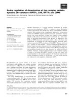
Fig. 1. Comparison between plant RMR proteins and mammalian PA-TM-RING proteins. (A) Structures of a typical plant RMR protein the
rice OsRMR1, BP-80, the pea VSR, two mammalian PA-TM-RING proteins MmRNF13 and MmGRAIL. RMR is predicted to be a type I inte-
gral transmembrane protein containing an N-terminal signal peptide (SP) and a PA domain at its N-terminus, a single TMD and a long CT with
aC
3
H
2
C
3
RING finger domain. The conserved PA and RING domains among the plant RMRs and the mammalian PA-TM-RING family pro-
teins are highlighted in boxes. The two conserved Asn-linked glycosylation sites in the lumenal domain of the plant RMR (OsRMR1) are indi-
cated by asterisks. (B) Amino acid sequence comparisons of TMD and CT regions of selective AtRMRs, OsRMRs and PA-TM-RING H2
proteins from mouse, chicken and humans. Gray boxes indicate highly conserved residues. (C) Phylogenetic analysis of selective plant RMR
and PA-TM-RING proteins using neighbor-joining algorithm with 1000 cycles of bootstrap resampling as indicated. (D) Phylogenetic analysis
of the five Arabidopsis RMRs (AtRMRs) and the two rice RMR (OsRMRs) using neighbor-joining algorithm with 1000 cycles of bootstrap re-
sampling as indicated.
H. Wang et al. Plant RMR proteins
FEBS Journal 278 (2011) 59–68 ª 2010 The Authors Journal compilation ª 2010 FEBS 61
ter protein comprised of a lumenal proaleurain repor-
ter domain linked to AtRMR2 transmembrane
sequence and cytoplasmic tail (designated Re-R-R).
Proaleurain is the precursor of aleurain, a vacuolar
cysterine protease from barley that is processed into its
mature form in lytic PVC. The report was expressed,
and its traffic was compared with that of a similar
reporter proteins, but in one case containing the pea
BP-80 transmembrane sequence and cytoplasmic tail
(designated Re-B-B, known to traffic from ER to
Golgi to lytic PVC), and in a second case containing
the BP-80 transmembrane sequence but with the
alpha-tonoplast intrinsic protein (a PSV marker) cyto-
plasmic tail (designated Re-B-alpha, known to traffic
directly from ER to a PSV PVC). The proaleurain
reporter moiety would be proteolytically processed by
a specific maturase [48,49] if it reached the lytic PVC,
and traffic into the Golgi would be assessed by evalu-
ating whether the reporter protein acquired complex
modifications to its two Asn-linked oligosaccharide
chains [13]. The results are summarized in Table 1,
and document that the RMR reporter protein entered
the Golgi apparatus because it acquired complex
glycans, but it did not traffic to the lytic PVC [34].
Thus, RMR proteins trafficked through the Golgi
apparatus in a pathway distinct from that of BP-80,
and were directed to a protein storage vacuole equiva-
lent in the suspension cultured protoplasts that also
contained alpha-tonoplast intrinsic protein, whereas in
plant seed embryos the RMR proteins were concen-
trated in internal membrane arrays in PSVs.
The growing pollen tube is an ideal single-cell model
system to study protein trafficking and their functions
in the secretory and endocytic pathways in plants. The
dynamics and function of BP-80 and secretory carrier
membrane protein (SCAMP) were also recently char-
acterized in growing lily (Lilium longiflorum) pollen
tube [25]. SCAMP localized to early endosomes,
plasma membrane and cell plate in plant cells [50].
Both lily BP-80 ⁄ VSR and SCAMP cDNAs (termed
LIVSR and LISCAMP respectively) were cloned and
used to make green fluorescent protein (GFP) fusions
for transient expression under the control of the pollen
specific promoter ZM13 in germinating lily pollen
tubes via particle bombardment for protein trafficking
studies. Interestingly, GFP–LISCAMP was mainly
concentrated in the tip region (Fig. 2A), which is
enriched with plant endocytic vesicles and early
endosomes 50–200 nm in size [50]. By contrast, GFP–
BP-80 ⁄ GFP–LIVSR were found to locate throughout
the pollen tubes except the apical clear zone region
(Fig. 2B) and were concentrated in 0.2-lm diameter
punctate organelles that represent prevacuolar com-
partments for the lytic vacuole. In addition, microin-
jection of VSR or SCAMP antibodies significantly
reduced the growth rate of the lily pollen tubes [25].
Because VSRs mediate vacuolar protein transport [51],
whereas SCAMPs may play roles in endocytosis
[50,52] as well as cell plate formation [48], these results
together suggest that both VSR and SCAMP are
required for pollen tube growth, likely working
together in regulating protein trafficking and mem-
brane flow in the secretory and endocytic pathways
which need to be coordinated in order to support
pollen tube elongation.
RMR proteins may also function in pollen tube
growth because microarray data analysis of gene
expression in Arabidopsis (GENEVESTIGATOR,
shows
that AtRMR3 is highly expressed in pollen compared
with other AtRMRs in various tissues (unpublished
results). We have thus recently taken a similar
approach to study the dynamics and distribution of
GFP-tagged RMR proteins using the same pollen tube
transient expression system. As shown in Fig. 2C,
when transiently expressed in a tobacco pollen tube,
a weak GFP–AtRMR3 signal was diffusely distributed
throughout the length of the growing pollen tube but
Table 1. Exploration and determination of RMR or VSR protein trafficking via reporter fusion protein. TMD, transmembrane domain;
CT, cytoplasmic tail; a-TIP, alpha-tonoplast intrinsic protein; LIVSR, lily vacuolar sorting receptor; LISCAMP, lily secretory carrier membrane
protein; GFP, green fluorescent protein; TGN, trans-Golgi network; ER, endoplasmic reticulum; NA, not determined.
Reporter protein
Complex
glycan
Proaleurain
maturation Trafficking pathway
Lumenal proaleurain reporter domain + BP-80 TMD and CT (Re-B-B) Yes Yes ER to Golgi to lytic PVC
Lumenal proaleurain reporter domain + AtRMR TMD and CT (Re-R-R) Yes No ER to Golgi to storage PVC
Lumenal proaleurain reporter domain + BP-80 TMD + a-TIP CT (Re-B-alpha) No No ER to storage PVC
LIVSR + GFP NA NA ER to Golgi to lytic PVC to lytic
vacuole in pollen tube
LlSCAMP + GFP NA NA PM to apical endocytic vesicels to
TGN to lytic vacuole in pollen tube
Plant RMR proteins H. Wang et al.
62 FEBS Journal 278 (2011) 59–68 ª 2010 The Authors Journal compilation ª 2010 FEBS
missing from the tip region, and concentrated within
some large 1–2-lm organelles (Fig. 2C) that were
mobile (data not shown), a pattern that was different
from those of GFP–LlSCAMP (Fig. 2A). Given the
known association of RMR proteins with protein stor-
age vacuoles or their PVCs in other plant systems, we
tentatively identify these structures as pollen tube PSVs
or their PVCs, although a firm identification will
require further colocalization studies with markers for
other organelles and ⁄ or immunogold EM studies.
Role of RMR proteins as sorting
receptors
The ability of the AtRMR2 lumenal domain to bind
potential protein ligands was evaluated using the
recombinant protein expressed in insect suspension cul-
ture cells from which it was secreted into the culture
medium and purified [44,45]. It should be noted that
all RMR lumenal domains contain two conserved sites
for Asn-linked glycosylation (Fig. 1A), and use of the
insect cell expression system allowed assurance that
proper glycosylation would be achieved [44]. This con-
sideration was relevant because the relatively large size
of such glycans would impose steric limitations on
interactions of the relatively small RMR protein with
potential ligands.
The experimental approach evaluated interactions
with two distinct types of known vacuolar sorting
determinant sequences. The first type is the NPIR
(Asn-Pro-Ile-Arg) motif recognized by the VSR pro-
teins, whereas the second type is demonstrated by two
different C-terminal propeptide sequences representing
the class of targeting signals that have no apparent
sequence conservation but the function of which
requires placement at the C-terminus of ligand proteins
[31,53]. It had been hypothesized that the latter direc-
ted proteins into the pathway to PSVs [54], and subse-
quent studies using genetic approaches in Arabidopsis
identified a specific SNARE complex, important for
membrane fusion in eukaryotes, to be essential for
traffic through the pathway required for vacuolar tar-
geting of ligands carrying C-terminal vacuolar sorting
determinants (defined as the PSV pathway), but not
the pathway for traffic to a lytic vacuole [55,56].
Park et al. [44] assessed binding of the AtRMR2
lumenal domain to synthetic peptides of defined
sequences that were coupled to agarose beads. AtRMR2
bound specifically to known C-terminal vacuolar sorting
determinant sequences, but only if they were presented
with a free C-terminus. Interestingly, binding of the
RMR protein to these C-terminal sorting determinant
sequences was not pH dependent; in contrast to the
interaction of BP-80 with its sequence-specific ligands,
the RMR protein could not be eluted from the peptide–
agarose beads by treatment at pH 4. In addition, specific
binding was blocked by the C-terminal addition of two
Gly residues, a modification known to prevent function
in vacuolar sorting [53]. Specific binding to peptides car-
rying sequence-specific sorting determinants was not
observed. Thus, RMR proteins specifically bind to pep-
tides corresponding to sorting determinants for the PSV
pathway, which is distinct from the pH-dependent
BP-80 ⁄ AtVSR1 sorting pathway to the lytic vacuole.
A
B
C
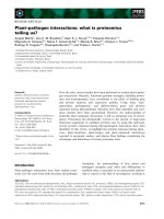
Fig. 2. Dynamics distribution of RMR vs.
VSR and SCAMP in growing lily pollen tube.
GFP fusions constructs with the lily
secretory carrier membrane protein 4
(GFP–LlSCAMP4) (A), the lily vacuolar
sorting receptor 2 (GFP–LlVSR2) (B) and the
Arabidopsis RMR3 (GFP–AtRMR3) (C) were
transiently expressed in growing lily pollen
tubes (A ⁄ B) or tobacco pollen tube (C)
respectively via particle bombardment,
followed by confocal imaging as previously
described [25]. Scale bar, 25 lm.
H. Wang et al. Plant RMR proteins
FEBS Journal 278 (2011) 59–68 ª 2010 The Authors Journal compilation ª 2010 FEBS 63
The functional relevance of these in vitro binding
results was further tested by expressing pairs of recom-
binant proteins, each representing a receptor lumenal
domain and a soluble ligand carrying complementary
halves of the GFP molecule, by transient expression in
tobacco suspension culture protoplasts. In this bimolec-
ular fluorescence complementation assay [57], reconsti-
tution of a fluorescent GFP molecule occurs only when
the two halves are brought into close proximity through
interaction of the recombinant proteins to which they
are attached. The obtained results indicated that BP-80
preferentially interacted with the vacuolar targeting
sequence of lytic vacuole marker proaleurain rather
than the C-terminal propeptide of the PSV marker
chitinase [57]. Conversely, AtRMR2 preferentially
interacted with the chitinase C-terminal propeptide but
not with the proaleurain targeting sequence. These
results were consistent with the in vitro binding assay
results and indicated that the AtRMR2 lumenal domain
could interact in a specific manner with the chitinase
C-terminal vacuolar sorting determinant in vivo.
In a separate series of experiments, the reporter pro-
tein Re-R-R with either GFP or monomeric red fluor-
escent protein (mRFP) inserted into its cytoplasmic
tail was transiently expressed in the suspension culture
protoplasts. Consistent with previous findings that
endogenous RMR proteins were internalized into PSVs
in developing seed embryos, Re-R-R tagged with either
fluorescent molecule was present in small punctate
cytoplasmic organelles, but also was internalized into
the lumen of the protoplasts’ central vacuoles. Thus,
traffic of these proteins, which as previously shown
[34] was determined by sequences in the AtRMR2
cytoplasmic tail, resulted in the cytoplasmic tails con-
taining the fluorescent tags being transferred from the
cytoplasm to the vacuole lumen.
A different study used the lumenal domain of
AtRMR1 expressed in bacterial system for binding
studies [46]. Those authors found that the At-
RMR1 protein bound to C-terminal vacuolar sorting
sequences but not to sequence-specific sorting
sequences, and that binding was pH dependent and
was abolished at pH 4. In addition, they presented
data that argued for recycling of the AtRMR1 protein
in transient expression experiments in Arabidopsis sus-
pension culture protoplasts. These results and those
obtained for AtRMR2, as well as experiments localiz-
ing endogenous RMR proteins in vivo [33,34], appear
to be contradictory. However, the possibility remains
that AtRMR1 has substantially different ligand-bind-
ing properties and patterns of traffic within cells.
Future genetic study using knockout mutants of
individual AtRMRs or coexpression of AtRMR1 and
AtRMR2 in the same cells may be able to address
these differences.
Spatial regulation of ligand sorting by
RMR proteins in the Golgi apparatus
From studies using developing pea cotyledons, Hinz
et al. have provided elegant data to argue for traffic of
seed-storage proteins in DVs, separate from BP-80 pro-
teins which are predominantly present in CCVs. Using
quantitative analyses at the electron microscope level,
those authors demonstrated that globulin-type storage
proteins form aggregates in the cis-Golgi that partition
at the periphery of cisternae and then move sequen-
tially towards the trans-face where they bud off as
DVs. By contrast, BP-80 receptors were localized pre-
dominantly at the trans-Golgi and were associated with
CCVs [58]. They therefore proposed a novel model
whereby spatial regulation of sorting within the Golgi
apparatus might explain how traffic of storage proteins
to PSVs could be separated from traffic of proteins
destined to be carried by CCVs to the lytic PVC.
In a subsequent study, those authors quantitatively
analyzed the distribution of AtRMR2, Arabidopsis
AtVSR proteins (BP-80 homologs) and the storage
protein cruciferin in the Golgi apparatus and vesicles
during Arabidopsis embryo development [33]. In con-
trast to Otegui et al. (2006) [59], but consistent with
prior results in the pea system, cruciferin was present
predominantly at the periphery in the cis and medial
cisternae and in DVs. AtVSR labeling was predomi-
nantly at the trans-face and in CCV, with very small
amounts associated with DVs. By contrast, labeling
for AtRMR2 in the Golgi and DVs was very similar
to that for cruciferin. These results were interpreted to
support the concept that RMR proteins were associ-
ated with sorting of storage protein aggregates into
DVs. Consistent with findings from other studies,
labeling for AtRMR2 on organelles representing PVCs
was predominantly internal, providing further support
that these proteins are internalized into organelle
lumens during their traffic to the PSV. Such internali-
zation would remove the possibility that AtRMR2
could recycle back to the Golgi apparatus to partici-
pate in more than one round of ligand sorting.
How could RMR proteins serve as efficient sorting
receptors if they do not recycle? The aggregation
model for storage protein sorting [58] may provide an
explanation. By interacting with an aggregate of many
storage protein molecules as the aggregate is sorted
into a DV, a limited number of RMR proteins could
participate in DV coat protein formation and effi-
ciently promote sorting [33,44].
Plant RMR proteins H. Wang et al.
64 FEBS Journal 278 (2011) 59–68 ª 2010 The Authors Journal compilation ª 2010 FEBS
The process of internalization of RMR proteins into
prevacuolar organelles would result in removal of
cytoplasmic tails of the proteins from the cytoplasm.
The RING-H2 domain found in mammalian RMR
protein homologs has been shown to function as a
ubiquitin–protein ligase [37,38]. There is no direct
AB
Fig. 4. Working model of RMR proteins in plants. (A) Subcellular localization and dynamics of RMRs in developing seeds. In developing
tomato and tobacco seeds, RMR is found in the crystalloid of PSV, the storage PVC or DIP organelle; whereas in developing Arabidopsis
seeds RMR were found in DVs [34]. (B) Subcellular localization and dynamics of RMR, VSR and SCAMP in growing pollen tube. Shown is a
working model on the localization, dynamics and possible functional roles of VSR, SCAMP and RMR proteins in germinating pollen tubes.
SCAMP is highly enriched in the apical region of the pollen tube which is missing the VSR [25]. In addition to a possible ER–Golgi–trans-
Golgi network–PVC ⁄ multivesicular body–vacuole transport pathway [25], VSR ⁄ BP-80 could also reach the plasma membrane from the trans-
Golgi network and then internalize because VSR was also found in PM in addition to multivesicular body or PVC in immunogold EM study
(our unpublished results). Similarly, SCAMP could reach the plasma membrane from either Golgi or trans-Golgi network and internalize from
the plasma membrane via endocytosis colocalizing with the internalized endocytic marker FM4-64. The SCAMP-positive small vesicles
enriched in the apical region are believed to be derived directly from the Golgi apparatus or via trans-Golgi network and endocytic vesicles
from plama membrane. RMR may mediate protein transport from Golgi apparatus and reach a yet-to-be identified storage organelle or PVC
distinct from the SCAMP-positive trans-Golgi network ⁄ early endosome and the VSR-positive multivesicular body ⁄ PVC in the same growing
pollen tube. Both VSR and SCAMP were found to reach the vacuole lumen in immunogold EM, presumably for degradation [25].
A
B
Fig. 3. Evidence for the presence of ubiquitin in protein storage vacuole crystalloid. Immunogold EM labeling [24] with anti-ubiquitin sera
was performed on ultrathin sections prepared from high-pressure freezing ⁄ frozen substituted developing tobacco seed embryo cells. A typi-
cal PSV in these cells contains three distinct subcompartments (crystalloid, matrix and globoid as indicated) (A), in which gold particles are
mainly found in the crystalloid as indicated by arrows (B). No labeling with secondary antibody alone was observed (data not shown).
H. Wang et al. Plant RMR proteins
FEBS Journal 278 (2011) 59–68 ª 2010 The Authors Journal compilation ª 2010 FEBS 65
experimental evidence that plant RMR proteins have
a similar ubiquitin–protein ligase activity although it is
reasonable to hypothesize so. Consistent with this
possibility, our preliminary studies using immunogold
EM labeling with anti-ubiquitin sera on ultrathin sec-
tions of cells from developing tobacco seed embryos
prepared by high-pressure freezing ⁄ frozen substitution
demonstrated positive labeling in the protein storage
vacuole crystalloid (Fig. 3). This result would be
consistent with the concept that RMR proteins are
internalized into the PSV as it develops, and intermo-
lecular ubiquitination might help explain the observa-
tion that ‘crystalloid’ proteins from Brassica napus
were cross-linked in a manner that resisted treatment
with disulfide reducing agents [47]. Such hypothesis of
ubiquitin-mediated cross-linking during internalization
of proteins into the PSV could be tested in future
experiments by isolation and biochemical analysis of
PSVs.
Both the mammalian GRAIL and RNF13 proteins
affect complex functions in cells where they are
expressed. In the case of the RNF13 protein, the cyto-
plasmic tail is cleaved from attachment to the TMD
during traffic to endosomes; the now free cytoplasmic
tail with its ubiquitin–protein ligase activity has been
postulated to provide a mechanism for activation of
signaling pathways that would affect cell functions and
fate [37]. Although genes encoding the beta and
gamma secretase proteases that are hypothesized to
participate in such a cleavage process [37] are not pres-
ent in plant genomes [60], it is possible that some other
mechanism for cleavage of plant RMR protein cyto-
plasmic tails within the basic region separating the
transmembrane and RING-H2 domains conserved in
both plant and mammalian proteins (as indicated in
Fig. 1A,B) might exist. Thus there may be an advan-
tage to the cell to have these relatively abundant pro-
teins removed from the cytoplasm as they reach the
terminus of their trafficking pathway. Whether the free
tail would participate in some signaling process
remains to be tested experimentally.
Conclusion and future perspective
In conclusion, Fig. 4A summaries the subcellular local-
ization, trafficking and possible function of RMRs in
developing seeds, where RMR-mediated storage
protein sorting is achieved via concentration sorting in
storage PVC [or dark intrinsic protein (DIP) organ-
elles] or DVs (Fig. 4A). In addition, the three integral
memebrane proteins, RMR, VSR and SCAMP, show
distinct patterns of subcellular localization and dynam-
ics in the same growing pollen tubes (Fig. 4B), indicat-
ing their distinct functional roles and transport
pathways in plants. However, the native ligands or
cargo proteins for most VSRs and RMRs in plants
remain elusive.
To identify native cargo proteins for the Arabidopsis
AtVSRs and AtRMRs, we have recently developed
and used a transgenic Arabidopsis suspension culture
cell system expressing the N-terminus of VSR or RMR
(lacking its TMD and CT) so that the secreted
truncated VSR or RMR proteins would bring along
their native cargoes into the culture media to be identi-
fied by LC-MS ⁄ MS analysis [30], however, this
approach would be difficult to carry out for RMR
cargo identification if RMR binds to aggregates. Such
a biochemical⁄ cell biology approach for functional
characterization of VSR and RMR, as well as their
cargo proteins in plants, will likely generate novel
information to complement genetic approaches.
Published studies of the luminal domain of plant
RMRs suggest that these proteins function as sorting
receptors for transporting storage proteins to PSVs in
plants. However, the functional roles of the RMR
C-terminal RING-H2 domain remain largely unknown
compared with that of the mammalian PA-TMD-
RING proteins. Because the RING domains are highly
conserved between the plant RMR and the mamma-
lian PA-TMD-RING proteins (Fig. 1A–C) and
because ubiquitin was localized in the PSV crystalloid
where RMR proteins are concentrated (Fig. 3), it is
reasonable to hypothesize that plant RMR proteins
may also have a similar ubiquitin–protein ligase
activity. Such hypothesis can be tested via in vitro
ubiquitin–protein ligase activity analysis in future
experiments.
Acknowledgements
Our work has been supported by grants from the
Research Grants Council of Hong Kong
(CUHK488707, CUHK465708, CUHK466309, CUHK
466610 and HKUST6 ⁄ CRF ⁄ 08), UGC-AoE, CUHK
Schemes B ⁄ C.
References
1 Gurkan C, Koulov AV & Balch WE (2007) An evolu-
tionary perspective on eukaryotic membrane trafficking.
Adv Exp Med Biol 607, 73–83.
2 Hwang I & Robinson DG (2009) Transport vesicle for-
mation in plant cells. Curr Opin Plant Biol 12, 660–669.
3 Paris N, Stanley CM, Jones RL & Rogers JC (1996)
Plant cells contain two functionally distinct vacuolar
compartments. Cell 85, 563–572.
Plant RMR proteins H. Wang et al.
66 FEBS Journal 278 (2011) 59–68 ª 2010 The Authors Journal compilation ª 2010 FEBS
4 Paul MJ & Frigerio L (2007) Coated vesicles in plant
cells. Semin Cell Dev Biol 18, 471–478.
5 Pryer NK, Wuestehube LJ & Schekman R (1992)
Vesicle-mediated protein sorting. Annu Rev Biochem 61,
471–516.
6 Robinson DG, Jiang L & Schumacher K (2008) The
endosomal system of plants: charting new and familiar
territories. Plant Physiol 147, 1482–1492.
7 Robinson DG (1996) Clathrin-mediated trafficking.
Trends Plant Sci 1, 349–355.
8 Robinson DG & Depta H (1988) Coated vesicles. Annu
Rev Plant Physiol Plant Mol Biol 39, 53–99.
9 Boller T & Kende H (1979) Hydrolytic enzymes in the
central vacuole of plant cells. Plant Physiol 63, 1123–
1132.
10 Klionsky DJ, Herman PK & Emr SD (1990) The fungal
vacuole: composition, function, and biogenesis. Micro-
biol Rev 54, 266–292.
11 Cereghino JL, Marcusson EG & Emr SD (1995) The
cytoplasmic tail domain of the vacuolar protein sorting
receptor Vps10p and a subset of VPS gene products
regulate receptor stability, function, and localization.
Mol Biol Cell 6, 1089–1102.
12 Cooper AA & Stevens TH (1996) Vps10p cycles
between the late-Golgi and prevacuolar compartments
in its function as the sorting receptor for multiple yeast
vacuolar hydrolases. J Cell Biol 133, 529–541.
13 Jiang L & Rogers JC (1998) Integral membrane protein
sorting to vacuoles in plant cells: evidence for two path-
ways. J Cell Biol 143, 1183–1199.
14 Kirsch T, Paris N, Butler JM, Beevers L & Rogers JC
(1994) Purification and initial characterization of a
potential plant vacuolar targeting receptor. Proc Natl
Acad Sci USA 91, 3403–3407.
15 Kornfeld S (1992) Structure and function of the man-
nose 6-phosphate ⁄ insulin-like growth factor II recep-
tors. Annu Rev Biochem 61, 307–330.
16 Paris N, Rogers SW, Jiang L, Kirsch T, Beevers L,
Phillips TE & Rogers JC (1997) Molecular cloning and
further characterization of a probable plant vacuolar
sorting receptor. Plant Physiol 115, 29–39.
17 Niemes S, Langhans M, Viotti C, Scheuring D,
Yan MS, Jiang L, Hillmer S, Robinson DG & Pimpl P
(2009) Retromer recycles vacuolar sorting receptors
from the trans-Golgi network. Plant J 61, 107–121.
18 Oliviusson P, Heinzerling O, Hillmer S, Hinz G,
Tse YC, Jiang L & Robinson DG (2006) Plant retro-
mer, localized to the prevacuolar compartment and
microvesicles in Arabidopsis, may interact with vacuolar
sorting receptors. Plant Cell 18, 1239–1252.
19 Jiang L, Erickson AH & Rogers JC (2002) Multivesicu-
lar bodies: a mechanism to package lytic and storage
functions in one organelle? Trends Cell Biol 12, 362–
367.
20 Jiang L & Rogers JC (1997) Golgi to prevacuole-target-
ing mechanisms of a plant vacuolar sorting receptor.
Plant Physiol 114(Suppl.), 70.
21 Jiang L & Rogers JC (1999) Sorting of membrane
proteins to vacuoles in plant cells. Plant Sci
146, 55–67.
22 Jiang L & Rogers JC (1999) The role of BP-80 and
homologs in sorting proteins to vacuoles. Plant Cell 11,
2069–2071.
23 Paris N & Neuhaus JM (2002) BP-80 as a vacuolar
sorting receptor. Plant Mol Biol 50, 903–914.
24 Tse YC, Mo B, Hillmer S, Zhao M, Lo SW,
Robinson DG & Jiang L (2004) Identification of
multivesicular bodies as prevacuolar compartments in
Nicotiana tabacum BY-2 cells. Plant Cell 16, 672–693.
25 Wang H, Tse YC, Law AH, Sun SS, Sun YB, Xu ZF,
Hillmer S, Robinson DG & Jiang L (2010) Vacuolar
sorting receptors (VSRs) and secretory carrier mem-
brane proteins (SCAMPs) are essential for pollen tube
growth. Plant J 61, 826–838.
26 Kirsch T, Saalbach G, Raikhel NV & Beevers L (1996)
Interaction of a potential vacuolar targeting receptor
with amino- and carboxyl-terminal targeting determi-
nants. Plant Physiol 111, 469–474.
27 Cao X, Rogers SW, Butler J, Beevers L & Rogers JC
(2000) Structural requirements for ligand binding by a
probable plant vacuolar sorting receptor. Plant Cell 12,
493–506.
28 Luo X & Hofmann K (2001) The protease-associated
domain: a homology domain associated with multiple
classes of proteases. Trends Biochem Sci 26, 147–148.
29 Mahon P & Bateman A (2000) The PA domain: a
protease-associated domain. Protein Sci 9, 1930–1934.
30 Suen PK, Shen J, Sun SS & Jiang L (2010) Expression
and characterization of two functional vacuolar sorting
receptor (VSR) proteins, BP-80 and AtVSR4 from cul-
ture media of transgenic tobacco BY-2 cells. Plant Sci
179, 68–76.
31 Neuhaus JM & Paris N (2005) Plant vacuoles: from
biogenesis to function. Plant Cell Monogr 1, 63–82.
32 Hinz G, Hillmer S, Ba
¨
umer M & Hohl I (1999) Vacuo-
lar storage proteins and the putative sorting receptor
BP-80 exit the Golgi apparatus of developing pea coty-
ledons in different transport vesicles. Plant Cell 11,
1509–1524.
33 Hinz G, Colanesi S, Hillmer S, Rogers JC &
Robinson DG (2007) Localization of vacuolar transport
receptors and cargo proteins in the Golgi apparatus of
developing Arabidopsis embryos. Traffic 8, 1452–1464.
34 Jiang L, Phillips TE, Rogers SW & Rogers JC (2000)
Biogenesis of the protein storage vacuole crystalloid.
J Cell Biol 150, 755–769.
35 Akuzawa N, Kurabayashi M, Ohyama Y, Arai M &
Nagai R (2000) Zinc finger transcription factor Egr-1
activates Flt-1 gene expression in THP-1 cells on induc-
H. Wang et al. Plant RMR proteins
FEBS Journal 278 (2011) 59–68 ª 2010 The Authors Journal compilation ª 2010 FEBS 67
tion for macrophage differentiation. Arterioscler
Thromb Vasc 20, 377–384.
36 Tranque P, Crossin KL, Cirelli C, Edelman GM &
Mauro VP (1996) Identification and characterization of
a RING zinc finger gene (C-RZF) expressed in chicken
embryo cells. Proc Natl Acad Sci USA 93, 3105–3109.
37 Bocock J, Carmicle S, Chhotani S, Ruffolo MR,
Chu H & Erickson AH (2009) The PA-TM-RING
protein RING finger protein 13 is an endosomal inte-
gral membrane E3 ubiquitin ligase whose RING finger
domain is released to the cytoplasm by proteolysis.
FEBS J 276, 1860–1877.
38 Kriegel MA, Rathinam C & Flavell RA (2009) E3
ubiquitin ligase GRAIL controls primary T cell activa-
tion and oral tolerance. Proc Natl Acad Sci USA 106,
16770–16775.
39 Stone SL, Hauksdottir H, Troy A, Herschleb J, Kraft E
& Callis J (2005) Functional analysis of the RING-type
ubiquitin ligase family of Arabidopsis. Plant Physiol
137, 13–30.
40 Stone SL, Williams LA, Farmer LM, Vierstra RD &
Callis J (2006) KEEP ON GOING, a RING E3 ligase
essential for Arabidopsis growth and development, is
involved in abscisic acid signaling. Plant Cell 18, 3415–
3428.
41 Jin X, Cheng H, Chen J & Zhu D (2010) RNF13:
an emerging RING finger E3 ubiquitin ligase important
in cell proliferation. FEBS J 278, 78–84.
42 Bocock JP, Carmicle S, Sicar M & Erickson AH(2010)
Trafficking and proteolytic processing of RNF13,
a model PA-TM-RING family endosomal membrane
ubiquitin ligase. FEBS J 278, 69–77.
43 Whiting CC, Su LL, Lin JT & Fathman CG (2010)
GRAIL: a unique mediator of CD4 T-lymphocyte
unresponsiveness. FEBS J 278, 47–58.
44 Park JH, Oufattole M & Rogers JC (2007) Golgi-medi-
ated vacuolar sorting in plant cells: RMR proteins are
sorting receptors for the protein aggregation ⁄ membrane
internalization pathway. Plant Sci 172, 728–745.
45 Park JH, Rogers SW, Paris N & Rogers JC (2002)
RMR proteins as sorting receptors for the protein
storage vacuole pathway. American Society of Plant
Biologists Annual Meeting (abstract). http://
216.133.76.127/pb2002/public/m13/1016.html.
46 Park M, Lee D, Lee G-J & Hwang I (2005) AtRMR1
functions as a cargo receptor for protein trafficking
to the protein storage vacuole. J Cell Biol 170, 757–
767.
47 Gillespie JE, Rogers SW, Deery M, Dupree P & Rogers
JC (2005) A unique family of proteins associated with
internalized membranes in protein storage vacuoles of
the Brassicaceae. Plant J 41, 429–441.
48 Halls CE, Rogers SW & Rogers JC (2005) Purification
of a proaleurain maturation protease. Plant Sci 168,
1267–1279.
49 Holwerda BC, Galvin NJ, Baranski TJ & Rogers JC
(1990) In vitro processing of aleurain, a barley vacuolar
thiol protease. Plant Cell 2, 1091–1106.
50 Lam SK, Siu CL, Hillmer S, Jang S, An G, Robinson
DG & Jiang L (2007) Rice SCAMP1 defines clathrin-
coated, trans-Golgi-located tubular–vesicular structures
as an early endosome in tobacco BY-2 cells. Plant Cell
19, 296–319.
51 Jiang L & Rogers JC (2003) Sorting of lytic enzymes
in the plant Golgi apparatus. Annu Plant Rev 9
, 114–
140.
52 Lam SK, Tse YC, Robinson DG & Jiang L (2007)
Tracking down the elusive early endosome. Trends
Plant Sci 12, 497–505.
53 Dombrowski JE, Schroeder MR, Bednarek SY &
Raikhel NV (1993) Determination of the functional
elements within the vacuolar targeting signal of barley
lectin. Plant Cell 5, 587–596.
54 Okita TW & Rogers JC (1996) Compartmentation of
proteins in the endomembrane system of plant cells.
Annu Rev Plant Physiol Plant Mol Biol 47, 327–350.
55 Bassham DC & Raikhel NV (2000) Plant cells are not
just green yeast. Plant Physiol 122, 999–1001.
56 Sanmartin M, Ordonez A, Sohn EJ, Robert S, Sanchez-
Serrano JJ, Surpin MA, Raikhel NV & Rojo E (2007)
Divergent functions of VTI12 and VTI11 in trafficking
to storage and lytic vacuoles in Arabidopsis. Proc Natl
Acad Sci USA 104, 3645–3650.
57 Kerppola T (2008) Biomolecular fluorescence comple-
mentation (BiFC) analysis as a probe of protein interac-
tions in living cells. Annu Rev Biophys 37, 465–487.
58 Hillmer S, Movafeghi A, Robinson DG & Hinz G
(2001) Vacuolar storage proteins are sorted in the
cis-cisternae of the pea cotyledon Golgi apparatus.
J Cell Biol 152, 41–50.
59 Otegui MS, Herder R, Schulze J, Jung R & Staehelin
LA (2006) The proteolytic processing of seed storage
proteins in Arabidopsis embryo cells starts in the
multivesicular bodies. Plant Cell 18, 2567–2581.
60 Khandelwal A, Chandu D, Roe CM, Kopan R &
Quatrano RS (2007) Moonlighting activity of presenilin
in plants is independent of gamma-secretase and evolu-
tionarily conserved. Proc Natl Acad Sci USA 104,
13337–13342.
Plant RMR proteins H. Wang et al.
68 FEBS Journal 278 (2011) 59–68 ª 2010 The Authors Journal compilation ª 2010 FEBS